Embed Size (px)
Citation preview

The MalariaCare Toolkit
Tools for maintaining high-quality malaria case management services
National malaria microscopy competency assessment and accreditation course:
Facilitator’s manual for Zambia
Note: Zambia’s national program was the first under the MalariaCare project to implement a national malaria microscopy competency assessment program to assess routine malaria
microscopists. The program was adapted from the World Health Organization External Competency Assessment of Malaria Microscopy course. Other countries may find such an exercise useful for
building competency in malaria microscopy and strengthening quality assurance systems. In such cases, this example may be adapted for the specific context.
Download all the MalariaCare Tools from: www.malariacare.org/resources/toolkit
ii
Contents
1.0 Introduction to the malaria microscopist competency assessment ......................................................... 1
1.1 WHO external competency assessment overview ................................................................................. 1
1.2 Zambian Competency Assessment overview ........................................................................................ 2
2.0 Introduction to the national competency assessment facilitator’s manual ................................................. 3
2.1 Aim of this manual ................................................................................................................................ 3
2.2 Goal ....................................................................................................................................................... 3
2.3 Objectives .............................................................................................................................................. 3
3.0 Role of the facilitator .................................................................................................................................. 4
3.1 Facilitator responsibilities ..................................................................................................................... 4
3.2 Using this guide ..................................................................................................................................... 4
4.0 Preparing for the workshop ........................................................................................................................ 5
4.1 Workshop duration ................................................................................................................................ 5
4.2 Plan with national and subnational authorities ...................................................................................... 5
4.3 Participant selection and characteristics ................................................................................................ 5
4.4 Venue selection and length of course .................................................................................................... 5
4.5 Required supplies, equipment, and materials ........................................................................................ 5
4.6 Course agenda and timetable ................................................................................................................. 6
4.7 Calendar of activities for preparation .................................................................................................... 6
5.0 Course monitoring and evaluation .............................................................................................................. 8
6.0 National competency assessment course structure ..................................................................................... 9
6.1 Training methods ................................................................................................................................... 9
6.2 Course structure overview ..................................................................................................................... 9
7.0 Pre-assessment overview .......................................................................................................................... 11
7.1 Theory-based knowledge .................................................................................................................... 11
7.2 Blood film preparation ........................................................................................................................ 11
7.3 Malaria microscopy ............................................................................................................................. 11
8.0 Assessment overview ............................................................................................................................... 13
8.1 Assessing participant preparation of thick and thin blood films ......................................................... 13
8.2 Assessing participant malaria microscopy competency ...................................................................... 14
8.2.1 Proposed slide sets for the microscopy competency assessment ................................................ 14
8.2.2 Conducting the malaria microscopy assessment ......................................................................... 14
8.2.3 Scoring the malaria microscopy assessment ............................................................................... 15
9.0 Final scoring ............................................................................................................................................. 16
10.0 Annexes .................................................................................................................................................. 17
Annex 1. Equipment and reagents required for training ........................................................................... 17
Annex 2. Learning units ............................................................................................................................ 19
Annex 3. Resources ................................................................................................................................... 23
1
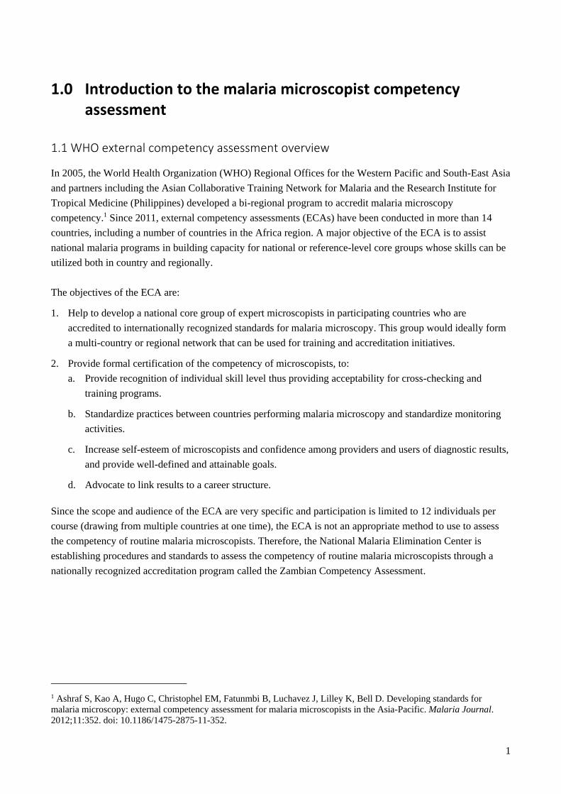
1.0 Introduction to the malaria microscopist competency assessment
1.1 WHO external competency assessment overview
In 2005, the World Health Organization (WHO) Regional Offices for the Western Pacific and South-East Asia
and partners including the Asian Collaborative Training Network for Malaria and the Research Institute for
Tropical Medicine (Philippines) developed a bi-regional program to accredit malaria microscopy
competency.1 Since 2011, external competency assessments (ECAs) have been conducted in more than 14
countries, including a number of countries in the Africa region. A major objective of the ECA is to assist
national malaria programs in building capacity for national or reference-level core groups whose skills can be
utilized both in country and regionally.
The objectives of the ECA are:
1. Help to develop a national core group of expert microscopists in participating countries who are
accredited to internationally recognized standards for malaria microscopy. This group would ideally form
a multi-country or regional network that can be used for training and accreditation initiatives.
2. Provide formal certification of the competency of microscopists, to:
a. Provide recognition of individual skill level thus providing acceptability for cross-checking and
training programs.
b. Standardize practices between countries performing malaria microscopy and standardize monitoring
activities.
c. Increase self-esteem of microscopists and confidence among providers and users of diagnostic results,
and provide well-defined and attainable goals.
d. Advocate to link results to a career structure.
Since the scope and audience of the ECA are very specific and participation is limited to 12 individuals per
course (drawing from multiple countries at one time), the ECA is not an appropriate method to use to assess
the competency of routine malaria microscopists. Therefore, the National Malaria Elimination Center is
establishing procedures and standards to assess the competency of routine malaria microscopists through a
nationally recognized accreditation program called the Zambian Competency Assessment.
1 Ashraf S, Kao A, Hugo C, Christophel EM, Fatunmbi B, Luchavez J, Lilley K, Bell D. Developing standards for
malaria microscopy: external competency assessment for malaria microscopists in the Asia-Pacific. Malaria Journal.
2012;11:352. doi: 10.1186/1475-2875-11-352.
2
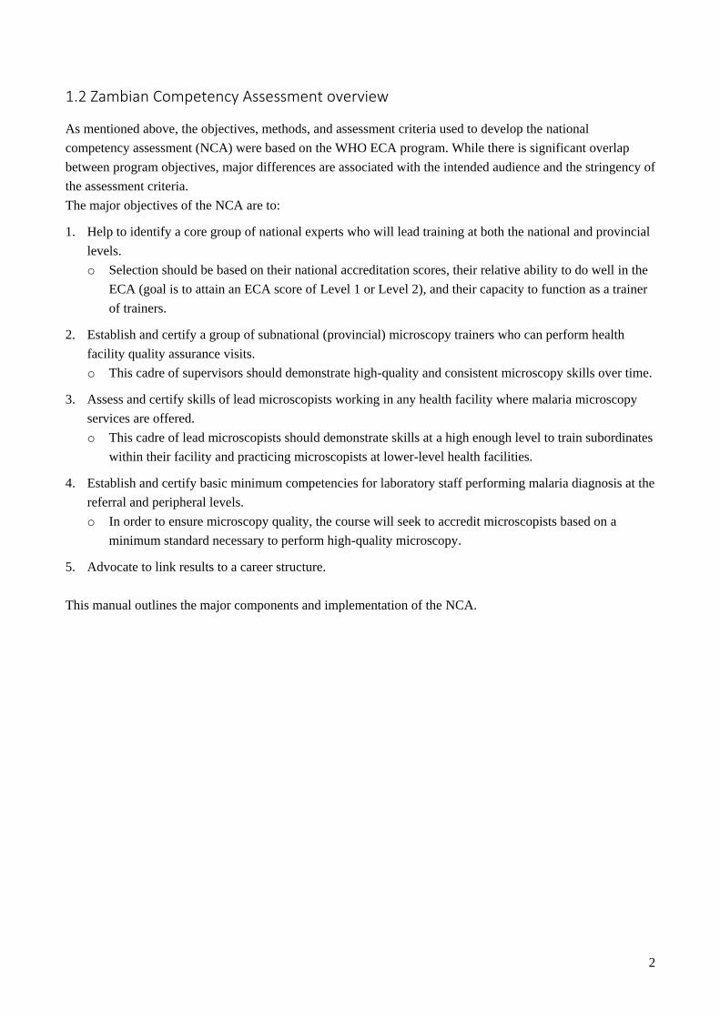
1.2 Zambian Competency Assessment overview
As mentioned above, the objectives, methods, and assessment criteria used to develop the national
competency assessment (NCA) were based on the WHO ECA program. While there is significant overlap
between program objectives, major differences are associated with the intended audience and the stringency of
the assessment criteria.
The major objectives of the NCA are to:
1. Help to identify a core group of national experts who will lead training at both the national and provincial
levels.
o Selection should be based on their national accreditation scores, their relative ability to do well in the
ECA (goal is to attain an ECA score of Level 1 or Level 2), and their capacity to function as a trainer
of trainers.
2. Establish and certify a group of subnational (provincial) microscopy trainers who can perform health
facility quality assurance visits.
o This cadre of supervisors should demonstrate high-quality and consistent microscopy skills over time.
3. Assess and certify skills of lead microscopists working in any health facility where malaria microscopy
services are offered.
o This cadre of lead microscopists should demonstrate skills at a high enough level to train subordinates
within their facility and practicing microscopists at lower-level health facilities.
4. Establish and certify basic minimum competencies for laboratory staff performing malaria diagnosis at the
referral and peripheral levels.
o In order to ensure microscopy quality, the course will seek to accredit microscopists based on a
minimum standard necessary to perform high-quality microscopy.
5. Advocate to link results to a career structure.
This manual outlines the major components and implementation of the NCA.
3
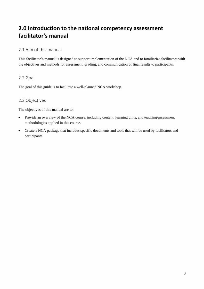
2.0 Introduction to the national competency assessment facilitator’s manual
2.1 Aim of this manual
This facilitator’s manual is designed to support implementation of the NCA and to familiarize facilitators with
the objectives and methods for assessment, grading, and communication of final results to participants.
2.2 Goal
The goal of this guide is to facilitate a well-planned NCA workshop.
2.3 Objectives
The objectives of this manual are to:
Provide an overview of the NCA course, including content, learning units, and teaching/assessment
methodologies applied in this course.
Create a NCA package that includes specific documents and tools that will be used by facilitators and
participants.
4
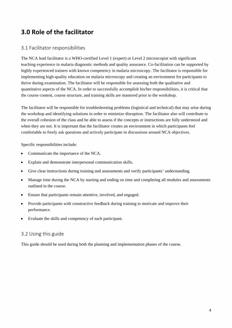
3.0 Role of the facilitator
3.1 Facilitator responsibilities
The NCA lead facilitator is a WHO-certified Level 1 (expert) or Level 2 microscopist with significant
teaching experience in malaria diagnostic methods and quality assurance. Co-facilitation can be supported by
highly experienced trainers with known competency in malaria microscopy. The facilitator is responsible for
implementing high-quality education on malaria microscopy and creating an environment for participants to
thrive during examination. The facilitator will be responsible for assessing both the qualitative and
quantitative aspects of the NCA. In order to successfully accomplish his/her responsibilities, it is critical that
the course content, course structure, and training skills are mastered prior to the workshop.
The facilitator will be responsible for troubleshooting problems (logistical and technical) that may arise during
the workshop and identifying solutions in order to minimize disruption. The facilitator also will contribute to
the overall cohesion of the class and be able to assess if the concepts or instructions are fully understood and
when they are not. It is important that the facilitator creates an environment in which participants feel
comfortable to freely ask questions and actively participate in discussions around NCA objectives.
Specific responsibilities include:
Communicate the importance of the NCA.
Explain and demonstrate interpersonal communication skills.
Give clear instructions during training and assessments and verify participants’ understanding.
Manage time during the NCA by starting and ending on time and completing all modules and assessments
outlined in the course.
Ensure that participants remain attentive, involved, and engaged.
Provide participants with constructive feedback during training to motivate and improve their
performance.
Evaluate the skills and competency of each participant.
3.2 Using this guide
This guide should be used during both the planning and implementation phases of the course.
5
4.0 Preparing for the workshop
This section describes the various activities and concepts the facilitator needs to consider when planning the
course.
4.1 Workshop duration
The course is conducted over five consecutive days.
4.2 Plan with national and subnational authorities
It is important to communicate training plans and intentions with planners at both the national and subnational
levels, as their approval and endorsement of the activity is critical for strong attendance and implementation.
These communications are especially important when identifying and selecting the most appropriate
participants. In addition, it is helpful to understand the different possible logistical constraints from province
to province (e.g., availability of equipment or supplies, power supply).
4.3 Participant selection and characteristics
The identified staff should be working mainly in the malaria/parasitology section of the laboratory and
performing routine malaria microscopy. Health facilities such as central and provincial-level hospitals that
serve a large proportion of the population may be given the highest priority.
4.4 Venue selection and length of course
An appropriate venue should be chosen based on the following specifications:
A room that can accommodate both teaching and practical laboratory work, or separate rooms for teaching
and practical laboratory work.
One functioning microscope available per participant.
Electricity and/or access to a reliable and well-fueled power generator.
A facility to provide morning and afternoon tea and lunch to the participants.
Transport to collect additional supplies and secretarial assistance.
Access to printing and photocopying is an added advantage.
Nearby accommodations for facilitators and participants.
4.5 Required supplies, equipment, and materials
Supplies and equipment should be organized well in advance of the training. Microscope quality should be
inspected by the facilitators to ensure that all necessary components are available (e.g., 100X immersion oil
6
objective, etc.). Extra microscope bulbs should be kept on hand. A list of equipment, supplies, and reagents
required for this training can be found in Annex 1.
4.6 Course agenda and timetable
In order to carry out a successful NCA, it is critical that facilitators become familiar with the course agenda. If
there is more than one facilitator, it will be important to identify which learning units will be conducted by
which facilitator based on areas of expertise. Assessment exercises should be well planned the evening before.
In order to become familiar with the course, it is important to review the sequence of all learning and
assessment modules.
The following activities should be conducted in order to become familiar with the course content and
structure:
Review the curriculum and assessment requirements.
Review the timetable and plan ahead for practical assessments.
Decide on the number of participants (ideally not to exceed 20).
Determine the space needed (lecture and laboratory space) to accommodate participants.
Confirm the number of facilitators and responsibilities for each facilitator.
Decide which modules each facilitator will teach.
Review the list of supplies, equipment, and other materials required and initiate procurement.
Consider the time it will take each day/evening to grade daily malaria microscopy competency
assessments.
Plan other activities, such as opening and closing ceremonies, certification, and follow-up.
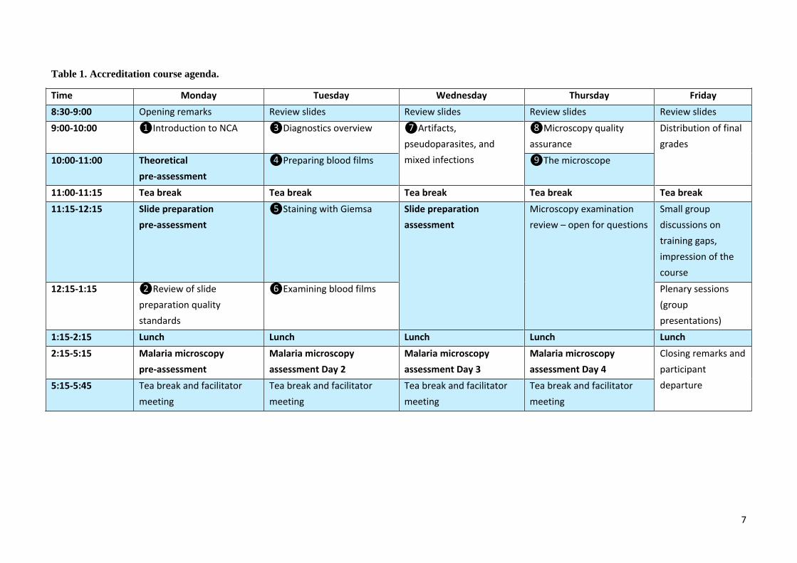
Table 1 on the following page outlines the course agenda and timetable.
4.7 Calendar of activities for preparation
A well-conceived work plan and calendar outlined with clear roles and responsibilities is essential for
staying organized and prepared. Each facilitator should consult both the work plan and calendar to see
which activities he/she was assigned.
7
Table 1. Accreditation course agenda.
Time Monday Tuesday Wednesday Thursday Friday
8:30-9:00 Opening remarks Review slides Review slides Review slides Review slides
9:00-10:00 ❶Introduction to NCA ❸Diagnostics overview ❼Artifacts,
pseudoparasites, and
mixed infections
❽Microscopy quality
assurance
Distribution of final
grades
10:00-11:00 Theoretical
pre-assessment
❹Preparing blood films ❾The microscope
11:00-11:15 Tea break Tea break Tea break Tea break Tea break
11:15-12:15 Slide preparation
pre-assessment
❺Staining with Giemsa Slide preparation
assessment
Microscopy examination
review – open for questions
Small group
discussions on
training gaps,
impression of the
course
12:15-1:15 ❷Review of slide
preparation quality
standards
❻Examining blood films Plenary sessions
(group
presentations)
1:15-2:15 Lunch Lunch Lunch Lunch Lunch
2:15-5:15 Malaria microscopy
pre-assessment
Malaria microscopy
assessment Day 2
Malaria microscopy
assessment Day 3
Malaria microscopy
assessment Day 4
Closing remarks and
participant
departure 5:15-5:45
Tea break and facilitator
meeting
Tea break and facilitator
meeting
Tea break and facilitator
meeting
Tea break and facilitator
meeting
8
5.0 Course monitoring and evaluation
This course utilizes a 360-degree approach to assessment, meaning the course participants are evaluated as
well as the facilitators.
Overall evaluation of course participants
Pre-assessments (theory and practical).
Observation by facilitators.
Daily competency assessments to measure performance in slide reading during the course.
Post-assessments.
Course evaluation by participants
At the end of the course, the participants will be asked to fill out a standard evaluation form. The evaluation is
conducted anonymously and addresses both technical and logistical issues.

9
6.0 National competency assessment course structure
The NCA can be opened by a representative from the central level; for example, from the National Malaria
Elimination Center or the National Reference Laboratory. The opening statement should be given by a well-
respected opinion leader who is recognized nationally in the field of malaria diagnostics in order to increase
the motivation of the participants. Additionally, this individual should effectively express the strong interest
and commitment of the Minister of Health in the NCA program.
6.1 Training methods
The facilitators will need to become familiar with the different training methods used during this course. Each
method will require a specific approach in terms of logistics and preparation.2 It is expected that each
facilitator will apply the relevant communication skills and knowledge to each training/assessment method.
The major learning tools used during this course are as follows:
Pre-assessments: To measure baseline knowledge of theory and slide preparation.
Learning units: Composed of a lecture using a corresponding PowerPoint presentation.
Competency assessments: For preparation of malaria blood films and malaria and malaria microscopy.
This activity requires advanced planning during facilitator meetings.
Facilitator meetings: To review the day’s activities, troubleshoot any problems, plan for the next day’s
activities, assemble slide sets, and grade the daily microscopy assessment.
Post-assessments
6.2 Course structure overview
The NCA will be conducted over a period of five consecutive days and is composed of both learning and
assessment modules. The learning modules will be didactic and focus on the baseline knowledge participants
possess and seek to improve upon. Pre-assessment of relevant malaria theoretical knowledge, blood film
preparation, and malaria microscopy will occur on Day 1. Days 2 through 4 will comprise both learning units
(Annex 2) and assessment modules for preparing malaria blood films and malaria microscopy assessment of
skills (detection, species identification, and parasite quantitation). Final grades will be presented on Day 5,
and group discussions will be held on how to implement lessons learned once participants return to their field
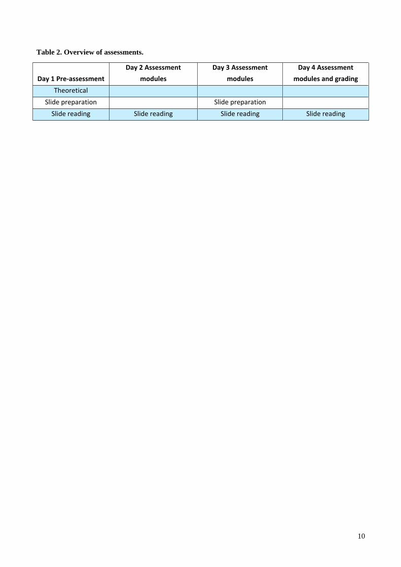
work. Table 2 below provides an overview of assessments that will require planning, such as preparation of
slides or reagents the day before.
2 World Health Organization, Special Programme for Research and Training in Tropical Diseases. Course Facilitators
Guide: Training of Trainers on Malaria Diagnostic Testing using RDTs. Draft; May 2013.
10
Table 2. Overview of assessments.
Day 1 Pre-assessment
Day 2 Assessment
modules
Day 3 Assessment
modules
Day 4 Assessment
modules and grading
Theoretical
Slide preparation Slide preparation
Slide reading Slide reading Slide reading Slide reading
11
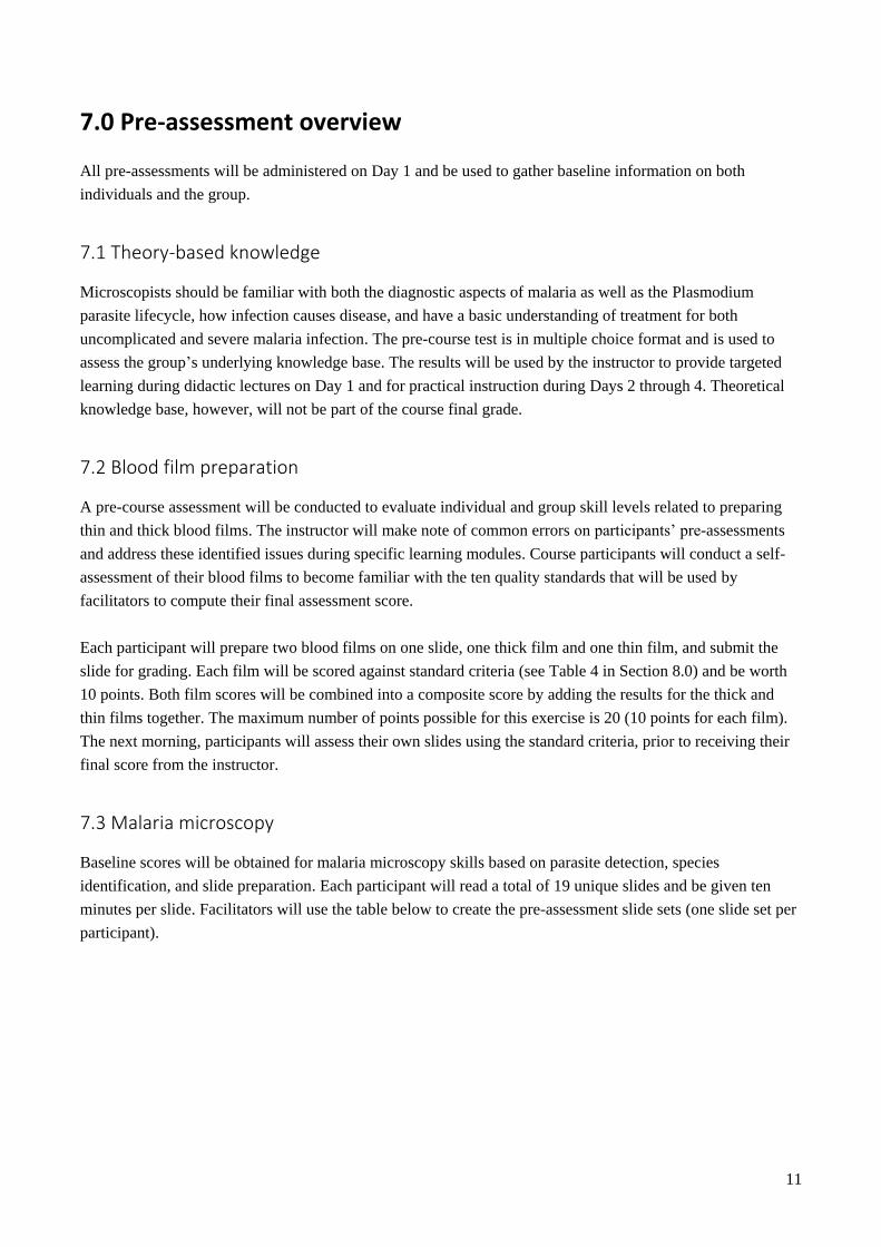
7.0 Pre-assessment overview
All pre-assessments will be administered on Day 1 and be used to gather baseline information on both
individuals and the group.
7.1 Theory-based knowledge
Microscopists should be familiar with both the diagnostic aspects of malaria as well as the Plasmodium
parasite lifecycle, how infection causes disease, and have a basic understanding of treatment for both
uncomplicated and severe malaria infection. The pre-course test is in multiple choice format and is used to
assess the group’s underlying knowledge base. The results will be used by the instructor to provide targeted
learning during didactic lectures on Day 1 and for practical instruction during Days 2 through 4. Theoretical
knowledge base, however, will not be part of the course final grade.
7.2 Blood film preparation
A pre-course assessment will be conducted to evaluate individual and group skill levels related to preparing
thin and thick blood films. The instructor will make note of common errors on participants’ pre-assessments
and address these identified issues during specific learning modules. Course participants will conduct a self-
assessment of their blood films to become familiar with the ten quality standards that will be used by
facilitators to compute their final assessment score.
Each participant will prepare two blood films on one slide, one thick film and one thin film, and submit the
slide for grading. Each film will be scored against standard criteria (see Table 4 in Section 8.0) and be worth
10 points. Both film scores will be combined into a composite score by adding the results for the thick and
thin films together. The maximum number of points possible for this exercise is 20 (10 points for each film).
The next morning, participants will assess their own slides using the standard criteria, prior to receiving their
final score from the instructor.
7.3 Malaria microscopy
Baseline scores will be obtained for malaria microscopy skills based on parasite detection, species
identification, and slide preparation. Each participant will read a total of 19 unique slides and be given ten
minutes per slide. Facilitators will use the table below to create the pre-assessment slide sets (one slide set per
participant).

12
Table 3. Pre-assessment slide set composition.
Pre-assessment slide set
#
unique
slides
# slides parasite
detection Species ID # of counting slides
Plasmodium falciparum (Pf)
Plasmodium vivax (Pv)
Placental malaria (Pm)
9 5 slides of:
2 Pf 50-100 p/µl
1 Pf 100-200 p/µl
1 Pv (density N/A)
1 Pm (density N/A)
5 slides of:
Same as in previous
column
4 slides of:
1 Pf 500-1,000 p/µl
1 Pf 5,000-10,000 p/µl
1 Pf 10,000-50,000 p/µl
1 Pf 50,000-100,000 p/µl
Negative 7 7 - -
Mixed:
Plasmodium falciparum (Pf)
Plasmodium ovale (Po)
3 3 3 slides of:
Pf/Po (density N/A)
-
Total # unique slides 19 15 8 4
Results will be returned the following morning for a detailed review using the microscope. Participants also
will be given one hour to review their answers against the slides.
13
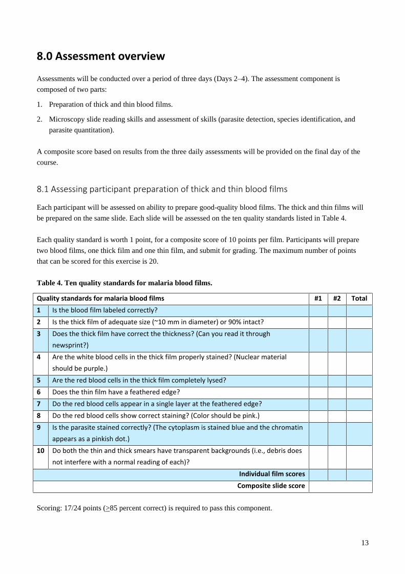
8.0 Assessment overview
Assessments will be conducted over a period of three days (Days 2‒4). The assessment component is
composed of two parts:
1. Preparation of thick and thin blood films.
2. Microscopy slide reading skills and assessment of skills (parasite detection, species identification, and
parasite quantitation).
A composite score based on results from the three daily assessments will be provided on the final day of the
course.
8.1 Assessing participant preparation of thick and thin blood films
Each participant will be assessed on ability to prepare good-quality blood films. The thick and thin films will
be prepared on the same slide. Each slide will be assessed on the ten quality standards listed in Table 4.
Each quality standard is worth 1 point, for a composite score of 10 points per film. Participants will prepare
two blood films, one thick film and one thin film, and submit for grading. The maximum number of points
that can be scored for this exercise is 20.
Table 4. Ten quality standards for malaria blood films.
Quality standards for malaria blood films #1 #2 Total
1 Is the blood film labeled correctly?
2 Is the thick film of adequate size (~10 mm in diameter) or 90% intact?
3 Does the thick film have correct the thickness? (Can you read it through
newsprint?)
4 Are the white blood cells in the thick film properly stained? (Nuclear material
should be purple.)
5 Are the red blood cells in the thick film completely lysed?
6 Does the thin film have a feathered edge?
7 Do the red blood cells appear in a single layer at the feathered edge?
8 Do the red blood cells show correct staining? (Color should be pink.)
9 Is the parasite stained correctly? (The cytoplasm is stained blue and the chromatin
appears as a pinkish dot.)
10 Do both the thin and thick smears have transparent backgrounds (i.e., debris does
not interfere with a normal reading of each)?
Individual film scores
Composite slide score
Scoring: 17/24 points (>85 percent correct) is required to pass this component.
14
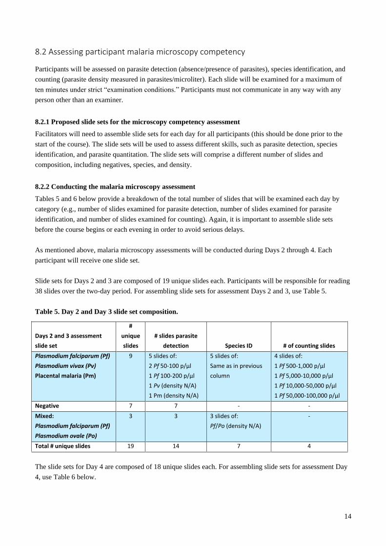
8.2 Assessing participant malaria microscopy competency
Participants will be assessed on parasite detection (absence/presence of parasites), species identification, and
counting (parasite density measured in parasites/microliter). Each slide will be examined for a maximum of
ten minutes under strict “examination conditions.” Participants must not communicate in any way with any
person other than an examiner.
8.2.1 Proposed slide sets for the microscopy competency assessment
Facilitators will need to assemble slide sets for each day for all participants (this should be done prior to the
start of the course). The slide sets will be used to assess different skills, such as parasite detection, species
identification, and parasite quantitation. The slide sets will comprise a different number of slides and
composition, including negatives, species, and density.
8.2.2 Conducting the malaria microscopy assessment
Tables 5 and 6 below provide a breakdown of the total number of slides that will be examined each day by
category (e.g., number of slides examined for parasite detection, number of slides examined for parasite
identification, and number of slides examined for counting). Again, it is important to assemble slide sets
before the course begins or each evening in order to avoid serious delays.
As mentioned above, malaria microscopy assessments will be conducted during Days 2 through 4. Each
participant will receive one slide set.
Slide sets for Days 2 and 3 are composed of 19 unique slides each. Participants will be responsible for reading
38 slides over the two-day period. For assembling slide sets for assessment Days 2 and 3, use Table 5.
Table 5. Day 2 and Day 3 slide set composition.
Days 2 and 3 assessment
slide set
#
unique
slides
# slides parasite
detection Species ID # of counting slides
Plasmodium falciparum (Pf)
Plasmodium vivax (Pv)
Placental malaria (Pm)
9 5 slides of:
2 Pf 50-100 p/µl
1 Pf 100-200 p/µl
1 Pv (density N/A)
1 Pm (density N/A)
5 slides of:
Same as in previous
column
4 slides of:
1 Pf 500-1,000 p/µl
1 Pf 5,000-10,000 p/µl
1 Pf 10,000-50,000 p/µl
1 Pf 50,000-100,000 p/µl
Negative 7 7 - -
Mixed:
Plasmodium falciparum (Pf)
Plasmodium ovale (Po)
3 3 3 slides of:
Pf/Po (density N/A)
-
Total # unique slides 19 14 7 4
The slide sets for Day 4 are composed of 18 unique slides each. For assembling slide sets for assessment Day
4, use Table 6 below.
15
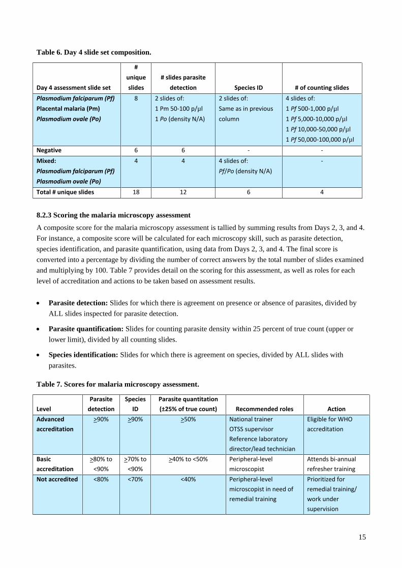
Table 6. Day 4 slide set composition.
Day 4 assessment slide set
#
unique
slides
# slides parasite
detection Species ID # of counting slides
Plasmodium falciparum (Pf)
Placental malaria (Pm)
Plasmodium ovale (Po)
8 2 slides of:
1 Pm 50-100 p/µl
1 Po (density N/A)
2 slides of:
Same as in previous
column
4 slides of:
1 Pf 500-1,000 p/µl
1 Pf 5,000-10,000 p/µl
1 Pf 10,000-50,000 p/µl
1 Pf 50,000-100,000 p/µl
Negative 6 6 - -
Mixed:
Plasmodium falciparum (Pf)
Plasmodium ovale (Po)
4 4 4 slides of:
Pf/Po (density N/A)
-
Total # unique slides 18 12 6 4
8.2.3 Scoring the malaria microscopy assessment
A composite score for the malaria microscopy assessment is tallied by summing results from Days 2, 3, and 4.
For instance, a composite score will be calculated for each microscopy skill, such as parasite detection,
species identification, and parasite quantification, using data from Days 2, 3, and 4. The final score is
converted into a percentage by dividing the number of correct answers by the total number of slides examined
and multiplying by 100. Table 7 provides detail on the scoring for this assessment, as well as roles for each
level of accreditation and actions to be taken based on assessment results.
Parasite detection: Slides for which there is agreement on presence or absence of parasites, divided by
ALL slides inspected for parasite detection.
Parasite quantification: Slides for counting parasite density within 25 percent of true count (upper or
lower limit), divided by all counting slides.
Species identification: Slides for which there is agreement on species, divided by ALL slides with
parasites.
Table 7. Scores for malaria microscopy assessment.
Level
Parasite
detection
Species
ID
Parasite quantitation
(±25% of true count) Recommended roles Action
Advanced
accreditation
>90% >90% >50% National trainer
OTSS supervisor
Reference laboratory
director/lead technician
Eligible for WHO
accreditation
Basic
accreditation
>80% to
<90%
>70% to
<90%
>40% to <50% Peripheral-level
microscopist
Attends bi-annual
refresher training
Not accredited <80% <70% <40% Peripheral-level
microscopist in need of
remedial training
Prioritized for
remedial training/
work under
supervision
16
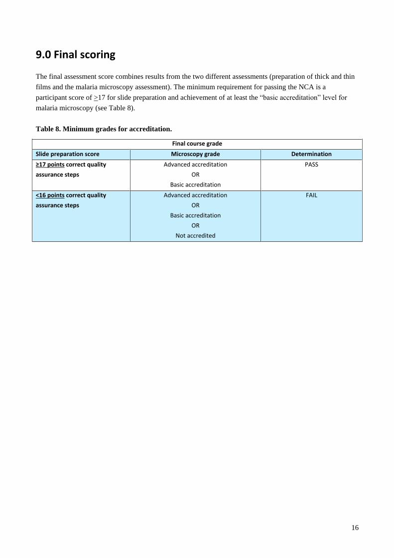
9.0 Final scoring
The final assessment score combines results from the two different assessments (preparation of thick and thin
films and the malaria microscopy assessment). The minimum requirement for passing the NCA is a
participant score of >17 for slide preparation and achievement of at least the “basic accreditation” level for
malaria microscopy (see Table 8).
Table 8. Minimum grades for accreditation.
Final course grade
Slide preparation score Microscopy grade Determination
≥17 points correct quality
assurance steps
Advanced accreditation
OR
Basic accreditation
PASS
<16 points correct quality
assurance steps
Advanced accreditation
OR
Basic accreditation
OR
Not accredited
FAIL
17
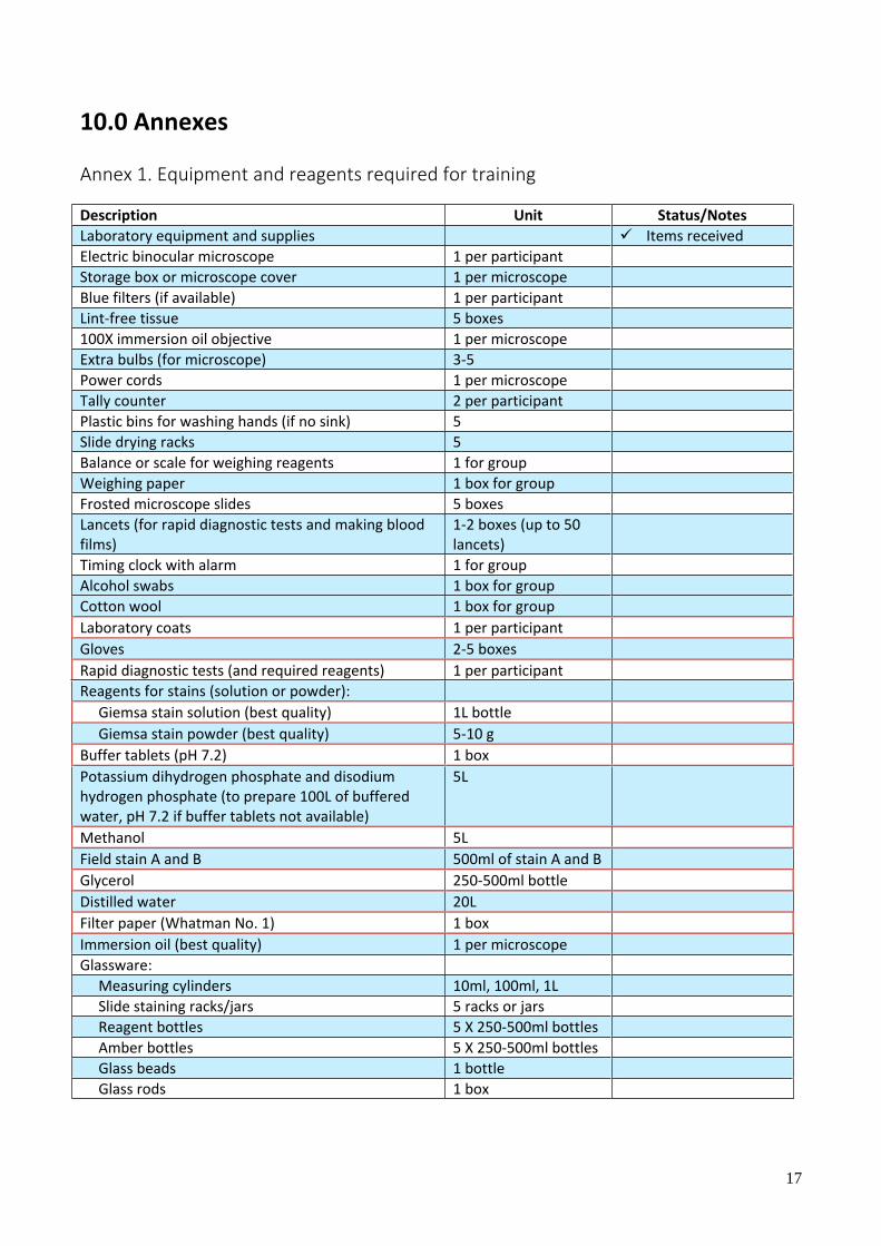
10.0 Annexes
Annex 1. Equipment and reagents required for training
Description Unit Status/Notes
Laboratory equipment and supplies Items received
Electric binocular microscope 1 per participant
Storage box or microscope cover 1 per microscope
Blue filters (if available) 1 per participant
Lint-free tissue 5 boxes
100X immersion oil objective 1 per microscope
Extra bulbs (for microscope) 3-5
Power cords 1 per microscope
Tally counter 2 per participant
Plastic bins for washing hands (if no sink) 5
Slide drying racks 5
Balance or scale for weighing reagents 1 for group
Weighing paper 1 box for group
Frosted microscope slides 5 boxes
Lancets (for rapid diagnostic tests and making blood films)
1-2 boxes (up to 50 lancets)
Timing clock with alarm 1 for group
Alcohol swabs 1 box for group
Cotton wool 1 box for group
Laboratory coats 1 per participant
Gloves 2-5 boxes
Rapid diagnostic tests (and required reagents) 1 per participant
Reagents for stains (solution or powder):
Giemsa stain solution (best quality) 1L bottle
Giemsa stain powder (best quality) 5-10 g
Buffer tablets (pH 7.2) 1 box
Potassium dihydrogen phosphate and disodium hydrogen phosphate (to prepare 100L of buffered water, pH 7.2 if buffer tablets not available)
5L
Methanol 5L
Field stain A and B 500ml of stain A and B
Glycerol 250-500ml bottle
Distilled water 20L
Filter paper (Whatman No. 1) 1 box
Immersion oil (best quality) 1 per microscope
Glassware:
Measuring cylinders 10ml, 100ml, 1L
Slide staining racks/jars 5 racks or jars
Reagent bottles 5 X 250-500ml bottles
Amber bottles 5 X 250-500ml bottles
Glass beads 1 bottle
Glass rods 1 box
18
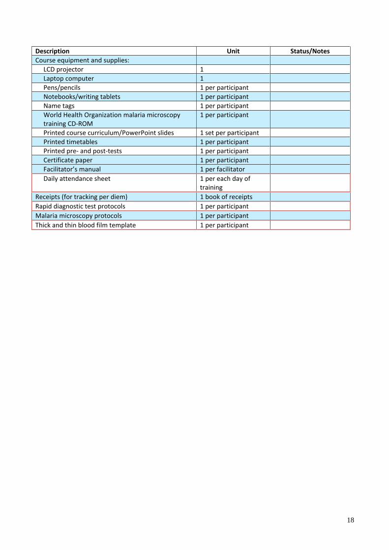
Description Unit Status/Notes
Course equipment and supplies:
LCD projector 1
Laptop computer 1
Pens/pencils 1 per participant
Notebooks/writing tablets 1 per participant
Name tags 1 per participant
World Health Organization malaria microscopy training CD-ROM
1 per participant
Printed course curriculum/PowerPoint slides 1 set per participant
Printed timetables 1 per participant
Printed pre- and post-tests 1 per participant
Certificate paper 1 per participant
Facilitator’s manual 1 per facilitator
Daily attendance sheet 1 per each day of training
Receipts (for tracking per diem) 1 book of receipts
Rapid diagnostic test protocols 1 per participant
Malaria microscopy protocols 1 per participant
Thick and thin blood film template 1 per participant
19
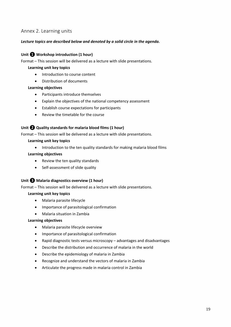
Annex 2. Learning units
Lecture topics are described below and denoted by a solid circle in the agenda.
Unit ❶ Workshop introduction (1 hour)
Format – This session will be delivered as a lecture with slide presentations.
Learning unit key topics
Introduction to course content
Distribution of documents
Learning objectives
Participants introduce themselves
Explain the objectives of the national competency assessment
Establish course expectations for participants
Review the timetable for the course
Unit ❷ Quality standards for malaria blood films (1 hour)
Format – This session will be delivered as a lecture with slide presentations.
Learning unit key topics
Introduction to the ten quality standards for making malaria blood films
Learning objectives
Review the ten quality standards
Self-assessment of slide quality
Unit ❸ Malaria diagnostics overview (1 hour)
Format – This session will be delivered as a lecture with slide presentations.
Learning unit key topics
Malaria parasite lifecycle
Importance of parasitological confirmation
Malaria situation in Zambia
Learning objectives
Malaria parasite lifecycle overview
Importance of parasitological confirmation
Rapid diagnostic tests versus microscopy – advantages and disadvantages
Describe the distribution and occurrence of malaria in the world
Describe the epidemiology of malaria in Zambia
Recognize and understand the vectors of malaria in Zambia
Articulate the progress made in malaria control in Zambia
20
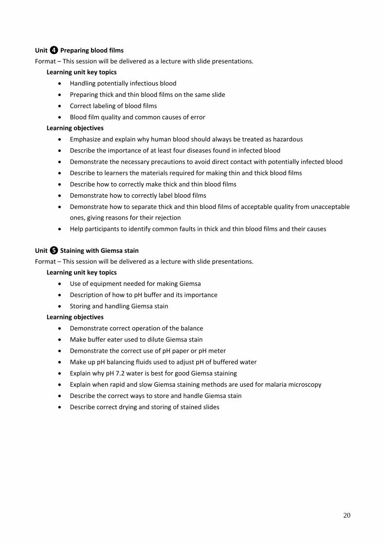
Unit ❹ Preparing blood films
Format – This session will be delivered as a lecture with slide presentations.
Learning unit key topics
Handling potentially infectious blood
Preparing thick and thin blood films on the same slide
Correct labeling of blood films
Blood film quality and common causes of error
Learning objectives
Emphasize and explain why human blood should always be treated as hazardous
Describe the importance of at least four diseases found in infected blood
Demonstrate the necessary precautions to avoid direct contact with potentially infected blood
Describe to learners the materials required for making thin and thick blood films
Describe how to correctly make thick and thin blood films
Demonstrate how to correctly label blood films
Demonstrate how to separate thick and thin blood films of acceptable quality from unacceptable
ones, giving reasons for their rejection
Help participants to identify common faults in thick and thin blood films and their causes
Unit ❺ Staining with Giemsa stain
Format – This session will be delivered as a lecture with slide presentations.
Learning unit key topics
Use of equipment needed for making Giemsa
Description of how to pH buffer and its importance
Storing and handling Giemsa stain
Learning objectives
Demonstrate correct operation of the balance
Make buffer eater used to dilute Giemsa stain
Demonstrate the correct use of pH paper or pH meter
Make up pH balancing fluids used to adjust pH of buffered water
Explain why pH 7.2 water is best for good Giemsa staining
Explain when rapid and slow Giemsa staining methods are used for malaria microscopy
Describe the correct ways to store and handle Giemsa stain
Describe correct drying and storing of stained slides

21
Unit ❻ Examining blood films
Format – This session will be delivered as a lecture with slide presentations.
Learning unit key topics
Normal blood components
Methods for slide examination
Recognizing and identifying other blood parasites
Learning objectives
List the four major components of normal blood
Demonstrate each method used for examining a thick and thin blood film for malaria parasites
Recognize and classify the normal components of blood
Name the main parts of a white blood cell
Unit ❼ Artifacts, pseudoparasites, and mixed infections
Format – This session will be delivered as a lecture with slide presentations.
Learning unit key topics
Recognize common artifacts
Recognize pseudoparasites
Recognize and identify other blood parasites
Learning objectives
Recognition of artifacts: stain deposits, dust, salts, scratches on slides
Recognition of pseudoparasites: fungi, bacteria
Recognition of mixed infections: thick and thin blood films
Recognition of other blood parasites: Borrelia species, trypanosomes, microfilariae, differences
from malaria parasites
Unit ❽ Microscopy quality assurance
Format – This session will be delivered as a lecture with slide presentations.
Learning unit key topics
Supportive supervision
Quality assurance
Quality control
Learning objectives
Explain the importance of supervision
Explain ways in which work can be supervised
Describe what must be provided for supervisors to allow them to supervise their work effectively
Describe methods of internal quality control for malaria microscopy
22
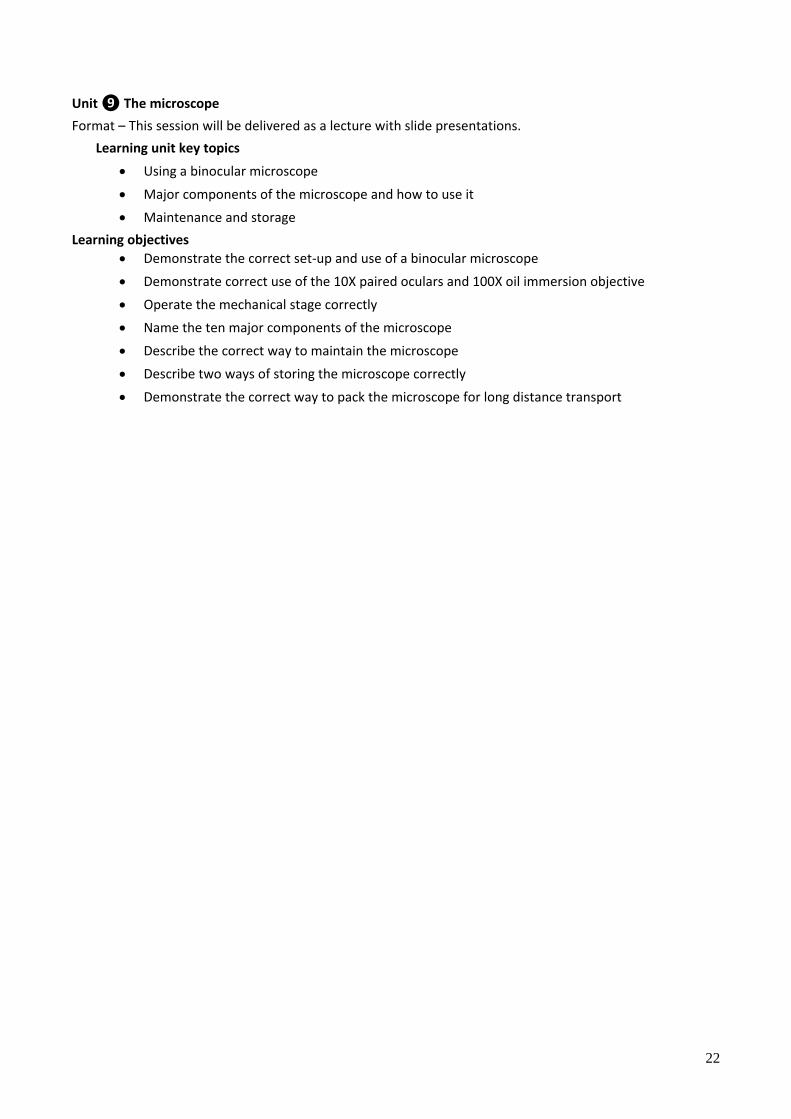
Unit ❾ The microscope
Format – This session will be delivered as a lecture with slide presentations.
Learning unit key topics
Using a binocular microscope
Major components of the microscope and how to use it
Maintenance and storage
Learning objectives
Demonstrate the correct set-up and use of a binocular microscope
Demonstrate correct use of the 10X paired oculars and 100X oil immersion objective
Operate the mechanical stage correctly
Name the ten major components of the microscope
Describe the correct way to maintain the microscope
Describe two ways of storing the microscope correctly
Demonstrate the correct way to pack the microscope for long distance transport
23
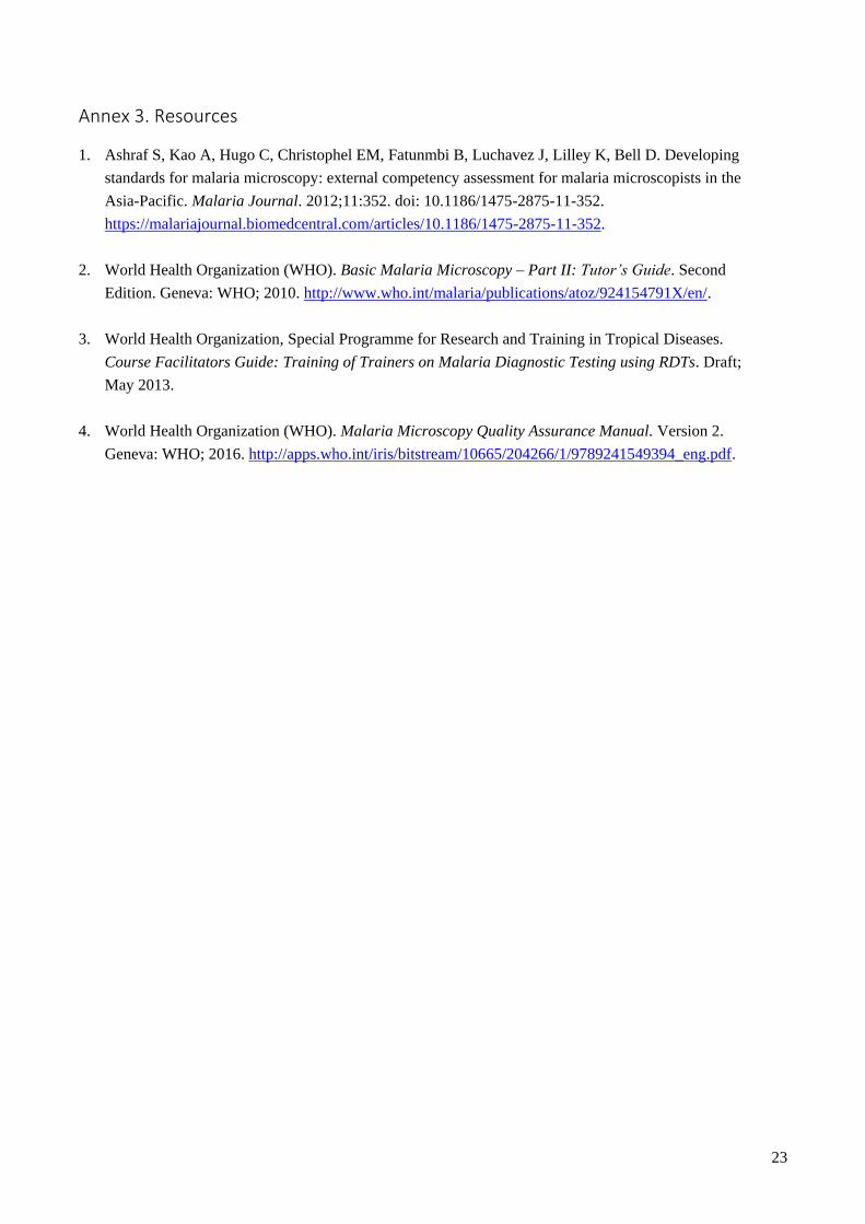
Annex 3. Resources
1. Ashraf S, Kao A, Hugo C, Christophel EM, Fatunmbi B, Luchavez J, Lilley K, Bell D. Developing
standards for malaria microscopy: external competency assessment for malaria microscopists in the
Asia-Pacific. Malaria Journal. 2012;11:352. doi: 10.1186/1475-2875-11-352.
https://malariajournal.biomedcentral.com/articles/10.1186/1475-2875-11-352.
2. World Health Organization (WHO). Basic Malaria Microscopy ‒ Part II: Tutor’s Guide. Second
Edition. Geneva: WHO; 2010. http://www.who.int/malaria/publications/atoz/924154791X/en/.
3. World Health Organization, Special Programme for Research and Training in Tropical Diseases.
Course Facilitators Guide: Training of Trainers on Malaria Diagnostic Testing using RDTs. Draft;
May 2013.
4. World Health Organization (WHO). Malaria Microscopy Quality Assurance Manual. Version 2.
Geneva: WHO; 2016. http://apps.who.int/iris/bitstream/10665/204266/1/9789241549394_eng.pdf.