Embed Size (px)
Citation preview
Nanotube Functionalization and Therapeutic Applications
by Alberto Bianco
Immunologie et Chimie Thérapeutiques, CNRS, Strasbourg, France
NanoSOFT (Roscoff), 21-25 May 2007
Immunologie et Chimie Thérapeutiques

Carbone amorphe Graphite Diamant
Fullerène Nanotube de carbone
The Allotropic Forms of Carbon

Zigzag direction
Armchair direction
STRIP OF A GRAPHENE SHEET ROLLED INTO A TUBE
Carbon Nanotube Folding
Movies from: http://www.photon.t.u-tokyo.ac.jp/~maruyama/nanotube.html

Types of Carbon Nanotubes
Single-walled carbon nanotube (SWNT) presents only one graphene layer
Multi-walled carbon nanotube (MWNT) presents several graphitic concentric layers
MWNTSWNT
10 µmHUMAN HAIR
MWNT
0.1 µm
250 nm200 nm
Ø = 1.4 - 100 nmL = 1 - several µm
Ø = 0.4 - 2 nmL = 20 - 1000 nm
BundlesØ = 10 - 30 nmL = 1 - 50 µm

Organic Functionalization of Carbon Nanotubes
CO2H
HNO3/H2SO4
HO2CCO2H
HO2C
CO2H
CO2H
Functionalization using the oxidation/cut method

CO2H
HO2CCO2H
HO2C
CO2H
CO2H
CO2-R
R-O2CCO2-R
R-O2C
CO2-R
CO2-RR-X
Organic Functionalization of Carbon Nanotubes
Functionalization using the oxidation/cut method
Functionalization using the addition/cycloaddition approach
R-X
R R
R
R
RR

101 103 104102 10510-1 107 108106
Nanometers
Water
Glucose
Antibody
Virus
Bacteria
Tennis ball
O
H
HO
H
HO
H
OH
OHHH
HO
Nanoscale devices are of the same scale of biologically important molecules
The Right Size of Nano-objects
Cancer cell
Nanodevices:• Nanoparticles• Dendrimers• Quantum Dots• Carbon nanotubes

- Carbon nanotubes as substrates for neuronal and cell growth
- Carbon nanotubes for tissue engineering and implants
- Carbon nanotubes as biosensors for sugars, antigens, proteins, antibodies
- Carbon nanotubes as substrates for engineered cell membrane surfaces
- Carbon nanotube cell uptake
- Carbon nanotubes for the delivery of therapeutic molecules
Biomedical Applications of Carbon Nanotubes

Since carbon nanotubes are metallic or semi-conductors, can they help to connect neurons which do not communicate because damaged?
Courtesy of M. Prato and L. Ballerini
Neuronal network Carbon nanotube network
Substrates for Neuronal and Cell Growth

Substrates for Neuronal and Cell Growth
Lovat V et al. Nano Lett. 2005, 5, 1107
MWNT remain adherent to the glass surface in the condition of cell culture
Neonatal hyppocampal neurons grow on the dispersed MWNT, with dendrites and axons extended across the tubes
Neurites travel in close contact with the nanotubes
Spontaneous postsynaptic current (PSC) shows the formation of functional synapses and it is indicative of neuronal network efficiency: neurons grown on the CNT display a 6-fold increase on the frequency of spontaneous PSC
Growing neuronal circuits on CNT grids promote an increase in the network operation
NCH3
CH2(CH2)4CH3
350 °CCH3(CH2)5CHO
CH3NHCH2COOH130 °C, DMF

Substrates for Neuronal and Cell Growth
Electric events are not related to an increase of surviving neurons in the presence of CNT
Cell morphology (A-D, G, H) does not present differences when the cell are cultured on glass or on CNT
Control
CNT
Density of neurons (E) and the number of neurite/cell (F) are equal when the cells are cultured on glass or on CNT

Carbon Nanotubes for Tissue Engineering
MacDonald R et al. J. Biomed. Mater. Res. 74A 2005, 489
Collagen CNT
CNT incorporated into collagen fibrils
Mechanical properties of carbon nanotubes
- Composite materials
- Reinforced fibers
- Collagen matrix with embedded CNT
Why collagen (Type I)?
- Important physical and biochemical functions
- Promise as matrix for regenerative medicine
Applications as scaffold for
- Tissue engineering
- Biosensor components
- Useful medical devices (to provide electrical signal for cardiac muscle)
Gelation and formation of a cell-seeded tissue construct

Analysis of the morphology of the cells embedded within collagen-CNT gels after 3 days : no cell morphology alteration and cell viability was above 85% along 7 days
(Red: nucleus; green actin cytoskeleton)
SEM analysis of collagen-CNT matrices (0.4 wt% SWNT) : presence of fine fibers (arrowed) distinct from the typical banded fibers of collagen. CNT strongly interact with collagen forming intersections and blends.
Carbon Nanotubes for Tissue Engineering

Carbon nanotubes as biosensors for sugars, antigens, proteins, antibodies

Electronic Sensors for the Detection of Antibodies
Chen RJ et al. PNAS 2003, 100, 4984
Carbon nanotubes as a platform for:
- Studying of surface/protein or protein/protein binding
- Electrical sensing of specific biomolecules
- Detecting clinically important species (i.e. antibodies)
Non-specific binding (NSB) of proteins
Proteins simply adsorb onto carbon nanotubes via hydrophobic interactions
Verification via AFM
Demonstration of irreversibility using quartz crystal microbalance (QCM) (a decrease of resonance frequency indicates mass uptake by CNT surface; level of detection down to 10 nM)
Similarly, conductance changes (FET) allow to detect protein absorption with a level of detection down to 100 pM

Approach to avoid non-specific binding (NSB) of proteins
Proteins do NOT adsorb onto carbon nanotubes coated with polyethylene (PEO) chains
Verification via AFM
Carbon nanotubes coated with PEO chains form stable water suspensions
No change detected with QCM
No change on conductance
Electronic Sensors for the Detection of Antibodies

Electronic Sensors for the Detection of AntibodiesSpecific bindingbetween biotin-PEOcoated CNT and SA (SA: streptavidin)
QCM detection Electricdetection
Specific binding betweenU1A-PEO coated CNT and10E3 (antigen/antibody)
QCM detection Electricdetection
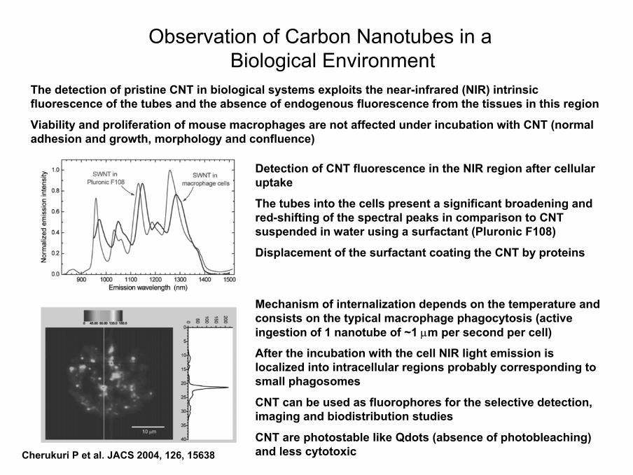
Observation of Carbon Nanotubes in a Biological Environment
Cherukuri P et al. JACS 2004, 126, 15638
The detection of pristine CNT in biological systems exploits the near-infrared (NIR) intrinsic fluorescence of the tubes and the absence of endogenous fluorescence from the tissues in this region
Viability and proliferation of mouse macrophages are not affected under incubation with CNT (normal adhesion and growth, morphology and confluence)
Detection of CNT fluorescence in the NIR region after cellular uptake
The tubes into the cells present a significant broadening and red-shifting of the spectral peaks in comparison to CNT suspended in water using a surfactant (Pluronic F108)
Displacement of the surfactant coating the CNT by proteins
Mechanism of internalization depends on the temperature and consists on the typical macrophage phagocytosis (active ingestion of 1 nanotube of ~1 µm per second per cell)
After the incubation with the cell NIR light emission is localized into intracellular regions probably corresponding to small phagosomes
CNT can be used as fluorophores for the selective detection, imaging and biodistribution studies
CNT are photostable like Qdots (absence of photobleaching) and less cytotoxic

Carbon nanotubes as substrates for engineered cell membrane surfaces

Biomimetic Cell Surface Engineering
Chen X et al. Angew. Chem. Int. Ed. 2004, 43, 6112
Biomimetic approach can be used to bridge nanomaterials and biological systems
Surface modification of carbon nanotubes using glycosylated polymers designed to mimic natural cell surface glycans (mucins)
Integration of carbon nanotubes into physiological conditions
Improvement of biocompatibilty
Receptor-mediated cell-cell recognition studies

Biomimetic Cell Surface Engineering
Chen X et al. JACS 2006, 128, 6292
Mucins, which are present at the cell surface, serve a dual role:
1) Molecular recognition
2) Resistance to biofouling (biofouling is the undesirable accumulation of microorganisms, plants and animals on artificial surfaces)
Do CNT coated with α-GalNAc interact with the receptor at the cell surface?
HPA is a hexavalent lectin capable of cross-linking cells and glycoproteins

Strategy to probe for biological process
Monitoring the variation in a cell’s local environment by following the changes in the electrical, mechanical and optical properties of CNT
Biomimetic Cell Surface Engineering
Interaction between CNT coated with α-GalNAc and the cell receptors
Specificity of the interaction between CNT coated with mucin mimetics and the cell receptors
Cytotoxic effect of the complexes on the cell growth

Biomimetic Bacterial Surface Engineering
Gu L et al. Chem Commun 2005, 874
O
OH
H
H
HO
H
HOHH
O
OH
NH
O= = D-galactose-binding protein
Gal-SWNT E. coliE. coli
Functionalization of CNT with galactose and interaction with galactose-binding protein at the bacterial surface
Capture of pathogenic E. coli cells by sugar-modified carbon nanotubes

Interaction of Pathogens with Antibody-CarbonNanotube Conjugates
Elkin T. et al. ChemBioChem 2005, 6, 640
The development of immuno-nanotubes is based on the conjugation of oxidized CNT to bovine serum albumin (BSA) and direct adsorption of an specific antibody (anti E. coli Ab)
Capture of pathogenic E. coli cells by antibody-modified carbon nanotubes Green E. coli Red Ab-CNT Yellow
overlapping
Immuno-CNT-E. Coli interactions are specific and through the antibody bound to the nanotubesand the antigens on the cell surface
Potential development of CNT devices for rapid and ultrasensitive detection of pathogens
Control

Carbon nanotube cell uptake

Do Carbon Nanotubes Penetrate into the Cells?
OO
NH
N
OO
NH
N
OO
NH
N
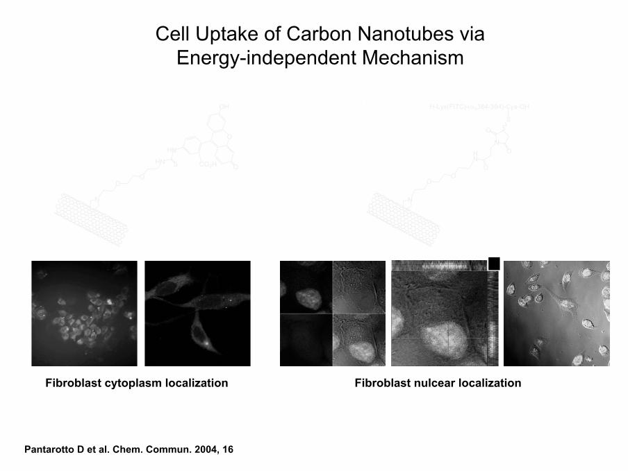
Functionalized carbon nanotubes penetrate into the cells following two mechanisms:
i) via an energy-independent mechanism (passive insertion and diffusion through lipid bilayerof the cell membrane – nanoneedle penetration)(Pantarotto D et al. Chem. Commun. 2004, 16; Angew Chem. Int Ed. 2004, 43, 5242; Cai D et al. Nat. Meth. 2005, 2, 449)
ii) via an energy-dependent mechanism (endocytosis, phagocytosis)
(Kam NWS et al. JACS 2004, 126, 6850; JACS 2005, 127, 6021; Cherukuri P et al. JACS 2004, 126, 15638)
OO
HN
N
NO
O
O
S
H-Lys(FITC)-(αs384-394)-Cys-OH
Cell Uptake of Carbon Nanotubes via Energy-independent Mechanism
OO
N
HN S
HN
CO2H
O
O
OH
Pantarotto D et al. Chem. Commun. 2004, 16
Fibroblast cytoplasm localization Fibroblast nulcear localization

Capacity of Functionalized Carbon Nanotubes to Penetrate Different Cell Types
MOD-K
E. coli C. neo-formansS. cere-visiae
OOHN
O
NH
HN
HO2C
OO OH
S
NO O NH3
+Cl-
N
O
OHO
O
OH OH
OH
OH OH
OHOH
O
OHO
H2NOH
O
OO
OONH
O
NH
HN
HO2C
OO OH
S
HN
N
O
O
HN NH
CO2H
O OHO
S
NO O NH3
+Cl-
NO O
HN
S
HN
HO2C
O
O
OH
Jurkat
MacrophagesLymphocytes BLymphocytes T
Kostarelos K et al. Nature Nanotech. 2007, 2, 108

Cells Incubated at 4°C Cells treated with DNPCells treated with NaN3
Functionalized carbon nanotubes penetrate into the cells at 4°C
Functionalized carbon nanotubes penetrate into the cells in the presence of the inhibitors of endocytosis NaN3 and DNP (2,4-dinitrophenol)
Cell Uptake of Carbon Nanotubes via Energy-independent Mechanism

Pantarotto D et al. Angew. Chem. Int. Ed. 2004, 43, 5242
Direct Visualization of Carbon Nanotubes into the Cells
OO
NH3+Cl-
N

Nanoneedle Cell Penetration of Carbon Nanotubes
CELL MEDIUM
CYTOPLASM
OO
NH3+Cl-
Nchromatin
internalised MWNT
nuclear membrane
Golgi’s complex
Functionalized carbon nanotubes penetrate like nanoneedles piercing the cell membrane without inducing cell death
Cell
Cell
CNT
F
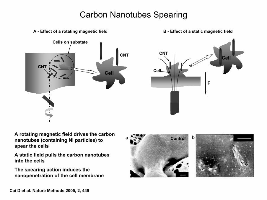
B - Effect of a static magnetic field
Carbon Nanotubes Spearing
Cai D et al. Nature Methods 2005, 2, 449
A - Effect of a rotating magnetic field
Cell
Cells on substate
CNT
CNT
A rotating magnetic field drives the carbon nanotubes (containing Ni particles) to spear the cells
A static field pulls the carbon nanotubesinto the cells
The spearing action induces the nanopenetration of the cell membrane 1 µm
500 nmControl

Kam NWS et al. JACS 2004, 126, 6850
COOH
NHHN
O OHO
S
ONH
NHHN
O
O
Alexa Fluor Streptavidin
NH
O
NH
O
NH
O
OH
O
Carbon nanotubes (100-1000 nm) conjugated to FITC (a) or fluorescent streptavidin (60 kD) via biotin (b)
The protein-CNT non covalent complexes are uptaken by the cells and they are localized into the endosomes (c)
The protein-CNT do not penetrate at 4 °C (d)
The free protein does not penetrate
Penetration is time dependent (after 4 h the fluorescence reaches the maximum) and dose dependent
The penetration is independent of the cell type and it is via endocytosis leading to the accumulation into the cytoplasm
Cell Uptake of Carbon Nanotubes via Energy-dependent Mechanism

Kam NWS et al. JACS 2005, 127, 6021
Cell Uptake of Carbon Nanotubes via Energy-dependent Mechanism
Proteins spontaneously adsorb onto oxidized carbon nanotubes via non specific binding
Non covalent protein-CNT conjugates are transported via endocytosis into different cell lines
After release from the endosomes, the proteins express their biological functions as demonstrated for example by induction of apoptosis (programmed cell death) using cytochrome-c (Cyt-c)
Complexes between CNT and different proteins
CNT BSA
spA Cyt-c

Cell Uptake of Carbon Nanotubes via Energy-dependent Mechanism
CNT affect the transportation of protein cargos into the cells
Proteins are internalized by CNT because they do not intrinsically penetrate into the cells
Protein-CNT are localized into the cytoplasmatic endosomes
No fluorescence is detected into the nucleus
Endocytosis is inhibited at 4°C, where little uptake is observed in comparison to 37°C
Intracellular internalization of protein-CNT
Proteins do not penetrate into the cells
4°C37°C

Carbon nanotubes for the delivery of therapeutic molecules
Functionalized Carbon Nanotubes as New Vectorsfor the Delivery of Therapeutic Molecules
• Functionalization of carbon nanotubes with nucleic acids and proteins• Application on bio-macromolecule delivery
• Functionalization of carbon nanotubes with bioactive peptides• Application on vaccine delivery
• Functionalization of carbon nanotubes with small bioactive molecules• Application on drug delivery
• Formation of supramolecular complexes based on charge interactions• Application on gene delivery

Carbon Nanotube Transporters: DNA
Kam NWS et al. PNAS 2005, 102, 11600
Development of carbon nanotubes for:
- DNA delivery
- Cancer therapy
The biological system are transparent in the near-infrared (NIR) region (700-1100 nm) while carbon nanotubes have a strong optical absorbance in the same spectral region
When DNA functionalized carbon nanotubes penetrate into the cells, NIR laser pulses trigger endosomal rupture and subsequent translocation of DNA into the nucleus
When carbon nanotubes are functionalized with molecules targeting cancer cells a continuous NIR irradiation causes the death of those cells that internalized the nanotubes
Carbon nanotubes (~150 nm) functionalized with a 15-mer ssDNA labeled with Cy3 green dye

DNA-CNT are internalized by the cells and accumulate into the cytoplasm
The penetration mechanism is energy-dependent because at at 4°C the cellular uptake is minimum
Carbon Nanotube Transporters: DNA
Irradiation by applying repeated, short (few secondes) laser NIR pulses provokes the release of DNA from the endosomes and DNA nuclear translocation (yellow color indicates co-localization into the nucleus)
Increase of fluorescence is an indicative of DNA unwrapping and release due to endosomal break
Continuous NIR pulses (808 nm) increase the temperature of the solution eventually triggering cell death

Carbon Nanotube Transporters: Proteins
Kam NWS et al. JACS 2005, 127, 6021
Cytochrome-c (Cyt-c) is a 12 kD protein which induces or activates apoptosis (programmed cell death) when injected into the cells
Cyt-c-CNT complexes are uptaken via endocytosis
The treatment of the cells with chloroquine induces the rupture of the endosomes and the release of the protein into the cytoplasm
Cell apoptosis occurs after Cyt-c migration into the cytoplasm
Carbon nanotubes are able to deliver active proteins which eventually display their biological function
Punctuated fluorescence indicates vesicle localization
Diffused and uniform fluorescence indicates vesicle release by action of chloroquine

Carbon Nanotube for Anticancer Therapy
Irradiation by applying repeated, short laser NIR pulses changes the morphology of the cell that have uptake the CNT-DNA complexes
Cells not incubated with the CNT-DNA complexes are undamaged by NIR pulses
Prolonged irradiation (2 minutes) induces extensive death of cells with CNT-DNA complexes
Cell death is accompanied by CNT aggregation, even visible to the naked eye
Kam NWS et al. PNAS 2005, 102, 11600

Carbon Nanotube for Anticancer Therapy
Cell death provoked by NIR irradiation is exploited for cancer therapy
Carbon nanotubes are functionalized with specific ligands that recognize and target cancer cells
Folate receptors (FR) are markers of cancer cells
Irradiation of overexpressing FR cells = cell dead
Irradiation of normal cells = cell alive
Cancer cells internalize the fluorescente tubes functionalized with folic acid
Normal cells shows minimal internalization because of PEG effect (inertness or blocking of non specific binding)

Liu Z et al. Nature Nanotechology 2007, 2, 47
Carbon nanotubes are functionalized with specific ligands which are recognized by tumor cells
Carbon Nanotubes for Anticancer Therapy

Delivery of peptide-based synthetic vaccines

0.45 nm1.34 nm
1.45 nm
1.36
nm
Molecular model of a helical peptide-CNT conjugate
Carbon Nanotubes as Suitable Scaffold for the Presentation of Antigens to the Immune System
Development of vaccines based on synthetic peptides
- Peptide antigens are poorly immunogenic
- Conjugation to protein carriers (BSA) are necessary to improve the Abs production
- The carrier presents the peptide to the immune system in the correct conformation
- Protein carrier are intrinsically immunogenic
- Generated Abs present low specificity
- Inert carriers system are ideal

Peptide Antigens Conjugated to Carbon Nanotubes for Synthetic Vaccine Delivery
O O
HN
N
7
N
O
O
O
SN
1
NH2
O
O
4) Ac-Cys-GSGVRGDFGSLAPRVARQL
1) Boc-Lys(Boc)-OH, DIC/HOBt in DMF, 3h
2) TFA, 2h
(Ac-Cys-FMDV) in H2O, 9h
Ac-Cys-FMDV
N O N
OO
O
O
O
3) in DMF, 3h
ONH
HN N
O
O
OAc-Cys-FMDV
S
O O
HN
N
6
NO
O
O
ON
O
O
N
O
O
O
S
2) Ac-Cys-GSGVRGDFGSLAPRVARQL (5)
1) (4) in DMF, 3h
(Ac-Cys-FMDV) in H2O, 6h
Ac-Cys-FMDV
Foot-and-Mouth Disease Virus (FMDV)Pantarotto et al. J. Am. Chem. Soc. 2003, 125, 6160 Pantarotto et al. Chem. Biol. 2003, 2003, 10, 961

Immunological Characterization of Peptide-Carbon Nanotubes
Antigenicity is the capacity of a peptide antigen to be specifically recognized by an antibody
The peptide antigen should adopt the correct conformation when bound to the carrier system
The conformation should be the same as in the case of the native structure present in the protein, which contains the peptide epitope
Immunogenicity is the capacity of a peptide antigen to elicit an immune response
Molecular model of the complex between a helical peptide-CNT conjugate and an antibody fragment (Fab: Fragment antigen binding)

Antigenic Characterization of Peptide-Carbon Nanotubes
Carbon nanotubes are a good multi-presentation system since they are able to present the peptide antigen with the correct conformation for the antibody recognition
Carbon nanotubes do not perturb the secondary structure of the peptide
Surface plasmon resonance (SPR) analysis
0
40
80
0 200 400 600Time (s)
RU
120
Bis FMDV-NT 7
Mono FMDV-NT 6
FMDV-peptide
Ac-NT

Immunogenic Characterization of Peptide-Carbon Nanotube Conjugates
FMDV(141-159)-BSA
Control-peptide-BSA
CNT
Coating antigens
- High antibody (Ab) responses
- Bis-conjugate generates higher levels of Abs than the mono-conjugate
- Carbon nanotubes devoid of the peptide are non immunogenic (no antibody production)
0
1
2
3
4
5
6
Freepeptide
Monoconjugate 6
Bisconjugate 7
Log1
0 an
tibod
ytit
er
FMDV(141-159)-BSA Control-peptide-BSA CNT

Delivery of nucleic acids

Delivery of Nucleic Acids by Carbon Nanotubes
Single-stranded DNA sequences are able to wrap around carbon nanotubes
- DNA assisted dispersion and and separation of carbon nanotubes
- Sequence dependent DNA carbon nanotube sorting
Zheng et al. Nat. Mater. 2003, 2, 338
Carbon nanotubes for the delivery of DNA, plasmid DNA, RNA
- Gene transfer applications
- Genetic vaccination

Complexes Based on Positive-negative Charge Interactions
1 base = 1 negative charge Positive charged carbon nanotube
⊕
⊕
⊕ ⊕
⊕
ΘΘΘ
ΘΘ
ΘΘ
Θ ΘΘ
ΘΘ
Θ
ΘΘΘΘ
ΘΘ
Θ Θ
ΘΘ
ΘΘ
++
Cationic macromolecules, such as peptides, dendrimers and liposomes in general achieve effective delivery of DNA leading to pronounced toxic effects at cellular level

Pantarotto et al. Angew. Chem. Int. Ed. 2004, 43, 5242Singh et al. J. Am. Chem. Soc. 2005, 127, 4388
N
NH3+ Cl-
O
O
10 nm 50 nm
III
Potential of Carbon Nanotubes on Gene Delivery
Plasmid DNA condenses on the surface of the cationic carbon nanotubes forming supercoiled and globular-like structures

Gel Electrophoresis of f-CNT:DNA Complexes
- A shift in the gel is indicative of the formation of the complex and of its stability
- A strong decrease in the fluorescence intensity and an increase in the upward shift of the free DNA bands is correlated to an increase of the +/- charge ratio
- Cationic single-walled CNT are not able to fully condense DNA
- Cationic multi-walled CNT are most efficient in condensing DNA
A. SWNT-NH3+
1 2 3 4
OCSC
B. SWNT-Lys-NH3+
1 2 3 4
OCSC
C. MWNT-NH3+
1 2 3 4
OCSC
1:1 2:1 10:1Control 1:1 2:1 10:1Control 1:1 2:1 10:1Control+/-

Delivery and Expression of Plasmid DNA by f-CNT
0.0E+00
2.0E+04
4.0E+04
6.0E+04
8.0E+04
1.0E+05
1.2E+05
Naïve DNAonly
SWNT:DNA1:1
SWNT:DNA2:1
SWNT:DNA4:1
SWNT:DNA6:1
SWNT:DNA8:1
SWNT:DNA10:1
B-g
alEx
pres
sion
(RLU
/wel
l)
- The charge ratio between NH3+ at the SWNT surface and the phosphates of the DNA backbone is a
determinant factor of the resulting levels of gene expression
- SWNT:DNA +/- charge ratios between 2:1 and 6:1 offer 5 to 10 times higher levels of gene expression compared to DNA alone
- This first generation of functionalized carbon nanotubes is less effective for transfection in vitrothan lipid:DNA system

Gao L. et al. ChemBioChem 2006, 7, 239
Characterization and Expression of Plasmid DNA by f-CNT MWNTox-NH3
+ MWNTox-NH3+-DNA
Formation of the complex detected by gel electrophoresis shift
Cationic carbon nanotubes display a reduced cellular toxicity in comparison to other transfection systems
Cationic carbon nanotubes are able to transfect the cells although less efficiently than lipofectamine
The level of transfection is dependent on the +/-charge ratio

Liu Y. et al. Angew. Chem. Int. Ed. 2005, 44, 4782
Delivery of Plasmid DNA Using Carbon Nanotubes
Alternative approach for the solublization and the preparation of cationic carbon nanotubes: conjugation of CNT to polyethylene imine (PEI)
A
B
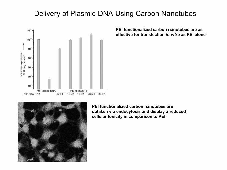
Delivery of Plasmid DNA Using Carbon Nanotubes
PEI functionalized carbon nanotubes are as effective for transfection in vitro as PEI alone
PEI functionalized carbon nanotubes are uptaken via endocytosis and display a reduced cellular toxicity in comparison to PEI

Delivery of therapeutic agents to cells

Zhu Y et al. JACS 2005, 127, 9875
Carbon Nanotubes for Boron Neutron Capture TherapyAn ideal therapy for cancer would be one whereby all tumor cells are selectively destroyed without damaging normal tissues
Most of the cancer cells should be destroyed, either by the treatment itself or with the help from the body's immune system, otherwise the danger exists that the tumor may reestablish itself
The promise of a new experimental cancer therapy with some indication of its potential efficacy has led to work on an approach called boron neutron capture therapy (BNCT)
BNCT is a binary radiation therapy modality that brings together two components that when kept separate have only minor effects on cells. The first component is a stable isotope of boron (boron-10) that can be concentrated in tumor cells by attaching it to tumor seeking compounds. The second is a beam of low-energy neutrons. Boron-10 in or adjacent to the tumor cells disintegrates after capturing a neutron and the high energy heavy charged particles produced destroy only the cells in close proximity to it, primarily cancer cells, leaving adjacent normal cells largely unaffected
Development of water soluble carborane-CNT conjugates able to target cancer cellsHNH
Me
HNH
Me
EtO
EtO
Na+
Na+
N
N
Me
Me
Me
N3NaOH
EtOH
CNT
o-DCB

Mammalian carcinoma cells (EMT6) are transplanted into the right flank of mice
Maximum boron concentration is achieved on the tumor cells after 30 h in comparison to blood and tissues (lung, liver, spleen)
The amount is slightly lower than the desired level for effective BNCT [30 µm(boron)/g(tissue)]
The low concentration on the different organs supports the preferential uptake by tumor cells
The efficient accumulation is guaranteed by the nanotube delivery capacity since the free carboranederivatives generally show no adsorption or retention on tumor cells
The mechanism of targeting of carborane-CNT is unknown
A passive accumulation of macromolecular drugs in tumor cells is favored by an increased vascular permeability and a decrease in lymphatic drainage system in cancer cells
Saline solution DMSO
Carbon Nanotubes for Boron Neutron Capture Therapy

Carbon Nanotubes for Drug Delivery
The conjugation of a drug to carbon nanotubes might have several advantages:
i) Increase of the solubility of the molecule
ii) Decrease of the aggregation phenomena
iii) Improvement of the efficacy owing to the internalization capacity of CNT
iv) Modulation of the drug activity against different types of cells (mammalian, bacterial, fungal)
v) Reduction of the amount of drug administered

Wu W et al. Angew. Chem. Int. Ed. 2005, 44, 6258
Delivery of Antibiotics by Carbon Nanotubes
Amphotericin B (AmB) is a potent antifungal agent for the treatment of chronic fungal infections
AmB is highly toxic for mammalian cells (likely because of the formation of aggregates which reduce the solubility in water)
New approach to carbon nanotube functionalization: preparation of nanotubes carrying one or more therapeutic agent with:
i) Recognition capacity
ii) Optical signal for imaging
iii) Specific targeting
OONH
O
NH
HN
HO2C
OO OH
S
N
O
O OH
O
OHOH
OH
OHOH
OHHO
O
O OH
NH2OH
O
O O
HN
FITC
AmB

0
10
20
30
40
50
60
MWNT
4 (1 µ
g/ml)
% A
popt
otic
and/
or d
ead
cells
MWNT
4 (2 µ
g/ml)
MWNT
4 (5 µ
g/ml)
MWNT
4 (10 µ
g/ml)
MWNT
4 (20 µ
g/ml)
Contro
l
MWNT
4 (40 µ
g/ml)
AmB(1
0 µg/m
l)
N
O
O OH
O
OHOH
OH
OHOH
OHHO
O
O OH
NH2OH
O
O O
OONH
O
NH
HN
HO2C
OO OH
S
HN
The conjugation of AmB to carbon nanotubes clearly reduces the toxic affect of the antifungal agent on mammalian cells (Jurkat cells)
Effect of AmB Conjugated to Carbon Nanotubes

Antifungal Activity of AmB Conjugated to Carbon Nanotubes
*The MIC corresponds to the lowest concentration of compound that inhibited visible growth of the organism. Results given are mean values of two independent determinations performed in duplicate. C.i.: clinical isolate. In this table, the MIC values for MWNT-AmB and SWNT-AmB refer to the concentration of AmB in the conjugates (approximately one third by weight).
Minimum inhibitory concentration (MIC)* in µg/ml
C. parapsilosis ATCC90118
C. famata (c.i.) C. albicans (c.i.)
C. neoformans ATCC90112
S. cerevisiae (c.i)
AmB 20 20 > 80 5 2.5 SWNT-NH3
+ > 80 > 80 > 80 > 80 > 80 MWNT-AmB 1.6 0.8 6.4 0.8 0.8 SWNT-AmB 1.6 1.6 13.8 0.8 1.6
AmB-CNT preserve a high antifungal activity
When equal amount of free and conjugated AmB are administered the CNT conjugates are more potent against certain strains
The reduced mammalian cell cytotoxicity and the increased antimycotic activity can be explained with:
i) Rapid internalization of AmB into the cytoplasm by the CNT reduces the possibility of disruption of the cell membrane
ii) Prevention of aggregation
iii) Increased solubility of the drug
iv) Binding to CNT and the presence of multiple copies of AMB per CNT favor the interaction of the drug with the fungal membrane
An appropriate conjugation increases the effectiveness of a drug (i.e. AmB) decreasing its cytotoxicity

Health and Environment Risks of Nanomaterials

Report: 253 Pages
CARBON NANOTUBES: 157 times
Report: 64 Pages
CARBON NANOTUBES: 17 times
Reports on the Health and Environment Impact of Nanomaterials

Advantages of Functionalized Carbon Nanotubes
• Control of the functionalization
• Lack of immunogenicity
• Reduced toxicity
• No apparent tissue/organ accumulation
Factors to be Carefully Addressed
• Quality of the starting material (carbon nanotubes)
• Control of the preparations
• Long-term toxicity
Conclusions and Perspectives