Embed Size (px)
Citation preview

ARTICLE
Received 31 Oct 2015 | Accepted 9 Nov 2015 | Published 9 Dec 2015
Nanoscale origins of the damage tolerance of thehigh-entropy alloy CrMnFeCoNiZiJiao Zhang1, M.M. Mao1, Jiangwei Wang2, Bernd Gludovatz3, Ze Zhang1, Scott X. Mao1,2, Easo P. George4,
Qian Yu1 & Robert O. Ritchie3,5
Damage tolerance can be an elusive characteristic of structural materials requiring both high
strength and ductility, properties that are often mutually exclusive. High-entropy alloys are of
interest in this regard. Specifically, the single-phase CrMnFeCoNi alloy displays tensile
strength levels of B1 GPa, excellent ductility (B60–70%) and exceptional fracture toughness
(KJIc4200 MPaOm). Here through the use of in situ straining in an aberration-corrected
transmission electron microscope, we report on the salient atomistic to micro-scale
mechanisms underlying the origin of these properties. We identify a synergy of multiple
deformation mechanisms, rarely achieved in metallic alloys, which generates high strength,
work hardening and ductility, including the easy motion of Shockley partials, their interactions
to form stacking-fault parallelepipeds, and arrest at planar slip bands of undissociated
dislocations. We further show that crack propagation is impeded by twinned, nanoscale
bridges that form between the near-tip crack faces and delay fracture by shielding the
crack tip.
DOI: 10.1038/ncomms10143 OPEN
1 Department of Materials Science & Engineering, Center of Electron Microscopy and State Key Laboratory of Silicon Materials, Zhejiang University, Hangzhou310027, China. 2 Department of Mechanical Engineering & Materials Science, University of Pittsburgh, Pittsburgh, Pennsylvania 15261, USA. 3 MaterialsSciences Division, Lawrence Berkeley National Laboratory, Berkeley, California 94720, USA. 4 Institute for Materials, Ruhr University, 44801 Bochum,Germany. 5 Department of Materials Science & Engineering, University of California, Berkeley, California 94720, USA. Correspondence and requests formaterials should be addressed to Q.Y. (email: [email protected]) or to R.O.R. (email: [email protected]).
NATURE COMMUNICATIONS | 6:10143 | DOI: 10.1038/ncomms10143 | www.nature.com/naturecommunications 1
Damage tolerance is arguably the most importantmechanical property of structural materials for manydesign applications, as it defines the combination of
strength and toughness, that is, the ability of a material to resistfracture in the inevitable presence of flaws1–5. However, in manymaterials, particularly metallic alloys, toughness comes at theexpense of strength5. Intrinsic toughness is promoted byextensive plastic deformation, which requires the easy motionand multiplication of dislocations (that can compromisestrength), yet contradictorily requires significant hardening togenerate strength and ductility by resisting dislocation motionand delaying plastic (necking) instabilities2,5,6. Despite this‘conflict’, the ability to resist cracking in metals and alloysunder mechanical loading can be affected by the prevailing modesof plastic deformation and microstructure at the crack tip.Generally, dislocation glide is the primary deformationmechanism at low homologous temperatures in crystallinematerials where dislocations move by locally breaking andreforming crystal bonds. In traditional metallurgy, variousalloying elements are usually added to the principal element todevelop required mechanical properties7,8. The solute atomslocally introduce different crystal bonds with varied strengths andalso cause local lattice distortion, thereby offering resistance todislocation motion; as a result, strength is generally enhanced butductility can be reduced.
Interestingly, a prominent member of the new class ofhigh-entropy alloys (HEAs), CrMnFeCoNi, which forms a face-centered cubic (fcc) solid solution1,9–12, has been found to displayexcellent damage tolerance with tensile strengths of B1 GPa (refs1,10,11), ductilities of B60–70% (refs 1,10,11), and fracturetoughness values exceeding 200 MPaOm (ref. 1), properties thatare comparable to the very best cryogenic steels, specificallyaustenitic stainless steels13 and high-Ni steels14–17. At relativelylow strains (B2%), deformation in CrMnFeCoNi occurs on thenormal fcc {111}o1104 slip systems by planar dislocation glide;both 1
2 110h i type dislocations and stacking faults (SFs) have beenobserved suggesting the splitting of some perfect dislocations intoShockley partials (1
6 112h i type)11. At cryogenic temperatures andat higher strains (B20%), nano-twinning becomes an additionaldeformation mechanism1,11. Twinning has also been observedafter severe plastic deformation at room temperature byhigh-pressure torsion18 as well as after rolling12. It has beenargued that since the combination of dislocation slip andtwinning provides a steady source of strain hardening to inhibitnecking1,10,11, ductility is enhanced together with strength to giveexcellent toughness (by intrinsic toughening)1,5,19. Similararguments have been advanced to explain the extensiveductility of the so-called twinning-induced plasticity (TWIP)steels20–22.
However, the detailed mechanisms and crack tip processesresponsible for the high toughness of this high-entropy alloy haveremained unclear. In this regard, understanding its defectbehaviour, which is affected by such factors as the stacking-faultenergy (SFE) and friction stress, is crucial. In this high-entropyalloy, the SFE is low and has been calculated to beB20–25 mJ m� 2 (ref. 23). In addition, given the apparentlyrandom arrangement of its constituent atoms, which form a solidsolution24 down to the atomic scale18,25, the alloy is expected tooffer a relatively high resistance to dislocation motion (frictionstress), especially at low homologous temperatures where theclosely spaced barriers to dislocation motion (high concentrationsof solute atoms) cannot be easily overcome by thermalactivation10,11,26.
In the present study, we attempt to identify the fundamentalsource of the excellent toughness of this high-entropy alloyat the nanoscale using in situ straining experiments in an
aberration-corrected transmission electron microscope (TEM) toexamine the microstructural evolution in the immediate vicinityof a crack. By imaging as close as within a few hundrednanometres of the crack tip, we identify multiple deformationmechanisms that are activated at different stages of deformationand act synergistically to contribute to the ultrahigh toughness.Deformability is initially afforded by the motion of the Shockleypartial dislocations and the corresponding formation of SFs.However, as the applied stress increases, perfect dislocations startto move but their motion is observed to be extremely difficult.They move in localized bands containing arrays of many closelypacked dislocations. These bands act as strong barriers for partialdislocation motion, which creates an outstanding strengtheningeffect. Strengthening is also caused by parallelepiped sessilevolume defects formed by the interaction of partials slipping ondifferent planes that block the motion of other dislocations.Finally, in the later stages of deformation, we see evidence of thecreation of nanoscale bridges that span the crack-tip region anddeform by nano-twinning, and as such are believed to be strongand ductile. As these nanobridges carry load that would beotherwise used to propagate the crack, they provide a potentmeans to inhibit crack advance (by extrinsic toughening5).
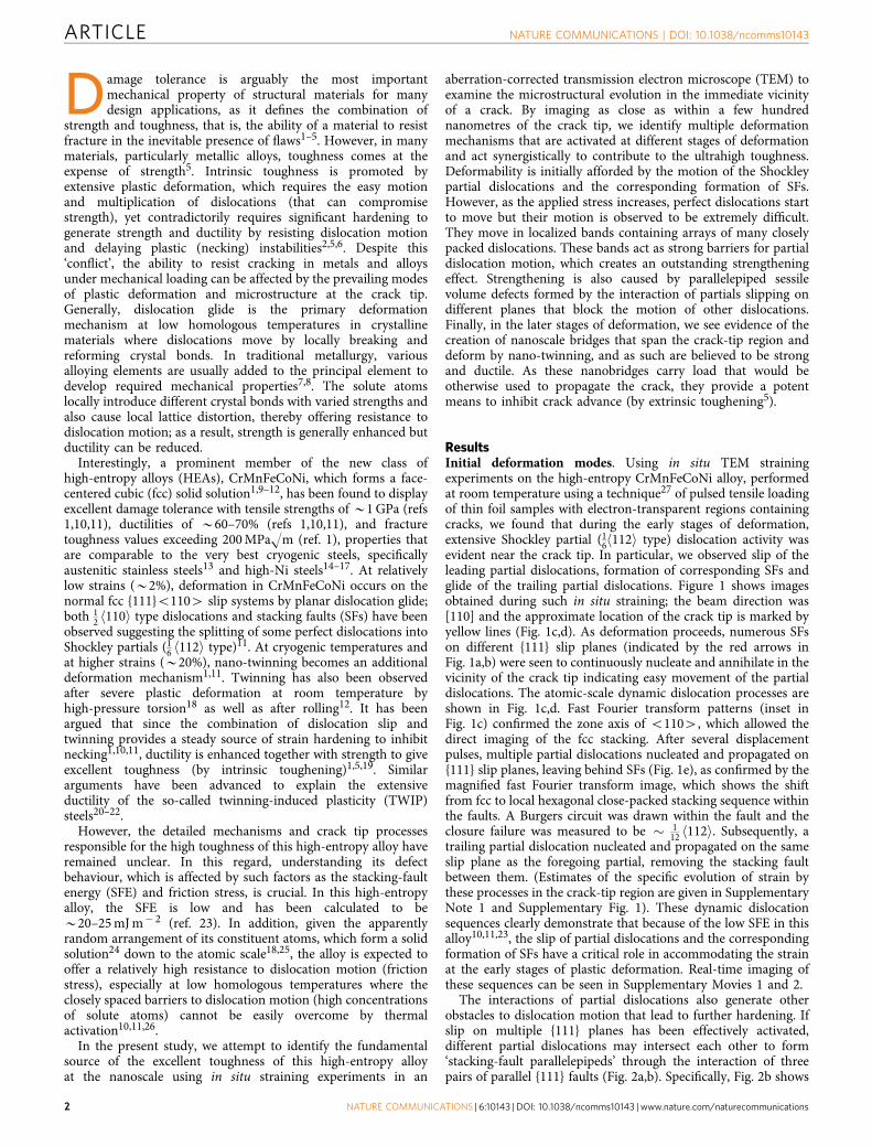
ResultsInitial deformation modes. Using in situ TEM strainingexperiments on the high-entropy CrMnFeCoNi alloy, performedat room temperature using a technique27 of pulsed tensile loadingof thin foil samples with electron-transparent regions containingcracks, we found that during the early stages of deformation,extensive Shockley partial (1
6 112h i type) dislocation activity wasevident near the crack tip. In particular, we observed slip of theleading partial dislocations, formation of corresponding SFs andglide of the trailing partial dislocations. Figure 1 shows imagesobtained during such in situ straining; the beam direction was[110] and the approximate location of the crack tip is marked byyellow lines (Fig. 1c,d). As deformation proceeds, numerous SFson different {111} slip planes (indicated by the red arrows inFig. 1a,b) were seen to continuously nucleate and annihilate in thevicinity of the crack tip indicating easy movement of the partialdislocations. The atomic-scale dynamic dislocation processes areshown in Fig. 1c,d. Fast Fourier transform patterns (inset inFig. 1c) confirmed the zone axis of o1104, which allowed thedirect imaging of the fcc stacking. After several displacementpulses, multiple partial dislocations nucleated and propagated on{111} slip planes, leaving behind SFs (Fig. 1e), as confirmed by themagnified fast Fourier transform image, which shows the shiftfrom fcc to local hexagonal close-packed stacking sequence withinthe faults. A Burgers circuit was drawn within the fault and theclosure failure was measured to be � 1
12 112h i. Subsequently, atrailing partial dislocation nucleated and propagated on the sameslip plane as the foregoing partial, removing the stacking faultbetween them. (Estimates of the specific evolution of strain bythese processes in the crack-tip region are given in SupplementaryNote 1 and Supplementary Fig. 1). These dynamic dislocationsequences clearly demonstrate that because of the low SFE in thisalloy10,11,23, the slip of partial dislocations and the correspondingformation of SFs have a critical role in accommodating the strainat the early stages of plastic deformation. Real-time imaging ofthese sequences can be seen in Supplementary Movies 1 and 2.
The interactions of partial dislocations also generate otherobstacles to dislocation motion that lead to further hardening. Ifslip on multiple {111} planes has been effectively activated,different partial dislocations may intersect each other to form‘stacking-fault parallelepipeds’ through the interaction of threepairs of parallel {111} faults (Fig. 2a,b). Specifically, Fig. 2b shows
ARTICLE NATURE COMMUNICATIONS | DOI: 10.1038/ncomms10143
2 NATURE COMMUNICATIONS | 6:10143 | DOI: 10.1038/ncomms10143 | www.nature.com/naturecommunications

TEM images of the same stacking-fault parallelepiped viewedfrom different tilting angles. With this fault structure, two partialdislocation systems are activated, each of them on two parallelplanes to form a dislocation rhomboid, which is a volume defectaround which the tangling of dislocations occurs. Thus, althoughthe movement of partial dislocations is relatively easy andprovides initial deformability in this alloy, their interaction canlead to significant strain hardening through the formation of suchthree-dimensional stacking-fault defects that are both stable and aformidable obstacle for other dislocations to overcome.
Subsequent deformation modes. As the displacement applied bythe straining stage increased, perfect dislocations started to move.In stark contrast to the relatively easy motion of the partials, themovement of perfect (undissociated, 1
2 110h i type) dislocationswas observed to be quite difficult. Our in situ TEM observations
demonstrate that the motion of these dislocations is not smooth;rather, they move in tiny segments with extremely low velocity.As a consequence, close-packed dislocation arrays form and theperfect dislocations move in localized bands leading to planarslip, as shown in real time in Supplementary Movie 3. Figure 3ashows a series of TEM images at different times of dislocations ina slip band in the crack-tip region captured from the in situstraining experiments showing that even when multiple dis-placement pulses were applied, the movement of the perfectdislocations was extremely slow. It is apparent that the motion ofundissociated dislocations in this high-entropy alloy needs toovercome continuous activation barriers, which presumablyaccounts for the high friction stress. The localized bands of planarslip with slow-moving perfect dislocations act as barriers to, andsignificantly impede the motion of, the fast moving partial dis-locations (as can be seen in real time in Supplementary Movie 4).The partial dislocations stopped in front of the bands of planar
Tens
ile a
xis
SFs
t=18.1s t=62.1s
t=1.2st=0s
BD= (110)
Initial structure SFs formationABCBCABCBC
1/12<112>Crack tipTe
nsile
axis
BD= (110)
SFs
71°
SFs
a b
c d e
Crack tip
Figure 1 | Partial dislocation activity and stacking-fault formation. (a,b) Bright-field TEM images that show the formation of SFs (indicated by the
red arrows) at the crack tip (top left-hand corner) under in situ loading of the CrMnFeCoNi high-entropy alloy (scale bar, 50 nm). Beam direction is [110].
(c,d) High-resolution TEM images captured from the in situ high-resolution TEM movie (scale bar, 2 nm). The formation of multiple SFs at the crack tip
(bottom left-hand corner) was observed at the atomic scale. (e) is the magnified inverse fast Fourier transform image showing the atomic structure and
stacking sequence (marked in yellow) of the SFs, surrounded by the red box in Fig. 1d. (The rapid motion of the Shockley partial dislocations and
corresponding formation of SFs near the crack tip can be seen in real time in Supplementary Movies 1 and 2).
X=0° X=25°a b
Figure 2 | Stacking-fault parallelepipeds. (a) TEM image of the stacking-fault parallelepiped structure (with faces lying on three sets of {111} planes; scale
bar, 200 nm). (b) shows the same stacking-fault parallelepiped at different x-tilting angles (scale bar, 500 nm). When slip on multiple {111} planes has been
effectively activated, different partial dislocations may intersect each other to form such stacking-fault parallelepipeds whose faces comprise three pairs of
parallel {111} faults. These stable volume defects can lead to significant strain hardening; they present a formidable obstacle for other dislocations to
overcome.
NATURE COMMUNICATIONS | DOI: 10.1038/ncomms10143 ARTICLE
NATURE COMMUNICATIONS | 6:10143 | DOI: 10.1038/ncomms10143 | www.nature.com/naturecommunications 3

slip, resulting in a significant pile-up of partial dislocations.Figure 3b is a bright-field TEM image showing the interactionbetween partial dislocations and a localized band of planar slip,which blocks the partial dislocations and makes their subsequentmotion more difficult. Additional force is needed for those partialdislocations to overcome the barrier. Extremely strong andcomplex dislocation interactions were observed. Such interactionsare considered to be one of the important strengtheningmechanisms during plastic deformation of this high-entropyalloy.
Extrinsic toughening by crack-tip bridging. In addition toidentifying a sequence of deformation modes in this alloy that canexplain its intrinsic toughness based on its inherent deformabilityand strain hardening, that is, its ductility and strength, ouratomic-resolution imaging during in situ straining also revealedcertain unusual mechanisms of extrinsic toughening for a metallicmaterial in the immediate vicinity (within a few hundrednanometres) of the crack tip (Fig. 4). First, nanovoids wereobserved to form, in the absence of second-phase particles, in the‘semi-cohesive zone’ in the vicinity of the crack tip, fromthe intersection of two {111} slip planes at the later stages ofdeformation. Interestingly, the formation of these nanoscale- tosubmicron-sized voids resulted in the creation of numerousnanoscale fiber-like regions that bridged the crack faces for abouta few hundred nanometres behind the crack tip (Fig. 4a).Consequently, the opening of the crack was accompanied bymany individual nanoscale deformation processes within thesenanobridges. For example, a large proportion of these bridgesdisplayed significant elongation when twinning shear was foundto be the primary deformation mode that accommodated strain.Figure 4b shows the typical response to the tensile loading of thenanobridges spanning the crack; deformation twinning wasobserved during elongation of the bridge, as shown in thehigh-resolution TEM image of this region taken during the in situtest (Fig. 4c). The formation and twinning sequence in thenanobridges is also shown in Supplementary Fig. 2 and in realtime in the Supplementary Movies 5 and 6.
As our in situ TEM testing was performed using a single-tiltholder, we conducted further evaluation of the atomic structure of
the deformation twins using an aberration-corrected Titan TEMwith a double-tilt holder. Figure 4d shows the representativepost-mortem atomic structure of the nanoscaled deformationtwins, marked by the red arrow in Fig. 4b and viewed along the[110] direction, revealing a typical {111} twin structure with ahighly coherent twin boundary, characteristic of fcc metallicmaterials28. As analysed in the Supplementary Note 2, the energydissipation achievable by twinning deformation can be muchlarger than by dislocation slip in the nanobridges. We believethat such deformation twinning-accommodated extension ofthe nanobridges spanning the crack provides a potent, yetunexpected, extrinsic toughening mechanism during the latestages of deformation, which impedes crack extension and furthercontributes to the excellent damage tolerance of this high-entropyalloy.
DiscussionUsing in situ atomic/nanoscale experimental observations, wehave found that the exceptional damage tolerance of theCrMnFeCoNi high-entropy alloy can be principally associatedwith a novel synergistic sequence of plastic deformationmechanisms at different stages of strain that are the source ofits excellent combination of strength, ductility and toughness.
In general, toughness or fracture resistance can be consideredto result from two classes of mechanisms: ‘intrinsic toughening’mechanisms, which are primarily associated with plasticity(and the generation of ductility) and operate ahead of the cracktip to provide resistance to microstructural damage, and ‘extrinsictoughening’ mechanisms, which operate principally in the wakeof the crack tip to inhibit fracture by ‘shielding’ the crack from theapplied driving force5. Intrinsic toughening is most effective inductile materials; it is inherent to the material and is effective ininhibiting both the initiation and growth of cracks. Extrinsictoughening, for example, crack bridging, conversely, is theprimary source of toughening in brittle materials; it isdependent on the crack size and geometry, and are onlyeffective in inhibiting crack growth5. One of the unique aspectsof this high-entropy alloy is that it can generate fractureresistance from both these classes of mechanisms; thenumerous plasticity and hardening mechanisms creating both
12
34
12
34
Planar slip band
Parti
al d
islo
catio
ns
+0 s
+96 s
a b
Figure 3 | Slow planar slip of perfect dislocations. (a) TEM images represent the dynamic process of the planar slip of undissociated 12 110h i type
dislocations; their motion was observed to be slow and quite difficult (shown in real time in Supplementary Movie 3), in contrast to the easy motion of the16 112h i type partials (shown in Fig. 1). Scale bar, 200 nm. (b) A bright-field TEM image showing the blocking of partial dislocations by the localized band of
planar slip (scale bar, 500 nm). As shown in Supplementary Movie 4, partial dislocations move fast but can be abruptly arrested at the localized bands of
planar slip containing arrays of many closely packed perfect dislocations. The strong interaction between them provides a significant hardening effect.
ARTICLE NATURE COMMUNICATIONS | DOI: 10.1038/ncomms10143
4 NATURE COMMUNICATIONS | 6:10143 | DOI: 10.1038/ncomms10143 | www.nature.com/naturecommunications

strength and ductility by intrinsic means, and the near-tip crackbridging, which is extremely rare in a ductile metallic material,providing additional toughening extrinsically.
Specifically, at the early stages of deformation, the easy motionof partial dislocations provides deformability and hence ductilityto the material. At later stages, the mobility of partial dislocationsis significantly affected since the slow motion of undissociateddislocations leads to the formation of localized bands ofplanar slip, which act as strong barriers for the motion of partialdislocations; moreover, dislocation barriers can result fromthe formation of stacking-fault parallelepipeds, that is, volumedefects created by the interaction of the partials. Consequently,significant strain hardening is expected (that is significantlydifferent from that in TWIP steels, where dislocation–dislocationand dislocation–twinning interactions are considered to be themajor sources of hardening). Eventually, at the final stages ofdeformation, crack propagation appears to be impeded by thepresence of twinned nanoscale fiber-like bridges across the crack,which act to shield the crack tip and provide a source of extrinsictoughening. We believe that the excellent strength, ductility andtoughness of this high-entropy alloy results directly from thissynergy of intrinsic (plasticity related) and extrinsic (shieldingrelated) mechanisms5, which (unlike traditional toughening
mechanisms involving mainly precipitates and/or boundaries)primarily result from its special material characteristics of a lowaverage SFE with possible variation of local SFE (that modulatesthe behaviour of full and partial dislocations locally)10,11 and highlattice friction (that provides strengthening from the increasedresistance to the planar slip of dislocations and its influence onthe motion of partial dislocations), the low SFE furthercontributing to the remarkable formation of nanoscale crackbridges within a few hundred nanometres of the crack tip.(Further deliberations on the controlling factors for thissynergy of deformation mechanisms are described inSupplementary Note 3).
In light of the unique nature of the origin of the tensile strengthand ductility and the resulting excellent fracture toughnessdisplayed by this high-entropy alloy, further optimization ofthese mechanisms may present new directions for the futuredesign of advanced metallic materials with unprecedenteddamage tolerance.
MethodsMaterial processing and sample preparation. The high-entropy CrMnFeCoNialloy was processed, as described previously1, by arc-melting high-purity elementalstarting materials and drop casting into rectangular-cross-section copper moulds,
(111) Plane
(111) Plane
(111
) P
lane
(111
) P
lane
VoidsVoids
NanotwinNanotwinMatrixMatrix
TwinTwin
Nano fibers t=9 s
t=41 s
NanotwinNanotwin
Tension
a b
dc
Figure 4 | Crack bridging via near-tip twinned nanobridges. (a) Bright-field TEM image of a growing crack during in situ straining of the CrMnFeCoNi
high-entropy alloy, showing the formation of nano/submicron voids at the intersection of two slip systems, which then grow along two {111} slip bands
(scale bar, 200 nm). The crack tip is located B500 nm away from the right-lower corner of this image. The inset in a is a schematic of the structure
at the crack tip. (b) two TEM images captured from the in situ TEM movie (Supplementary Movie 6) that show the nanoscale tensile loading of nano
‘fibres’ that bridge the crack in the near-tip region (scale bar, 200 nm). Nanotwins can be seen to form in some of the fibres, enhancing their ductility and
resulting in their significant elongation. (c) High-resolutionTEM image of deformation twinning taken during in situ TEM tensile test (scale bar, 5 nm).
(d) HAADF-STEM image of the atomic structure of the deformation nanotwins, showing characteristic {111} twins (scale bar, 2 nm).
NATURE COMMUNICATIONS | DOI: 10.1038/ncomms10143 ARTICLE
NATURE COMMUNICATIONS | 6:10143 | DOI: 10.1038/ncomms10143 | www.nature.com/naturecommunications 5

followed by cold forging and cross rolling at room temperature into sheetsroughly 10 mm thick. Recrystallization at 800 �C resulted in an equiaxed grainstructure with an average grain size of B6 mm. Samples were prepared from therecrystallized sheet in the form of rectangular pieces (2.5� 6.0� 1.0 mm) cut outusing electric discharge machining. They were then ground and mechanicallypolished with SiC papers down to a thickness of B80mm. To produce electron-transparent regions for observation and analyses during the in situ TEM tensiletests, the mechanically polished specimens were further thinned using ion millingor jet polishing until a hole appeared in the middle of the foils. We used twodifferent methods to rule out any influences from specific sample preparationmethods. Ion milling was performed using a voltage of 5 kV. Twin jet polishing wasconducted at B10 �C using a current/voltage of 13 mA/12 V.
The thinned high-entropy alloy foils were glued on stainless-steel substrates asshown schematically by the dashed horizontal rectangle in Supplementary Fig. 3.The substrate contained two circular holes for the loading pins of the strainingstage and a narrow rectangular window in the center for transmission of theelectron beam and to ensure that the high-entropy alloy film stretched(approximately) uniaxially across it with minimum rotation. This set-up is similarto that used in a previous study29.
Characterization methods. The in situ TEM tensile tests were conducted at roomtemperature with a Gatan model 654 single-tilt straining holder, in a FEI TecnaiG2 F20 TEM operating at 200 kV. Only samples that were well attached to thesubstrate, about 12, free of contamination from the sample preparation procedureand did not rotate or bend during deformation were selected for detailed TEMinvestigations. The tensile loading was accomplished by applying intermittentdisplacement pulses manually through a trigger switch that activated a motor in thestraining holder, resulting in an axial displacement rate of B1.0 mm s� 1 duringeach pulse. During the holding period between pulses, the specimen remained in astrained state at a fixed displacement. Time-resolved TEM and high-resolutionTEM images of the regions of interest were recorded with a Gatan CCD camera at10 frames per second. The ion milling and jet polishing processes produced manytiny cracks in the electron-transparent region around the hole, but our analysesfocused mainly on the microstructure near those cracks that were roughlyperpendicular to the loading direction. The regions in front of the crack tip weremonitored during the straining process. The thickness of the TEM foils was notuniform; the regions closest to the hole were the thinnest. Since the thickness of thefoil increases gradually, the cracks grew from the thin area (electron-transparentregion) to the thick area (non-electron transparent). However, the plasticdeformation of the crack tip area always made the tip region thinner than the areaaround it. Nanovoids and nanobridges then formed in the ‘semi-cohesive’ zone inthe crack-tip region.
It should be noted here that such ultrahigh-resolution observations have to bemade at the nanoscale to permit any characterization of mechanisms so close to thecrack tip. Accordingly, thin foil samples must be used which, under load, cannotdisplay the full constraint of a bulk sample.
References1. Gludovatz, B. et al. A fracture-resistant high-entropy alloy for cryogenic
applications. Science 345, 1153–1158 (2014).2. Lu, K., Lu, L. & Suresh, S. Strengthening materials by engineering coherent
internal boundaries at the nanoscale. Science 324, 349–352 (2009).3. Demetriou, M. D. et al. A damage-tolerant glass. Nat. Mater. 10, 123–128
(2011).4. Greer, A. L. Metallic glasses: damage tolerance at a price. Nat. Mater. 10, 88–89
(2011).5. Ritchie, R. O. The conflicts between strength and toughness. Nat. Mater. 10,
817–822 (2011).6. Zhu, Y. T. & Liao, X. Nanostructured metals: retaining ductility. Nat. Mater. 3,
351–352 (2004).7. Akhtar, A. & Teghtsoonian, E. Substitutional solution hardening of magnesium
single crystals. Philos. Mag. 25, 897–916 (1972).8. Kariya, Y. & Otsuka, M. Mechanical fatigue characteristics of Sn-3.5 Ag-x
(x¼ Bi, Cu, Zn and In) solder alloys. J. Electron. Mater. 27, 1229–1235 (1998).9. Cantor, B., Chang, I. T. H., Knight, P. & Vincent, A. J. B. Microstructural
development in equiatomic multicomponent alloys. Mater. Sci. Eng. A375–377, 213–218 (2004).
10. Gali, A. & George, E. P. Tensile properties of high- and medium-entropy alloys.Intermetallics 39, 74–78 (2013).
11. Otto, F. et al. The influences of temperature and microstructure on the tensileproperties of a CoCrFeMnNi high-entropy alloy. Acta Mater. 61, 5743–5755(2013).
12. Stepanov, N. et al. Effect of cryo-deformation on structure and properties ofCoCrFeNiMn high-entropy alloy. Intermetallics 59, 8–17 (2015).
13. Mills, W. J. Fracture toughness of type 304 and 316 stainless steels and theirwelds. Int. Mater. Rev. 42, 45–82 (1997).
14. Strife, J. R. & Passoja, D. E. The effect of heat treatment on microstructureand cryogenic fracture properties in 5Ni and 9Ni steel. Metall. Trans. A 11,1341–1350 (1980).
15. Syn, C., Morris, J. W. & Jin, S. Cryogenic fracture toughness of 9Ni steelenhanced through grain refinement. Metall. Trans. A 7, 1827–1832 (1976).
16. Read, D. T. & Reed, R. P. Fracture and strength properties of selected austeniticstainless steels at cryogenic temperatures. Cryogenics 21, 415–417 (1981).
17. Shindo, Y. & Horiguchi, K. Cryogenic fracture and adiabatic heating ofaustenitic stainless steels for superconducting fusion magnets. Sci. Technol. Adv.Mater. 4, 319–326 (2003).
18. Schuh, B. et al. Mechanical properties, microstructure and thermal stability of ananocrystalline CoCrFeMnNi high-entropy alloy after severe plasticdeformation. Acta Mater. 96, 258–268 (2015).
19. Launey, M. E. & Ritchie, R. O. On the fracture toughness of advanced materials.Adv. Mater. 21, 2103–2110 (2009).
20. Grassel, O., Kruger, L., Frommeyer, G. & Meyer, L. High strength Fe–Mn–(Al,Si) TRIP/TWIP steels development—properties—application. Int. J. Plast. 16,1391–1409 (2000).
21. Frommeyer, G., Brux, U. & Neumann, P. Supra-ductile and high-strengthmanganese-TRIP/TWIP Steels for high energy absorption purposes. ISIJ Int.43, 438–446 (2003).
22. Vercammen, S., Blanpain, B., De Cooman, B. & Wollants, P. Cold rollingbehaviour of an austenitic Fe–30Mn–3Al–3Si TWIP-steel: the importance ofdeformation twinning. Acta Mater. 52, 2005–2012 (2004).
23. Zaddach, A. J., Niu, C., Koch, C. C. & Irving, D. L. Mechanical properties andstacking fault energies of NiFeCrCoMn high-entropy alloy. JOM 65, 1780–1789(2013).
24. Otto, F., Yang, Y., Bei, H. & George, E. P. Relative effects of enthalpy andentropy on the phase stability of equiatomic high-entropy alloys. Acta Mater.61, 2628–2638 (2013).
25. Laurent-Brocq, M. et al. Insights into the phase diagram of the CrMnFeCoNihigh entropy alloy. Acta Mater. 88, 355–365 (2015).
26. Wu, Z., Bei, H., Pharr, G. M. & George, E. P. Temperature dependence of themechanical properties of equiatomic solid solution alloys with face-centeredcubic crystal structures. Acta Mater. 81, 428–441 (2014).
27. Li, B., Sui, M., Li, B., Ma, E. & Mao, S. Reversible twinning in pure aluminum.Phys. Rev. Lett. 102, 205504 (2009).
28. Christian, J. W. & Mahajan, S. Deformation twinning. Prog. Mater. Sci. 39,1–157 (1995).
29. Shan, Z. et al. Grain boundary mediated plasticity in nanocrystalline nickel.Science 305, 654–657 (2004).
AcknowledgementsThis work was supported in China by grants from the Chinese 1000-Youth-Talent Planand the State Key Program for Basic Research of China (No. 2015CB659300) (for Z.J.Z.,M.M.M., J.W., Z.Z., S.X.M. and Q.Y.) and in the US by US Department of Energy, Officeof Science, Office of Basic Energy Sciences, Materials Sciences and Engineering Division,through the Materials Science and Technology Division at Oak Ridge NationalLaboratory (for E.P.G.) and the Mechanical Behaviour of Materials program (KC13)at the Lawrence Berkeley National Laboratory (for B.G. and R.O.R.).
Author contributionsQ.Y. and R.O.R. conceived the study, Z.J.Z. and M.M.M. performed the experiments(directly supervised by J.W., Z.Z., S.X.M. and Q.Y.), B.G., E.P.G., R.O.R. and Q.Y.analysed the results and wrote the manuscript.
Additional informationSupplementary Information accompanies this paper at http://www.nature.com/naturecommunications
Competing financial interests: The authors declare no competing financial interests.
Reprints and permission information is available online at http://npg.nature.com/reprintsandpermissions/
How to cite this article: Zhang, Z. et al. Nanoscale origins of the damage tolerance of thehigh-entropy alloy CrMnFeCoNi. Nat. Commun. 6:10143 doi: 10.1038/ncomms10143(2015).
This work is licensed under a Creative Commons Attribution 4.0International License. The images or other third party material in this
article are included in the article’s Creative Commons license, unless indicated otherwisein the credit line; if the material is not included under the Creative Commons license,users will need to obtain permission from the license holder to reproduce the material.To view a copy of this license, visit http://creativecommons.org/licenses/by/4.0/
ARTICLE NATURE COMMUNICATIONS | DOI: 10.1038/ncomms10143
6 NATURE COMMUNICATIONS | 6:10143 | DOI: 10.1038/ncomms10143 | www.nature.com/naturecommunications