Embed Size (px)
Citation preview

Nanoscale Biological Interactions: Recognition
& MotorsL. A. Reuter
Biology Department
Winona State University

How is biology involved in nano?
• Much of biology at the subcellular level involves nanoscale interactions
• New ways to study molecular interactions
• New ways to deliver drugs
• New interfaces with living tissue
• Biological ways to make nanomaterials
• Principles of self-assembly
• (Two more examples will be given later!)
A Few Examples:

How is biology involved in nano?• Much of biology at the subcellular level
involves nanoscale interactions:– Synthesis of biomolecules– Movement of vesicles– Movement of cells– Movement of tissues
• New ways to study molecular interactions• New ways to deliver drugs• New interfaces with living tissue• Biological ways to make nanomaterials• Principles of self-assembly

How is biology involved in nano?• Much of biology at the subcellular level
involves nanoscale interactions:• New ways to study molecular interactions
– Quantum dots for stable fluorescence– Nano biomolecule modifications– Nanoparticles for labeling molecules– Carbon nanotubes for improved electron
emission
• New ways to deliver drugs• New interfaces with living tissue• Biological ways to make nanomaterials• Principles of self-assembly

How is biology involved in nano?• Much of biology at the subcellular level
involves nanoscale interactions:• New ways to study molecular interactions• New ways to deliver drugs
– Liposomes– Viral capsid vectors– Protein aggregates– Functionalized nanomaterials– Encapsulated cells
• New interfaces with living tissue• Biological ways to make nanomaterials• Principles of self-assembly

How is biology involved in nano?• Much of biology at the subcellular level
involves nanoscale interactions:• New ways to study molecular interactions• New ways to deliver drugs• New interfaces with living tissue
– Nano connections for nervous system– Nano connections for connective tissue– Nano connections for regeneration– Nano coatings for tissue compatibility– Nano coatings for microbe incompatibility
• Biological ways to make nanomaterials• Principles of self-assembly

How is biology involved in nano?• Much of biology at the subcellular level
involves nanoscale interactions:• New ways to study molecular interactions• New ways to deliver drugs• New interfaces with living tissue• Biological ways to make nanomaterials
– Plasmid systems– Viral systems– Bacterial systems– Hybridomas– Other ??
• Principles of self-assembly

How is biology involved in nano?• Much of biology at the subcellular level
involves nanoscale interactions:
• New ways to study molecular interactions
• New ways to deliver drugs
• New interfaces with living tissue
• Biological ways to make nanomaterials
• Principles of self-assembly– Membranes– Central dogma: DNA RNA Protein– Chaperons/ Chaperonins – Vesicular Coats

How is biology involved in nano?• Much of biology at the subcellular level
involves nanoscale interactions:
• New ways to study molecular interactions
• New ways to deliver drugs
• New interfaces with living tissue
• Biological ways to make nanomaterials
• Principles of self-assembly
• Energy transduction• Motion
Two Biggies coming up:

How is biology involved in nano?• Much of biology at the subcellular level
involves nanoscale interactions:
• New ways to study molecular interactions
• New ways to deliver drugs
• New interfaces with living tissue
• Biological ways to make nanomaterials
• Principles of self-assembly
• Energy transduction• Motion

The End (for tonight)

Figure 10-18 F1 and F0 Components of the Bacterial F0F1ATP Synthase
ATP Synthase (Top View) ATP Synthase (Side View)
• Chapter: 10• Movie: F1_top_sp_2.mov• Description: ATP synthase 3D structure.
Watch a top view of the conformational changes in the ATPase F1 complex during one 360° rotation of the stalk. The three subunits are shown in yellow, the three subunits in red+green, and the stalk in blue+grey. Notice how each rotation consists of three successive 120° movements of the stalk, causing large domain shifts in the and subunits. Courtesy of George Oster and Hongyun Wang.


Salmonella
1974 rotary motor
10 nm dia. x 10,000 nm long helix
20,000 rpm; reverses within 1 msec
10-16 wattsproton motive force
80% efficiency
~30 flagellar proteins, several to 10s of K
Return

Bio-Molecule modification
Proteins can be engineered with single amino acid substitutions!
Enzyme active sites can be engineered!
A new rule has evolved for directing evolution!
Return

Nano labels
• NanoGold
• Antibodies
• Fluorescent Molecules
• Enzymes
• Radionuclides
• ---

NanoGold• Why GOLD?• There are a number of advantages: • may safely be used before osmium tetroxide staining (silver is
dissolved by the oxidizing agent O4; gold is stable) • lower backgrounds than silver in some cases; autonucleation
minimal even after 1-2 hours • for SEM, gold gives a much better backscatter signal than
silver • compatible with physiological buffers (silver precipitates with
chloride ion, as in PBS buffer; gold does not) • reaction is less pH senstitive than silver, and GoldEnhance is
near neutral for best structural preservation of biological samples (many silver enhancers have a pH of 3-4) excellent shelf life
• low viscosity for easy and accurate mixing of components

Structure of undecagold showing the [Au11] core, coordinated ligands and peripheral reactive group (maleimide).
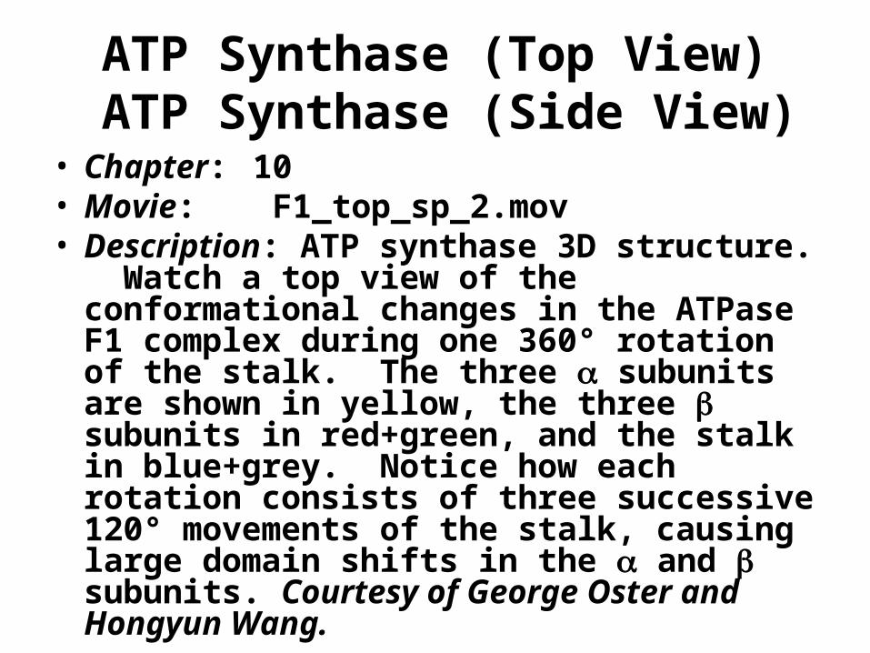
Undecagold• Features of Undecagold
• Ultra small 0.8 nm gold core.
• Use to prepare the smallest possible gold probes.
• Highest possible resolution.
• Site specific covalent labeling with choice of reactivities.

Undecagold• Undecagold (Au11) is smaller than Nanogold®, with a core of
11 gold atoms only 0.8 nm in diameter. It is ideal for ultra-high-resolution EM work such as scanning transmission electron microscopy, or for resolving elements of large structures by TEM in conjunction with image processing. Undecagold has been used to see the biotin binding sites on avidin to 1 nm resolution by electron microscopy. It is prepared in a form with one reactive arm for cross-linking to a specific site on a target molecule, and is available with different reactivities for labeling different sites. Note: Single undecagold clusters are not routinely visualized directly in the TEM. Undecagold may be seen upon image processing of protein helices and crystals, or visualized en masse if there is a bulk deposition such as staining of an organelle. Also, undecagold develops more slowly and with less final silver deposition than Nanogold®. Therefore, for many applications we recommend Nanogold®.

Structure of Alexa Fluor®* 594 FluoroNanogold Fab' and Streptavidin conjugates, showing covalent attachment.

Autocatalysis of gold deposition to increase particle size for detection.

Comparison with enzymatic detection: Light micrographs of formalin-fixed serial sections of cervical carcinoma, in situ hybridized for HPV-16/18 using a biotinylated probe (Pathogene-HPV kit, Enzo) (bar=10 micrometers) (1) direct detection using streptavidin-peroxidase-DAB; (2) Sirect detection using Nanogold®-streptavidin followed by development with GOLDENHANCE (Micrographs courtesy of Prof. G. W. Hacker. Medical Research Coordination Center, University of Salzburg, Austria).
streptavidin-peroxidase-DAB
Nanogold®-streptavidin -GOLDENHANCE

Left: Structure of Alexa Fluor® 488 and Nanogold® - Fab', showing covalent attachment of components. Right: Fluorescent staining obtained using combined combined Alexa Fluor® 488 and Nanogold® - Fab' tertiary probe. The specimen is a slide from the NOVA Lite ANA HEp-2 test, an indirect immunofluorescent test system for the screening and semi-quantitative determination of anti-nuclear antibodies (ANA) in human serum (see ). The slide was stained using positive pattern control human sera, a Mouse anti-Human secondary antiboidy, and combined Alexa Fluor® 488 and Nanogold® - Fab' tertiary probe. Specimens were washed with PBS (30 minutes) between each step, then blocked by the addition of 7 % nonfat dried milk to the tertiary antibody solution (original magnification 400 X).

Localization of caveolin-1a in ultrathin cryosection of human placenta using a new FNG; caveolin 1 alpha is primarily located to caveolae in placental endothelial cells. One-to-one correspondence is found between fluorescent spots and caveola labeled with gold particles (right). Ultrathin cryosections collected on formvar film-coated nickel EM grids were incubated with chicken anti-human caveolin-1a IgY for 30 min at 37°C, then with biotinylated goat anti-chicken F(ab)2 (13 mg/ml) for 30 min at 37°C, then stained with ALEXA-594 FluoroNanogold-Streptavidin (1:50 dilution) for 30 min at room temperature. Non-specific sites on cryosections were blocked with 1% milk - 5% fetal bovine serum-PBS for 30 minutes at room temperature (figure courtesy of T. Takizawa, Ohio State University, Columbus, OH).

Liposomes: LDL Structure
LDL enters cells by receptor-mediated endocytosis
or lipid-soluble drugs
Target-directing ligand
Return

Protein Aggregates
• Glue Molecules • Target ligand• Toxic Payload• Lytic Enzyme
• Deliver drug to target• Poison target• Prevent drug excretion• Remove drug
Glue
TargetLigandToxic
PayloadLyticEnzyme
Return

Functionalized Nanomaterials
• Tissue compatibility for:– Axonal regeneration– Blood vessel stents– Artificial kidneys– ect.


Functionalized Nanomaterials• The researchers demonstrated the composite heparin membrane
with nanopores could work as an artificial kidney, or dialyzer, by filtering the blood and maintaining its flow. The presence of this blood-compatible dialyzer could potentially eliminate the need for systemic administration of heparin to the patient during kidney dialysis, the researchers say.
The heparin-coated membranes are described in a paper titled “Ionic Liquid-Derived Blood Compatible Membranes for Kidney Dialysis,” published online Apr. 24 in advance of print in the Journal of Biomedical Materials Research.
“These heparin composite membranes and fibers and coated carbon nanotubes are an enabling technology,” says Saravanababu Murugesan, a recent doctoral graduate in chemical and biological engineering at Rensselaer and lead author of the paper. “Our results show these novel materials have great promise in the development of improved medical devices that are blood
Return
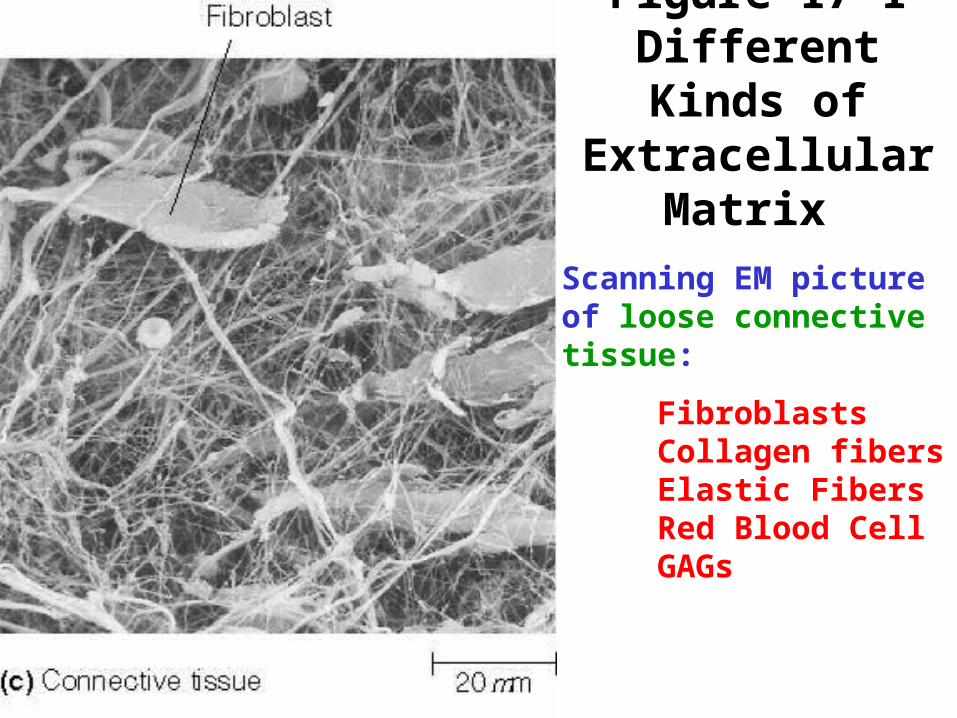
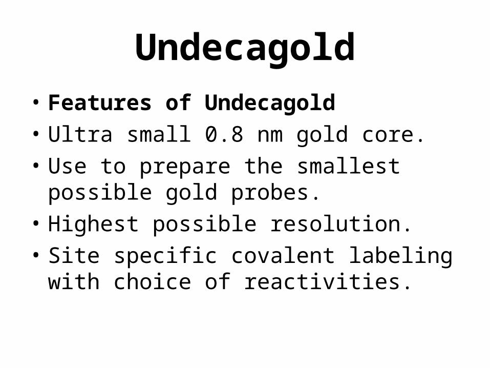
Figure 17-1 Different Kinds of
Extracellular Matrix
Scanning EM picture of loose connective tissue:
FibroblastsCollagen fibersElastic FibersRed Blood CellGAGs

Loose connective tissue

Dense Regular Connective Tissue

Figure 17-2 The Structure of Collagen
Protein is high in amounts of: Glycine Hydroxylysine Hydroxyproline
Glycine is in the axis of the triple helix, the only aa small enough to fit there
Tension strength of a 1 mm diameter fiber is ~9 kg.67 nm repeat distance of cross striationsStretch < 5% breaking

Figure 17-3 Collagen Assembly
270 to 300 nm long,1.5 nm diameter
67 nm repeat distance, ~270 molecules x-sec
After action of procollagen peptidase

Collagen is formed in a
fibroblast groove.
Return

Figure 12-12 Exocytosis
Microtubular movement of vessicles
Fusigenic proteins/ receptors
Ca++ 2nd messenger trigger
Polarized secretion on some parts of plasma membrane only (e.g.: apical)

Figure 12-13 Endocytosis

Figure 12-14 Phagocytosis
Opsin – coat on particle to be ingested
Opsinization – coating is often achieved with antibodies, which promote phagocytosis
Phagocytes are specialized for phagocytosis.
Phagocytic vesicles fuse with endosomes containing acid hydrolyases
Lysosome vs. endosomePhagolysosome vs. lysosome

Figure 12-15 Receptor-Mediated
EndocytosisCoated pit = up to 20% of surface area of plasma membrane
Adaptor protein
Clathrin
Dynamin
EGF (epidermal growth factor) undergoes receptor-mediated endocytosis (down regulation)

Figure 12B-2 Visualization of LDL Binding

Figure 12B-2 Visualization of LDL Binding

The yolk region of a chicken egg is a single cell
Yolk proteins,Vitellogen,Vitellogenins, are made in hepatocytes and follicle cells
Figure 12-16 Receptor-Mediated Endocytosis of Yolk Protein by a Chicken Oocyte

Figure 12-16 Receptor-Mediated Endocytosis of Yolk Protein by a Chicken Oocyte

Figure 12-16 Receptor-Mediated Endocytosis of Yolk Protein by a Chicken Oocyte

Figure 12-16 Receptor-Mediated Endocytosis of Yolk Protein by a Chicken Oocyte

Receptor-mediated endocytosis.
• Chapter: 12• Movie: nbt929-S3.mov Clathrin Assembly• Description: Receptor-mediated endocytosis.
Watch as a receptor-ligand complex is internalized by receptor-mediated endocytosis in a chinese hamster ovary cell. A cell expressing a fusion of the epidermal growth factor (EGF) receptor with green fluorescent protein is treated with a fluorescently-labelled EGF ligand (red). Notice the rapid internalization of the receptor-ligand complex by endocytosis. Used by permission of the Nature Publishing Group: http://www.nature.com/.
Return

Figure 3.13
Internal Structures of an Animal Cell {ER & Ribosome}

Figure 3.15
Endoplasmic Reticulum (ER) and Ribosome

Figure 7-10 Movements of Phospholipid Molecules Within Membranes
Kinds of phospholipids differ.
Saturation of phospholipids differ
Lateral motion w neighbor – 10M/sec; several microns/sec
Phospholipid translocators or Flipases enhance Transverse diffusion
Resting membrane flip-flop = 1/few hours to <1/weekReturn

Figure 24-16 Fusion of Cancer Cells with Normal
Cells

Figure 24B-1 The Monoclonal Antibody
Technique
Return

Figure 11-8 Noncyclic Electron Flow in Oxygenic Phototrophs
Z scheme, Non-cyclic electron flow
Mn-stabilizing protein
Pheophytin (2 protons iso Mg++)
plastoquinone
Oxygen-evolving complexplastocyanin
ferredoxin
Ferredoxin-NADP+ reductase
4

Figure 11-9 A Model of the Orientation of the Major Energy Transduction Complexes Within the Thykaloid Membrane
Water photolysis
plastoquinone
plastocyanin(Cu-containing peripheral prot.)
FerredoxinFNR Ferredoxin NADP+ Reductase
CFoCF1 complex
PMF is due mainly to 2 pH unit difference
Atrazine binds to plastoquinone-binding protein;Inhibits ET in PSII

Figure 10-15 The Flow of Electrons Through Respiratory Complexes I, III, and IV with Concomitant Directional Proton Pumping
43P 11P13P
3 or 4 protons => 1 ATP

Figure 8-14 The Bacteriorhodopsin Proton Pump of Halobacteria

Figure 11-10 Cyclic Electron Flow
Cyclic system allows for variation in the amounts of ATP and NADPH used by a cell
Pumps Protons!
Cyclic system produces ATP.
Cyclic phosphorylation.
No water used
No O2 released
phylloquinonechlorophyll a
Iron-sulfur centers
ferredoxin
plastocyanin
cyto-chromes
Return