Embed Size (px)
Citation preview

�������� ����� ��
Assessment of in vivo Systemic Toxicity and Biodistribution of Iron-dopedSilica Nanoshells
Natalie Mendez, Alexander Liberman, Jacqueline Corbeil, ChristopherBarback, Robert Viveros, James Wang, Jessica Wang-Rodriguez, Sarah L.Blair, Robert Mattrey, David Vera, William Trogler, Andrew C. Kummel Ph.D.
PII: S1549-9634(16)30188-5DOI: doi: 10.1016/j.nano.2016.10.018Reference: NANO 1460
To appear in: Nanomedicine: Nanotechnology, Biology, and Medicine
Received date: 5 May 2016Revised date: 10 October 2016Accepted date: 18 October 2016
Please cite this article as: Mendez Natalie, Liberman Alexander, Corbeil Jacqueline, Bar-back Christopher, Viveros Robert, Wang James, Wang-Rodriguez Jessica, Blair Sarah L.,Mattrey Robert, Vera David, Trogler William, Kummel Andrew C., Assessment of in vivoSystemic Toxicity and Biodistribution of Iron-doped Silica Nanoshells, Nanomedicine:Nanotechnology, Biology, and Medicine (2016), doi: 10.1016/j.nano.2016.10.018
This is a PDF file of an unedited manuscript that has been accepted for publication.As a service to our customers we are providing this early version of the manuscript.The manuscript will undergo copyediting, typesetting, and review of the resulting proofbefore it is published in its final form. Please note that during the production processerrors may be discovered which could affect the content, and all legal disclaimers thatapply to the journal pertain.
ACC
EPTE
D M
ANU
SCR
IPT
ACCEPTED MANUSCRIPTAssessment of in vivo Systemic Toxicity and Biodistribution of Iron-doped
Silica Nanoshells
Natalie Mendez1, Alexander Liberman
1, Jacqueline Corbeil
2, Christopher Barback
2, Robert Viveros
1,
James Wang1, Jessica Wang-Rodriguez
3, Sarah L. Blair
4, Robert Mattrey
2, David Vera
2, William Trogler
5,
Andrew C. Kummel5*
1. Department of Materials Science and Engineering, Nanoengineering, and Chemical Engineering,
University of California, San Diego, California, USA
2. Department of Radiology, University of California, San Diego, California, USA
3. Department of Pathology, University of California, San Diego, California, USA
4. Department of Surgery, University of California, San Diego, California, USA
5. Department of Chemistry and Biochemistry, University of California, San Diego, California,
USA, *[email protected]
* Corresponding author: Andrew C. Kummel, Ph.D., University of California, San Diego
Chemistry & Biochemistry, 9500 Gilman Drive #0358, La Jolla, CA 92093-0358
Email : [email protected], Phone : 1-858-534-3368, Fax : 1-858-534-2063
Abstract; 149 words
Complete manuscript word count: 5,011
Number of figures: 9
Number of references: 46
Tables: 1
This research was supported by NIH IMAT 1R33CA177449-01A1 and the NIH—Cross Training
Translation Cancer Researchers in Nanotechnology (CRIN) Support (NIH Grant No. 3 R25 CA
153915-03S1). Individual student funding was provided by the NCI Research Supplements to
Promote Diversity in Health Related Research Fellowship (NIH Grant No. 1R33CA177449-
01A1). The authors thank Dr. K. Pestonjamasp and the rest of the Cancer Center Microscopy
Core Facility at UCSD (NCI Grant No. P30 CA23100) and the UCSD Histology and
Immunohistochemistry core facility.
A.C. Kummel and W.C. Trogler have an equity interest in Nanocyte Medical, Inc., a company
that may potentially benefit from the research results, and also serve on the company’s Scientific
Advisory Board. S. L. Blair has a family member with an equity interest in Nanocyte Medical,
Inc., a company that may potentially benefit from the research results. The terms of this
arrangement have been reviewed and approved by the University of California, San Diego in
accordance with its conflict of interest policies.

ACC
EPTE
D M
ANU
SCR
IPT
ACCEPTED MANUSCRIPTAssessment of in vivo Systemic Toxicity and
Biodistribution of Iron-doped Silica Nanoshells
Natalie Mendez1, Alexander Liberman
1, Jacqueline Corbeil
2, Christopher Barback
2, Robert
Viveros1, James Wang
1, Jessica Wang-Rodriguez
3, Sarah L. Blair
4, Robert Mattrey
2, David Vera
2,
William Trogler5, Andrew C. Kummel
5*
1. Department of Materials Science and Engineering, Nanoengineering, and Chemical
Engineering, University of California, San Diego, California, USA
2. Department of Radiology, University of California, San Diego, California, USA
3. Department of Pathology, University of California, San Diego, California, USA
4. Department of Surgery, University of California, San Diego, California, USA
5. Department of Chemistry and Biochemistry, University of California, San Diego,
California, USA, *[email protected]
Abstract
Silica nanoparticles are an emerging class of biomaterials which may be used as diagnostic and
therapeutic tools for biomedical applications. In particular, hollow silica nanoshells are attractive
due to their hollow core. Approximately 70% of a 500nm nanoshell is hollow, therefore more
particles can be administered on a mg/kg basis compared to solid nanoparticles. Additionally,
their nanoporous shell permits influx/efflux of gases and small molecules. Since the size, shape,
and composition of a nanoparticle can dramatically alter its toxicity and biodistribution, the
toxicology of these nanomaterials was assessed. A single dose toxicity study was performed in
vivo to assess the toxicity of 500nm iron-doped silica nanoshells at clinically relevant doses of
10-20 mg/kg. This study showed that only a trace amount of silica was detected in the body 10
weeks post-administration. The hematology, biochemistry and pathological results show that the
nanoshells exhibit no acute or chronic toxicity in mice.
Keywords: Silica nanoparticles, toxicity, biodistribution, biomaterials, toxicology,
nanomaterials
ACC
EPTE
D M
ANU
SCR
IPT
ACCEPTED MANUSCRIPTAbbreviations: ICP-OES, inductively coupled plasma optical emission spectroscopy; IV,
intravenous; mesoporous silica nanoparticle, MSN; Tetramethyl orthosilicate, TMOS; energy
dispersive X-ray spectroscopy, EDX; complete blood count, CBC; haematoxylin and eosin,
H&E; scanning electron microscopy, SEM; high intensity focused ultrasound, HIFU; alanine
transaminase, ALT; alkaline phosphatase, ALP; blood urea nitrogen, BUN; phosphate buffered
saline, PBS; SiO2, silica; Fe-SiO2, iron-silica
Background
Silica (SiO2) nanomaterials have gained much attention due to their broad range of potential
biomedical applications which include bio-imaging, drug delivery, biosensors, and therapeutic
ablation.1-3
Characteristics that make SiO2 nanoparticles a promising material are their well-
established surface modification chemistry, high surface area, large pore volume, high stability
in storage, and in vivo stability.4, 5
Furthermore, porous SiO2 nanoshells have gained considerable
interest as in vivo imaging agents due to their potential to improve cancer diagnosis and their
ability to provide guidance during surgery.6-8
SiO2 nanoshells can be used as carriers for contrast
agents such as a fluorophore,9, 10
perfluorocarbon,8, 11
and superparamagnetic material.12-14
Many studies have evaluated the toxicity of various SiO2 nanoparticle formulations. The size,
shape, morphology, charge, and surface properties of a nanoparticle can dramatically affect its
biodistribution and toxicology.3 For instance, solid SiO2 materials with amorphous particle
morphology may cause hemolysis of red blood cells, which raises concerns regarding their safety
in the clinic.15, 16
Yu et al. (2011) investigated the impact of SiO2 nanoparticle design on cellular
toxicity and hemolytic activity up to 500 µg/ml by evaluating the effect of geometry, size,
porosity, and surface charge of SiO2 nanoparticles in vitro.17
It was shown that the toxicity of
SiO2 nanoparticles is influenced by surface charge and pore size and that cellular toxicity is cell-
type dependent. Their studies showed that solid nonporous nanoparticles had a far greater

ACC
EPTE
D M
ANU
SCR
IPT
ACCEPTED MANUSCRIPThemolytic activity compared to mesoporous silica nanoparticles (MSNs), which showed no
hemolytic toxicity for all geometries, at doses below 100 μg/mL.
Several in-vivo studies have been performed to assess the toxicity of SiO2 nanoparticles. They
show that SiO2 nanoparticles can cause inflammatory responses and hepatotoxicity which are
influenced by factors such as dose, particle size, surface area, charge, and particle formulation 9,
17-21. For example, Yu et al. investigated the acute toxicity of 120 nm spherical MSNs when
administered intravenously (IV)22
; the maximum tolerated dose for these particles was 30 mg/kg.
Nishimori et al. investigated the relationship between particle size and toxicity using nonporous
spherical SiO2 particles with diameters of 70 nm, 300 nm and 1000 nm23
. Their findings showed
that 70 nm nanoparticles induced liver injury at 30 mg/kg, while 300 nm and 1000 nm SiO2
particles exhibited no biochemical or histological tissue damage even at 100 mg/kg. This shows
that the size of the nanoparticle can significantly alter the toxicity even for the same particle
formulation.
The biodistribution of nanoparticles provides some information of the excretion and
degradability of the administered particles. Several studies have examined the biodistribution of
SiO2 particles, which have shown them to primarily accumulate in mononuclear phagocyte
system (MPS) organs such as the liver, lungs and spleen. Studies have shown that SiO2
nanoparticles can be excreted from the body; the excretion is influenced by particle formulation,
size, dose, and surface modification, among other factors 4, 11, 24, 25
. It is expected that these
factors will also influence the toxicity.
Before new nanomaterial formulations can be applied safely in a clinical setting, their
biocompatibility and biodistribution need to be assessed. Despite the numerous studies of the
toxicology of various forms of SiO2 nanoparticles, the toxicity of each formulation must be

ACC
EPTE
D M
ANU
SCR
IPT
ACCEPTED MANUSCRIPTassessed before being translated to the clinic, since nanoparticle properties depend strongly on
their composition, size, shape, and surface chemistries20
. In the present study, the toxicity and
long-term biodistribution of calcined 500 nm SiO2 nanoshells and Fe-SiO2 nanoshells, which
have been shown to be biodegradeable in vitro, are investigated when administered IV to mice at
a clinically relevant dose. One attractive characteristic of hollow SiO2 nanoshells is that due to
their hollow core, more particles can be administered on a per mass basis. Approximately 70% of
the nanoshell is hollow, therefore, on average an equivalent of 3X more particles can be
administered on a mg/kg basis compared to solid nanoparticles of the same size. The particles are
calcined, which offers long term stability for storage, and their nanoporous shell wall permits
influx and efflux of drugs, gases, and other small molecules. These characteristics allow for this
platform to potentially be used as an ultrasound contrast agent or for drug delivery.6-8
Other
platforms such as solid nanoparticles cannot carry gases due to their solid core, therefore solid
nanoparticles do not have color Doppler ultrasound capabilities. In addition, 500nm nanoshells
have demonstrated to have good ultrasound imaging capabilities which is partly the reason why
their toxicity is being evaluated in this study. By understanding the toxicology effects of new
nanomaterial formulations, nanoparticles can be designed for decreased toxicity thereby creating
more efficacious and safe therapeutic and diagnostic agents.
Materials and Methods
Materials
Tetramethyl orthosilicate (TMOS) was purchased from Sigma Aldrich Corp (St. Louis,
Missouri). 500 nm amino-polystyrene beads were acquired from Polysciences Inc (Warrington,
Pennsylvania). Iron (III) ethoxide was acquired from Gelest Inc (Moorisville, Pennsylvania).
Potassium hydroxide was purchased from Fischer-Scientific (Pittsburg, Pennsylvania). 500 mM
aqueous KOH was obtained by dissolving the KOH pellets in Milli-Q water. Nitric acid (HNO3)
ACC
EPTE
D M
ANU
SCR
IPT
ACCEPTED MANUSCRIPTwas provided by EMD (Billerica, Massachusetts) at 67 – 70% purity and 1% HNO3 was prepared
by diluting the stock HNO3 with MilliQ water. The multi-element standard solution for silicon
calibration was provided by SPEX CertiPrep (Metuchen, New Jersey). The yttrium standard
solution for internal calibration was purchased from Agilent Technologies (Santa Clara,
California). Blood collection tubes coated in either heparin or EDTA were purchased from BD
Biosciences (Franklin Lakes, New Jersey).
Six week old female Swiss Webster mice were provided by Charles River Laboratories,
weighing between 20-25 grams each. Mice were fed a commercial pelleted diet (Harlan Tekland)
and kept at 22°C in UCSD approved animal housing with a 12 hour light/dark cycle. All animal
care and procedures were approved by the UCSD Institutional Animal Care and Use Committee.
All animals in these studies were treated with humane care.
Methods
Nanoshell Synthesis
Fe-SiO2 and pure SiO2 nanoshells were synthesized as previously reported.6, 7
Fe-SiO2
nanoshells were synthesized by mixing of 2.7 ul of TMOS, 10 ul of iron ethoxide (20 mg/ml in
ethanol), and 50 ul of amino-polystyrene templates in 1 ml of absolute ethanol for 5 hours on a
vortex mixer at 3000 RPM. After mixing, the particles are centrifuged and washed three times in
ethanol and left to dry overnight. Dry particles were then calcined for 18 hours at 550 C. Pure
SiO2 particles were synthesized by mixing 3 ul of TMOS and 50 ul of polystyrene templates in 1
ml of absolute ethanol for 5 hours. After mixing, the particles were centrifuged and washed
three times in ethanol and left to dry overnight. The dry particles were subsequently calcined for
18 hours at 550 °C. Particle synthesis and quality was confirmed by scanning electron
microscopy (SEM) and transmission electron microscopy (TEM). The diameter and shell
thickness of nanoshells was quantified by measuring the diameter and length of the shell
ACC
EPTE
D M
ANU
SCR
IPT
ACCEPTED MANUSCRIPTthickness taken from high resolution TEM images and analyzed using ImageJ. The percentage of
hollow space occupied by the nanoshells was determined by calculating the volume of the
hollow nanoshell using the outer diameter and subtracting the volume of the inner diameter of
the nanoshell. SEM images were acquired using a FEI/Phillips XL30 FEG ESEM with an
accelerating voltage of 10 kV. Energy dispersive X-ray spectroscopy (EDX) was performed on
the same Oxford EDX attachment and analyzed with INCA software. TEM images were
acquired using a JEOL 1200 EX II TEM.
Single Dose Toxicity
A single dose toxicity study was performed over the course of 10 weeks in healthy female
Swiss white mice. Four groups were compared (n = 10) to determine if the nanoshells had any
measureable toxicity. Group 1 received a 100 µl injection of Fe-SiO2 nanoshells at 4 mg/ml
(approximately 20 mg/kg), group 2 received 100 µl of SiO2 nanoshells at 4 mg/ml, group 3
received 100ul of SiO2 nanoshells at 2 mg/ml (approximately 10 mg/kg) which is the particle
number/ml equivalent of group 1 (Fe-SiO2 at 4mg/ml), and group 4 was a control group which
received no particles. All injections were performed IV via the tail vein. 100 µl of blood was
drawn with a submandibular lancet from each mouse weekly for the first four weeks and then
biweekly for the following six weeks. From each group, 5 mice had their blood allocated into
vacutainer tubes containing EDTA for Complete Blood Count (CBC) analysis and the other 5
mice had their blood allocated into vacutainer tubes contain heparin for serum chemistry
analysis. CBC and Serum Chemistry analyses were performed by UCSD Animal Care Program
Diagnostic Laboratory and STAT Labs. After 10 weeks, the animals were sacrificed. Three
animals from each group were used for long-term biodistribution studies, and the remaining
animals were used for pathological investigation after sectioning and H&E staining.
Inductively Coupled Plasma Optical Emission Spectroscopy (ICP-OES Analysis)

ACC
EPTE
D M
ANU
SCR
IPT
ACCEPTED MANUSCRIPTICP-OES was used to detect nanoshells retained in mouse organs. One mouse was injected
with 100 µl of a 4 mg/mL aqueous nanoshell solution. Control mice were sacrificed 24 hours
after the mice received the nanoshell injection, and the organs were collected. To analyze the
organs for silicon content, the organs were incubated in 7 ml of aqueous 500 mM KOH and bath
sonicated for 48 hours. 0.5 ml of each digested sample was drawn out and acidified using 2.5 ml
of 1% HNO3. The samples were centrifuged for 15 minutes at 3000 rpm, and 0.5 ml of a 300
ppb yttrium standard solution was added to each 2.5 ml aliquot of a supernatant sample. The
samples were finally briefly centrifuged immediately before ICP-OES analysis as described in a
previous study26
.
Histological Analysis
Tissue samples were fixed in 10% formalin and submitted to the UCSD histology core facility
for paraffin block processing, sectioning and Hematoxylin and Eosin (H&E) staining. The slides
were imaged using a Nikon Eclipse E600 microscope. The slides were randomized and analyzed
by the Chief of Pathology and Laboratory Medicine at the VA Medical Center, La Jolla. Focal
inflammation was quantified by counting the number of inflammatory foci per organ for each
group.
Statistical Analysis
Serum chemistry and hematology markers were compared by using multiple unpaired two-
tailed t-tests to determine statistical significance (alpha = 0.05). Mean values were calculated for
each time point in order to perform multiple t-tests as described by J. N. Matthews, et al.27
Statistical analysis was performed by using GraphPad Prism software version 6. A total number
of 5 mice per group were used for the study.
Results
ACC
EPTE
D M
ANU
SCR
IPT
ACCEPTED MANUSCRIPTCharacterization of Nanoshells
The SiO2 and Fe-SiO2 nanoshells were hollow and spherical in shape with an average diameter
of 497 nm ± 13 and 455 nm ± 14, respectively, as shown by SEM and TEM in Figure 1. The
images show that the SiO2 and Fe-SiO2 nanoshells are fairly uniform in size with no visible
holes, cracks, or other deformations. The diameters and shell thicknesses of nanoshells were
determined by using TEM images. The incorporation of iron does not greatly affect the shell
thickness or morphology compared to pure SiO2 nanoshells; the shell thickness for Fe-SiO2 was
27 nm ± 4 and 33 nm ± 5 for SiO2 nanoshells. EDX showed that Fe-SiO2 nanoshells have an
average 5 mol % of iron doped into the nanoshell matrix. The incorporation of iron makes the
nanoshells biodegradable in human serum, and attractive for biomedical applications.28
Serum
transferrin has been shown to bind to the iron-doped nanoshells, which may induce degradation
and dissolution of any residual nanoshells that are not excreted from the body29
. In addition,
500nm Fe-SiO2 nanoshells may be filled with gases giving them ultrasound capabilities as shown
in Figure 2.
Single Dose Acute Toxicity
The 500 nm nanoshells evaluated in this study have been developed for use as contrast agents
when filled with perfluoropentane gas or as ablative agents when combined with high intensity
focused ultrasound (HIFU).2, 6, 30
It is important to thoroughly evaluate the toxicity of new
compounds before the translation to clinical trials. Therefore, the acute toxicity of SiO2 and Fe-
SiO2 nanoshells was assessed in order to evaluate their safety in vivo. Mice received an IV
injection of 400 µg of SiO2 (20 mg/kg), 400 µg of Fe-SiO2 (20 mg/kg), 200 µg of SiO2 (10
mg/kg), or Phosphate Buffered Saline (PBS). 10 mg/kg was assessed for SiO2 because it is the
equivalent of the Fe-SiO2 by particle concentration. Blood was collected over the course of 10
weeks and serum chemistry and hematology analysis was performed.

ACC
EPTE
D M
ANU
SCR
IPT
ACCEPTED MANUSCRIPTNanoparticles have been shown to have different pharmacological actions when administered
IV, depending on factors such as size, shape, charge, and composition. In particular,
nanoparticles usually preferentially accumulate in the liver.3 Therefore it is important to evaluate
if nanoparticle administration affects liver function. For serum chemical analysis, albumin,
bilirubin, alanine transaminase (ALT), and alkaline phosphatase (ALP) were evaluated to assess
hepatic function as shown in Figure 3. The serum chemistry levels for mice which received Fe-
SiO2 or SiO2 did not differ significantly from the control group. Multiple t-tests were performed
for statistical analysis and all groups showed no statistically significant difference between the
control and nanoshell dosed groups (alpha = 0.05). This data indicates that a systemic dose of 20
mg/kg (4mg/ml) of SiO2 or Fe-SiO2 nanoshells show no acute hepatic toxicity over the course of
10 weeks.
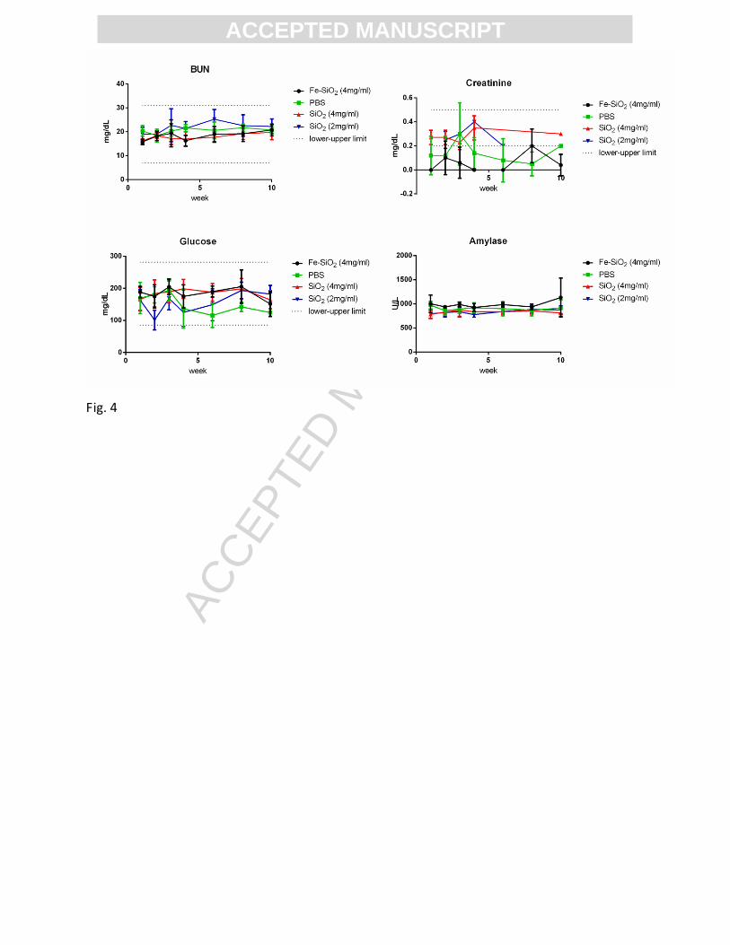
Nanoparticles have been shown capable of being cleared through the renal system.4 Therefore
blood urea nitrogen (BUN) and creatinine were measured to determine if IV administration of
500 nm SiO2 and Fe-SiO2 nanoshells alters renal function. Pancreatic function was also assessed
by measuring amylase and glucose as shown in Figure 4. The SiO2 nanoshell group had a
significantly higher serum creatinine levels compared to the control group (p=0.003 for 4mg/ml
p=0.01 for 2mg/ml). However, for all groups the values were still within the normal range for
serum creatinine values reported by Charles River for female Swiss Webster mice.
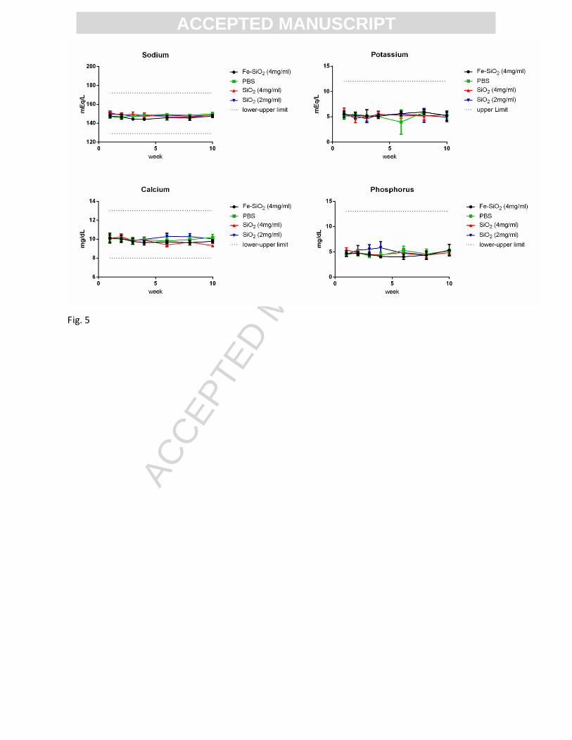
Serum analytes such as sodium (Na), potassium (K), calcium (Ca), and phosphorus (P) were
measured in order to evaluate any electrolyte imbalances of mice post injection as shown in
Figure 5. There was a significantly higher serum Na levels for mice that received Fe-SiO2 when
compared to the control group (p= 0.002). Nevertheless, when compared to Na values reported
by Charles River, the Na values measured in this study fell within the normal range of the 95%

ACC
EPTE
D M
ANU
SCR
IPT
ACCEPTED MANUSCRIPTconfidence interval. Overall, the general health of mice administered with Fe-SiO2 and SiO2
nanoshells IV was unaffected by the particles.
Amorphous SiO2 particles have been shown to cause hemolysis of red blood cells which raise
safety concerns about their use in the clinic.15, 16
The interaction of SiO2 nanoparticles and blood
components impacts cellular toxicity and hemolytic activity, which depend on different
nanoshell formulations such as composition, size, and charge. Therefore, hematology analysis of
500 nm Fe-SiO2 and SiO2 nanoshells was performed in order to assess blood chemistry, blood-
forming organs, and blood diseases. Neutrophils, lymphocytes, hemoglobin (Hb), and
eosinophils were measured to assess hematological abnormalities after nanoshell administration
over the course of ten weeks and as a potential indicator of an immune or inflammatory reaction,
as shown in Figure 6. Other analytes such as monocytes, red blood cells (RBC), nucleated
RBCs/100WBC, mean corpuscular volume (MCV), mean corpuscular hemoglobin concentration
(MCHC), and mean platelet volume (MPV) were also measured (See Figure S1). There was no
significant difference observed between Fe-SiO2 nanoshells, SiO2 nanoshells at 2 mg/ml or 4
mg/ml, and the PBS vehicle group for any of the analytes. In addition, all measured values fell
within the 95% CI values reported by Charles River. These findings are a good indication that
SiO2 and Fe-SiO2 nanoshells are generally safe when administered systemically at the indicated
doses. It is likely that the physical characteristics of the nanoshells including their hollow core,
surface composition, and size contributed to their low toxicity.
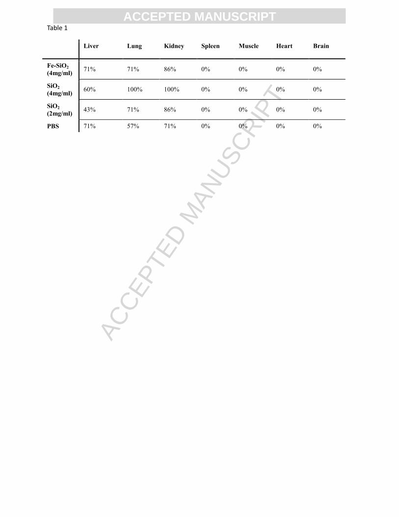
Haematoxylin and eosin (H&E) histological analysis was performed on liver, lung, kidney,
spleen, muscle, and heart tissue samples 10 weeks after administration of Fe-SiO2 or SiO2
nanoshells to check for inflammation or any abnormalities, as shown in Figure 7. The pathology
revealed some mild chronic inflammation in the liver, lung, and kidney in all groups as
evidenced by focal lymphocytic infiltrates. Representative sections show lymphocytic infiltrate

ACC
EPTE
D M
ANU
SCR
IPT
ACCEPTED MANUSCRIPTin the perisinusoidal distribution in the liver, peri-bronchial in the lung, and peri-vascular in the
kidney. Histological analysis was also performed on the brain and showed no pathological
changes when compared to the PBS control (See Figure S2). The number of mice with focal
inflammation in each group was not significantly different in mice treated with nanoshells when
compared to mice injected with PBS as summarized in Table 1.
In addition, the organs of naive mice which has no isoflurane anesthesia exposure nor blood
drawn were evaluated. Mild focal inflammation was observed in the lung and kidney, and acute
inflammation was observed in the liver which shows that mild inflammation is typical in naive
Swiss mice as shown in Figure 7e. Similarly, Plummer et al. 31
showed that the control group as
well as rats exposed to inhaled isoflurane anesthesia for a prolonged period of time showed
hepatic focal inflammation suggesting that mild focal inflammation is often observed in healthy
laboratory rodents. It is possible that repeat bleeding and isoflurane anesthesia are probable
factors contributing to increased focal inflammation observed in our experimental and PBS
control mice groups.32
No multinucleated cells were observed in the spleen. No abnormalities were noted for any of
the H&E sections and no apparent differences were observed between mice treated with Fe-SiO2
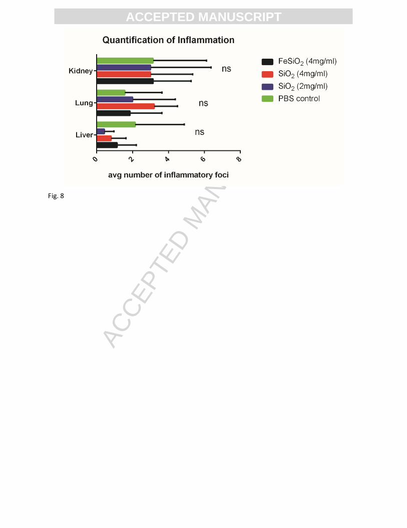
or SiO2 nanoshells and the PBS group. The number of inflammatory foci was quantified to
evaluate the degree of focal inflammation per group as shown in Figure 8. There were no
significant differences between groups treated with Fe-SiO2 or SiO2 nanoshells and the control
group. These results showed that there was no significant damage to major organs (liver, lungs,
heart, kidney, and spleen) when compared to control mice. This indicates that Fe-SiO2 and SiO2
nanoshells can potentially be safely used in the clinic by IV dosing at the given doses.
ACC
EPTE
D M
ANU
SCR
IPT
ACCEPTED MANUSCRIPTThe biodistribution of Fe-SiO2 and SiO2 nanoshells was performed 24 hours or 10 weeks after
systemic administration, and ICP-OES was used to measure the amount of elemental Si in each
organ, as shown in Figure 9.
For SiO2 nanoshells, 42% of total injected nanoshells accumulated in the liver, which is
consistent with previous studies.7 It is important to note that nearly 57% of the total injected dose
was not detected in any organs or the blood 24 hours after administration; this may be due to
excretion. After 10 weeks post administration, only 2% of nanoshells were detected in the liver.
Similarly, the biodistribution of Fe-SiO2 was studied 24 hours and 10 weeks post-injection.
Interestingly, when SiO2 nanoshells were doped with iron, only 5% of the total injected dose was
present in the liver, while 8% was detected in the lungs. Nearly 88% of the total injected dose
was undetectable in the organs or the blood after only 24 hours. After 10 weeks post
administration, only 4% of nanoshells were detected in organs, mostly in the liver.
In a previous study, whole body scintigraphy in a mouse showed that after IV administration, a
large portion of Fe–SiO2 and SiO2 nanoshells localized in the liver when monitored immediately
post injection and at 1 hour, 24 hours, and 72 hours.7 After 72 hours, organ gamma counting
showed a substantial amount of signal in the liver, and some in the kidney and spleen, but very
little signal was detected elsewhere. The results in the present study show that the nanoshells
are retained mostly by the liver and suggest that incorporation of iron can alter the
biodistribution of SiO2 nanoshells. These findings show that fewer nanoshells were retained
within 24 hours when they were doped with iron. It is postulated that iron doping leads to
transferrin interactions in the body which may lead to faster biodegradation as seen in previous
studies28
.

ACC
EPTE
D M
ANU
SCR
IPT
ACCEPTED MANUSCRIPT
Discussion
The kidney is capable of removing molecules from the blood by filtering particles through the
glomerular capillary wall, also known as glomerular filtration. Renal filtration depends on
surface charge and size, where molecules with a hydrodynamic diameter greater than 8 nm are
typically not capable of glomerular filtration and therefore are not excreted via the renal system
33, 34; however, some studies have suggested that particles larger than 8 nm can be excreted
through the urine.4 Nevertheless, it is highly unlikely that a large number or intact 500 nm SiO2
nanoshells are being excreted in the urine unless the particles were first broken down to smaller
components before renal excretion.
The liver catabolizes particles in the blood through phagocytosis followed by biliary
excretion.34
Phagocytic Kupffer cells, specialized macrophages located in the liver, are highly
effective at recognizing and removing unwanted substances from the blood, including nano- and
microparticles. Kupffer cells selectively phagocytose opsonized particulates which have been
coated with complement proteins for recognition.35, 36
Particles are subsequently metabolized and
excreted as bile via the biliary system. Passage through the liver can make metabolites smaller
and more polar which can result in reabsorption in the blood leading to excretion via the renal
system however, renal clearance is observed at lower rates. In the present study, it is most likely
that the particles are being cleared via the hepatic system. It is hypothesized that iron
incorporation can alter the uptake, metabolism, and clearance of the nanoshells. It is probable
that the interaction of serum proteins is different for Fe-SiO2 and SiO2 nanoshells, which may
result in a different extent of opsonization for each formulation. The altered surface protein
coating of the Fe-SiO2 nanoshells may change the extent of Kupffer cell phagocytosis and
hepatic excretion.

ACC
EPTE
D M
ANU
SCR
IPT
ACCEPTED MANUSCRIPTThe main site of transferrin synthesis is in the liver.
37 Transferrin is an iron transport protein
that delivers Fe3+
from the duodenum and macrophages to all tissues by circulating in the
blood.38
Human transferrin binds iron as Fe3+
with a high affinity; therefore, the incorporation of
Fe3+
into the SiO2 nanoshell structure likely facilitates the decomposition of Fe-SiO2 nanoshells
by degradation, which results in hepatic clearance. The biodegradation of Fe-SiO2 in FBS and
human serum was shown in previous studies,28
consistent with the in vivo results shown in the
present study. More recent studies show that Fe-SiO2 nanoshells undergo binding/endocytosis in
cells that overexpress the transferrin receptor and that their uptake can be blocked with addition
of holotransferrin.29
This suggests that the surface of the Fe-SiO2 nanoshells may also be bound
to transferrin in vivo. Not only are hepatocytes involved in iron metabolism, but the liver
endothelium has been shown to be rich in the transferrin receptor, which enhance endocytosis of
the Fe-SiO2 nanoshells.39-41
The liver endothelium has an increasingly recognized role in billiary
excretion.42
A small percentage of Fe-SiO2 nanoshells was observed in the lung, whereas only trace
amounts of non-doped SiO2 nanoshells were seen. Previous studies have shown that SiO2
nanoparticles can cause an inflammatory response in the lung.33
Kusaka et al. showed that 30 nm
SiO2 nanoparticles administered intratracheally to mice caused more severe lung inflammation
than did 3000 nm SiO2 particles.43
The present study shows that there was no additional long-
term lung damage after 500 nm Fe-SiO2 and SiO2 nanoshells were administered IV when
compared to the control group, which may be due to their larger size and/or the difference in
route of administration. Several studies have been performed in order to assess the toxicity of
SiO2 nano- and microparticles11, 25, 44, 45
46
which showed that the SiO2 particle biodistribution
depends on several factors such as charge, size, and surface modification.

ACC
EPTE
D M
ANU
SCR
IPT
ACCEPTED MANUSCRIPTThese nanoshells are expected to be used as contrast agents for cancer diagnosis or for
ultrasound guidance during surgery. In previous studies, the nanoshells have shown to be
stationary markers in vivo and can be imaged intermittently over the course of 10 days in a
tumor-bearing mouse model, and continuously for 45 minutes when administered
intratumorally.30
In the event that the nanoshells escape the tumor or are administered IV, this
study has shown that the nanoshells show no apparent systemic toxicity and can be safely cleared
from the body after a single dose. In addition, these nanoshells may also be used in combination
with HIFU for ablative therapy.2
These results show that the nanoshells presented in this study are safe when administered
systemically allowing exploitation of the hollow structure and calcined shells. Approximately
70% of the nanoshell is hollow, therefore, on average an equivalent of 3X more particles can be
administered on a mg/kg basis compared to solid nanoparticles of the same size. The particles are
calcined, which offers long term stability for storage, and their nanoporous shell wall can permit
influx and efflux of drugs, gases, and other small molecules. Although future pharmacokinetic
studies including blood circulation and clearance should be performed, this study indicates that
500 nm nanoshells are safe when administered IV at the doses studied.
Corresponding Author
9500 Gilman Drive, La Jolla, CA 92093-0332 Email: [email protected].
Acknowledgements
The authors thank the Cancer Center Microscopy Core Facility at UCSD and the UCSD
Histology and Immunohistochemistry core facility.
References
ACC
EPTE
D M
ANU
SCR
IPT
ACCEPTED MANUSCRIPT1. Tan, W.; Wang, K.; He, X.; Zhao, X. J.; Drake, T.; Wang, L.; Bagwe, R. P.
Bionanotechnology based on silica nanoparticles. Medicinal Research Reviews 2004, 24, 621-
638.
2. Liberman, A.; Wu, Z.; Barback, C. V.; Viveros, R. D.; Wang, J.; Ellies, L. G.; Mattrey,
R. F.; Trogler, W. C.; Kummel, A. C.; Blair, S. L. Hollow iron-silica nanoshells for enhanced
high intensity focused ultrasound. Journal of Surgical Research 2014, 190, 391-398.
3. Liberman, A.; Mendez, N.; Trogler, W. C.; Kummel, A. C. Synthesis and surface
functionalization of silica nanoparticles for nanomedicine. Surface Science Reports 2014, 69,
132-158.
4. Huang, X.; Li, L.; Liu, T.; Hao, N.; Liu, H.; Chen, D.; Tang, F. The Shape Effect of
Mesoporous Silica Nanoparticles on Biodistribution, Clearance, and Biocompatibility in Vivo.
ACS Nano 2011, 5, 5390-5399.
5. Chen, Y.; Chen, H.; Shi, J. In vivo bio-safety evaluations and diagnostic/therapeutic
applications of chemically designed mesoporous silica nanoparticles. Adv Mater 2013, 25, 3144-
76.
6. Liberman, A.; Martinez, H. P.; Ta, C. N.; Barback, C. V.; Mattrey, R. F.; Kono, Y.; Blair,
S. L.; Trogler, W. C.; Kummel, A. C.; Wu, Z. Hollow silica and silica-boron nano/microparticles
for contrast-enhanced ultrasound to detect small tumors. Biomaterials 2012, 33, 5124-5129.
7. Liberman, A.; Wu, Z.; Barback, C. V.; Viveros, R.; Blair, S. L.; Ellies, L. G.; Vera, D.
R.; Mattrey, R. F.; Kummel, A. C.; Trogler, W. C. Color Doppler Ultrasound and Gamma
Imaging of Intratumorally Injected 500 nm Iron–Silica Nanoshells. ACS Nano 2013, 7, 6367-
6377.
8. Martinez, H. P.; Kono, Y.; Blair, S. L.; Sandoval, S.; Wang-Rodriguez, J.; Mattrey, R. F.;
Kummel, A. C.; Trogler, W. C. Hard shell gas-filled contrast enhancement particles for colour
Doppler ultrasound imaging of tumors. Medchemcomm 2010, 1, 266-270.
9. Park, J.-H.; Gu, L.; von Maltzahn, G.; Ruoslahti, E.; Bhatia, S. N.; Sailor, M. J.
Biodegradable luminescent porous silicon nanoparticles for in vivo applications. Nat Mater
2009, 8, 331-336.
10. Wu, P.; He, X.; Wang, K.; Tan, W.; Ma, D.; Yang, W.; He, C. Imaging breast cancer
cells and tissues using peptide-labeled fluorescent silica nanoparticles. Journal of nanoscience
and nanotechnology 2008, 8, 2483-2487.
11. Decuzzi, P.; Godin, B.; Tanaka, T.; Lee, S. Y.; Chiappini, C.; Liu, X.; Ferrari, M. Size
and shape effects in the biodistribution of intravascularly injected particles. Journal of
Controlled Release 2010, 141, 320-327.
12. Ji, X.; Shao, R.; Elliott, A. M.; Stafford, R. J.; Esparza-Coss, E.; Bankson, J. A.; Liang,
G.; Luo, Z.-P.; Park, K.; Markert, J. T. Bifunctional gold nanoshells with a superparamagnetic
iron oxide-silica core suitable for both MR imaging and photothermal therapy. The Journal of
Physical Chemistry C 2007, 111, 6245-6251.
13. Erogbogbo, F.; Yong, K.-T.; Hu, R.; Law, W.-C.; Ding, H.; Chang, C.-W.; Prasad, P. N.;
Swihart, M. T. Biocompatible magnetofluorescent probes: luminescent silicon quantum dots
coupled with superparamagnetic iron (III) oxide. ACS nano 2010, 4, 5131-5138.
14. Thorek, D. L.; Chen, A. K.; Czupryna, J.; Tsourkas, A. Superparamagnetic iron oxide
nanoparticle probes for molecular imaging. Annals of biomedical engineering 2006, 34, 23-38.
15. Slowing, I. I.; Wu, C.-W.; Vivero-Escoto, J. L.; Lin, V. S. Y. Mesoporous Silica
Nanoparticles for Reducing Hemolytic Activity Towards Mammalian Red Blood Cells. Small
2009, 5, 57-62.
16. Nash, T.; Allison, A. C.; Harington, J. S. Physico-Chemical Properties of Silica in
Relation to its Toxicity. Nature 1966, 210, 259-261.
17. Lu, X.; Tian, Y.; Zhao, Q.; Jin, T.; Xiao, S.; Fan, X. Integrated metabonomics analysis of
the size-response relationship of silica nanoparticles-induced toxicity in mice. Nanotechnology
2011, 22, 055101.
ACC
EPTE
D M
ANU
SCR
IPT
ACCEPTED MANUSCRIPT18. Cho, M.; Cho, W.-S.; Choi, M.; Kim, S. J.; Han, B. S.; Kim, S. H.; Kim, H. O.; Sheen, Y.
Y.; Jeong, J. The impact of size on tissue distribution and elimination by single intravenous
injection of silica nanoparticles. Toxicology Letters 2009, 189, 177-183.
19. Liu, T.; Li, L.; Teng, X.; Huang, X.; Liu, H.; Chen, D.; Ren, J.; He, J.; Tang, F. Single
and repeated dose toxicity of mesoporous hollow silica nanoparticles in intravenously exposed
mice. Biomaterials 2011, 32, 1657-1668.
20. Chen, Y.; Chen, H.; Shi, J. In Vivo Bio-Safety Evaluations and Diagnostic/Therapeutic
Applications of Chemically Designed Mesoporous Silica Nanoparticles. Advanced Materials
2013, 25, 3144-3176.
21. Zhang, H.; Dunphy, D. R.; Jiang, X.; Meng, H.; Sun, B.; Tarn, D.; Xue, M.; Wang, X.;
Lin, S.; Ji, Z.; Li, R.; Garcia, F. L.; Yang, J.; Kirk, M. L.; Xia, T.; Zink, J. I.; Nel, A.; Brinker, C.
J. Processing Pathway Dependence of Amorphous Silica Nanoparticle Toxicity: Colloidal vs
Pyrolytic. Journal of the American Chemical Society 2012, 134, 15790-15804.
22. Yu, T.; Greish, K.; McGill, L. D.; Ray, A.; Ghandehari, H. Influence of Geometry,
Porosity, and Surface Characteristics of Silica Nanoparticles on Acute Toxicity: Their
Vasculature Effect and Tolerance Threshold. ACS Nano 2012, 6, 2289-2301.
23. Nishimori, H.; Kondoh, M.; Isoda, K.; Tsunoda, S.-i.; Tsutsumi, Y.; Yagi, K. Silica
nanoparticles as hepatotoxicants. European Journal of Pharmaceutics and Biopharmaceutics
2009, 72, 496-501.
24. He, Q.; Zhang, Z.; Gao, F.; Li, Y.; Shi, J. In vivo Biodistribution and Urinary Excretion
of Mesoporous Silica Nanoparticles: Effects of Particle Size and PEGylation. Small 2011, 7,
271-280.
25. Souris, J. S.; Lee, C.-H.; Cheng, S.-H.; Chen, C.-T.; Yang, C.-S.; Ho, J.-a. A.; Mou, C.-
Y.; Lo, L.-W. Surface charge-mediated rapid hepatobiliary excretion of mesoporous silica
nanoparticles. Biomaterials 2010, 31, 5564-5574.
26. Viveros, R. D.; Liberman, A.; Trogler, W. C.; Kummel, A. C. Alkaline and ultrasonic
dissolution of biological materials for trace silicon determination. Journal of Vacuum Science &
Technology B 2015, 33, 031803.
27. Matthews, J. N.; Altman, D. G.; Campbell, M. J.; Royston, P. Analysis of serial
measurements in medical research. British Medical Journal 1990, 300, 230-235.
28. Pohaku Mitchell, K. K.; Liberman, A.; Kummel, A. C.; Trogler, W. C. Iron(III)-Doped,
Silica Nanoshells: A Biodegradable Form of Silica. Journal of the American Chemical Society
2012, 134, 13997-14003.
29. Mitchell, K. P.; Sandoval, S.; Cortes-Mateos, M. J.; Alfaro, J. G.; Kummel, A. C.;
Trogler, W. C. Self-assembled targeting of cancer cells by iron (iii)-doped, silica nanoparticles.
Journal of Materials Chemistry B 2014, 2, 8017-8025.
30. Liberman, A.; Wang, J.; Lu, N.; Viveros, R. D.; Allen, C. A.; Mattrey, R. F.; Blair, S. L.;
Trogler, W. C.; Kim, M. J.; Kummel, A. C. Mechanically Tunable Hollow Silica Ultrathin
Nanoshells for Ultrasound Contrast Agents. Advanced Functional Materials 2015, 25, 4049-
4057.
31. Plummer, J.; Hall, P. d. l. M.; Jenner, M.; Ilsley, A.; Cousins, M. Effects of chronic
inhalation of halothane, enflurane or isoflurane in rats. British journal of anaesthesia 1986, 58,
517-523.
32. Liu, H.; Xiao, X.; Sun, C.; Sun, D.; Li, Y.; Yang, M. Systemic inflammation and multiple
organ injury in traumatic hemorrhagic shock. Front Biosci 2015, 20, 927-33.
33. Deen, W. M.; Lazzara, M. J.; Myers, B. D. Structural determinants of glomerular
permeability. Am J Physiol Renal Physiol 2001, 281, F579-96.
34. Longmire, M.; Choyke, P. L.; Kobayashi, H. Clearance properties of nano-sized particles
and molecules as imaging agents: considerations and caveats. Nanomedicine 2008, 3, 703-17.
35. Jaeschke, H.; Farhood, A.; Bautista, A. P.; Spolarics, Z.; Spitzer, J. J. Complement
activates Kupffer cells and neutrophils during reperfusion after hepatic ischemia. American
Journal of Physiology - Gastrointestinal and Liver Physiology 1993, 264, G801-G809.
ACC
EPTE
D M
ANU
SCR
IPT
ACCEPTED MANUSCRIPT36. Owens Iii, D. E.; Peppas, N. A. Opsonization, biodistribution, and pharmacokinetics of
polymeric nanoparticles. International Journal of Pharmaceutics 2006, 307, 93-102.
37. Brissot, P.; Wright, T. L.; Ma, W. L.; Weisiger, R. A. Efficient clearance of non-
transferrin-bound iron by rat liver. Implications for hepatic iron loading in iron overload states.
Journal of Clinical Investigation 1985, 76, 1463-1470.
38. Crichton, R. R.; Charloteaux-Wauters, M. Iron transport and storage. European Journal
of Biochemistry 1987, 164, 485-506.
39. Brieland, J. K.; Clarke, S. J.; Karmiol, S.; Phan, S. H.; Fantone, J. C. Transferrin: a
potential source of iron for oxygen free radical-mediated endothelial cell injury. Archives of
biochemistry and biophysics 1992, 294, 265-270.
40. Morgan, E. H.; Baker, E. Iron uptake and metabolism by hepatocytes. Fed Proc 1986, 45,
2810-6.
41. Soda, R.; Tavassoli, M. Liver endothelium and not hepatocytes or Kupffer cells have
transferrin receptors. Blood 1984, 63, 270-276.
42. Smedsrød, B.; Pertoft, H.; Gustafson, S.; Laurent, T. C. Scavenger functions of the liver
endothelial cell. Biochemical Journal 1990, 266, 313-327.
43. Kusaka, T.; Nakayama, M.; Nakamura, K.; Ishimiya, M.; Furusawa, E.; Ogasawara, K.
Effect of silica particle size on macrophage inflammatory responses. PLoS One 2014, 9.
44. Yu, T.; Hubbard, D.; Ray, A.; Ghandehari, H. In vivo biodistribution and
pharmacokinetics of silica nanoparticles as a function of geometry, porosity and surface
characteristics. Journal of Controlled Release 2012, 163, 46-54.
45. He, X.; Nie, H.; Wang, K.; Tan, W.; Wu, X.; Zhang, P. In Vivo Study of Biodistribution
and Urinary Excretion of Surface-Modified Silica Nanoparticles. Analytical Chemistry 2008, 80,
9597-9603.
46. Kumar, R.; Roy, I.; Ohulchanskky, T. Y.; Vathy, L. A.; Bergey, E. J.; Sajjad, M.; Prasad,
P. N. In Vivo Biodistribution and Clearance Studies Using Multimodal Organically Modified
Silica Nanoparticles. ACS Nano 2010, 4, 699-708.
Figure Captions
Figure 1. SEM and TEM images of 500nm a) Fe-SiO2 and b) SiO2 nanoshells.
Figure 2. 500nm SiO2 and Fe-SiO2 nanoshells have ultrasound imaging capabilities under color Doppler
and CPS mode.
Figure 3. Serum chemistry analysis for hepatic function. Mice were injected with 100 µl of Fe-SiO2
nanoshells at 4mg/ml (●), SiO2 at 4mg/ml (▲), SiO2 nanoshells at 2mg/ml (▼), or PBS (■). Albumin,
bilirubin, alanine transaminase (ALT), and alkaline phosphatase (ALP) were measured in mice over the
course of 10 weeks to evaluate hepatic function. Serum chemistry did not differ significantly between
control and treated groups. Data is shown as average ± SD, where n=5.
Figure 4. Serum chemistry analysis for renal and pancreatic function. Mice were injected with 100 µl of
Fe-SiO2 nanoshells at 4mg/ml (●), SiO2 at 4mg/ml (▲), SiO2 nanoshells at 2mg/ml (▼), or PBS (■).
Blood urea nitrogen (BUN), creatinine, glucose and amylase were measured in mouse serum over the
course of 10 weeks to evaluate general renal and pancreatic function. Serum chemistry did not differ
significantly between control and treated groups. Data is shown as average ± SD, where n=5.

ACC
EPTE
D M
ANU
SCR
IPT
ACCEPTED MANUSCRIPTFigure 5. Serum chemistry analysis for electrolyte imbalance. Mice were injected with 100 µl of Fe-SiO2
nanoshells at 4mg/ml (●), SiO2 at 4mg/ml (▲), SiO2 nanoshells at 2mg/ml (▼), or PBS (■). Sodium,
potassium, calcium, and phosphorus, were measured in mouse serum over the course of 10 weeks to
evaluate electrolyte imbalance. Serum chemistry did not differ significantly between control and treated
groups. Data is shown as average ± SD, where n=5.
Figure 6. Mouse hematology. Mice were injected with 100 µl of Fe-SiO2 nanoshells at 4mg/ml (●), SiO2
at 4mg/ml (▲), SiO2 nanoshells at 2mg/ml (▼), or PBS (■). Blood was collected over the course of 10
weeks and hematology tests were performed to assess blood, blood-forming organs, and blood diseases.
No significant difference was observed between Fe-SiO2 nanoshells, SiO2 nanoshells, and PBS control.
Data is shown as average ± SD, where n=5.
Figure 7. Histological analysis of Fe-SiO2 and SiO2 nanoshells. A single dose acute toxicity study was
performed over the course of 10 weeks in healthy female Swiss white mice. Five groups were compared
to determine if the nanoshells had any measureable toxicity; a) Fe-SiO2 (400 µg), b) SiO2 (400 µg), c)
SiO2 (200 µg) d) PBS e) and naive group. Images are shown at 100X. Mild focal inflammation is
observed in the liver, lung and kidney for all groups. Arrows are pointing to inflammation foci.
Figure 8. Quantification of focal inflammation in liver, lung, and kidney. Focal inflammation was
quantified by counting the total number of focal inflammation foci for each mouse and computing the
average per group. Multiple t-tests showed no statistical significance (ns) when compared to the control
group.
Figure 9. Biodistribution of SiO2 after IV administration. 500 nm SiO2 nanoshells and Fe-SiO2
nanoshells were administered IV to mice. Mice were sacrificed and organs were collected 24 hours or 10
weeks post-treatment. ICP-OES analysis was used to detect silica content in each organ. High
accumulation of SiO2 nanoshells was observed 24 hours post administration, and clearance occurred after
10 weeks. Fe-SiO2 nanoshells showed reduced uptake in the liver but some uptake in the lung after
24hours. Both SiO2 and Fe-SiO2 showed very low levels of detectable silica in organs 10 weeks after
administration. Data is shown as average ± SD.
Table 1. Inflammation in major mouse organs. Pathological evaluation of mouse liver, lung, kidney,
spleen, muscle, heart, and brain showed mild inflammation in the liver, lung, and kidney of all groups.
The percentage of mice which showed at least one focal point of inflammation in each organ is
summarized per organ (n=5 per group). Multiple t-test analysis showed no statistical significance when
compared to the control group.

ACC
EPTE
D M
ANU
SCR
IPT
ACCEPTED MANUSCRIPT
ACC
EPTE
D M
ANU
SCR
IPT
Fig. 1

ACC
EPTE
D M
ANU
SCR
IPT
ACCEPTED MANUSCRIPT
ACC
EPTE
D M
ANU
SCR
IPT
Fig. 2

ACC
EPTE
D M
ANU
SCR
IPT
ACCEPTED MANUSCRIPT
Fig. 3
ACC
EPTE
D M
ANU
SCR
IPT
ACCEPTED MANUSCRIPT
Fig. 4
ACC
EPTE
D M
ANU
SCR
IPT
ACCEPTED MANUSCRIPT
Fig. 5

ACC
EPTE
D M
ANU
SCR
IPT
ACCEPTED MANUSCRIPT
Fig. 6

ACC
EPTE
D M
ANU
SCR
IPT
ACCEPTED MANUSCRIPT
ACC
EPTE
D M
ANU
SCR
IPT
Fig. 7
ACC
EPTE
D M
ANU
SCR
IPT
ACCEPTED MANUSCRIPT
Fig. 8

ACC
EPTE
D M
ANU
SCR
IPT
ACCEPTED MANUSCRIPT
Fig. 9
ACC
EPTE
D M
ANU
SCR
IPT
ACCEPTED MANUSCRIPTTable 1
Liver Lung Kidney Spleen Muscle Heart Brain
Fe-SiO2
(4mg/ml) 71% 71% 86% 0% 0% 0% 0%
SiO2
(4mg/ml) 60% 100% 100% 0% 0% 0% 0%
SiO2
(2mg/ml) 43% 71% 86% 0% 0% 0% 0%
PBS 71% 57% 71% 0% 0% 0% 0%

ACC
EPTE
D M
ANU
SCR
IPT
ACCEPTED MANUSCRIPTGraphical Abstract
Many studies have evaluated the toxicity of various silica nanoparticle formulations. The size, shape,
morphology, charge, and surface properties of a nanoparticle can dramatically affect its biodistribution
and toxicology. In the present study, 500nm hollow silica nanoshells showed no acute or chronic toxicity
in mice after intravenous administration. In addition, the nanoshells were retained mostly by the liver
and were undetectable after 10 weeks. Incorporation of iron altered the biodistribution of SiO2
nanoshells.
AC
CEP
TED
MAN
USC
RIP
T