Embed Size (px)
Citation preview
Survey on basic knowledge about exposure and potential environmental and health risks for selected nanomaterials Sonja Hagen Mikkelsen, Erik Hansen and Trine Boe Christensen
COWI A/S, Denmark Anders Baun and Steffen Foss Hansen
DTU Environment Mona-Lise Binderup
DTU Food
Environmental Project No. 1370 2011 Miljøprojekt

The Danish Environmental Protection Agency will, when opportunity
offers, publish reports and contributions relating to environmental
research and development projects financed via the Danish EPA.
Please note that publication does not signify that the contents of the
reports necessarily reflect the views of the Danish EPA.
The reports are, however, published because the Danish EPA finds that
the studies represent a valuable contribution to the debate on
environmental policy in Denmark.
3
Table of Contents
PREFACE 7
EXECUTIVE SUMMARY 9
INTRODUCTION 13
1.1 OVERVIEW OF TYPES OF NANOMATERIALS 13 1.2 NANOMATERIALS IN CONSUMER PRODUCTS 15 1.3 SPECIAL CHARACTERISTICS OF NANOMATERIALS VS. BULK
MATERIALS 16 1.4 USE OF NANOMATERIALS IN DENMARK 18
1.4.1 Industry and products 18 1.4.2 Results from selected Nordic surveys 19
1.5 INDUSTRY SURVEY 22
2 NANOMATERIALS SURVEY 23
2.1 SELECTION OF NANOMATERIALS FOR THE SURVEY 23 2.2 NANOMATERIALS PROFILES 24
2.2.1 Manufacturing and applications 24 2.2.2 Ecotoxicological and toxicological profiles 25 2.2.3 Relevant exposures 25 2.2.4 Risk profiles 26
3 FULLERENES - C60 27
3.1 GENERAL CHARACTERISTICS 27 3.2 MANUFACTURING PROCESSES 27 3.3 USES 27
3.3.1 Main applications 27 3.3.2 Results from industry survey 28
3.4 ECO-TOXICOLOGICAL PROFILE 28 3.5 TOXICOLOGICAL PROFILE 29
3.5.1 ADME studies 30 3.5.2 Short term toxicity 32 3.5.3 Irritation and corrosion 33 3.5.4 Skin and respiratory sensitisation 33 3.5.5 Repeated dose toxicity 33 3.5.6 Mutagenotoxicity/genotoxicity 34 3.5.7 Carcinogenicity 35 3.5.8 Reproductive toxicity 36 3.5.9 In vitro toxicity studies 36 3.5.10 Summary 36
3.6 EXPOSURE SCENARIOS 37 3.7 RISK PROFILE 38
3.7.1 Environment 38 3.7.2 Human health 39
3.8 SUMMARY SHEET FOR C60 40
4 TITANIUM DIOXIDE - TIO2 43
4.1 GENERAL CHARACTERISTICS 43
4
4.2 MANUFACTURING PROCESSES 43 4.3 USES 43
4.3.1 Main applications 43 4.3.2 Results from industry survey 44
4.4 ECO-TOXICOLOGICAL PROFILE 45 4.5 TOXICOLOGICAL PROFILE 46
4.5.1 ADME studies 47 4.5.2 Short term toxicity 47 4.5.3 Irritation and corrosion 48 4.5.4 Skin and respiratory sensitization (in vitro and in vivo) 49 4.5.5 Repeated dose toxicity, short term, sub-chronic and long term 49 4.5.6 Mutagenicity/genotoxicity 50 4.5.7 Carcinogenicity 51 4.5.8 Reproductive toxicity, developmental toxicity and teratogenicity 51 4.5.9 Summary 52
4.6 EXPOSURE SCENARIOS 53 4.7 RISK PROFILE 54
4.7.1 Environment 54 4.7.2 Human health 54
4.8 SUMMARY SHEET FOR TIO2 55
5 ZERO VALENT IRON - NZVI 59
5.1 GENERAL CHARACTERISTICS 59 5.2 MANUFACTURING PROCESSES 59 5.3 USES 59
5.3.1 Main applications 59 5.3.2 Results from industry survey 59
5.4 ECO-TOXICOLOGICAL PROFILE 59 5.5 TOXICOLOGICAL PROFILE 60 5.6 EXPOSURE SCENARIOS 61 5.7 RISK PROFILE 61
5.7.1 Environment 61 5.7.2 Human health 62
5.8 SUMMARY SHEET FOR NANO-SCALE ZERO-VALENT IRON 62
6 CERIUM DIOXIDE - CEO2 65
6.1 GENERAL CHARACTERISTICS 65 6.2 MANUFACTURING PROCESSES 65 6.3 USES 65
6.3.1 Main applications 65 6.3.2 Results from industry survey 65
6.4 ECO-TOXICOLOGICAL PROFILE 66 6.5 TOXICOLOGICAL PROFILE 66
6.5.1 Uptake of CeO2 into cells 66 6.5.2 In vitro toxicity- lung models 67 6.5.3 Dermal models 67 6.5.4 Mechanistic studies - Oxidative stress 68 6.5.5 Summary 69
6.6 EXPOSURE SCENARIOS 69 6.7 RISK PROFILE 69 6.8 SUMMARY SHEET FOR CEO2 70
7 SILVER - AG 73
7.1 GENERAL CHARACTERISTICS 73 7.2 MANUFACTURING PROCESSES 73 7.3 USES 73
5
7.3.1 Main applications 73 7.3.2 Results from industry survey 74
7.4 ECO-TOXICOLOGICAL PROFILE 74 7.5 TOXICOLOGICAL PROFILE 75
7.5.1 ADME studies 76 7.5.2 Acute toxicity 78 7.5.3 Irritation and corrosion 78 7.5.4 Sensitisation 78 7.5.5 Repeated dose toxicity 78 7.5.6 Mutagenicity 80 7.5.7 Carcinogenicity 80 7.5.8 Reproductive toxicity and developmental toxicity 80 7.5.9 Biological mechanism 81 7.5.10 Summary 81
7.6 EXPOSURE SCENARIOS 82 7.7 RISK PROFILE 83
7.7.1 Environment 83 7.7.2 Human health 83
7.8 SUMMARY SHEET FOR NANO-SILVER 87
8 NANOCLAY 89
8.1 GENERAL CHARACTERISTICS 89 8.2 MANUFACTURING PROCESSES 89 8.3 USES 90
8.3.1 Main applications 90 8.3.2 Results from industry survey 90
8.4 ECO-TOXICOLOGICAL PROFILE 91 8.5 TOXICOLOGICAL PROFILE 91
8.5.1 In vivo studies 91 8.5.2 In vitro studies 92 8.5.3 Summary 93
8.6 EXPOSURE SCENARIOS 93 8.7 RISK PROFILE 93
8.7.1 Environment 93 8.7.2 Human health 93
8.8 SUMMARY SHEET FOR NANOCLAY 94
9 SILICIUM DIOXIDE SIO2 95
9.1 GENERAL CHARACTERISTICS 95 9.2 MANUFACTURING PROCESSES 95 9.3 USES 95
9.3.1 Main applications 95 9.3.2 Results from industry survey 96
9.4 ECO-TOXICOLOGICAL PROFILE 96 9.5 TOXICOLOGICAL PROFILE 97
9.5.1 ADME studies 98 9.5.2 Short term toxicity 98 9.5.3 Irritation and corrosion 100 9.5.4 Skin and respiratory sensitization (in vitro and in vivo) 101 9.5.5 Repeated dose toxicity, short term, sub-chronic and long term 101 9.5.6 Mutagenicity/genotoxicity 102 9.5.7 Carcinogenicity 103 9.5.8 Reproductive toxicity, developmental toxicity and teratogenicity 103 9.5.9 In vitro studies 103 9.5.10 Summary 104
9.6 EXPOSURE SCENARIOS 104

6
9.7 RISK PROFILE 105 9.7.1 Environment 105 9.7.2 Human health 105
9.8 SUMMARY SHEET FOR SIO2 106
10 EXPOSURE AND RISK POTENTIAL 109
11 REFERENCES 113
ANNEX 1 NANO TERMINOLOGY AND ACRONYMS 131
ANNEX 2 COMPANIES THAT COMMERCIALISE NANOTECHNOLOGY AND / OR NANO MATERIALS IN DENMARK 135
7
Preface
Development of nanomaterials opens opportunities for new product types with many special technological features. There is, however, also expressed concern for nanomaterials health and environmental aspects, where lack of concrete knowledge can be a major problem in the regulation of nanomateri-als.
The Danish EPA (DEPA) has already initiated several projects which have highlighted the nanomaterials that can be found in products on the Danish market (Consumer Survey No. 81, 2007 / Forbrugerprojekt nr. 81, 2007 (Danish version)) and the nanomaterials used in the Danish industry (Envi-ronmental Project No. 1206, 2007).
For a number of nanomaterials and products specific knowledge and experi-ence are lacking and though nanomaterials are covered by the existing chemi-cal legislation there is an ongoing debate on how risk assessments of nanoma-terials best can be carried out. Chemical control is predominantly covered by common EU legislation, and work is currently carried out in both the EU and the OECD to assess whether the methods used for hazard and risk assessment are able to handle nanomaterials or if nanomaterials in certain cases possess specific properties, such that the methods and technical tools of regulation should be adjusted accordingly.
In principle REACH also covers nanomaterials, as the regulation covers chemical substances, but work is carried out in relation to REACH in order to clarify various issues concerning the definition, identification, registration and assessment of nanomaterials.
Denmark has taken several initiatives related to research and knowledge gen-eration concerning the possible environmental and health effects of nanomate-rials and there are also a number of knowledge institutions in Denmark, work-ing to examine these effects. Both Danish and foreign knowledge institutions have contributed to build up considerable knowledge about potential expo-sures to nanomaterials and the associated risks.
For the individual citizen or company a major source of current knowledge on nanomaterials is the DEPA website, which provides an overview of both nanomaterials, nanomaterials in consumer products, current research related to environmental and health effects, and regulation of the materials. Turning to the individual applications of nanomaterials, knowledge on exposure and possible health and environmental effects is to a large extent missing.
DEPA has therefore initiated this project to provide an overview of the exist-ing knowledge about seven of the most common nanomaterials, their envi-ronmental and health properties, the use of those nanomaterials and the pos-sibility of exposure of humans and the environment.
The Danish Environmental Protection Agency has contracted with COWI A/S in collaboration with DTU Environment and DTU Food to carry out this survey on basic knowledge about exposure and potential environmental and health risks for selected nanomaterials.

8
The study has been guided by a steering group consisting of Flemming Inger-slev, Poul Bo Larsen, Magnus Løfstedt and Katrine Bom, the Danish Envi-ronmental Protection Agency, Sonja Hagen Mikkelsen, COWI A/S, Anders Baun, DTU Environment and Mona-Lise Binderup, DTU Food.
This report was prepared by Erik Hansen and Sonja Hagen Mikkelsen (Pro-ject Manager), COWI A/S, Denmark and Anders Baun and Steffen Foss Hansen, DTU Environment and Mona-Lise Binderup, DTU Food. Trine Boe Christensen, COWI has contributed to the development of input for in-formation material to be presented on the Danish EPA homepage. The study was conducted during a period from September 2010 to March 2011.
9
Executive Summary
Background and Objective
Danish Environmental Protection Agency (DEPA) has initiated the study "Survey on basic knowledge about exposure and potential environmental and health risks for selected nanomaterials". The objective of the study is to pro-vide an overview of the applications of the most commonly used or wide-spread nanomaterials and to identify areas most likely to have health or envi-ronmental problems associated with their use. Characterisation and Selection of nanomaterials
Nanomaterials are often defined as materials having one or more external di-mensions in the nanoscale (1 nm to 100 nm) or materials which are nanos-tructured (possessing a structure comprising contiguous elements with one or more dimensions in the nanoscale but excluding any primary atomic or mo-lecular structure). There is yet no scientific consensus on the more precise categorization of nanomaterials.
Seven nanomaterials have been selected for the study. Focus is on the core particles without surface functionalisation. The seven nanomaterials are:
Titanium dioxide
Cerium dioxide
Fullerenes (Carbon balls)
Silver
Zero-valent iron
Silicium dioxide
Nanoclay
Selection was made based on expected application volumes, potential human and environmental exposure from consumer products and the expected bio-logical effects. Carbon nanotubes are covered by another similar study, and are therefore not selected here, although they would qualify based on the se-lection criteria. Use of nanomaterials in Denmark
There is no single source of information that provides an overview of the use of nanomaterials and products in Denmark or in the EU for that matter. Pieces of information are, however, available from databases and previous studies initiated by DEPA. This information has in this project been reviewed together with results from other studies carried out in the Nordic countries and including estimates on relevant consumer applications and uses of the selected nanomaterials. A considerable part of the nanomaterial-containing
10
products are found to be sold from web shops in Denmark and abroad but an increasing part is sold from ordinary shops.
A limited industry survey on the industrial use of the selected nanomaterials in Denmark has been conducted. The objective of this survey was to confirm the use of the nanomaterials in question in Denmark, and to develop a rough es-timate of the consumption.
The survey was carried out among identified actors dominating the markets for the selected nanomaterials and their typical applications. The relevant ac-tors were asked about the uses and the amounts of the nanomaterials. Focus for the survey was on obtaining information for the most dominant field of application and not to cover all different use areas.
The outcome of our survey can be summarized as follows:
Titanum dioxide, nanoclay and silicium dioxide are all materials used in most significant quantities in Denmark.
The use of nanosilver has not been confirmed, but indications exist that some products/brands may contain nanosilver.
The use of cerium dioxide has not been confirmed either. It is not used by leading marked actors in Denmark.
No information was available on fullerenes and zero-valent iron. Nanomaterial profiles
A profile for each of the selected materials was then developed. For each ma-terial the focus has been on the general characteristics and manufacture of the nanomaterials, their current uses (mainly focussed at consumer products), and hazard profiles (ecotoxicity and human toxicity). Furthermore the pro-files include sections discussing relevant exposures from consumer products and considerations regarding the related risk.
Each nanomaterial profile is summarised in a 'summary sheet' containing the key findings and also emphasising areas where information is lacking. The general picture is that the specific knowledge base is limited and that more information is needed for sufficient characterisation of the nanomaterials and for illustration of the relevant (eco)toxicological endpoints. In addition more information is required with regard to fate, behaviour and kinetics of the dif-ferent nanoparticles and crucial to the assessment of the relevant risks is an agreed methodology for risk assessment.
Conclusive risk assessments were therefore not possible to develop within the framework of the present project. Based on the reviewed literature the seven selected nanomaterials were not found to exhibit new and completely un-known risks to the consumer or to the environment in the current application. Products in the form of liquids or free particles are expected to give rise to the highest exposures in the environment and to humans, particularly those liq-uids that are intended to come in direct contact with the body, and the poten-tial risk is likely to increase with increased exposure. However, as the applica-bility of the existing exposure and risk assessment methodology has been chal-
11
lenged in relation to nanomaterials, there are still areas that need to be ex-plored - especially for engineered nanomaterials.
A key question in relation to risk and safety assessment of nanomaterials as raised in Stone et al. (2010) is to which extent the existing knowledge base about toxicity and risk related to the bulk counterparts can be used in the evaluation of the nanomaterials. In other words, it is the question of whether the risk information can be scaled from bulk substances to the nano-form tak-ing the size of the nanoparticles into account or whether it is the small size that triggers the nano-specific behaviour and effects.
Based on the reviewed literature there are some indications that scaling of tox-icity could be relevant for the more chemically intert materials as TiO2 and SiO2 whereas e.g. carbon-based materials like fullerenes where surface-modifications are introduced are more likely to acquire nano-specific proper-ties. This is an area that needs further clarification before firm conclusions can be made. Relevant for this discussion is also the fact that many nanoscale par-ticles (e.g. silver, nanoclay, TiO2 and SiO2) are naturally occurring particles that have been used for decades. However, these materials may also be modi-fied with different surface coating, which can alter there physical-chemical properties and toxicity.

12
13
Introduction
1.1 Overview of types of nanomaterials
Nanoparticles can originate from primary sources (natural), from secondary sources (artificial) or they can be engineered nanoparticles.
Naturally occurring nanoparticles comprise small particles from e.g. volcanic ashes, particles formed during combustion processes and also some biological molecules like RNA and DNA. Artificial nanoparticles comprise particles from e.g. diesel exhaust or by-products from industrial production whereas engineered or manufactured particles are designed with a specific purpose.
Nanomaterials are often defined as materials having one or more external di-mensions in the nanoscale (1 nm to 100 nm) or materials which are nanos-tructured (possessing a structure comprising contiguous elements with one or more dimensions in the nanoscale but excluding any primary atomic or mo-lecular structure).
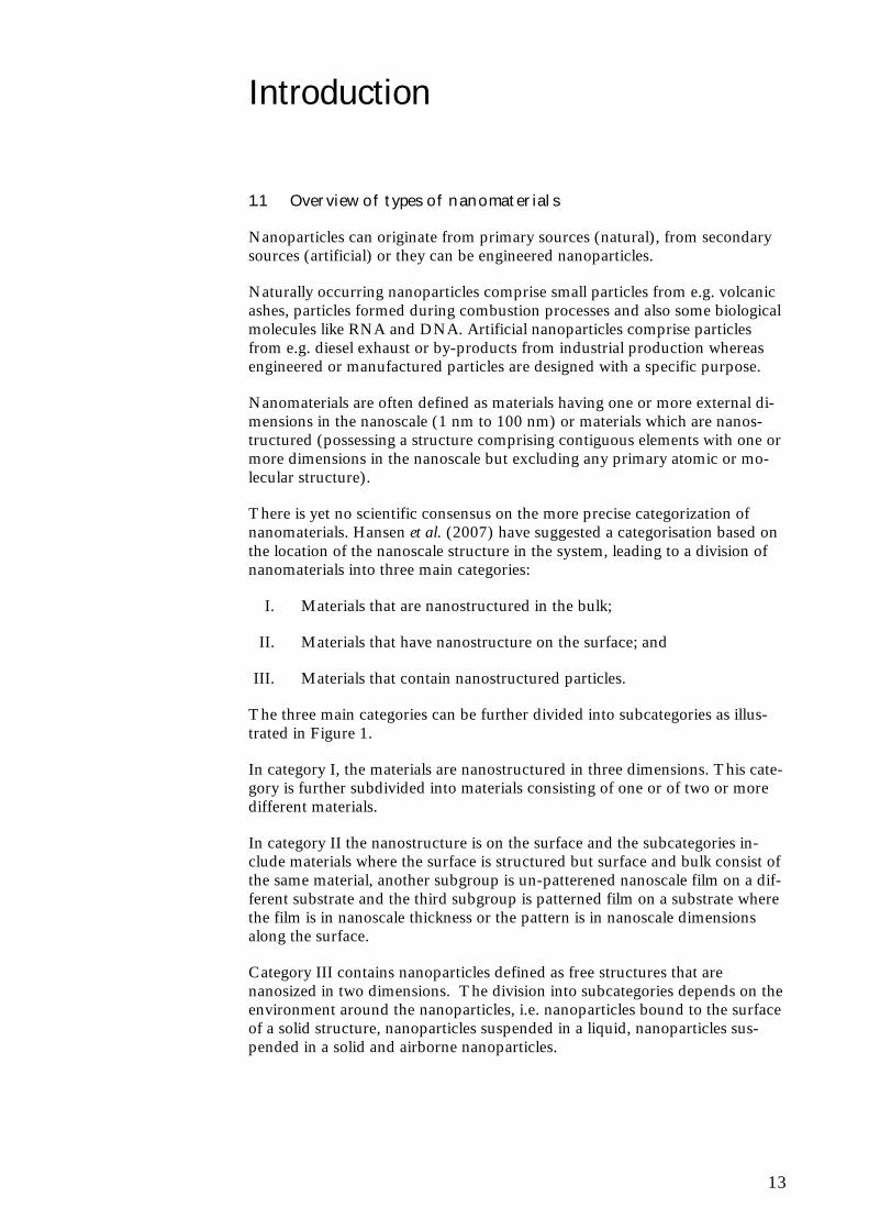
There is yet no scientific consensus on the more precise categorization of nanomaterials. Hansen et al. (2007) have suggested a categorisation based on the location of the nanoscale structure in the system, leading to a division of nanomaterials into three main categories:
I. Materials that are nanostructured in the bulk;
II. Materials that have nanostructure on the surface; and
III. Materials that contain nanostructured particles.
The three main categories can be further divided into subcategories as illus-trated in Figure 1.
In category I, the materials are nanostructured in three dimensions. This cate-gory is further subdivided into materials consisting of one or of two or more different materials.
In category II the nanostructure is on the surface and the subcategories in-clude materials where the surface is structured but surface and bulk consist of the same material, another subgroup is un-patterened nanoscale film on a dif-ferent substrate and the third subgroup is patterned film on a substrate where the film is in nanoscale thickness or the pattern is in nanoscale dimensions along the surface.
Category III contains nanoparticles defined as free structures that are nanosized in two dimensions. The division into subcategories depends on the environment around the nanoparticles, i.e. nanoparticles bound to the surface of a solid structure, nanoparticles suspended in a liquid, nanoparticles sus-pended in a solid and airborne nanoparticles.
14
Figure 1 The categorization framework for nanomaterials as suggested by Hansen et al. (2007)
This categorisation closely corresponds to the classification of nanomaterials according to dimensions which are not confined to the nanoscale range, where nanomaterials can be classified as zero-dimensional (0-D), one-dimensional (1-D), two-dimensional (2-D) and three-dimensional (3-D).
0-D nanomaterials are those materials where all dimensions are nanoscale, most commonly nanoparticles; 1-D nanomaterials have one dimension out-side the nanoscale, e.g. nanotubes, -rods and -wires; 2-D nanomaterials have two dimensions outside the nanoscale, e.g. nanofilms and graphene-based composites; and 3-D nanomaterials have three dimensions outside the nano-scale, e.g. heterogeneous nanostructures like mesoporous carbonbased com-posites and nanostructured networks.
The DEPA report (Environmental Project No. 1206, 2007) distinguishes be-tween six types of nanomaterials based on the shape characteristics of the ma-terials:
• Nanoparticles with all three dimensions in the nanoscale range (e.g. TiO2 nanoparticles);
• Nanofibres and tubes with at least two dimensions in the nanoscale range and an aspect ratio of more than 3 (primarily carbon nanotubes);
• Nanostructured surfaces (protrusions or grooves in the nanoscale range);
• Nanofilm (coatings with layers thinner than 100 nm e.g. cured film on glass);
• Nano Flakes with at least one dimension in the nanoscale range (e.g. nano-clay (silicate) and the materials used in semiconductor elements);
• Nanoporous structures with pore sizes in the nanoscale range (such as ceramic materials used as catalysts).
The most commonly available engineered nanomaterials can be organised into four types (EPA, 2007):
15
Carbonbased materials Carbonbased materials are composed mainly of carbon and commonly shaped as hollow spheres, ellipsoids, or tubes. Spherical and ellipsoidal carbon nanomaterials are referred to as fullerenes, while cylindrical ones are called nanotubes. These particles have many potential applications, including im-proved films and coatings, stronger and lighter materials, and applications in electronics.
Metal-based materials These nanomaterials include quantum dots, nanogold, nanosilver, zero valent iron and metal oxides, such as titanium dioxide, and cerium oxide. A quan-tum dot is a closely packed semiconductor crystal comprised of hundreds or thousands of atoms, and whose size is on the order of a few nanometers to a few hundred nanometers. Changing the size of quantum dots changes their optical properties.
Dendrimers These nanomaterials are nanosized polymers built from branched units. The surface of a dendrimer has numerous chain ends, which can be tailored to perform specific chemical functions. This property could also be useful for catalysis. Also, because three-dimensional dendrimers contain interior cavities into which other molecules could be placed, they may be useful for drug de-livery.
Composites Composites combine nanoparticles with other nanoparticles or with larger, bulk-type materials. Nanoparticles, such as nanosized clays, are already being added to products ranging from auto parts to packaging materials, to enhance mechanical, thermal, barrier and flame-retardant properties.
1.2 Nanomaterials in consumer products
Nanomaterials which are already widely used in various consumer products and therefore also are focus for research into environmental, health and safety aspects of the materials include:
Carbon tubes and fullerenes. Carbon materials have a wide range of uses, ranging from composites for use in vehicles and sports equipment, to in-tegrated circuits for electronic components.
Cerium dioxide. Nano cerium is being investigated for uses ranging from drug delivery to automobile catalytic converters. Currently a major use in some countries is as a diesel fuel additive to reduce exhaust particulates and increase fuel mileage.
Titanium dioxide. Nano titanium dioxide is currently used in many products. Depending on the type of particle, it may be found in sun-screens, cosmetics, food additives and paints and coatings. It is also being investigated for use in removing contaminants from drinking water.
Silicium dioxide. Silicium dioxide is like titanium dioxide used in many products including sunscreens, cosmetics, paints and cement. Silicium dioxide is also used in the food industry.
16
Silver. Nanosilver has long been known for its antimicrobial properties. Nanosilver is being incorporated into textiles and other materials to eliminate bacteria and odour from clothing, food packaging and other items where antimicrobial properties are desirable.
Iron. While nano-scale iron is being investigated for many uses, including “smart fluids” for uses such as optics polishing and as better-absorbed iron nutrient supplement, one of its more-prominent current applications is for remediation of polluted groundwater and soil.
Zinc oxide. Zinc oxide is very UV-stable and is used as sunscreen in cosmetic products and UV-stabiliser in plastics. Zinc oxide also exhibits anti-bacterial properties which are utilised in pharmaceutical applications.
Nanoclay. Nanoclay (aluminium silicon oxide) is used as additive for re-inforced plastics improving both mechanical and thermal properties as well as barrier characteristics. It is used in food packaging where it re-duces the permeation rate of oxygen through the packaging material and more recently it has also been used as synergist flame retardant to substi-tute the halogen-containing flame retardants.
Other types of nanomaterials are gold nanoparticles and dendrimers, but they are mainly explored in relation to medical and other more specialised applica-tions, although patents are already filed for the application of dendrimers in various cosmetic products.
Examples of consumer products (PEN, 2011) that use nanomaterials are:
Health and Fitness: toothpaste, toothbrush, tennis racket, air filter, sunscreen, antibacterial socks, cosmetics, waste and stain resistant pants, golf clubs, wound dressings, pregnancy tests, bath and sports towels
Electronics and Computers: computer displays, computer hardware, games
Home and Garden: paint, antimicrobial pillows, stain resistant cush-ions, humidifiers, cleaners, fabric softeners
Food and Beverage: non-stick coatings, antimicrobial refrigerator, ca-nola oil, food storage containers, packaging
Other: coatings, lubricants
1.3 Special characteristics of nanomaterials vs. bulk materials
The special characteristics and properties of nanomaterials are largely attrib-uted to the small size and the very large surface area to volume ratio. This makes a large fraction of the atoms available on the surface and results in more surface-dependent material properties which again may enhance or modify the properties of the bulk material.
It should, however, be stressed that many conventional bulk chemicals with various applications in e.g. consumer products, foodstuffs and construction materials also contain a naturally occurring nanosized fraction. This is the case for titanium dioxide, silicium dioxide and clays. Other examples include
17
products containing nanosilver particles, which have been commercially avail-able for over 100 years, and have been used in applications as pigments, pho-tographics, wound treatment, conductive/antistatic composites, catalysts and as biocides (Nowack et al., 2011).
Nanomaterials in general are known to have many novel properties that are already utilised or explored for use in different products and technologies and that allow new areas of application of these materials. These properties in-clude mechanical, thermal, biological, optical and chemical properties, which may also affect the potential exposure to the materials, as well as the health end environmental effects from that exposure. Information on potential haz-ards to health and environment is therefore urgently needed.
A key challenge in relation to characterisation of the materials is that the dif-ferent nanomaterials exist in various forms, sizes and shapes and cannot be described by a unique set of parameters. Their small sizes, complex struc-tures, potential property changes during synthesis and use, sensitivity to and interaction with the surrounding environment also add to the challenge of characterization and providing sufficient information for assessing potential risks of the nanomaterials. Furthermore, information available in the literature is often very scarce and not always reported in a form relevant for risk assess-ment.
Key physical-chemical parameters (list of endpoints) to take into account, when testing specific manufactured nanomaterials for human health and envi-ronmental safety within phase one of the OECD testing program include (OECD, 2010):
Agglomeration and/or aggregation
Water solubility / Dispersability
Crystalline phase
Dustiness
Crystallite size
Particle size distribution - dry and in relevant media
Specific surface area
Zeta potential (surface charge)
Surface chemistry (where appropriate)
Photocatalytic activity
Pour density
Porosity
Octanol-water partition coefficient, where relevant
Redox potential
18
Radical formation potential
Other relevant physical-chemical properties and material characterisa-tion information (where available)
Further studies investigating the relation between these parameters and the toxicity of nanomaterials are needed, and will need to be addressed at some point in the evaluation and risk assessment of nanomaterials.
The current EU legislative framework for chemicals covers in principle the potential health, safety and environmental risks posed by nanomateri-als. However, there is also a recognised need to modify this legislation, in order to reflect the specific properties of nanomaterials, and the need for more elaborate characterisation of the nanomaterials compared to the conventional bulk form to include e.g. the specific surface properties. The existing testing requirements for bulk chemicals may also not be adequate in all areas of toxicity, and the same is the situation with regard to classifi-cation and labelling of substances and mixtures and thereby also toxicity-based thresholds based on these criteria.
REACH provides the overarching legislation applying to the manufacture, placing on the market, and use of substances on their own, in preparations or in articles. The current view from the Commission is that the legislation in place to a large extent covers risks in relation to nanomaterials, and that the risks can be dealt with under the current legislative framework includ-ing REACH. However, modification is expected for example with regard to thresholds used in some legislation and with regard to testing methods, test guidelines and risk assessment of nanomaterials.
1.4 Use of nanomaterials in Denmark
1.4.1 Industry and products
There is no single source of information that provides an overview of the use of nanomaterials and products in Denmark or in the EU for that matter. Pieces of information are, however, available.
Recently, the Nanowerk published an online Company & Labs directory1 with 4,196 links to labs, associations, networks and companies. This directory in-cludes only companies and labs that work with and/or commercialise nanotechnology and/or nanomaterials and does not include entities that only have “nano” in their name.
According to Nanowerk there are 18 commercial companies in Denmark in-volved in various fields of nanotechnology. The majority are relatively new and very specialised companies and some have emerged from university re-search. One or two do not seem to be operational anymore in December 2010 based on their homepage information. The list of companies is presented in Annex 1.
1 http://www.nanowerk.com/nanotechnology/research/nanotechnology_links.php
19
1.4.2 Results from selected Nordic surveys
Conclusions regarding consumer products from three recent surveys from Denmark and Norway on products containing nanomaterials or based on nanotechnology are presented in this section.
Survey on production and application of nanomaterials in Danish industry
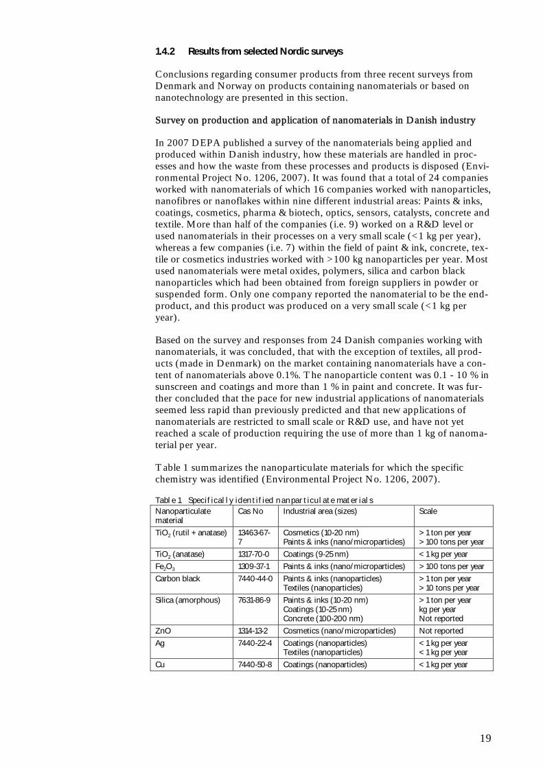
In 2007 DEPA published a survey of the nanomaterials being applied and produced within Danish industry, how these materials are handled in proc-esses and how the waste from these processes and products is disposed (Envi-ronmental Project No. 1206, 2007). It was found that a total of 24 companies worked with nanomaterials of which 16 companies worked with nanoparticles, nanofibres or nanoflakes within nine different industrial areas: Paints & inks, coatings, cosmetics, pharma & biotech, optics, sensors, catalysts, concrete and textile. More than half of the companies (i.e. 9) worked on a R&D level or used nanomaterials in their processes on a very small scale (<1 kg per year), whereas a few companies (i.e. 7) within the field of paint & ink, concrete, tex-tile or cosmetics industries worked with >100 kg nanoparticles per year. Most used nanomaterials were metal oxides, polymers, silica and carbon black nanoparticles which had been obtained from foreign suppliers in powder or suspended form. Only one company reported the nanomaterial to be the end-product, and this product was produced on a very small scale (<1 kg per year).
Based on the survey and responses from 24 Danish companies working with nanomaterials, it was concluded, that with the exception of textiles, all prod-ucts (made in Denmark) on the market containing nanomaterials have a con-tent of nanomaterials above 0.1%. The nanoparticle content was 0.1 - 10 % in sunscreen and coatings and more than 1 % in paint and concrete. It was fur-ther concluded that the pace for new industrial applications of nanomaterials seemed less rapid than previously predicted and that new applications of nanomaterials are restricted to small scale or R&D use, and have not yet reached a scale of production requiring the use of more than 1 kg of nanoma-terial per year.
Table 1 summarizes the nanoparticulate materials for which the specific chemistry was identified (Environmental Project No. 1206, 2007).
Table 1 Specifically identified nanparticulate materials Nanoparticulate material
Cas No Industrial area (sizes) Scale
TiO2 (rutil + anatase) 13463-67-7
Cosmetics (10-20 nm) Paints & inks (nano/microparticles)
> 1 ton per year > 100 tons per year
TiO2 (anatase) 1317-70-0 Coatings (9-25 nm) < 1 kg per year
Fe2O3 1309-37-1 Paints & inks (nano/microparticles) > 100 tons per year
Carbon black 7440-44-0 Paints & inks (nanoparticles) Textiles (nanoparticles)
> 1 ton per year > 10 tons per year
Silica (amorphous) 7631-86-9 Paints & inks (10-20 nm) Coatings (10-25 nm) Concrete (100-200 nm)
> 1 ton per year kg per year Not reported
ZnO 1314-13-2 Cosmetics (nano/microparticles) Not reported
Ag 7440-22-4 Coatings (nanoparticles) Textiles (nanoparticles)
< 1 kg per year < 1 kg per year
Cu 7440-50-8 Coatings (nanoparticles) < 1 kg per year
20
The particle size of the used materials varies from approximately 10 nm and more and in the case of metaloxide (pigments in the paints & ink industry), silica (concrete industry) and zinc oxide (cosmetics) the medium particulate particle size is >> 100 nm. The companies estimated particle size distribution of the used materials is so broad that an unknown fraction of the particles falls within the usual definition of nanoparticles.
Commercialised nanoproducts in Denmark
In 2007, DEPA initiated a survey in order to identify consumer products available to the Danish consumer (Consumer Survey No. 81, 2007). The sur-vey was based on interviews and questionnaires submitted to stakeholders in Denmark, internet searches and follow-up on search results of consumer products in the Consumer Product Inventory maintained by the Project of Emerging Nanotechnologies at the Woodrow Wilson Centre in Washington, DC, USA.
As there is no legal requirement for producers or importers of products to de-clare the content of nanomaterials, it is not possible to be certain, that a pro-ducer or importer who uses the prefix ‘nano’ in association with a product are referring to a content of nanoparticles, or if a nanomaterial is formed during use or whether it is the technology behind the product that is ‘nano’ (Con-sumer Survey No. 81, 2007).
The survey found that 243 products based on a nanomaterial were available on the Danish consumer market. The searches for Danish importers and dis-tributors of products in the Woodrow Wilson database and Danish web shops selling these articles showed that two out of three products registered in the U.S.A. in general are for sale in Denmark (Consumer Survey No. 81, 2007).
The report further concludes, that more than two thirds of the products on the Danish market (i.e. 154 products) – are various liquid products, partly for surface treatment of a great number of materials such as glass, concrete, metal (especially car maintenance) glass fibres and textiles, and partly for skin pro-tection products, especially sun lotions. The remaining products are in par-ticular sporting goods- and clothing, which account for 60 out of the 99 re-maining products (Consumer Survey No. 81, 2007).
More than half of the consumer products on the Danish market are products from Europe. Out of the 135 European products on the Danish market, al-most 100 come from Germany. The remaining products originate from United Kingdom, Finland and France. Three products are sun lotions formu-lated in Denmark. In 202 out of the 243 products it was not possible to iden-tify the nanomaterial in the product. Of the 41 known nanomaterials, half of them were found in cosmetic products (six products with zinc oxide and 13 with titanium dioxide), 10 with antibacterial silver in textiles and home appli-ances, and 12 with carbon tubes or balls (seven with carbon tubes in sporting goods and five with fullerenes in cosmetics) (Consumer Survey No. 81, 2007).
As part of the survey it was found, that a considerable part of the consumer products are sold in web shops in Denmark and abroad, especially products for surface treatment within the product types `Car care products and acces-sories’, ‘Home and gardening’ and ‘Personal care and sports equipment’, but a smaller and increasing part is found in ordinary shops. A group of paints contain ‘carbon black’ (20-100 nm) as colouring agent or silica (down to
21
approx. 10 nm) as thickening agent. Both these materials have been used for a number of years, but are only now recognized as nanomaterials. In the Danish Product Register a great number of individual products are registered with carbon black (approx. 9,500) or silicium dioxide (approx. 15,500). The regis-trations do, however, not include information about whether these substances include particles in the nanoscale. The registered amounts used in paints are 483 tons carbon black and 622 tons silicium dioxide. The individual products containing these materials have not been further analysed (Consumer Survey No. 81, 2007).
With regard to potential exposure of consumers the survey concludes that in most products containing nanomaterials on the Danish market the nanomate-rials are suspended in liquid and that these products constitute the greatest likelihood of exposure of the consumer. These products include products for surface treatment and cosmetics. No products with free nanoparticles were identified (Consumer Survey No. 81, 2007).
Survey on production, import and use of nanomaterials in Norway
In the survey carried out in 2010 based on questionnaires sent to companies expected to produce, import or use nanomaterials, 27 out of 162 responded positively. Nanomaterials were primarily titanium dioxide, polymers, carbon nanotubes and carbon nanofibres mostly in powder form. Other forms in-cluded suspensions, composite materials and films. Particle size was reported to be in the range of 20 to 100 nm for all types of powder (Norwegian Labour Inspection Authority, 2010).
The Norwegian Product Register has in mid 2009 started voluntary registra-tion of products containing nanomaterials. Registration covers in general all products with a hazard classification and produced or imported in volumes of 100 kg or more. Cosmetics are not included. By 20 June 2010 19 products containing nanomaterials were registered of which 13 were consumer prod-ucts. Typical products were paints and lacquers, car care products, products for impregnation and windscreen wash (Norwegian Labour Inspection Au-thority, 2010).
Use of nanomaterials in Sweden in 2008 - analysis and prognosis
The Swedish Chemicals Agency has published a survey on the use of nano-materials in Sweden in 2008 (KemI, 2009). According to this survey, the nanomaterials in Swedish nanoproducts can be categorised as 7 % ceramic materials, 13 % carbon-based, 7 % metals, and 5 % polymers in terms of the number of products. It is also stated in the report that it is difficult to get a true picture of nanoproducts that are on the market. This is partly due to the fact, that in most cases it is not obvious from the product in-formation, that the product contains a nanomaterial, nor what material it is, if they do. Another problem is that consumers are purchasing the nano-products directly on the Internet.
An overview from an English summary of the report of examples of nanomaterials in products on the Swedish market is shown in Table 2.
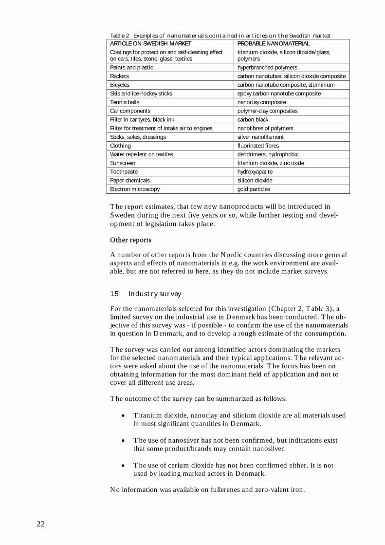
22
Table 2 Examples of nanomaterials contained in articles on the Swedish market ARTICLE ON SWEDISH MARKET PROBABLE NANOMATERIAL
Coatings for protection and self-cleaning effect on cars, tiles, stone, glass, textiles
titanium dioxide, silicon dioxide/glass, polymers
Paints and plastic hyperbranched polymers
Rackets carbon nanotubes, silicon dioxide composite
Bicycles carbon nanotube composite, aluminium
Skis and ice-hockey sticks epoxy carbon nanotube composite
Tennis balls nanoclay composite
Car components polymer-clay composites
Filler in car tyres, black ink carbon black
Filter for treatment of intake air to engines nanofibres of polymers
Socks, soles, dressings silver nanofilament
Clothing fluorinated fibres
Water repellent on textiles dendrimers, hydrophobic
Sunscreen titanium dioxide, zinc oxide
Toothpaste hydroxyapatite
Paper chemicals silicon dioxide
Electron microscopy gold particles
The report estimates, that few new nanoproducts will be introduced in Sweden during the next five years or so, while further testing and devel-opment of legislation takes place.
Other reports
A number of other reports from the Nordic countries discussing more general aspects and effects of nanomaterials in e.g. the work environment are avail-able, but are not referred to here, as they do not include market surveys.
1.5 Industry survey
For the nanomaterials selected for this investigation (Chapter 2, Table 3), a limited survey on the industrial use in Denmark has been conducted. The ob-jective of this survey was - if possible - to confirm the use of the nanomaterials in question in Denmark, and to develop a rough estimate of the consumption.
The survey was carried out among identified actors dominating the markets for the selected nanomaterials and their typical applications. The relevant ac-tors were asked about the use of the nanomaterials. The focus has been on obtaining information for the most dominant field of application and not to cover all different use areas.
The outcome of the survey can be summarized as follows:
Titanium dioxide, nanoclay and silicium dioxide are all materials used in most significant quantities in Denmark.
The use of nanosilver has not been confirmed, but indications exist that some product/brands may contain nanosilver.
The use of cerium dioxide has not been confirmed either. It is not used by leading marked actors in Denmark.
No information was available on fullerenes and zero-valent iron.
23
2 Nanomaterials survey
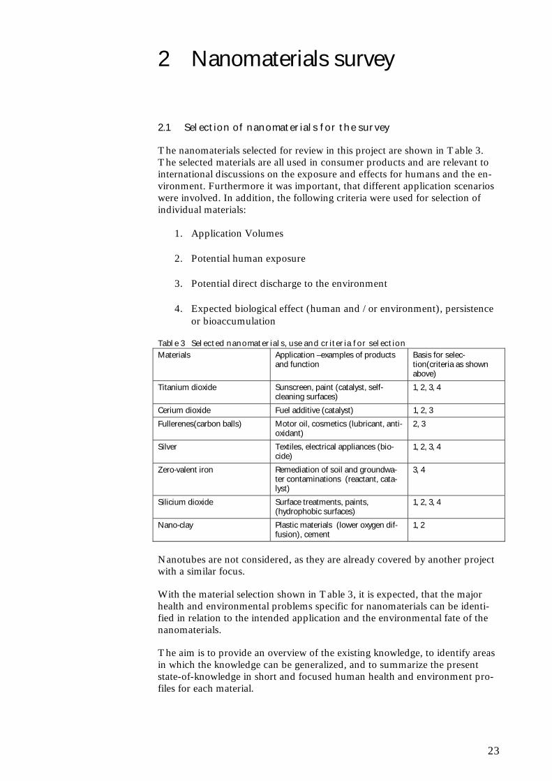
2.1 Selection of nanomaterials for the survey
The nanomaterials selected for review in this project are shown in Table 3. The selected materials are all used in consumer products and are relevant to international discussions on the exposure and effects for humans and the en-vironment. Furthermore it was important, that different application scenarios were involved. In addition, the following criteria were used for selection of individual materials:
1. Application Volumes
2. Potential human exposure
3. Potential direct discharge to the environment
4. Expected biological effect (human and / or environment), persistence or bioaccumulation
Table 3 Selected nanomaterials, use and criteria for selection Materials Application –examples of products
and function Basis for selec-tion(criteria as shown above)
Titanium dioxide Sunscreen, paint (catalyst, self-cleaning surfaces)
1, 2, 3, 4
Cerium dioxide Fuel additive (catalyst) 1, 2, 3
Fullerenes(carbon balls) Motor oil, cosmetics (lubricant, anti-oxidant)
2, 3
Silver Textiles, electrical appliances (bio-cide)
1, 2, 3, 4
Zero-valent iron Remediation of soil and groundwa-ter contaminations (reactant, cata-lyst)
3, 4
Silicium dioxide Surface treatments, paints, (hydrophobic surfaces)
1, 2, 3, 4
Nano-clay Plastic materials (lower oxygen dif-fusion), cement
1, 2
Nanotubes are not considered, as they are already covered by another project with a similar focus.
With the material selection shown in Table 3, it is expected, that the major health and environmental problems specific for nanomaterials can be identi-fied in relation to the intended application and the environmental fate of the nanomaterials.
The aim is to provide an overview of the existing knowledge, to identify areas in which the knowledge can be generalized, and to summarize the present state-of-knowledge in short and focused human health and environment pro-files for each material.
24
To focus the characterisation, it is the pure form of the nanomaterials that is discussed in the following, thereby ignoring that various kinds of doping and coatings might exists. Furthermore, the overview of the key characteristics is primarily based on the form of the nanomaterials that is commercially avail-able.
2.2 Nanomaterials profiles
In chapter 2-7 a profile for each of the selected materials will be developed. For each material the focus has been on the general characteristics and manu-facturing of the nanomaterials, their current uses (mainly focussed at con-sumer products), and hazard profiles (ecotoxicity and human toxicity).
2.2.1 Manufacturing and applications
The manufacturing processes and applications are described based on a litera-ture review, and the available information from the small industry survey is also included. Furthermore, an updated review of the commercially available consumer products in Denmark containing the selected nanomaterials is in-cluded in this report based on the methodology described in Consumer Pro-ject No. 81 (2007). Outset is taken in the online “Nanotechnology Consumer Products Inventory” maintained by the Project of Emerging Nanotechnolo-gies at the Woodrow Wilson International Center for Scholars. The Nanotechnology Consumer Products Inventory was launched in 2005 with the inclusion of 54 products – a number that one year later had increased to 356 products. In the years 2007-2009, the number of products listed in the inventory continued to increase (580 products in 2007, 803 products in 2008, and 1015 products in 2009). In the most recent update in March 2011, a total of 1317 products were listed as commercially available worldwide from a wide variety of producers and countries (Woodrow Wilson Inventory, 2011). For products to be included in the inventory it has to fulfil mainly the following conditions: The products can be purchased directly by the consumers or iden-tified by the producer or another source as based on nanotechnology and the information about nanomaterials in the product seems probable.
The database divides the products in a number of categories: Appliances (heating, cooling and air; large kitchen appliances, air cleaners and air condi-tion devices, domestic appliances, bio-up and textile protection products), Automotive (exterior) maintenance and accessories, Goods for children (ba-sics; toys and games), Electronics and computers (audio; cameras and film, computer hardware; display; mobile devices and communications, television; video), Foodstuffs (cooking, foodstuffs, storage, dietary supplement), Health and fitness (clothing, cosmetics, filtration, personal care, sporting goods, sun screen), Home and garden (cleaning, construction materials, home furnish-ings; luxury products; paint), and Surface treatment (overlapping several groups).
In order to identify products that contain nanomaterials, and which are com-mercially available in Denmark, it was investigated if the products registered in the database, are also marketed in Denmark or may be available through a web shop. This was done for the 2009 data since the most recent update co-incide with the termination of the present project.
It should be noted, however, that as there is no legal requirement to producers or importers of products to declare the content of nanomaterials, it is not pos-
25
sible to be certain that a producer or importer, who uses the prefix ‘nano’ in association with a product, is referring to a content of nanoparticles, a nano-meter thin surface layer that is formed upon use, a nanotechnological function expressed when used, or whether it is the technology behind the product that is ‘nano’. The term ‘nano’ may also be used in advertisement without a back-ground in real nanotechnology and this is also a factor that may bias attempts to make inventories of nano-based products. It should be emphasised that the information in the inventory is based only on information that can be found on the internet and therefore products solely sold by non-internet retailers are not included.
Using the same methodology as in Consumer Project No. 81 (2007), we iden-tified a total of 612 products to be available in Denmark (of the 1015 prod-ucts included in the database in 2009) primarily through various webshops compared to 243 products in 2007 as identified in Consumer Project No. 81 (2007). It is unknown, which nanomaterials are used in more than half of the products identified. For each of the selected nanomaterials, the results of this survey are included in the nanomaterials profiles in Chapter 2-7. It should be noted, that the methodology used, may for some product types lead to an un-derestimation of the actual number of products on the market and for others the number of “real” nano-based products may be overestimated.
2.2.2 Ecotoxicological and toxicological profiles
The sections on ecotoxicolgical and toxicological profiles are short summaries primarily of results of literature reviews published in the open peer-reviewed literature. In this respect the ENRHES review (Stone et al., 2010) proved to be an especially valuable source of information. Therefore, a number of sec-tions are extracted from the ENRHES review and updated to the state-of-knowledge by the end of 2010.
As nanospecific standard procedures for ecotoxicological and toxicological testing as well as nanospecific control of assays and assay conditions are not yet developed it can be difficult to compare and conclude about the available results. This is further complicated by the fact that characterisation of the par-ticles in biological systems is often insufficient and sometimes leads to con-flicting evidence in the literature.
2.2.3 Relevant exposures
With regard to the exposure, focus is on nanomaterials from consumer prod-ucts that can end up in the environment and the potential exposure routes relevant for consumers via direct exposure to the products containing the ma-terials or indirectly via the environment.
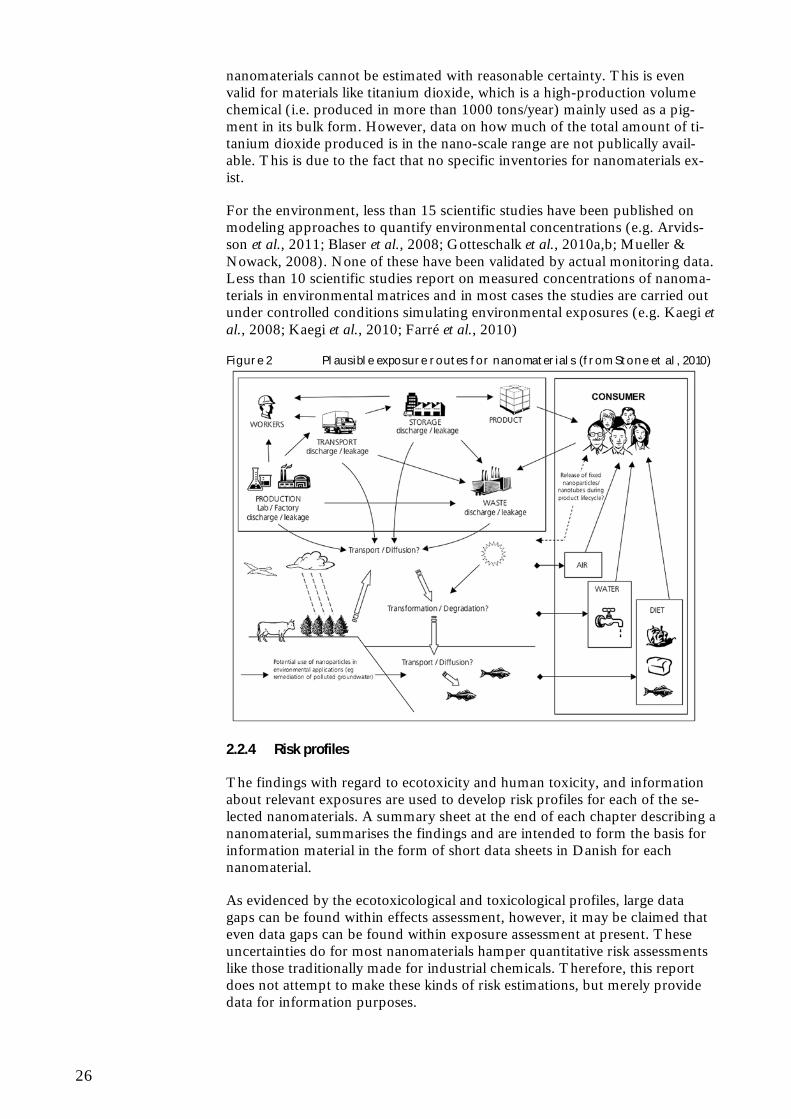
An illustration of the plausible exposure routes for nanomaterials from the ENRHES study is presented in Figure 2. It must be emphasized that at pre-sent the level of knowledge on actual environmental or human exposures to nanomaterials originating from consumer products is extremely limited. Even though it is likely that the methods for exposure assessment for “ordinary” chemicals can be transferred to nanomaterials in consumer products (Hansen et al., 2008), the pronounced lack of data hampers quantitative exposure as-sessments. The actual content of nanomaterials used in consumer products is often unknown and realistic human exposure scenarios are therefore difficult to evaluate in a quantitative way. For more generic overviews and model pre-dictions, it is a major limiting factor that the production volumes of specific
26
nanomaterials cannot be estimated with reasonable certainty. This is even valid for materials like titanium dioxide, which is a high-production volume chemical (i.e. produced in more than 1000 tons/year) mainly used as a pig-ment in its bulk form. However, data on how much of the total amount of ti-tanium dioxide produced is in the nano-scale range are not publically avail-able. This is due to the fact that no specific inventories for nanomaterials ex-ist.
For the environment, less than 15 scientific studies have been published on modeling approaches to quantify environmental concentrations (e.g. Arvids-son et al., 2011; Blaser et al., 2008; Gotteschalk et al., 2010a,b; Mueller & Nowack, 2008). None of these have been validated by actual monitoring data. Less than 10 scientific studies report on measured concentrations of nanoma-terials in environmental matrices and in most cases the studies are carried out under controlled conditions simulating environmental exposures (e.g. Kaegi et al., 2008; Kaegi et al., 2010; Farré et al., 2010)
Figure 2 Plausible exposure routes for nanomaterials (from Stone et al, 2010)
2.2.4 Risk profiles
The findings with regard to ecotoxicity and human toxicity, and information about relevant exposures are used to develop risk profiles for each of the se-lected nanomaterials. A summary sheet at the end of each chapter describing a nanomaterial, summarises the findings and are intended to form the basis for information material in the form of short data sheets in Danish for each nanomaterial.
As evidenced by the ecotoxicological and toxicological profiles, large data gaps can be found within effects assessment, however, it may be claimed that even data gaps can be found within exposure assessment at present. These uncertainties do for most nanomaterials hamper quantitative risk assessments like those traditionally made for industrial chemicals. Therefore, this report does not attempt to make these kinds of risk estimations, but merely provide data for information purposes.
27
3 Fullerenes - C60
3.1 General characteristics
Fullerenes consist of a family of soccer-ball shaped molecules with 60 or more carbon atoms arranged in closed spherical and elliptic structures as each car-bon atom bonded to three others in pentagonal and hexagonal rings. The size of a fullerene is approximately 1 nm. C60 has long been regarded as the poster child of nanotechnology, but the fullerenes furthermore consists of the C70, C76 and C84 (Health and Safety Executive, 2004). Fullerenes can furthermore con-sist of a number of layers which are called onions (Terrones & Terrones, 2003). C60-fullerenes are extremely stable and are extremely temperature and pressure resistant. Fullerenes have a tendency to agglomerate and form clus-ters of crystals, termed nanoC60 or nC60. Although normally considered to be an insoluble material, C60 can be surface modified making it more water solu-ble and thereby provide them with a great variation in their physico-chemical properties as well as biological activity (Stone et al. 2010). Modification can be through the attachment of e.g. hydrophilic groups, peptides, or carbonhy-drates which changes the properties of C60 (Sayes et al. 2004, European Commission, 2005).
3.2 Manufacturing processes
Different methods exists to produce fullerenes, but most large scale manufac-turers use a combustion process, which uses hydrocarbons as a raw material to produce C60. Toluene is fed together with oxygen to a low-pressure com-bustion chamber. The flame produces fullerene-enriched soot, which is ex-tracted and filtered (Takeara et al., 2004). Fullerenes are then extracted from the soot by solvents (e.g. chlorobenzene and toluene) in a tank where the in-soluble soot settle to the bottom while C60 are dissolved in the solution. Puri-fied fullerenes are collected after the solvent has evaporated and appear as a black powder. The separation of single size fullerenes (C60, C70) and the de-gree of purity determine the price of the final product.
3.3 Uses
3.3.1 Main applications
C60 has been applied in a number of different consumer products such as sports gear (badminton- and tennis rackets), cosmetics and personal care products (anti-aging, eyeliner, skin creams, etc.) and also lubricants (motor oil) (Franco et al., 2007, Consumer Survey No. 81, 2007). Other current uses include energy applications (such as fuel cells, solar cells and batteries), cata-lysts, polymer modifications and targeted drug delivery systems (Aschberger et al., 2010). In total five products have been identified to be commercially available on the Danish marked. While C60-containing cosmetics and personal care products can be bought online (Franco et al., 2007, Consumer Survey No. 81, 2007), only products related to sports gear have been identified on the Danish market. In these products C60 is added to strengthen the structure of e.g. tennis rackets where the C60-fullerenes are dispersed in a resin between
28
carbon fibers. The C60 content of e.g. sports gear and lubricants is unknown. For patents filed in US in regard to cosmetics, concentrations between 0.05 and 6 % are reported. Concentrations used are the highest in eyeliner and mascara with 6% and 5%, respectively (Boxall et al., 2008). In lubricants, fullerene soot containing approximately 3.2 % fullerenes is added to improve sliding between metal surfaces. With a soot content of 9.4 %, the concentra-tion of C60 is estimated at about 3 g/kg. The fullerene molecules are expected to be partly free during use and unintended release should therefore be con-sidered in an exposure assessment (Franco et al., 2007).
The production and use of fullerenes is assumed to be limited at present, but expected to grow significantly over the next decade. In Japan a large scale production plant has been opened recently with a production capacity of 40 tons per year (Aitken et al., 2006 and Fujitani et al., 2008 in Aschberger et al., 2009).
3.3.2 Results from industry survey
No information on fullerenes has been obtained.
3.4 Eco-toxicological profile
For the following overview of the ecotoxicological profile of C60-fullerenes it should be stressed that only a few ecotoxicological studies exist and only a part of these are aimed at the base-set organisms (fish, crustacean and algae) required for doing effects assessment according to REACH. Even fewer stud-ies report the results in terms of the endpoints and values listed in Information Requirements of the REACH (e.g., LC50, EC50, NOEC, LOEC) (ECHA, 2008).
The testing of C60 in ecotoxicological tests is difficult due to the very low wa-ter solubility of the compound. This has lead to the use of organic solvents, which has been shown to influence the ecotoxic response. Thus, studies have demonstrated that the degradation of tetrahydrofuran (THF), used as a sol-vent in many of the early ecotoxicity studies on C60, may in fact be the cause of the toxicity observed (Stone et al., 2010). In general, less adverse effects are observed when using water-stirred or sonicated C60 compared to C60 tested in the presence of tetrahydrofuran or dimethylsulfoxide as solvents. For instance, a number of studies observed no effect on the survival of larval zebrafish and fathead minnow after exposure to stirred and sonicated C60, whereas increased mortality and elevated lipid peroxidation was found after exposure to THF- C60 (Henry et al., 2007; Zhu et al., 2006a; Oberdörster et al., 2006). Evidence from studies on Daphnia magna also shows a large difference between the ecotoxicity of water-stirred and THF- C60 (Lovern and Klaper 2006; Lovern et al., 2007; Zhu et al., 2006). These interactions lead Stone et al. (2010) to the conclusions that ecotoxicological studies carried out with tetrahydrofuran do not have a high credibility.
In C60-suspensions prepared by long-term stirring, Zhu et al. (2008) observed no mortality or unusual behaviours of juvenile carp (Carassius auratus) after 32 days of exposure to between 0.04–1.0 mg/L. However, a significantly re-duction in the mean total length was observed after 32 d exposure to 0.2 mg/L of C60 and a significantly reduced body weight at 1.0 mg/ L. No detectable
29
effects were observed after exposure to 0.04 mg/L for 32 days. This might correspond to a NOEC of 0.04 mg/L and a LOEC of 0.2 mg/L for the length of juvenile carp and a LOEC of 1.0 mg/L for the body weight after exposure to C60 in water for 32 days.
For crustaceans, the 48 h lethality study by Lovern and Klaper (2006) re-ported on a great variation in mortality in Daphnia magna, but a LC50, 48h of 7.9 mg/L for water-sonicated C60 could be established. LOEC and NOEC were reported to be 0.5 mg/L and 0.2 mg/L, respectively.
In regard to survival and reproductive endpoints a significant reduction in the number of offspring after 21 days and delays in moulting of the carapace was observed by Oberdörster et al. (2006) after exposing Daphnia magna to 2.5 mg/L water-stirred C60. An increased cumulative mortality and significant de-lay in moulting and reduced offspring was reported as well after exposure to 1-5 mg/L for 21 days (Oberdörster et al. 2006).
For earthworms no significant mortality was found after consuming dry food spiked with 99.5% C60 concentrations of 1 g/kg dry food for up to 28 days (Scott-Fordsmand et al., 2008). Johansen et al. (2008) found that exposure to 50 mg/kg to 99.5% C60 aggregates caused a 60% inhibition of the number of bacterial colony-forming units CFUs in clay loam soil 3 hours after incorpora-tion.
No studies on bioaccumulation of fullerenes have been reported in the litera-ture. No studies on the degradability of fullerenes have been found in the lit-erature. The cage-like structure of C60 suggests very low biological degradabil-ity, however functionalisation (e.g. hydroxylation) may alter the degradability behaviour significantly. It has been suggested that C60 can be oxidised to C60fullerol through both abiotic- and biotic-mediated means (Schreiner et al., 2009). Two white rot basidiomycete fungi (Phlebia tremellosa and Trametes versicolor) has again been demonstrated by Schreiner et al. (2009) to metabo-lize and degrade C60-fullerol to CO2 after 32 weeks of decay, with minor amounts of the fullerol carbon incorporated into lipid biomass.
Finally, it should be mentioned that Baun et al. (2008) found that the pres-ence of C60 in toxicity tests increase the toxicity of phenanthrene. It was fur-thermore found that the uptake of phenanthrene in D. magna was faster in the presence of C60. A 1.7 times higher steady-state concentration was reached in the animals. However, a very fast clearance took place when animals were transferred to clean water resulting in no accumulation of phenanthrene (Baun et al., 2008).
3.5 Toxicological profile
Both in vitro and in vivo studies have been performed on fullerenes, but most of the studies have some limitations. A number of the toxicological studies with fullerenes are relatively old, and therefore they do not focus on the “nano” dimension of fullerenes. None of the studies are performed according to guidelines (e.g. OECD). Different dispersants used to enhance dispersion and to minimise cluster/crystal size can also influence the toxicity. The fact that a number of fullerene derivates are available, with different number of carbon atoms (e.g. C60 or C70), and different surface modifications used to render fullerenes water soluble can also complicate the toxicological evalua-tion of fullerenes.
30
A particular focus of the more recent studies has been to determine the anti-oxidant properties of fullerenes, and how to improve their dispersion within aqueous suspensions. The most relevant of the studies for risk assessment are summarised below and quoted from Stone et al. (2010) and Aschberger et al. (2009).
3.5.1 ADME studies
Determining the kinetics of fullerenes within the body, subsequent to expo-sure (via the lungs, gut and skin) is necessary to identify potential targets of fullerene toxicity, and thereby direct relevant in vitro assessments of their tox-icity at particular target sites. However, only few studies provide evidence for the absorption of fullerenes into the blood from their exposure site.
Absorption
Inhalation In a study by Baker et al. (2008) nano and microparticulete forms of fullere-nes were not detected in blood following inhalation by rats, suggesting that they do not translocate from their exposure site. A half life of 26 days for fullerenes nanoparticles was determined which is similar to microparticles (29 days) suggesting that similar elimination processes are involved during the removal from the lungs. However, the pulmonary deposition fraction was 50 % higher for the nano form compared to the non-nano form. It is necessary to note that the preparation method and therefore the form of the fullerene dis-persion could influence this data and therefore additional studies are required before this finding can be considered universal.
In a rat study no translocation of C60 to other organs was observed after intra-tracheal installation (3.3 mg/kg bw) or inhalation exposure (0.12 mg/m3), supporting that there is no absorption after inhalation (Shinohara et al., 2009)
In contrast Naota et al. (2009) suggested that nano C60 may be absorbed after installation. However, it is unclear whether the suspension induced oedema could have influenced the result.
Oral: After oral administration to rats and mice, C60 was not effectively absorbed, but instead the majority was excreted in the faeces within 48 hours. However, trace amounts of fullerene were observed in the urine, indicating that some fullerenes were able to pass through the gut wall (Yamago et al., 1995).
In a study by Folkmann et al. (2009) oxidative DNA damage was observed in liver and lung after oral exposure via gavage to C60 suspended in either saline or corn oil, indicating absorption via the oral route.
Dermal: In a study by Xia et al. (2010) it was shown that pristine nanoC60 can pene-trate deep into the stratum corneum both in vivo (tape stripping an tissue bi-opsies in weanling pigs) and in vitro (diffusion cell experiment). The absorp-tion was modulated by the solvent, in which C60 was dispersed. This observa-tion underlines the importance of taken the effect of the dispersion medium into account in risk assessment of C60.
31
After administration of C60 dissolved in squalane (Lipo-fullerene, LF-SQ) to human skin biopsies at concentrations as high as 223 ppm C60 in LF-SQ, C60 permeated into the epidermis but into the dermis, indicating that this prepara-tion of C60 will not be systemically available after dermal administration (Kato et al., 2009).
Other studies: Several in vitro studies have shown that fullerenes are taken up by different cell types often with oxidative and lethal consequences. Computer simulation has also been used to simulate uptake, but the relevance of these studies is still unknown (Stone et al., 2010).
Distribution
Inhalation, oral, dermal There is limited information on distribution to secondary organs, probably because there is no or low absorption.
Other routes - injection Following intraperitoneal injection into rats water soluble, polyalkylsulfonated C60 were transported via blood, accumulating in liver, spleen and kidney, with evidence of toxicity at sites of accumulation (Chen et al., 1998b).
After intravenous injection water soluble fullerenes were rapidly removed from the blood and accumulated primarily in liver, but also, presumably depending on their water solubility in kidney, lungs, spleen, heart and brain (Yamago et al., 1995).
Yamago et al. (1995) investigated the distribution of 14C labelled, water solu-ble C60 within rats, after intravenous injection. Subsequent to exposure, the fullerenes were rapidly removed from the blood (only 1.6% of the adminis-tered dose remained in the blood after an hour) and accumulated within the liver, which was the primary site of localisation, although some localisation was also evident within, for example the kidney, lungs spleen, heart and brain.
In a similar study, Bullard-Dillard et al. (1996) also exposed rats via intrave-nous exposure to pristine (unmodified) and a water soluble quaternary am-monium salt-derivatised C60. Clearance of C60 from the blood was again rapid. However, the clearance of quaternary ammonium salt-derivatised C60, was slower than of pristine C60 due to its water soluble character. Again the major-ity of the unmodified particles were contained within the liver (over 90%) at 120 minutes post exposure, with minimal accumulation within the spleen, lung and muscle. The water-soluble C60 had a wider tissue distribution, with only 50% of the administered dose evident within the liver, with the remaining dose contained in the spleen, lungs, muscle and cellular component of blood. After 120 hours, it was apparent that the majority (95%) of unmodified C60
still remained within the liver, with no evidence of elimination within urine or faeces, highlighting that the liver is a potential target for fullerene accumula-tion and toxicity.
Metabolism
The metabolism of fullerenes has been suggested to occur, following their ac-cumulation within Kupffer cells in the liver of rats (Gharbi et al., 2005). The metabolites have not yet been identified.
32
Elimination
The elimination of fullerenes in urine (Yamago et al., 1995) and faeces (Mori et al., 2006; Yamago et al., 1995) has been demonstrated in rat and mouse, suggesting that they may be eliminated, in part, from the body following ex-posure via a number of routes.
3.5.2 Short term toxicity
Oral Two studies were identified that show a very low toxicity of fullerenes subse-quent to oral exposure.
No lethality or other signs of toxicity in terms of behaviour or body weight were evident in rats after oral exposure at a dose of 2000 mg/kg of fullerite (a mixture of C60 and C70), during the observation period (up to 14 days) (Mori et al., 2006). Based on this study a NOAEL of 2000 mg/kg bw/day is sug-gested.
Chen et al. (1998b) demonstrated that polyalkylsulfonated (water soluble) C60
showed no effects subsequent to oral exposure of rats in acute (2500 mg/kg, single administration) and as a consequence it was considered to be not acutely toxic. A NOAEL of 2500 mg/kg bw/day is suggested.
In both studies only one dose was used without effect, and therefore the true oral acute NOAEL may be higher.
Inhalation/intracheal installaion A range of nanoparticles has been shown to induce pro-inflammatory effects in the lung. However, this does not seem to be the case with fullerenes.
In a study by (Baker et al., 2008) no inflammatory potential was observed in rats exposed to fullerenes following nasal inhalation at concentrations of 2.22 mg/m3 (nanoparticle, 55 nm diameter) and 2.35 mg/m3 (microparticle, 0.93 µm diameter) for 3 hours per day for 10 consecutive days, and following a single intratracheal instillation at concentrations between 0.2 and 3 mg/kg C60 or C60 (OH)24, for a period of up to 3 months following exposure (Sayes et al., 2007).
In a study by Roursgaard et al. (2008) where mice were exposed via intratra-cheal instillation to doses of 0.02 to 200 µg per mouse for 24 hours it was shown that at low concentrations (20 μg per mouse), fullerols (i.e. hydroxy-lated fullerenes) may have protective, anti-inflammatory properties probably due to the ability of fullerols to reduce ROS mediated inflammation, but at higher concentrations (200 μg/mouse) they exhibit a pro-inflammatory re-sponse.
Dermal Only one study on the potential skin effects of fullerenes was found. In a hu-man study Huczko et al. (1999) used patch testing to assess the skin irritant potential of fullerene soot within 30 volunteers (who reported irritation and allergic susceptibilities) for a 96 hour exposure time. No skin irritation was found.
33
Other routes
Intraperitoneal exposure Following intraperitoneal injection in mice and rats fullerenes induced anti-genic behaviour by stimulating the generation of antibodies (Chen et al., 1998) which were also able to interact with SWCNT (Erlanger et al., 2001). An LD50 of 600 mg/kg was determined via intraperitoneal injection to rats with water soluble, polyalkylsulfonated C60, in an acute (up to 1000 mg/kg, for 24 hours) or subacute setting (up to 60 mg/kg, with daily exposures for 12 consecutive days). The kidney was recognised as a primary site of fullerene elimination and toxicity (nephropathy) (Chen et al., 1998). Subsequent to intraperitoneal administration fullerenes have also been observed to accumu-late within Kupffer cells in the liver (Gharbi et al., 2005).
3.5.3 Irritation and corrosion
Skin The only identified investigation was a patch test model on humans by (Huczko et al., 1999) mentioned above
Eye A Draize rabbit eye irritation test was performed to reveal the potential toxic-ity of fullerenes to the eye. Instillation of a fullerene soot suspension (for up to 72 hours) was observed to have no toxicity within the eye (Huczko et al., 1999).
Inhalation No information has been identified on respiratory irritation.
3.5.4 Skin and respiratory sensitisation
No effects were seen in a human patch testing to assess the skin irritant poten-tial (Huczko et al., 1999). No other information on skin and respiratory tract sensitisation is identified. There are indications that C60 derivatives may act as sensitising agents following intraperitoneal exposure (Erlanger et al., 2001). The relevance of these findings to the skin and respiratory tract has to be in-vestigated.
3.5.5 Repeated dose toxicity
Oral No effects were observed after subacute (50 mg/kg daily for 12 days) oral ex-posure to polyalkylsulfonated (water soluble) C60 (Chen et al., 1998b). No information on effects after subchronic or chronic exposure has been identi-fied.
Inhalation In a subacute inhalation study 0.12 mg/m3 fullerenes did not induce signifi-cant inflammation and tissue injury during the inhalation exposure period (28 days) and after 3 months observation period. However, some genes associated with the immune system were up-regulated by C60 fullerene particles. It was concluded that fullerenes might not have severe pulmonary toxicity (Fujita et al., 2009). A LOAEC of 0.12 mg/m3 is proposed.
No inflammation was seen after 10 days nasal inhalation of 2.22 mg/m3 for 3 hours per day (Baker et al., 2008) and 3 months after a single intratracheal instillation of 3 mg/kg C60 or C60(OH)24 (Sayes et al., 2007). No information
34
after subchronic or chronic exposure has been identified. An acute NOAEC of 2.22 mg/m3 is proposed.
Dermal No information after repeated dermal exposure is identified.
3.5.6 Mutagenotoxicity/genotoxicity
In vitro data
Several in vitro genotoxicity studies have been performed during recent years in different cell lines and also a few in vivo studies. Different fullerene types have been tested as indicated in the text below.
Gene mutation in bacteria Mutagenicity was observed in the Salmonella typhimurium strains TA102, T104 and YG3003 (a repair deficient strain of TA102) but only after irradia-tion with visible light, and the effect was reduced in the presence of oxygen scavengers like β-carotene, indicating the formation of ROS by photo activa-tion of fullerenes (Sera et al., 1996). Highest tested concentration was 30 µg per plate.
No mutagenic effect was induced within a variety of Salmonella typhimurium and Escherichia Coli strains by a C60/C70 mixture (fullerite); up to 5000 µg per plate (Mori et al., 2006).
In a study performed according to OECD guideline 471 (Shinohara et al., 2009) with a well characterised stable suspension of C60 in 0.1% carboxy-methylcellulose sodium (CMC-Na) no mutagenic effect was observed in any strains either with or without metabolic activation and regardless of irradia-tion. The highest tested concentration was 1000 µg per plate which was the highest achievable. Diameters of the majority of particles were < 100 nm.
Lipo-fullerene (squalane and C60 ) was tested in 4 Salmonella strains and one E. Coli strain according to OECD guideline 471 up to 5000 µg per plate. No mutagenic effect was observed in this study either with or without metabolic activation.
Chromosomal aberration in mammalian cells No numerical or structural chromosomal aberrations were induced in CHL/IU hamster lung cells (Mori et al., 2006).
A dispersion of pristine C60 in saline containing 0.05% Tween 80 induced a concentration related increase of micronuclei in A549 human lung cells at concentrations from 0.02 to 200 µg/ml. The tested suspension contained mainly agglomerates with a wide distribution range (10.5 to 12914 nm) (Tot-suka et al., 2009).
A dispersion of pristine C60 in CMC-Na did not induce structural chromoso-mal aberrations in CHL/IL cells either with or without metabolic activation and irrespective of irradiation at concentration up to 200 µg/ml (Shinohara et al., 2009). The assay was claimed to be performed according to Japanese and OECD testing guidelines.
Genotoxic effect in mammalian cells Fullerenes have shown to induce DNA damage within human lymphocytes in
35
a Comet assay when exposed at concentrations ranging from 0.42 to 2100 µg/L for up to 6 hours (Dhawan et al.,. 2006).
C60 (0-200 µg/ml, 24 or 576 hours) did not increase the level of DNA strand breaks, but there was a slight induction of FPG sensitive sites/oxidised purines, using the Comet assay, which could be explained by a slight induc-tion of ROS both within cells and in a cell free medium that were observed (Jacobsen et al., 2008).
Mrdanovic et al. (2009) investigated the genotoxic and antigenotoxic effect of fullerenol (C60(OH24) on Chinese hamster ovary cells (CHO-K1). Fullerenol did not induce micronuclei (MN) or chromosomal aberrations (CA) at a wide range of concentrations (11 – 221 µM). Fullerenol had a protective effect (an-tigenotoxic) on both non damaged (control) and mitomycin C (MMC) dam-aged cells. A dose dependent decrease in MN frequency in non damaged cells was found after 24 h exposure after 3 h this effect was only observed at lower concentrations, but MN was lower at all concentrations and time points com-pared to controls. CA frequency was lowered after both short (3h) and long (24h) treatment, but lowest after 3h (all concentrations) and at low concentra-tions after 24 h. Fullerenol had antigenotoxic effect on MMC induced MN and CA at all concentrations and time points and most pronounced on MN after 24h treatment at low concentrations. These findings suggest an anti-oxidative effect at low fullerenol concentrations which may turn into a pro-oxidative effect at higher concentrations.
In vivo data
A single intragastric administration to rats of C60 suspended in either saline or corn oil induced oxidative damage measured as 8-oxodG in the liver and lung but not in colon. There was a significant effect of corn oil itself but there was no indication of interaction between the type of vehicle used and particle ex-posure. These data indicate that C60 is absorbed from the gastrointestinal tract to blood and circulated to secondary organs resulting in oxidative DNA dam-age (Folkmann et al., 2009).
C60 suspended in saline containing 0.05% Tween 80 single or multiple doses of 0.2 mg/kg was installed intratracheally to male C57BL/6 mice or gpt delta transgenic mice. After 3 hours, DNA damage was observed in the lung of male C57BL/6 mice in the alkaline comet assay. After 24 hour this effect was decreased, indicating DNA repair. C60 also induced gpt mutations in the lung but not in the kidney of transgenic mice the effect increased after multiple (x4) exposures. From the mutation spectrum obtained it is suggested that oxidative DNA damage may be involved in the mutagenic effect (Totsuka et al., 2009).
A bone marrow micronucleus assay using a stable suspension in 0.1% aque-ous Tween 80 of nanosized C60 was performed in ICR mice. Three doses (22, 45 and 88 mg/kg) were administered twice by gavage. There was no induction of micronuclei at any doses tested. However, there was also no indication that the substance did reach the bone marrow (no toxicity on bone marrow or general toxicity). The test was claimed to be performed according to OECD guideline 487 (Shinohara et al., 2009).
3.5.7 Carcinogenicity
No information on carcinogenic effects of fullerenes has been identified.
36
Some studies have reported anti-tumour effects of fullerenes in vivo and in vitro, depending on derivatisation, dispersion and light irradiation (Chen et al., 2005; Tabata et al., 1997; Zhu et al., 2008). It would appear that fullerenes can accumulate in tumours due to hyper permeability of tumour vasculature with very low toxicity to other organs. Light irradiation seems to be essential for tumour destructive effect to manifest.
Gd@C82(OH)22 following intraperitoneal administration has been demon-strated to inhibit the growth of malignant tumours within mice, and that this was due to their ROS scavenging activity (Yin et al., 2008).
3.5.8 Reproductive toxicity
Effect on fertility No in vivo studies have been identified
Developmental toxicity Only one in vivo mammalian study on the effects of fullerenes on the develop-ing embryo has been identified. Following intraperitoneal administration of polyvinylpyrrolidone solubilised C60 (up to 137 mg/kg) to pregnant mice, ef-fects like abnormal enlargement of the head, tail abnormalities and dead em-bryos at the higher doses were seen as well as shrunken membrane and nar-row blood vessels of the yolk sack (Tsuchiya et al., 1996). The NOAEL was 16.7 mg/kg. This study is of limited relevance due to low number of animals per exposure group and the unusual route of administration, using a relatively high exposure dose and covering only a small part of the pregnancy period.
3.5.9 In vitro toxicity studies
Some in vitro toxicity studies has been performed. However, since they are not relevant for the risk assessment they will not be mentioned here.
3.5.10 Summary
Fullerenes exist in a variety of forms (carbon atom number, surface modifica-tions, aggregations states, etc) and it is difficult to make generalisation about their toxic behaviour.
So far it has been shown that one of the main factors for differences in fullerene toxicity seems to be the water solubility with fullerenes of greater wa-ter solubility being less toxic.
Fullerenes are assumed not to be effectively absorbed and to remain at the deposition site (mainly lung and gut). A small amount may be absorbed through the gut wall. No information is available for possible dermal absorp-tion.
Fullerenes seem to have a very low toxicity after oral exposure and an acute NOAEL of 2000 mg/kg bw/day for fullerites and a sub-acute NOAEL of 50 mg/kg bw/day for polyalkylsulfonated C60 from a subacute toxicity study (12 days) are suggested.
Following exposure via the pulmonary route fullerenes were able to induce pro- or anti-inflammatory responses with the factors driving these effects still unknown. An acute NOAEC of 2.22 mg/m3 for acute inhalation is suggested as no inflammatory potential was seen at this concentration. In addition a LOAEC of 0.12 mg/m3 from a 28 day whole body inhalation study where
37
weak inflammation was observed (Fujita et al., 2009) is suggested for deriving a DNEL for chronic exposure.
Identified studies suggest that fullerenes may not have an irritating potential to skin and eyes. No conclusions can be made on the sensitising properties of fullerenes. Further clarification concerning an irritating and sensitising poten-tial of fullerenes is needed.
No sub-chronic or chronic studies with fullerenes for any of the exposure routes have been identified. The main effects associated with fullerene toxicity as shown by in vitro and acute toxicity studies are inflammatory and oxidative responses as well as anti-inflammatory and anti-oxidant properties. Further studies should focus on the sublethal effects following subchronic/chronic ex-posure via relevant exposure routes. For humans, relevant concentrations have to be determined based on a better operational exposure database. These studies should investigate the chronic effects of the observed inflammation. These effects could become more severe with prolonged period, there could, however, also be recovery of the organism. Prolonged inflammation could also lead to the release of factors which could induce systemic effects – however, there is no indication of that yet.
Identified in vitro and in vivo mutagenicity /genotoxicity tests have reported contradicting results. Appropriate in vitro and in vivo studies should confirm the presence or absence of a mutagenic effect and the conditions influencing the test results.
No information on carcinogenic effects of fullerenes has been identified and there are indications that fullerenes may have anti-tumorigenic effects which could be an indication that carcinogenicity is not an endpoint of concern for fullerenes. However, the identified information is not sufficient to reach this conclusion. The conclusion on whether carcinogenicity studies are needed is dependent on more information on toxicokinetic and mutagenicity.
Effects on female reproductive organs and embryos have been shown under quite artificial conditions. This endpoint should be re-evaluated once more information on the absorption and subchronic/chronic effects become avail-able.
One important issue to be solved is the question regarding what drives the pro- and anti-oxidative and inflammatory properties of fullerenes, to estimate the hazard of different fullerene types and maybe allow for a grouping of them with regard to their toxicity.
Full physicochemical characterisation is essential to allow comparisons be-tween fullerene toxicity and a risk assessment should focus on the fullerene type and concentrations to which humans are exposed.
3.6 Exposure scenarios
For the use of C60 in sports gear (e.g. badminton and tennis rackets) the po-tential exposure for both humans and the environment is expected to be very low during the use phase of the products. This is due to the fact the fullerenes are embedded in the products e.g. in a polymer. After disposal of the prod-ucts, fullerenes may be released, but since nothing is known today about re-

38
lease of nanomaterials during waste management operations (e.g. recycling, land filling, or incineration), exposure estimations cannot be made.
The highest concentrations of C60 in cosmetics have been reported for eyeliner and mascara (5-6%), but C60 is also used in day and night creams due to its acclaimed effectiveness as an antioxidant (Boxall et al., 2008). In the case of facial creams used both night and day, Hansen et al. (2008) estimated a worst-case exposure of 26 µg/kg bw/d assuming a content of 0.1% of nanopar-ticles, e.g. fullerenes. Though the concentrations in eyeliner and mascara are much higher than this, the amounts used are far lower. After use the facial creams etc. will be washed off and the content of fullerenes will be emitted to the wastewater treatment plant. Although the resulting concentrations are ex-pected to be very low indeed, it should be noted that fullerenes are not readily biodegradable (Hartman et al., 2010 – submitted), but may end up in the sludges from the wastewater treatment plant after agglomeration and settling. Fullerenes may also be stabilized by the presence of natural organic matter and stay as colloids in the water phase (Stone et al., 2010).
The use of C60 in motor oil makes exposure to both professionals and con-sumers possible. Environmental emissions of C60 from motor oil can mainly be anticipated during filling and change of the oil. C60 may also adhere to metallic components of the car and may therefore have to be taken into account when the car is disposed of.
Exposure to fullerenes will mainly occur via inhalation and the dermal route at the work place.
The main exposure route for consumers is expected to be the dermal route via cosmetics and personal care products and chronic exposure might be ex-pected. Oral exposure is expected to be low (except for potential usage for the treatment of cancer).
The exposure via sports gear (badminton- and tennis rackets) is expected to be negligible.
Based on current use levels, the human exposure via the environment is unlikely to be at detectable levels. However, this might increase in the future.
For all exposure scenarios, quantitative assessment is hampered by the lack of data on amounts, sources, and purity of fullerenes.
3.7 Risk profile
3.7.1 Environment
The ecotoxicity of bulk C60 fullerenes is at present very poorly documented. Only a few of the ecotoxicity studies have reported the results in terms of the endpoints and values listed in Information Requirements of REACH (ECHA, 2008).
In general, the acute toxicity of C60 seems to be low. However, a LC50, 48h-value of 7.9 mg/L for water-sonicated C60 has been reported. This would lead to a classification of C60 as “Aquatic Chronic 2”. In longer-term exposure studies (32 d) with fish a NOEC of 0.04 mg/L and a LOEC of 0.2 mg/L for the length of juvenile carp has been found.

39
It must be stressed, that the ecotoxicological testing of C60 is difficult, due to the very low water solubility of the compound and further method develop-ment may be needed. At present it seems clear, that ecotoxicological studies carried out with tetrahydrofuran as a solvent, do not have a high credibility and should not be used for hazard identification or risk assessment purposes.
Based on a number of assumptions, Boxall et al. (2008) estimated regional concentrations of fullerenes in water to be in the order of 0.3 µg/L. While this might be used as a first estimation of PEC, it must be stressed that this esti-mate is highly uncertain due to lack of data about the sources for fullerenes, amounts used, and the environmental transport and fate of fullerenes.
As concluded by Stone et al. (2010) the poor ecotoxicity database for fullere-nes implies that no reliable estimation of PNEC can be made. Given the qual-ity of the present PEC estimates and the lack of suitable ecotoxicity data for PNEC estimation, a quantitative risk characterization of fullerenes cannot be made at present.
3.7.2 Human health
After oral exposure fullerenes showed very low toxicity and an acute NOAEL > 2000 mg/kg bw/d for fullerites and > 2500 mg/kg bw/d for polyalkylsul-fonated C60 was suggested.
There is limited absorption of pristine fullerenes from the gut, and therefore no toxicity may be expected after oral exposure. However, functionalised fullerenes with a higher solubility may behave differently.
Since no oral exposure estimates are known no quantitative risk estimates can be performed but qualitatively very low risk via this exposure route is ex-pected.
A potential risk for consumers could arise from dermal exposure to skin creams due to their direct application and potential widespread use. However, no toxicity data on dermal effects has been identified, that could be used to derive a human NOAEL. Therefore no quantitative risk assessment is possi-ble. However, based on the current available information the risk is expected to be rather low.
Based on the assumption that the toxic effect of fullerenes is thresholded, a human indicative no-effect level (INEL) was derived (Stone et al., 2010 and Aschberger et al., 2010) for inhalation. The key study used for this assump-tion (Baker et al., 2008) is not a guideline study and was not performed for the purpose of risk assessment; therefore a proper DNEL cannot be derived.
A NOAC of 2.22 mg/m3 C60 was derived based on absence of inflammation in rats. This NOAC was modified according to ECHA (2008). A corrected NOAC for the general public (24 h exposure via inhalation from air pollu-
tion) and an INELchronic general public= 1.9 µ/m3 was derived.
No data have been found, that have shown adverse health effects towards fullerenes in consumer products. Although data from the present use in con-sumer products indicate a low potential for risk towards human health the data gaps with regard to toxicological properties as well as to exposure have to be acknowledged. Thus complete toxiciolgical data sets on 'pure' fullerenes as well as on the various functionalised fullerenes are still lacking. Furthermore,
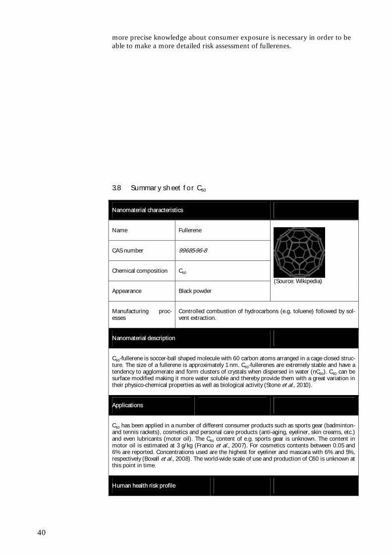
40
more precise knowledge about consumer exposure is necessary in order to be able to make a more detailed risk assessment of fullerenes.
3.8 Summary sheet for C60
Nanomaterial characteristics
Name Fullerene
CAS number 99685-96-8
Chemical composition C60
Appearance Black powder
(Source: Wikipedia)
Manufacturing proc-esses
Controlled combustion of hydrocarbons (e.g. toluene) followed by sol-vent extraction.
Nanomaterial description
C60-fullerene is soccer-ball shaped molecule with 60 carbon atoms arranged in a cage closed struc-ture. The size of a fullerene is approximately 1 nm. C60-fullerenes are extremely stable and have a tendency to agglomerate and form clusters of crystals when dispersed in water (nC60). C60 can be surface modified making it more water soluble and thereby provide them with a great variation in their physico-chemical properties as well as biological activity (Stone et al., 2010).
Applications
C60 has been applied in a number of different consumer products such as sports gear (badminton- and tennis rackets), cosmetics and personal care products (anti-aging, eyeliner, skin creams, etc.) and even lubricants (motor oil). The C60 content of e.g. sports gear is unknown. The content in motor oil is estimated at 3 g/kg (Franco et al., 2007). For cosmetics contents between 0.05 and 6% are reported. Concentrations used are the highest for eyeliner and mascara with 6% and 5%, respectively (Boxall et al., 2008). The world-wide scale of use and production of C60 is unknown at this point in time.
Human health risk profile
41
Fullerenes exist in a variety of forms (carbon atom number, surface modifications, aggregations states, etc) and it is difficult to make generalisation about their toxic behaviour.
So far it has been shown that one of the main factors for differences in fullerene toxicity seems to be the water solubility with fullerenes of greater water solubility being less toxic.
Fullerenes are assumed not to be effectively absorbed and to remain at the deposition site (mainly lung and gut). A small amount may be absorbed through the gut wall. The absorption via the skin is dependent on the fullerene type (functionallisation), the solvent/dispersant used and on skin properties, but the dermal absorption is generally assumed to be low. Fullerenes seem to have a very low toxicity after oral exposure and an acute NOAEL of 2000 mg/kg bw/day.
Following exposure via the pulmonary route fullerenes were able to induce pro- or anti-inflammatory responses with the factors driving these effects still unknown. An acute NOAEC of 2.22 mg/m3 for acute inhalation is suggested as no inflammatory potential was seen at this con-centration.
Identified studies suggest that fullerenes may not have an irritating potential to skin and eyes. No conclusions can be made on the sensitising properties of fullerenes. Further clarification concern-ing an irritating and sensitising potential of fullerenes is needed.
No sub-chronic or chronic studies with fullerenes for any of the exposure routes have been identi-fied. The main effects associated with fullerene toxicity as shown by in vitro and acute toxicity studies are inflammatory and oxidative responses as well as anti-inflammatory and anti-oxidant properties.
Identified in vitro and in vivo mutagenicity /genotoxicity tests have reported contradicting results. Appropriate in vitro and in vivo studies should confirm the presence or absence of a mutagenic effect and the conditions influencing the test results.
No information on carcinogenic effects of fullerenes has been identified and there are indications that fullerenes may have anti-tumorigenic effects which could be an indication that carcinogenicity is not an endpoint of concern for fullerenes. However, the identified information is not sufficient to reach this conclusion. The conclusion on whether carcinogenicity studies are needed is depend-ent on more information on toxicokinetic and mutagenicity
Effects on female reproductive organs and embryos have been shown under quite artificial condi-tions. This endpoint should be re-evaluated once more information on the absorption and sub-chronic/chronic effects become available.
Full physicochemical characterisation is essential to allow comparisons between fullerene toxicity and a risk assessment should focus on the fullerene type and concentrations to which humans are exposed.
With regard to the human health risk there is little information available with regard to the amounts of nano C60 used in different product types and thereby the extent of the exposure.
Environmental risk profile
In general the acute ecotoxicity of C60 seems to be low, however, a LC50, 48h-value of 7.9 mg/L has been reported. This would rank C60 as “Aquatic Chronic 2”. In longer-term exposure studies (32 d) with fish a NOEC of 0.04 mg/L and a LOEC of 0.2 mg/L for the length of juvenile carp has been found. Ecotoxicological testing of C60 is difficult due to the very low water solubility of the compound and further method development may be needed. Tests performed using organic sol-vents do not have a high credibility and should not be used for hazard identification or risk as-sessment purposes.
Boxall et al. (2008) estimated regional concentrations of fullerenes in water to be in the order of 0.3 µg/L. This estimate of the predicted environmental concentration (PEC) is highly uncertain due to lack of data about the sources for fullerenes, amounts used, and the environmental transport and fate of fullerenes.
As concluded by Stone et al. (2010) the poor ecotoxicity database for fullerenes implies that no reliable estimation of predicted no-effect concentration (PNEC) can be made. Given the quality of the present PEC estimates and the lack of suitable ecotoxicity data for PNEC estimation, a quanti-tative risk characterization of fullerenes cannot be made at present.

42
43
4 Titanium dioxide - TiO2
4.1 General characteristics
Titanium dioxide is the naturally occurring oxide of titanium. Often a distinc-tion is made by TiO2 manufacturers between pigmentary grade and ultrafine grade. The primary crystal size typically range from 150 to 300 nm for TiO2 of a pigmentary grade and the surface area typically ranges from 6 to 60 m2/g (coated and uncoated). The ultrafine grade typically has a primary crystal size from 10 to 150 nm, and a surface area of between 50 to 200 m2/g (coated and uncoated). The pigmentary TiO2 has a white colour and is therefore widely used in paints etc. The ultrafine TiO2 including nano-sized TiO2 is transpar-ent. A number of different crystal structures of TiO2 exist of which the rutile and anatase forms of TiO2 are the most important in relation to the use of TiO2 in consumer products. Nano-scale TiO2 particles have various forms and shapes and may be cicular (non-spherical) or approaching spherical forms. Nano-scale TiO2 may furthermore display complex and various ag-glomeration structures. In contrast to the bulk TiO2 (>100 nm) that is consid-ered chemically inert, nano-scale TiO2 can act as a photo-catalyst, that can generate reactive oxygen species upon illumination. Both UV-light and visible light can induce the catalytic activity of TiO2 and the anatase crystal form is a more efficient photocatalyst than the rutile form. The fotocatalytic effects can be avoided by surface treatment e.g. doping with other metals. Although nor-mally considered to be insoluble material, it can be made water dispersible by applying certain surface treatments. In dry form, TiO2 nanomaterials will tend to agglomerate due to interaction of the particle surfaces, but the degree of agglomeration is depending on the specific surface treatment, relative hu-midity, sample aging, etc. (ED & DuPont, 2007).
4.2 Manufacturing processes
A large number of manufacturing processes exist for the ultrafine grade, many of which use either titanium tetrachloride or titanyl sulfate as starting material. These processes include precipitation, thermal hydrolysis and flame hydroly-sis. For the ultrafine grade, the crystal may be further processed involving milling, then coating and milling again. Depending on the medium relevant to the application for marketing, a possible last dispersion step (with water / cosmetic oils) can be applied, for example for UV attenuation dispersion grades. If no further dispersion is carried out, the products obtained are UV attenuation powder grades. Both fine and ultrafine TiO2 may be surface treated to increase their applicability in products, e.g. to ensure a uniform dis-tribution in sunscreens or to optimize UV-absorption properties.
4.3 Uses
4.3.1 Main applications
TiO2 is one of the most widely used chemicals in the world. As the bulk form of the substance often contains a nano-sized fraction, nano- TiO2 has been
44
used for many years for different applications, without being identified as nano.
A wide range of applications exists, exploiting the various properties of TiO2 nanomaterials. The ability to scatter light can be manipulated by changing the size of TiO2 nanomaterials. Hence pigmentary TiO2 has been optimised for visible light, which is applicable in white opaque paints, whereas ultrafine TiO2 has been optimised for UV absorption and used in sunscreens. The abil-ity of TiO2 to absorb UV radiation can be manipulated through surface treat-ment which is applicable to degradation of pollutants (NOx or dirt) or protec-tion of paint films and polymers from degradation from other chemical spe-cies (ED & DuPont, 2007). Thus, nano-scale TiO2 is widely used in sun-screens and cosmetics due to the UV-absorption of the material.
In paints and for water treatment, nano-scale TiO2 is used as a photo-catalyst producing reactive oxygen that may degrade other organics. According to ED & DuPont (2007) the rutile form of nano-TiO2 is most often used in applica-tions where low interaction with the surrounding matrix is desired, whereas untreated anatase nano-TiO2 is most often used in photocatalyst applications.
A number of other and very diverse areas of application exist, such as oint-ments, toothpaste, catalysts, catalyst supports, adsorbents, delustrants, semi-conductors, etc. In total 21 products have been identified to be commercially available on the Danish marked through searches in the Woodrow Wilson In-stitute database. Most of these products fall into the category of 'Health and fitness'. In the category of 'Health and fitness' nano-TiO2 is mostly used in sunscreens (5), personal care products (4) and sporting goods (2), however, it must be stressed that these numbers are expected to underestimate the actual number of products in use in Denmark, since non-internet retailers are not included in the Woodrow Wilson Institute database. Thus, the number of sunscreen products and paints containing nano-TiO2 is expected to be higher than what the searches in the Woodrow Wilson Institute data base suggests. However, this can not be quantified, since the producers are not obliged to declare whether or not their products contain nanoparticles.
While TiO2 rank as one of the most used chemicals world-wide (mainly as a pigment), the tonnages of nano-scale TiO2 used nationally, in the EU or worldwide can at present not be estimated. This is due to the fact that no spe-cific inventories for nanomaterials exist. However, it should be mentioned, that in some consumer products, e.g. sunscreens, the percentage of nano-TiO2 may constitute several percent of the product (Barker & Branch, 2008; Hansen et al., 2008). TiO2 (E171), which may contain nano-scale TiO2, is used for whitening and brightening food, especially for confectionary, white sauces and dressings and certain powdered foods. TiO2 is also used in the pharmaceutical industry as and opacity agent.
It has been estimated, that in UK the dietary intake of TiO2 (non-nano + nano) is 5 mg/person per day (Powell et al, 2010).Given the range of possible applications of nano-scale TiO2 especially within photocatalysis (e.g. for self-cleaning surfaces and water treatment), the use of nanoTiO2 is anticipated to increase significantly in the near future.
4.3.2 Results from industry survey
The survey on TiO2 has been focused on the use of TiO2 as a pigment. TiO2 is known to be widely used as a white pigment in paint, plastics, mortar, ce-
45
ment, toothpaste, cosmetics and in most other cases, where a white pigment is needed. TiO2 is, however, also used in combination with other pigments to obtain different shades of e.g. grey, green etc.
The concentration of TiO2 differs depending on the material in question and the colour strength required. In paint the concentration of pigments will typi-cally be in the range of 0.1-10% (Environmental project No. 1206, 2007).
The consumption of TiO2 in Denmark may be roughly estimated to be above 1000 tons/year. An import of pigments and preparations containing TiO2 of about 1800 tonnes to Denmark was registered for 2009 2.
4.4 Eco-toxicological profile
While bulk-scale TiO2 is generally believed to be inert and not expected to be hazardous to the environment, nano-scale TiO2 concerns have been raised, since novel properties at the nano-scale, e.g. photo-catalytic activity, may change the hazard profile of TiO2.
The ecotoxicological profile of nano-scale titaniumdioxide (TiO2NP) is build upon original research papers, but it should be stressed that only a few ecotoxicological studies exist and that none of these have been aimed at verifi-cation/reproducing the effects found. Thus, it is still unknown whether the patterns observed for the ecotoxicity of TiO2NP later on will be proven to be single events.
For fish there are strong indications that even though the acute toxicity of TiO2 nanoparticles is low, effects are observed in the more detailed studies focussed at gills, brain, and intestine (Federici et al., 2007; Ramsden et al., 2009). This implies that other endpoints than the traditionally used ones may be relevant for nanoparticles.
For crustaceans, TiO2NP has been found to show both acute and long-term toxicity as indicated by the values reported by Zhu et al. (2010): LC50, 72h, of 2.02 mg/L and EC50, 21d (survival of offspring) of 0.46 mg/L. This would correspond to a classification in the hazard class “hazardous to the aquatic environment” according to the CLP3. However, it should be emphasized that the results of Zhu et al. (2010) are in contrast to other studies that have found LC50 values of around 2000 mg/L (Heinlaan et al., 2008) and >10 mg/L (Griffitt et al., 2008) and a 40% mortality at the 20 mg/L (Adams et al., 2006). In a 21-day study using Daphnia magna and 40 nm TiO2NP, Kim et al. (2010) found a significantly increased mortality at concentrations of 5 and 10 mg/L, but no reduction of the reproduction ability was observed. Kim et al. (2010) suggest that oxidative stress in D. magna due to reactive oxygen spe-cies may explain the TiO2NP toxicity. At present the large spread in the re-ported data makes it impossible to make a definitive conclusion on the toxicity of TiO2NP on crustaceans.
For algae, the lowest EC50, 72h, for TiO2 nanoparticles is 5.83 mg/L (Arouja et al., 2008) and this study also found that TiO2NP were more toxic than the 2 Danmarks Statistik, 2010 3 REGULATION (EC) No 1272/2008 OF THE EUROPEAN PARLIAMENT AND OF THE COUNCIL of 16 December 2008 on classification, labelling and packaging of substances and mixtures, amending and repealing Directives 67/548/EEC and 1999/45/EC, and amending Regulation (EC) No 1907/2006
46
corresponding bulk particles (EC50, 72h, estimated to 35.9 mg/L). NOEC of the bulk materials was found to be 10.1 mg/L for bulk TiO2as compared to 0.98 mg/L for TiO2NP (Aruoja et al. 2008). However, the low effect values found by Arouja et al. (2008) and Wang et al. (2008) may not be reliable, considering the experimental challenges in the algal test as described by Hartmann et al. (2010). It seems unlikely that Arouja et al. (2008) did not en-counter the same kind of problems with reproducibility and agglomeration during testing using uncoated TiO2 particles. Therefore, it is recommended that the lowest of the values found by Hartmann et al. (2010) is used in haz-ard assessment of TiO2NP, i.e. the EC50, 72h of 71.1 mg/L.
So far only three studies of bioaccumulation of TiO2 has been published. Zhu et al. (2010) found that 21 nm TiO2 nanoparticles were accumulating in D. magna and that it was increasing exposure time. Due to a relatively slow elimination of TiO2 from D. magna the authors report very high BCF values (>10,000) based on concentrations in daphnids versus water phase concen-trations (Zhu et al., 2010). However, by calculating BCF values from the up-take and depuration rates, BCF values <100 can be estimated. In fish, Ram-den et al. (2010) found Ti-accumulation in the gill, gut, liver, brain and spleen of rainbow trout (Oncorhynchus mykiss) after 8 weeks of dietary exposure to 10 or 100 mg/kg bw TiO2 nanoparticles. In some of these organs, especially the brain, no clearance of Ti occurred after exposure. No estimations of BCF were made in this study. After intravenous injection of TiO2 nanoparticles in Rainbow trout (Oncorhynchus mykiss), Scown et al. (2010) found increased levels of Ti in the kidneys and the Ti remained there for up to 21 days. Evi-dence of clearance was, however, noted after 90 days. Ti accumulation in the liver was 15 times lower than in the kidney, but no apparent clearance took place in the liver.
By definition metal oxide nanoparticles are not degradable and speciation of TiO2 (e.g. dissociation or complexation) is not likely to occur in aquatic media under normal conditions. Therefore, TiO2 particles (both bulk and NP) can be regarded as persistent in the sense that the particles will remain TiO2-particles, however a number of factors may influence the particle size and also the resulting bioavailability in aquatic media (Hartmann et al., 2010).
4.5 Toxicological profile
Due to its widespread exploitation, a great number of studies have focussed on revealing the toxicity of titanium dioxide (TiO2). Such studies have ad-dressed the consequences of exposure via a number of routes. TiO2 (bulk form) exposure via the pulmonary route has included the use as a control par-ticulate compared to pathogenic materials such as alpha-quartz, but has also been studied in relation to occupational exposures. Dermal penetration and toxicity of TiO2 particles have also formed the focus of a number of studies due to their inclusion within sunscreens and cosmetic products. In a few stud-ies toxicity was investigated after intraperitoneal and oral exposure. Humans are exposed orally to high concentrations of TiO2 particles via food. The toxicological evaluation of TiO2 is mainly quoted from ENHRES (Stone et al., 2010). In addition the following reviews were used; IARC, NIOSH (2005) and Jelnes et al., (1997).
As mentioned earlier, bulk TiO2 is considered chemically inert and of low tox-icity. Adverse lung effects associated with TiO2 are primarily a result of a gen-eral particle overload more than a specific toxic effect of TiO2. In relation to
47
nano-scale TiO2 it is therefore of particular interest to evaluate if the nano-forms have specific toxic properties.
4.5.1 ADME studies
It is necessary to consider the potential for particle absorption from the site of exposure into the blood, as this will dictate their systemic availability and dis-tribution within the body.
Absorption and distribution
No studies were identified investigating systemic absorption after exposure via inhalation of TiO2 nanoparticles. Many studies were performed on toxicity after inhalation, but the issue on systemic absorption has not been reported. There is evidence of pulmonary retention of TiO2 nanoparticles after expo-sure via inhalation (Ferin et al., 1992 and Bermudez et al., 2004). Wang et al. (2008a and 2008b) illustrated the transfer of TiO2 nanoparticles to the brain, following intranasal exposure, which occurred via their transport within neu-rones.
TiO2 nanoparticles were observed to translocate into the blood, following oral exposure, and thereafter distribute to secondary targets, including the liver, spleen, lungs, and kidneys (Wang et al., 2007, 2008). However, it is not pos-sible to evaluate the degree of absorption based on the data available.
Studies investigating dermal exposure of TiO2 in vivo (humans) and in vitro (porcine and human skin) suggest that penetration of nanoparticles past the stratum corneum is negligible, and therefore it is unlikely that the particles will access the circulation via this route for intact skin, whereas more knowledge has to be gained on damaged skin (Stone et al., 2010).
Metabolism and elimination
No evidence of TiO2 metabolism or elimination from the body could be iden-tified in the literature at this time.
4.5.2 Short term toxicity
Acute inhalation toxicity
In several studies there is clear evidence that nano-sized TiO2 is considerably more toxic than microsized TiO2 ( Ferin et al., 1992; Renwick et al., 2004; Chen et al. 2006; Inooue et al. 2008). In addition, it was found that the crys-tallinity (or the specific crystal form) of TiO2 nanoparticles is thought to influ-ence the toxicity, with the anatase form expected to be more toxic than the rutile form (Warheit et al., 2007). The pulmonary response to TiO2 is in-flammation (Ferin et al., 1992; Chen et al., 2006; Warheit et al., 2005; War-heit et al., 2007; Inoue et al., 2008; Renwick et al., 2004; Grassian et al., 2007), epithelial damage, increased permeability of the lung epithelium, oxi-dative stress and cytotoxicity (Renwick et al., 2004), and morphological al-teration within the lung (Chen et al., 2006). In the Renwick et al. study (2004) no toxicity was observed at 125 µg per rat corresponding to 0.5 µg/kg bw, as-suming a rat body weight of 250 g whereas toxicity (inflammation and histo-logical changes in the lung) was seen at 500 µg per rat (particle size 29 nm). A NOAEL for toxicity of 0.5 µg/kg bw was proposed by Stone et al. (2010).
48
Chen et al. (2006) exposed mice, via intratracheal instillation (0.1 and 0.5 mg per mouse), to nano (19-21 nm) and micro (180-250 nm) forms of TiO2 and determined their pulmonary toxicity 3 days, 1 week or 2 weeks post exposure. In mice exposed to TiO2 particles (size 19-21 nm) toxicity (inflammation and histological changes in the lung) was observed at the lowest dose of 100 μg per mouse (Chen et al., 2006). Despite the huge doses used, no pathological lesions were found in response to microparticulate TiO2.
Acute dermal toxicity
The impact of TiO2 exposure on the skin is especially relevant due to the in-clusion of such particles within sunscreens and cosmetics that are directly ap-plied to skin.
In relation to acute dermal toxicity, no in vivo studies have been identified. However, Kiss et al. (2008) and Jin et al. (2008) (as cited by Stone et al., 2010) have shown that TiO2 nanoparticles may affect different cell lines in vitro.
The findings from the available studies demonstrated that the penetration of microsized-TiO2 is negligible within the “healthy” skin. However, sunscreens are often applied to burnt, damaged and diseased skin, where the barrier func-tion of the stratum corneum will inevitably be impaired (Borm et al., 2006; Kiss et al., 2008) (as cited by Stone et al., 2010), which should be taken into consideration in future studies.
Acute oral toxicity
Although high human exposure via the oral route has been estimated (Powell et al., 2010) only one study could be identified that investigated acute oral ex-posure to TiO2. Wang et al. (2007) investigated the distribution and acute toxicity of nanoparticulate (25 and 80 nm) and microparticulate (155 nm) forms of TiO2 following the oral exposure (5 g/kg bw) in mice. TiO2 particles of all sizes were distributed to the liver, spleen, lungs, kidneys, thus providing evidence that they could be transported to other sites subsequent to exposure, due to their translocation into the blood. The primary target organ for nanoparticles was the liver (an inflammatory response was detected). Markers of cardiac damage were also observed to be elevated by TiO2 nanoparticles. Microparticulate TiO2 induced limited toxicity.
Acute intraperitoneally toxicity
The only study that used intraperitoneal injection (Chen et al., 2008) investi-gated the acute toxicity of TiO2 nanoparticles (80-100 nm) subsequent to in-traperitoneal injection in mice. The doses used were exceptionally high (rang-ing from 324 to 2592 mg/kg), and it is therefore not surprising that mortality was associated with exposure. Furthermore, TiO2 was observed to block pul-monary vessels, leading to thrombosis, with pathology also evident within the liver, spleen and kidneys. This study highlights the need to consider the use of relevant doses of particles, as the observed effects derived from the high dose and not the inherent toxicity of particles.
4.5.3 Irritation and corrosion
No studies specifically investigating irritation (dermal, eye and inhalation) have been identified. However, as TiO2 nanoparticles are used widely in sun
49
creams, they are not expected to be irritating to the skin. It is also not ex-pected that these nanoparticles are irritating following inhalation. 4.5.4 Skin and respiratory sensitization (in vitro and in vivo)
No studies specifically investigating sensitization (dermal, eye and inhalation) have been identified. However, as TiO2 nanoparticles are used widely in sun creams, they are not expected to be sensitizing to the skin. It is also not ex-pected that these nanoparticles are sensitizing following inhalation. 4.5.5 Repeated dose toxicity, short term, sub-chronic and long term
Repeated dose: inhalation
The greatest number of studies on TiO2 by far address the consequences of the exposure of the lungs, and in particular the size dependence of any effects. The studies performed on pulmonary exposure of TiO2 shows that the toxic-ity primarily was dictated by particle size, and crystal structure; whereby de-creasing particle size, and anatase forms of TiO2 enhanced particle toxicity.
In a 12 week inhalation study Ferin et al. (1992) (as cited by Stone et al, 2010) showed that TiO2 nanoparticles caused a pulmonary inflammation in rats, which micro-TiO2 (250 nm) did not at the same dose levels (thus con-firming the potency of TiO2 nanoparticles as discussed within the short term toxicity section).
In addition, it is suggested that the exposure scenario (including species used, exposure method and dose administered) can impact on the toxicity of TiO2 and other metal oxides. It has been shown in a 13 weeks (6 hours per day, 5 days per week, 21 nm particles) inhalation study that rats are more sensitive than mice and much more sensitive than hamsters Bermudez et al. (2004). As the rat is the most sensitive species it was used for risk assessment in the EN-RESH report. From this study a No Observed Adverse Effect Concentration (NOAEC) of 0.5 mg/m3 (500 μg/m3) was identified.
It has been shown in a 30 days study that both rutile and anatase TiO2 nanoparticles after nasal installation of 500 µg/mouse every other day for a total of 30 days can bypass the blood brain barrier and be translocated (via the olfactory nerve) to the brain, where they accumulated within the cerebral cor-tex, thalamus and hippocampus (main target). This resulted in morphological alterations and loss of neurones in the hippocampus, induction of oxidative stress and initiation of inflammation (Wang et al., 2008a, 2008b).
After chronic exposure (2 years) of rats to TiO2 nanoparticles, at an average exposure concentration of 10 mg/ m3, lung tumours were observed. However, the relevance of these data for risk assessment purposes are dubious due to the very high doses used (average exposure concentration of 10 mg/m3), and the high mortality: 60% after 24 months (control 40%) and 90% after 130 weeks (control 85%) (Heinrich et al., 1995).
Repeated dermal dose
No studies related to repeated dose toxicity via the dermal route were avail-able
50
Repeated oral dose
No studies related to repeated dose toxicity via the oral route were available
4.5.6 Mutagenicity/genotoxicity
There is little information available concerning in vivo genotoxicity of nano TiO2. Driscoll et al. (1997) investigated the mutagenic effect at the hprt locus in rat alveolar epithelial cells after intratracheal installation of anatase TiO2 (median diameter = 180 nm). TiO2 induced mutations at 100 mg/kg bw but not at 10 mg/kg bw. The potency of TiO2 was compared with α-quarts par-ticles (mean diameter = 900 nm) and carbon (mean diameter = 15 nm). Α-quarts was the most potent followed by cabon black and TiO2 being the less potent. All particles induced a neutrophilic inflammatory respond which cor-related with the relative genotoxic potency of the different materials. These findings indicate that inflammation may play a key role of the in vivo genotoxic effect of insoluble particles, supporting a non-linear (thresholded) relationship between particle exposure and mutagenicity.
In a recent paper Trouiller et al. (2009) investigated the genotoxicity, oxida-tive DNA damage and inflammation of nano-TiO2 in an in vivo study in mice. The crystal structure was a mixture of 75% anatase and 25% rutile TiO2 with a particle size of 21 nm. Male mice were dosed for 5 days in the drinking wa-ter with doses corresponding to 0, 50, 100 250 and 500 mg/kg bw. Pregnant dams were dosed in drinking water with 500 mg/kg bw for 10 days at gesta-tion days 8.5 to 18.5 post-coitum. In male mice, nano-TiO2 induced DNA single strand breaks measured by the comet assay at the highest dose tested (500 mg/kg/bw) and micronuclei in peripheral blood. DNA double strand breaks (DBS) was measures by λ-H2AX immunostaining assay in bone mar-row, which was the most sensitive of the assays involved, and showed an in-crease in DBS in a dose dependent manner. Oxidative DNA damage (8-OHdG) was measured in the liver at the highest dose tested, and a proin-flammatory response, measured as changes in cytokine expression, was seen in peripheral blood. In utero exposure of fetuses via the mother caused an in-crease in large deletions in offspring.
This in vivo study show that the investigated nano-TiO2 administrated orally is systemically distributed to different tissues such as blood, bone marrow, liver and even the embryo, where it can induce genotoxicity at relatively high exposure levels. The inflammatory response and oxidative damage in liver indicate that the mechanism behind the observed genotoxicty may be due to a secondary response following inflammation and oxidative stress.
Although the genotoxic effect observed after oral exposure might be thresh-olded, concern is raised due to its carcinogenic potential following chronic exposure (Heinrich et al., 1995), and because of the potential to induce ge-netic disorders in the offspring as indicated by Trouiller et al. (2009).
No in vitro genotoxicity/mutagenicity studies were identified using the classi-cal tests used in regulatory settings. However, some evidence of genotoxicity has been encountered within a number of in vitro studies.
Rahman et al. (2002) (as cited by Stone et al., 2010) investigated the potential for TiO2 to elicit chromosomal aberrations in SHE fibroblasts. Micronuclei were evident within cells exposed to nano (<20 nm), but not micro (>200 nm) TiO2 particles (up to 10 µg/cm2, for up to 72 hours). The nanoparticles
51
also triggered the induction of apoptosis within cells, which is recognised as a common response to DNA damage.
It was shown by Karlsson et al. (2008) (as cited by Stone et al., 2010) that TiO2 nanoparticles were able to elicit DNA damage, as determined by the Comet assay in A549 lung epithelial cells.
Nakagawa et al. (1997) investigated the genotoxicity of TiO2 nanoparticles (in rutile and anatase forms) in the absence or presence of UV light. Without UV light, TiO2 nanoparticles induced no, or very limited genotoxicity. However, in the presence of UV light, TiO2 elicited DNA damage and chromosome ab-errations (but no gene mutations). The type of cells used for the test is not mentioned. The genotoxic effect of TiO2 seems to be influenced by the crys-talline form with the anatase form being more potent that the rutile form.
The UV dependence of the genotoxic potential of TiO2 nanoparticles (in rutile and anatase forms) was also investigated by Theogaraj et al. (2007). In contrast to the previously discussed investigations, no chromosomal altera-tions were observed within CHO cells, either in the presence or absence of UV light.
The genotoxicty of TiO2 nanoparticles is thought to be driven by particle me-diated ROS production. The particles themselves are not thought to be inher-ently genotoxic, but may trigger genotoxicity via an indirect threshold driven inflammatory mechanism involving oxidative stress and in particular neutro-phil presence (Driscoll et al., 1997).
4.5.7 Carcinogenicity
Only one study on carcinogenicity was identified (Heinrich et al., 1995) and is described under repeated dose toxicity above. In this chronic inhalation study very high doses were used, and the study had a long duration, resulting in high death, also in the control group. Based on this data, NIOSH (2005) concluded, that TiO2 is carcinogenic in rats and that it cannot be excluded as carcinogenic in humans. It is expected that carcinogenicity occurs following pulmonary particle overload and thus has a threshold and that the effects are considered to be caused by the particle exposure rather than the specific chemical substance. The International Agency for Research on Cancer have assessed TiO2 (even the microform – if exposure is high enough) to be a Class 2B carcinogen (Possibly carcinogenic to humans) (IARC 2006). 4.5.8 Reproductive toxicity, developmental toxicity and teratogenicity
Evaluation of metal oxide nanoparticle effects on the reproductive system is limited to a small number of in vitro studies.
Komatsu et al. (2008) determined the potential for TiO2 nanoparticles, diesel exhaust particles and ultrafine carbon black to impair the male mouse repro-ductive system. This study evaluated the direct effect of nanoparticles on tes-tis-constituent cells, and examined the effect of TiO2 on mouse Leydig TM3 cells, the testosterone-producing cells of the testis. TiO2 was more cytotoxic to Leydig cells than the other carbon based particles used in the study. The pro-liferation of Leydig cells was suppressed transiently by treatment with TiO2.
52
TiO2 nanoparticles were taken up by Leydig cells, and in turn affected cell viability, proliferation and gene expression.
No literature examining nanoparticle effects on organs or cell types in the fe-male reproductive system were found.
4.5.9 Summary
The bulk (microsized) form of TiO2 is generally regarded as relatively inert, with a low toxic potential (Jelnes, 1997).
However, in several studies there is clear evidence that nano-sized TiO2 is more toxic than micro-sized TiO2. First of all nano-sized TiO2 is absorbed from the gastrointestinal tract and distributed to secondary target organs, whereas microsized TiO2 is not absorbed via this exposure route (Jelnes, 1997). It has been consistent within a wide range of investigations that the acute toxicity increases as particle size decreases. In addition the toxicity is dependent on the structure of the particles, with the anatase form being most toxic. The toxicity of TiO2 nano-particles has been demonstrated to be in-flammation, induction of oxidative stress, and histological changes in the tar-get organs. The primary target organ after inhalation is the lung. It is assumed (Stone et al., 2010) that the NOAEC is somewhere between 0.5 to 2 mg/m3. A few studies indicate the liver is the primary target organ after oral exposure, and that TiO2 exposure also may be related to cardiac damage.
There is indication from in vitro studies that nano-sized TiO2 but not micro-sized TiO2 is genotoxic, and that the genotoxic response is caused by oxida-tive stress, assuming a thresholded mechanism.
The genotoxic effect of in vitro of TiO2 has been confirmed in two in vivo studies, which also indicate that genotoxicity is due to inflammation and oxi-dative stress.
Although the genotoxic effect observed after oral exposure might be thresh-olded, concern is raised due to its carcinogenic potential following chronic exposure (Heinrich et al., 1995), and because of the potential to induce ge-netic disorders in the offspring as indicated by Trouiller et al. (2009).
The inflammogenic, oxidative and indirect genotoxic effects of nano-sized TiO2 are considered to be inherently linked.
Future needs for research
Although nano-sized TiO2 is one of the most well investigated nano sub-stances, several data gaps still exist in relation to toxicological evaluation.
None of the studies performed and reviewed in this survey is in accordance with the requirements in international accepted guidelines for toxicity testing accepted for regulatory purposes. The responses observed within a few stud-ies were measured following relatively high exposure doses. In order to clarify whether the observed effects, e.g. carcinogenicity, is of biological relevance, further studies following international accepted guidelines are needed. This is especially important because nano-sized TiO2 has shown genotoxic potential in vivo in different organs and even in the fetus after exposure of the mother. Therefore relevant reproductive studies are also needed.
53
For the TiO2 samples, variations in crystalline structure suggest that it might be difficult to compare across studies and to generate an overall conclusion. Clearer characterisation of the particles used is required within studies in or-der to identify the attributes of TiO2 that are most influential in driving toxic-ity.
It can also be suggested that systemic responses following pulmonary expo-sure are possible. However, this would require further investigations, which could also focus on determining the factors responsible for such a response, if the particles cannot directly access the circulation. In addition, the neuronal transport of nanoparticles has been demonstrated, which offers a potential route for nanoparticle distribution within the body, and enables them to by-pass the blood brain barrier, and thereby access the brain. Further studies for examining the consequences of this exposure route are needed to put these potential CNS effects in a risk assessment perspective.
Studies have mainly focused on dermal and pulmonary toxicity of particles, but there is an absence of data on the consequences of exposure to dam-aged/diseased skin, and few data are available on toxicity after oral exposure. Thus further data is needed. Studies on other relevant target organs include the liver, kidney, cardiovascular system and brain which are necessary due to the fact that nanoparticles are likely to become systemically or neuronally available. The liver could be highlighted as a priority due to the propensity of particles to accumulate in this organ.
4.6 Exposure scenarios
For consumers the highest direct exposure to nano-scale TiO2 is expected to be through the use of sunscreens and dermal contact is the most likely route of exposure unless the products are applied as sprays. For sunscreens containing 2% nano-scale TiO2, Hansen et al. (2008) estimated a worst-case exposure of 57 µg/kg bw/d for a 2-year old child.
TiO2 is also used in paints, varnishes and inks and coatings/surfaces to which consumers can be exposed either via the dermal or inhalation route (sprays).
The only consumer exposure data available, reported very low exposure (22 particles/m3, 55 nm) for release from coatings due to wear and tear, which will probably not constitute a risk.
TiO2 is used in spray applications, but no exposure data for consumers are available. Based on modelling of application of a spray-on sunscreen, a maxi-mum value of 3.5 g/m3 for short term exposure is estimated.
In addition humans are also exposed to high amounts of TiO2 via food, and an unknown part of this will be on nanoform. It has been estimated that in UK the dietary intake of TiO2 is 5 mg/person pr day (Powell et al., 2010).
This may raise concern because nano-sized TiO2 has a genotoxic potential after oral exposure, but presumably by a non-linear (thresholded) mechanism.
The widespread uses of TiO2 in consumer products like sunscreens, surface treatment and paints allow for emissions of TiO2 nanoparticles to the envi-ronment during and after application e.g. via wastewater or leaching of nanoparticles from treated surfaces.
54
In the study by Kaegi et al. (2008) it was found that TiO2 nanoparticles leached from painted house facades resulting in a concentration of 8 µg/L in urban runoff. Using modelling approaches, Müller and Nowack (2008) calcu-lated PEC-values between 0.7 and 16 µg/L in water and Boxall et al. (2008) estimated environmental concentrations of 24.5 µg/L TiO2 nanoparticles in water. Both of these modelling attempts are based on assumptions that cannot be validated, however, it should be noted that they give results in the same order of magnitude.
4.7 Risk profile
4.7.1 Environment
The ecotoxicity of bulk TiO2 (particles >100 nm, typically with a size around 300 nm) is at present very poorly documented. Thus, reliable comparisons between nano-sized and bulk TiO2 are difficult at present. Only a few of the ecotoxicity studies have reported the results in terms of the endpoints and values listed in Information Requirements of REACH (ECHA, 2008).
The lowest reported EC50 and LC50 values are in the range 1-10 mg/L (algae and crustaceans). Detailed studies on fish indicate that other endpoints than the traditionally used ones may be relevant and focus should be directed to-wards gills, brain, and intestines.
Bioaccumulation of TiO2NP has been shown in aquatic species, however, more research is needed to quantify this in terms of reliable BCF values. TiO2 particles (both bulk and NP) can be regarded as persistent in the sense that the particles will remain TiO2-particles upon release to the environment.
TiO2 nanoparticles have been found to be released to the environment from the different uses, e.g. outdoor paints and personal care products. Upon con-tact with water, uncoated TiO2 nanoparticles tend to aggregate and settle, however the presence of natural organic matter may have a stabilizing effect.
In Stone et al. (2010) a PNEC of 5.8 µg/L for freshwater has been estimated using an assessment factor of 1000. Based on comparisons with modelled PEC-values, the risk quotient for TiO2 nanoparticles (PEC/PNEC) assumes values from 0.1-4.2, indicating that TiO2 may give rise to toxicity in the aquatic environment. However, very large uncertainties exist not only with respect to obtaining valid ecotoxicological results for TiO2 nanoparticles (Hartmann et al., 2010), but also for estimating environmental concentrations and for taking bioavailability into account when estimating e.g. PNEC. The lack of information on production volumes, amounts of TiO2 nanoparticles in products, and market penetration makes all estimates of PEC very uncertain.
4.7.2 Human health
TiO2 nanoparticles were shown to be absorbed following oral exposure, and the main target organ seems to be the liver. There is however too few data to perform a risk characterisation for oral exposure.
There is substantial evidence that nano-TiO2 does not pass through healthy skin, and it can be concluded that healthy skin is most likely not a risk, but it might be a risk for damaged skin.
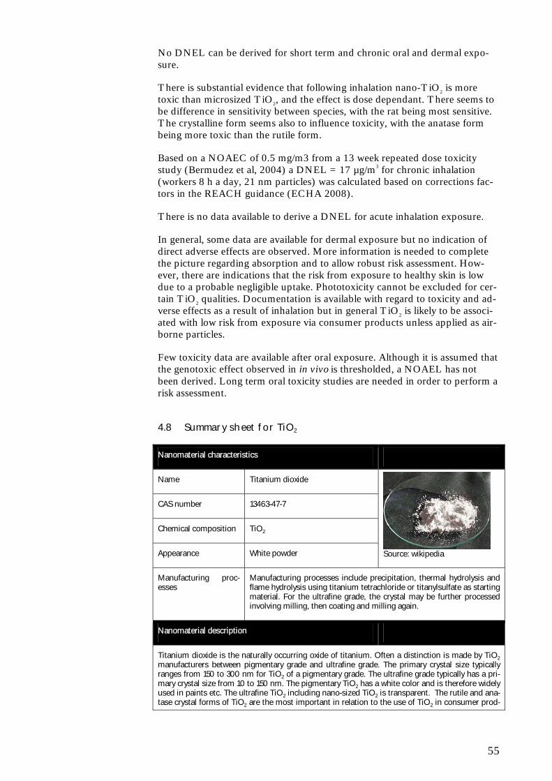
55
No DNEL can be derived for short term and chronic oral and dermal expo-sure.
There is substantial evidence that following inhalation nano-TiO2 is more toxic than microsized TiO2, and the effect is dose dependant. There seems to be difference in sensitivity between species, with the rat being most sensitive. The crystalline form seems also to influence toxicity, with the anatase form being more toxic than the rutile form.
Based on a NOAEC of 0.5 mg/m3 from a 13 week repeated dose toxicity study (Bermudez et al, 2004) a DNEL = 17 µg/m3 for chronic inhalation (workers 8 h a day, 21 nm particles) was calculated based on corrections fac-tors in the REACH guidance (ECHA 2008).
There is no data available to derive a DNEL for acute inhalation exposure.
In general, some data are available for dermal exposure but no indication of direct adverse effects are observed. More information is needed to complete the picture regarding absorption and to allow robust risk assessment. How-ever, there are indications that the risk from exposure to healthy skin is low due to a probable negligible uptake. Phototoxicity cannot be excluded for cer-tain TiO2 qualities. Documentation is available with regard to toxicity and ad-verse effects as a result of inhalation but in general TiO2 is likely to be associ-ated with low risk from exposure via consumer products unless applied as air-borne particles.
Few toxicity data are available after oral exposure. Although it is assumed that the genotoxic effect observed in in vivo is thresholded, a NOAEL has not been derived. Long term oral toxicity studies are needed in order to perform a risk assessment.
4.8 Summary sheet for TiO2
Nanomaterial characteristics
Name Titanium dioxide
CAS number 13463-47-7
Chemical composition TiO2
Appearance White powder
Source: wikipedia
Manufacturing proc-esses
Manufacturing processes include precipitation, thermal hydrolysis and flame hydrolysis using titanium tetrachloride or titanylsulfate as starting material. For the ultrafine grade, the crystal may be further processed involving milling, then coating and milling again.
Nanomaterial description
Titanium dioxide is the naturally occurring oxide of titanium. Often a distinction is made by TiO2 manufacturers between pigmentary grade and ultrafine grade. The primary crystal size typically ranges from 150 to 300 nm for TiO2 of a pigmentary grade. The ultrafine grade typically has a pri-mary crystal size from 10 to 150 nm. The pigmentary TiO2 has a white color and is therefore widely used in paints etc. The ultrafine TiO2 including nano-sized TiO2 is transparent. The rutile and ana-tase crystal forms of TiO2 are the most important in relation to the use of TiO2 in consumer prod-
56
ucts.
In contrast to the bulk TiO2 (>100 nm) that is considered chemically inert, nano-scale TiO2 and in particular the anatase form can act as a photo-catalyst that can generate reactive oxygen species upon illumination. Both UV-light and visible light can induce the catalytic activity of TiO2 and the anatase crystal form is a more efficient photocatalyst than the rutile form. Although normally con-sidered to be insoluble material can be made water dispersible by applying certain surface treat-ments. In dry form, TiO2 nanomaterials will tend to agglomerate due to interaction of the particle surfaces, but the degree of agglomeration is depended on the specific surface treatment, relative humidity, sample aging, etc. Both fine and ultrafine TiO2 may be surface treated to increase their applicability in products, e.g. to ensure a uniform distribution in sunscreens or to optimize UV-absorption properties.
Applications
A wide range of applications exists for TiO2 nanomaterials exploiting the various properties of TiO2 nanomaterials. Pigmentary TiO2 is widely used as a pigment in paints, whereas nano-scale TiO2 is widely used in sunscreens and cosmetics due to the UV-absorption of the material. In paints and for water treatment nano-scale TiO2 is used as a photo-catalyst producing reactive oxygen that may degrade organic contaminants. Finally, a number of other and very diverse set of applications ex-ists such as ointments, toothpaste, catalysts, catalyst supports, adsorbents, delustrants, semi-conductors, etc. In some consumer products, e.g. sunscreens, the percentage of nano TiO2 may constitute several percent of the product. TiO2 rank as one of the most used chemicals world-wide (mainly as a pigment), but the tonnages of nano-scale TiO2 used nationally, in the EU or world-wide can at present not be estimated. Given the range of possible applications of nano-scale TiO2, the use is anticipated to increase significantly in the near future.
Human health risk profile
The bulk (microsized) form of TiO2 is generally regarded as relatively inert, with a low toxic poten-tial. However, in several studies there is clear evidence that nano-sized TiO2 is more toxic than micro-sized TiO2. First of all, nano-sized TiO2 is absorbed from the gastrointestinal tract and dis-tributed to secondary target organs, whereas microsized TiO2 is not absorbed via this exposure route. It has been consistent within a wide range of investigations, that the acute toxicity increases as particle size decreases. In addition the toxicity is dependent on the structure of the particles, with the anatase form being most toxic. The toxicity of TiO2 nano-particles has been demonstrated to be inflammation, induction of oxidative stress, and histological changes in the target organs. The primary target organ after inhalation is the lung. It is assumed (Stone et al., 2010) that the NOAEC is somewhere between 0.5 to 2 mg/m3. A few studies indicate the liver is the primary tar-get organ after oral exposure, and that TiO2 exposure also may be related to cardiac damage.
There is indication from in vitro and in vivo studies that nano-sized TiO2 but not micro-sized TiO2 is genotoxic, and that the genotoxic response is caused by oxidative stress, assuming a thresh-olded mechanism.
The inflammogenic, oxidative and indirect genotoxic effects of nano-sized TiO2 are considered to be inherently linked.
Environmental risk profile
The bulk (microsized) form of TiO2 is generally regarded as relatively inert, with a low environ-mental risk. For nano TiO2 the lowest reported EC50 and LC50 values are in the range 1-10 mg/L (algae and crustaceans). Detailed studies on fish indicate that other endpoints than the tradition-ally used ones may be relevant and focus should be directed towards gills, brain, and intestines.
Bioaccumulation of TiO2NP has been shown in aquatic species, however, more research is needed to quantify this in terms of reliable BCF values. TiO2 particles (both bulk and NP) can be regarded as persistent in the sense that the particles will remain TiO2-particles upon release to the environ-ment.
TiO2 nanoparticles have been found to be released to the environment from the different uses, e.g. outdoor paints and personal care products. Upon contact with water, uncoated TiO2 nanoparticles tend to aggregate and settle, however, the presence of natural organic matter may have a stabiliz-ing effect.
In Stone et al. (2010) a PNEC of 5.8 µg/L for freshwater has been estimated using an assessment factor of 1000. Based on comparisons with modelled PEC-values, the risk quotient for TiO2 nanoparticles (PEC/PNEC) assumes values from 0.1-4.2, indicating that TiO2 may give rise to risk

57
in the aquatic environment. However, very large uncertainties exist not only with respect to obtain-ing valid ecotoxicological results for TiO2 nanoparticles, but also for estimating environmental concentrations. The lack of information on production volumes, amounts of TiO2 nanoparticles in products, and market penetration makes all estimates of PEC very uncertain.

58
59
5 Zero valent iron - nZVI
5.1 General characteristics
Nano-scale zero-valent iron is the nanoform of zero valent iron. In its most basic form it consists of spherical iron (Fe0) nanoparticles with individual par-ticle dimensions less than 100 nm. Substantial variations exist in the proper-ties of nZVI in regard to average particle size, particle size distribution, spe-cific surface area, surface charge and the presence of trace metals. nZVI may furthermore often be coated in order to prevent agglomeration and better con-trol their reactivity and mobility. Polymers, polyelectrolytes, and surfactants are among the main types of coatings used for nZVI, including starch, poly(acrylic acid) (PAA) , poly(styrene sulfonate), etc. The lifetime of these coatings is generally unknown (Grieger et al., 2010).
5.2 Manufacturing processes
A number of different synthesis methods exist including the Sol-gel methods or chemical solution deposition methods where the solution acts as the pre-cursor for an integrated network or gel as described by Chang et al. (2005), and the method of ferric iron reduction with sodium borohydride as described by Sun et al. (2006) and Li et al. (2006) (Grieger et al., 2010).
5.3 Uses
5.3.1 Main applications
Not many applications of nZVI are known to us at this point and no con-sumer products have been identified to be commercialized on the Danish market. In the nanoform, ZVI have significantly increased available reactive surface areas which have been found to subsequently enhance contaminant degradation reactions. This has been studied in pilot studies in in situ soil remediation at contaminated sites (PCBs, chlorinated organic solvents and organochlorine pesticides) not easily accessed by other treatment methods (Grieger et al., 2010). To the best of our knowledge no field scale applications of nZVI has been undertaken in Denmark. Some medical applications have also been reported, but it is the use in soil and groundwater remediation that is anticipated to be the major use of nZVI in the future.
5.3.2 Results from industry survey
No further information about nZVI has been available.
5.4 Eco-toxicological profile
For the following overview of the ecotoxicological profile of nano-scale zero-valent iron it should be stressed that only a few ecotoxicological studies exist and only one of these is aimed at the base-set organisms (fish, crustacean and algae) required for doing effects assessment according to REACH.
60
In a study with bacteria (E. coli), it was found that nZVI is bactericidal under anoxic conditions and that the toxicity was significantly lower under oxygen saturated conditions (Lee et al., 2008). It was suggested that Fe(II) released from nZVI contributes to toxicity and that the toxic mechanism is oxidative stress induced by reactive oxygen. nZVI was found to cause a significant physical disruption of cell membranes.
Based on extensive literature searches in ISI Web of Science it is found that no studies have been published in peer reviewed journals (until 22 October 2010) on the ecotoxicity of nZVI to algae and crustaceans. Only one peer-reviewed study (Li et al., 2009) has been carried out test with fish (Medaka, Oryzias latipes). It was found that nZVI caused disturbance in the oxidative defence system for embryos and adults, as well as oxidative damage in em-bryos. Observed effects occurred at concentrations down to 0.5 mg/L. In adult fish a disturbance of the antioxidant balance was also observed, but the fish recovered after the exposure. Li et al. (2009) also reported on changes in gills and intestine of adult fish. Another study – not peer reviewed – report on attachment of nZVI particles to the outer surfaces of D. magna and indicates that EC50 values will be in the mg/L range (EC50, 48 hours of 55 mg/L) (Oberdörster et al., 2006). In the same study, it was found that exposure of fathead minnow to 50 mg/L nZVI did not cause any mortality (Oberdörster et al., 2006).
No studies on the bioaccumulation of nZVI have been found as of 22 October 2010. By definition metal nanoparticles are not degradable. Oxidation of Fe0 is very likely to occur in aquatic media under normal conditions forming iron-hydroxides. Therefore, nZVI particles cannot be regarded as persistent in the sense that the particles will remain as the original nZVI particles entering the aqueous media. However, a number of environmental factors (like pH, ionic strength, natural organic matter) may influence the particle sizes and agglom-erate structures in the aquatic environment (Baalousha et al., 2008; Baalou-sha, 2009) and also nanoparticle characteristics will influence the behaviour. Thus, Schrick et al. (2004) showed that unsupported iron nanoparticles will agglomerate rapidly when added to water, but also that different coatings of the iron nanoparticles (e.g. anionic hydrophilic carbon, and polyacrylic acid) resulted in colloidal suspensions with slow settling rates (hours to days).
5.5 Toxicological profile
Only a very limited number of peer-reviewed and published studies on the toxicological effects of nZVI exist. However, there is evidence that nZVI may attach to cells and to cause histological and morphological changes (Grieger et al., 2010). Furthermore, some coatings have been found to decrease toxicity probably due to reduced adherence. The effects observed seem to be linked to the release of Fe(II) from nZVI followed by production of reactive oxygen species. This may lead to disruption of cell membranes and cell death. It has furthermore been found that the ageing of nZVI (i.e., oxidation of Fe0) re-duces the toxicity of nZVI (Grieger et al., 2010). Thus, the study by Phenrat et al. (2009) of the neurotoxic effects of nZVI, “aged” nZVI (oxidized), mag-netite nanoparticles, and surface-modified nZVI, found that “pristine” nZVI was the most toxic to rodent neuron and microglia cells. The authors found that this could largely be attributed to oxidative stress. Mitochondrial swell-ings as well as apoptosis were found when the cells were exposed to nZVI. While the surface-modified nZVI was found to be less toxic than the “pris-tine” nZVI, the coatings were found to potentially facilitate cytoplasmic and
61
nuclear cell entry. Keenan et al. (2009) studied the effects of nZVI on human bronchial epithelial cells and found that cell viability was affected negatively possibly due to an internal ROS production. Similar to other studies, ageing of the nZVI resulted in decreased cytotoxicity.
5.6 Exposure scenarios
The use nZVI involves a direct injection of nZVI nanoparticles in contami-nated ground water through drilled bore holes. In this way clean-up of hard-to-reach sites is made possible (Grieger et al., 2010). In the injections concen-trations 10 g/L of nZVI are typically used (Grieger et al., 2010). The reactiv-ity of nZVI has been estimated to last 4 - 8 weeks, but this does of course de-pend on the specific characteristics of the particles and surrounding environ-mental conditions (like pH, redox conditions and ionic strength).
Environmental exposure conditions are largely unknown to day, although some data is emerging (Grieger et al., 2010). In general, environmental migra-tion of uncoated nZVI has been estimated to be only a few centimeters (Tratnyek and Johnson, 2006; Saleh et al., 2008) as a result of aggrega-tion/agglomeration and deposition to other surfaces in the environment (Lowry and Casman, 2009). Environmental organisms that may be exposed to nZVI include bacteria, protozoa, and fungi, as well as other unicellular or-ganisms if nZVI migrates to surface waters. During and after injection, soil-dwelling organisms may also be exposed to nZVI around injection sites, and higher plants may potentially be exposed through extensive root systems reaching groundwater tables (Imada et al., 2008). Although iron occurs natu-rally in the environment, the fate and behaviour of the engineered nZVI may be different from the naturally occurring iron (Grieger et al., 2010). However, numerous highly complex environmental interactions may occur upon release and most of the processes involved in estimating environmental exposures for nZVI are largely unknown today (Grieger et al., 2010).
As use of nZVI is only identified in occupational settings the only very low exposure potential for consumers is if particles end up in drinking water.
5.7 Risk profile
5.7.1 Environment
As the current use or zero valent iron is very much restricted to ground water remediation, traditional risk assessment based on toxicity to fish, algae and crustaceans is of less relevance for this exposure scenario.
However, for nZVI very few ecotoxicological studies have been performed. It appears that the acute toxicity to aquatic organisms is relatively low, but sub-lethal effects have been observed at concentrations less than 1 mg/L. The ef-fects observed of nZVI in bacteria, under anoxic conditions, may be linked the release of Fe(II) and subsequent formation of reactive oxygen species. Under aerobic conditions nZVI toxicity is expected to be lower, due to a rapid oxida-tion of Fe0 to iron hydroxides (rust). At present the database is too limited to justify estimation of PNEC values.
Even though environmental exposure concentrations may be high at the site of injection, transformation and low transport ability seem to limit the risk of
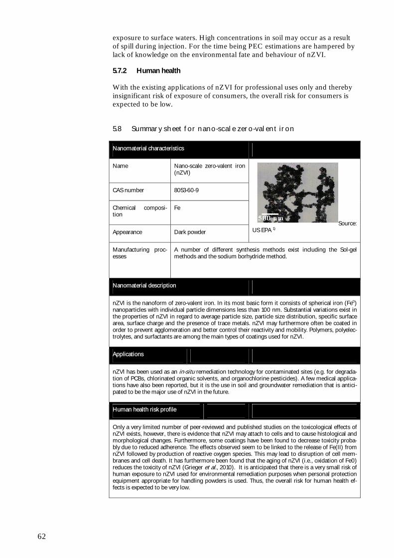
62
exposure to surface waters. High concentrations in soil may occur as a result of spill during injection. For the time being PEC estimations are hampered by lack of knowledge on the environmental fate and behaviour of nZVI.
5.7.2 Human health
With the existing applications of nZVI for professional uses only and thereby insignificant risk of exposure of consumers, the overall risk for consumers is expected to be low.
5.8 Summary sheet for nano-scale zero-valent iron
Nanomaterial characteristics
Name Nano-scale zero-valent iron (nZVI)
CAS number 8053-60-9
Chemical composi-tion
Fe
Appearance Dark powder
Source: US EPA 1)
Manufacturing proc-esses
A number of different synthesis methods exist including the Sol-gel methods and the sodium borhydride method.
Nanomaterial description
nZVI is the nanoform of zero-valent iron. In its most basic form it consists of spherical iron (Fe0) nanoparticles with individual particle dimensions less than 100 nm. Substantial variations exist in the properties of nZVI in regard to average particle size, particle size distribution, specific surface area, surface charge and the presence of trace metals. nZVI may furthermore often be coated in order to prevent agglomeration and better control their reactivity and mobility. Polymers, polyelec-trolytes, and surfactants are among the main types of coatings used for nZVI.
Applications
nZVI has been used as an in-situ remediation technology for contaminated sites (e.g. for degrada-tion of PCBs, chlorinated organic solvents, and organochlorine pesticides). A few medical applica-tions have also been reported, but it is the use in soil and groundwater remediation that is antici-pated to be the major use of nZVI in the future.
Human health risk profile
Only a very limited number of peer-reviewed and published studies on the toxicological effects of nZVI exists, however, there is evidence that nZVI may attach to cells and to cause histological and morphological changes. Furthermore, some coatings have been found to decrease toxicity proba-bly due to reduced adherence. The effects observed seem to be linked to the release of Fe(II) from nZVI followed by production of reactive oxygen species. This may lead to disruption of cell mem-branes and cell death. It has furthermore been found that the aging of nZVI (i.e., oxidation of Fe0) reduces the toxicity of nZVI (Grieger et al., 2010). It is anticipated that there is a very small risk of human exposure to nZVI used for environmental remediation purposes when personal protection equipment appropriate for handling powders is used. Thus, the overall risk for human health ef-fects is expected to be very low.

63
Environmental risk profile
For nZVI very few ecotoxicological studies have been performed. It appears that the acute toxicity to aquatic organisms is relatively low, but sub-lethal effects have been observed at concentrations less than 1 mg/L. The effects observed of nZVI in bacteria, under anoxic conditions, may be linked the release of Fe(II) and subsequent formation of reactive oxygen species. Under aerobic condi-tions nZVI toxicity is expected to be lower due to a rapid oxidation of Fe0 to iron hydroxides (rust). At present the database is too limited to justify estimation of PNEC values. Even though environ-mental exposure concentrations may be high at the site of injection, transformation and low transport ability seem to limit the risk of exposure to surface waters. High concentrations in soil may occur as a result of spill during injection. For the time being PEC estimations are hampered by lack of knowledge on the environmental fate and behavior of nZVI.
1)Http://cfpub.epa.gov/ncer_abstracts/index.cfm/fuseaction/display.abstractDetail/abstract/2172/report/2002

64
65
6 Cerium dioxide - CeO2
6.1 General characteristics
Cerium(IV) oxide (CeO2) is an oxide of the element cerium. The crystal form of CeO2 is cerianite and according to Wakefield et al. (2008) CeO2 is a ce-ramic compound, which means that it is inorganic and non-metallic. CeO2 is commercially available in a number of different size ranges below 100 nm (Nanowerk, 2010).
6.2 Manufacturing processes
It is not clear at this point in time how the nanoform of CeO2 is manufactured, but industrial bulk cerium is extracted from mined minerals, primarily mona-zite and bastnasite (Lide, 2009) whereas CeO2 are formed by thermal treat-ment processes.
6.3 Uses
6.3.1 Main applications
CeO2 has several uses exploiting the catalytic ability of CeO2 to adsorb and release oxygen (Deshpande et al., 2005) which can be used to coat the inside of self-cleaning ovens, in solar panels and for hydrogen production (Charvin et al., 2009), in fuel cells and in automobile catalysts (Lide, 2009). CeO2 has also been used as a fuel additive (Wakefield et al., 2008) and this use may be particularly important, since it may lead direct emissions during the use phase. CeO2 can also be used in polishing agents for glass mirrors (Prospect, 2010). It is not clear whether such uses are deployed in Denmark, however, no in-formation indicating that this is the case has been found. The production and use of CeO2 nanoparticles (CeO2-NP) is rapidly growing, and as mentioned above CeO2-NP have a number of applications of which the use as a diesel additive in the UK is among the most prominent ones (Rothen-Rutishauser et al., 2009; Wakefield et al., 2008). For this application CeO2-NP is mixed completely with the diesel (concentration: 5-8 ppm; average particle size: 8-10 nm) (Wakefield et al., 2008). The advantage of using CeO2-NP as a fuel cata-lyst is the improved engine combustion efficiency that results in reduced emis-sions of soot, CO and NOx. Furthermore, the fuel efficiency has been re-ported to increase by 8-9 % (Wakefield et al., 2008). While Boxall et al. (2007) mentions that CeO2 is used as a fuel additive in countries like the Phil-ippines, New Zealand and the UK, the amounts used are at present unknown.
6.3.2 Results from industry survey
The survey has been focused on the use of ceriumdioxide as additive to diesel oil. In this context the trade association for oil products in Denmark has been contacted. The association informs that ceriumdioxide is not used as additive in oil products by any of the oil companies operating in Denmark (EOF, 2010). No information is available on products supplied by smaller companies operating on the market in Denmark. Based on the information available it is
66
not possible to quantify the use of ceriumdioxide in Denmark. However, a major release from fuel is not expected in Denmark.
6.4 Eco-toxicological profile
For the following overview of the ecotoxicological profile of nano-scale ceriu-moxide (CeO2) it should be stressed that only three ecotoxicological studies were found and that only one of these found effects of CeO2.
Harper et al. (2008) investigated the toxicity of various metal oxides towards Danio rerio embryos and among these was CeO2. However, no effects were reported following exposure to up to 250 mg/L for 5 days.
Velzeboer et al. (2008) observed no measurable effects on neither the crusta-cean Chydorus sphaericus nor the freshwater algae Pseudokirchneriella Subcapi-tata exposed to a concentration of 100 mg/L CeO2 nanoparticles and tap wa-ter for 48 hours.
Van Hoecke et al. (2009) studied the toxicity of CeO2 NPs of three different sizes (14, 20, and 29 nm) towards green algae (Pseudokirchneriella subcapi-tata), two crustaceans species (Daphnia magna and Thamnocephalus platyu-rus), and zebra fish embryos (Danio rerio). They found no acute toxicity for the two crustaceans and fish embryos in test concentrations up to 1000, 5000, and 200 mg/L, respectively. For the freshwater green algae, EC10-values be-tween 2.6 and 5.4 mg/L were observed.
No studies on bioaccumulation of CeO2 have been found as of 22 October 2010.
By definition CeO2 nanoparticles are not degradable and speciation of CeO2 (e.g. dissociation or complexation) is not likely to occur in aquatic media un-der normal conditions. Therefore, CeO2 particles can be regarded as persis-tent in the sense that the particles will remain CeO2-particles, however, the sizes of the particles and hence their bioavailability may change due to ag-glomeration and aggregation in aquatic media.
6.5 Toxicological profile
Very few toxicological studies have been performed on nano-sized CeO2, and all studies performed were in vitro studies.
6.5.1 Uptake of CeO2 into cells
No ADME studies have been performed on cerium dioxide, but a study has been performed on lung epithelial cells.
The physico-chemical properties of particles have been illustrated to influence CeO2 uptake and adsorption onto proteins by Patil et al. (2007). The surface charge of particles was observed to influence the adsorption of proteins onto the particle surface. Specifically, BSA adsorbed onto the surface of positively charged CeO2 to the greater extent, when compared to negatively charged CeO2. Negatively charged nanoparticles were internalised by A549 cells to a greater extent. Electrostatic interactions were suggested to be the main driving force for the protein adsorption and cellular uptake of CeO2 nanoparticles.
67
Surface charge therefore appears to be a determining factor to particle uptake and protein adsorption.
In general, the uptake of metal oxides by a variety of cell types has been dem-onstrated on numerous occasions. The consequences of particle uptake into the cells are assumed to be oxidative stress followed by cytotoxicity and possi-bly genotoxicity. The physico-chemical properties of particles may influence their internalisation by cells; accordingly the importance of size and surface charge has the ability to influence particle uptake. In addition, the cell type under investigation has the potential to determine the mechanism of uptake and intracellular fate of particles.
6.5.2 In vitro toxicity- lung models
Pulmonary toxicity has been investigated in two different models: rat lung slices and a lung epithelial cell line.
CeO2 is used as a fuel additive to reduce particulate matter emissions from diesel engines. Fall et al. (2007) demonstrated that the CeO2 content of diesel exhaust fumes was unable to impact on the toxicity of CeO2supplemented diesel, within rat lung slices. On exposure of lung slices to freshly generated fumes, no alterations in cell viability, pro-inflammatory mediator expression (TNFα) and antioxidant enzyme activity (glutathione peroxidase, superoxide dismutase) were identified. However, it was demonstrated that the antioxidant enzyme catalase, and GSH were elevated, at low concentrations of CeO2, which is likely to derive as a defence response. Overall, it was concluded that CeO2could be tolerated at a high dose. In contrast, exposure of lung slices to non-supplemented fuel was able to moderately decrease ATP content, and decrease GSH levels. Therefore, it is implied that the addition of CeO2 intro-duces a protective effect.
Lin et al. (2006) investigated the effects of CeO2 (20 nm diameter) on the A549 epithelial cell line. Cells were exposed to CeO2 at concentrations of up to 23 µg ml-1 for up to 72 hours. A concentration and time dependent in-crease in cytotoxicity and ROS production, antioxidant (GSH and �-tocopherol) depletion and lipid peroxidation (indicated by malondialdehyde) were observed. This study therefore highlighted the oxidative and cytotoxic behaviour of CeO2. Similarly, in a study by Park et al. (2008a) the cytotoxicity and oxidative potential of CeO2 nanoparticles (15, 25, 35, 40 nm) to BEAS-2B epithelial cells was investigated, at concentrations up to 40 µg ml-1, for a period of up to 96 hours. CeO2 elicited a dose and time dependent decrease in cell viability within BEAS-2B cells, with all particle sizes eliciting a similar re-sponse. Cytosolic caspase 3 activation and chromosome condensation were also observed, suggesting that an apoptotic mode of cell death was involved. CeO2 exposure was also associated with enhanced cellular ROS production, and a decrease in cellular GSH, so that the cytotoxic response was assumed to be driven by oxidative stress. No cytotoxic response was observed within H9C2 cardiomyocyte or T98G brain fibroblast cells, indicating that some cell types may be more sensitive to the effects of CeO2.
6.5.3 Dermal models
Park et al. (2007) investigated the toxicity of nano (9 nm) and micro (320 nm) forms of CeO2 to the skin, in vitro. The EpiDerm skin model indicated that both forms of CeO2 tested were skin irritants. No cytotoxicity was ob-
68
served and CeO2 was not mutagenic in the Ames test. The toxicity observed within this study was not size dependant. 6.5.4 Mechanistic studies - Oxidative stress
Oxidative responses exhibited by metal oxide particles have been a focus of a number of studies, especially for TiO2, and its recurrence has prompted the suggestion that oxidative stress drives the inflammatory and cytotoxic re-sponses evident. In contrast, a number of investigations have suggested that cerium dioxide exhibit anti-oxidant, cytoprotective properties as shown below
Xia et al. (2008) investigated whether ZnO (13 nm), TiO2 (11 nm) and CeO2 (8 nm) oxidative stress development or particle dissolution contributed to their toxicity. RAW 264.7 macrophages and BEAS-2B epithelial cells were exposed to the nanoparticle panel for up to 15 hours, at concentrations up to 60 µg ml/L. Only ZnO particles were capable of inducing cytotoxicity, within both cell types. In addition, ZnO stimulated increased ROS production, which in turn, increased HO-1 expression, and activated the JNK signalling pathway, which paralleled the release of IL-8 and TNFα. Increased calcium release was also mediated by ZnO, and was associated with mitochondrial damage. These findings therefore imply that ZnO elicits an oxidant driven response that was responsible for the initiation of an inflammatory and cyto-toxic response. On the contrary, CeO2 was able to stimulate a cytoprotective response, whereby pre-treatment of cells with CeO2 protected against diesel exhaust particle mediated cell damage (which is known to be oxidant driven). All particle types were internalised by an endocytic mechanism, however, only ZnO accumulated within lysosomes, which was suggested to drive their ability to inflict oxidative injury, and promote particle dissolution. ZnO also induced ultrastructural alterations, including nuclear fragmentation, apoptotic body formation and mitochondrial disappearance. ZnO was therefore recognised as being the most toxic particle within the panel, however, this was thought to be accounted for, in part, by their dissolution and therefore release of Zn2+ ions. The ability of ZnO particles to generate ROS, oxidant injury, inflammation and cell death was demonstrated within this study, highlighting how these processes are inherently linked. The toxicity of the metal oxide particles was therefore suggested to be driven by their oxidant properties, which was de-pendent on their composition, dissolution and intracellular fate. In contrast CeO2 may exhibit cytoprotective response, suggesting that composition plays an important role in determining cellular responses to nanoparticles.
Neurones are particularly prone to oxidative stress (Das et al., 2007). As a result, Das et al. (2007) investigated the ability of CeO2 particles (3-5 nm) to exhibit neuroprotective effects, during the culture of rat spinal cord neurones. The survival of neurones was promoted by the inclusion of CeO2 (10 nM) within the cell culture medium, and this was thought to derive from the ability of CeO2 to scavenge free radicals, and thereby prevent against oxidative stress. The antioxidant behaviour of CeO2 was also suggested by the findings that CeO2 treatment improved neurone viability following H2O2 exposure. How-ever, further studies would be required to confirm this hypothesis.
In line with these findings, Schubert et al. (2006) investigated the neuropro-tective behaviour of CeO2 from oxidative stress mediated cell death. The HT22 neuronal cell line was exposed to CeO2 nanoparticles (6 and 12 nm) or microparticles (1 µm) for 20 hours, at concentrations up to 100 µg ml/L, and no detrimental impact on cell viability was observed. Pre-treatment with CeO2 particles were able to protect against glutamate mediated cell death, which is
69
known to be driven by oxidative stress. As a consequence, it was suggested that CeO2 particles acted as antioxidants, and that this property was not size dependent. This was confirmed by the finding that glutamate mediated in-creases in ROS production were diminished by CeO2 pre-treatment. The findings were also replicated using yttrium oxide particles. Therefore contrary to the findings that demonstrate CeO2 is toxic, this study illustrated that CeO2 may also exhibit antioxidant, cytoprotective properties.
6.5.5 Summary
No in vivo studies have been identified on cerium dioxide, and therefore no risk characterisation can be performed on this substance.
From in vitro studies it is evident that surface charge appears to be a deter-mining factor to particle uptake and protein adsorption.
There was indication from in vitro studies with rat lung slices, that cerium di-oxide had low toxicity and even had protective effect against oxidative stress produced by diesel exhaust particles
Contrary in the lung epithelial cell line A549 CeO2 induced oxidative stress and cytotoxicity. No cytotoxic response was observed within H9C2 cardio-myocyte or T98G brain fibroblast cells, indicating that some cell types may be more sensitive to the effects of CeO2.
6.6 Exposure scenarios
The use of cerium dioxide nanoparticles as a fuel additive may lead to a direct exposure of the general population through inhalation. Boxall et al. (2008) estimated an air concentration a distance of 5 m from roads of 0.6 ng/m3 and Park et al. (2008) estimated worst case concentrations of 5-25 ng/m3. These values are considered to be low when compared to occupational exposure concentrations (Stone et al., 2010). For water and soil extremely low pre-dicted environmental concentrations were estimated by Boxall et al. (2008), but it should be emphasised that the data and assumptions used are difficult to validate.
6.7 Risk profile
The only study on the ecotoxicity of CeO2 NPs showed no acute toxicity for crustaceans and fish embryos in test concentrations up to 1000, 5000, and 200 mg/L, respectively. For the freshwater green algae effects were seen at 10% inhibition CeO2 at concentrations between 2.6 and 5.4 mg/L for three different sizes of CeO2 NP. This indicates that CeO2 nanoparticles in the worst case can be classified as “Aquatic Chronic 2”, but more likely should be classified as “Aquatic Chronic 3”.
No studies of bioaccumulation of CeO2 have been found as of 22 October 2010. CeO2 particles can be regarded as persistent in the sense that the parti-cles will remain CeO2 -particles; however, the sizes of the particles may change due to agglomeration and aggregation in the environment.
With only one ecotoxicity study and very uncertain concentration estimates at hand, neither PNEC nor PEC can be estimated and therefore no quantitative risk characterisation can be carried out.
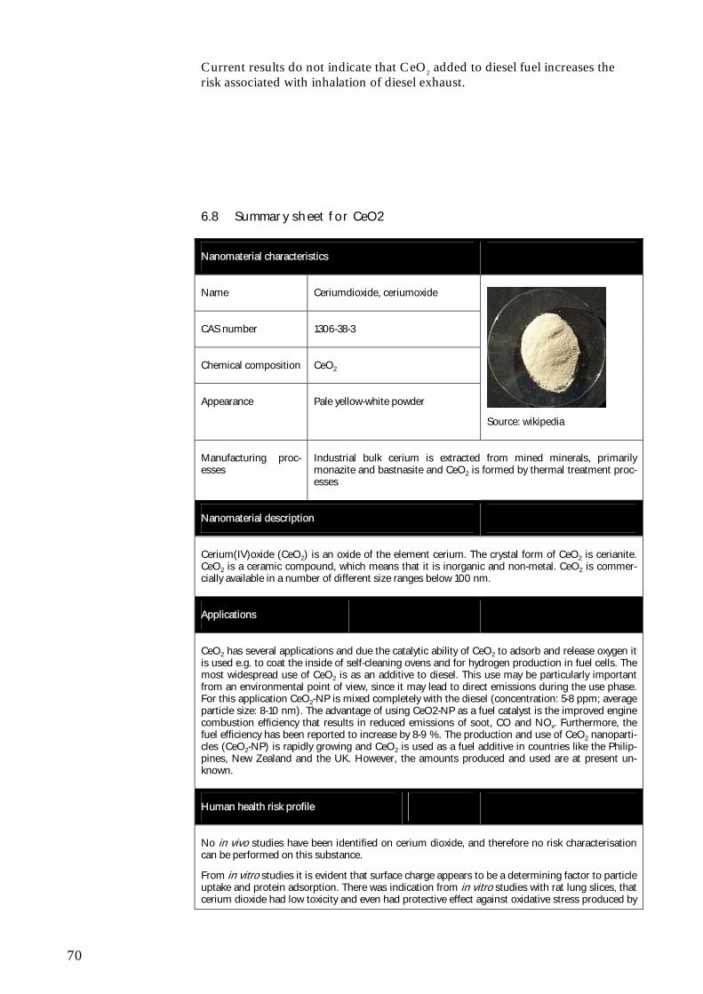
70
Current results do not indicate that CeO2 added to diesel fuel increases the risk associated with inhalation of diesel exhaust.
6.8 Summary sheet for CeO2
Nanomaterial characteristics
Name Ceriumdioxide, ceriumoxide
CAS number 1306-38-3
Chemical composition CeO2
Appearance Pale yellow-white powder
Source: wikipedia
Manufacturing proc-esses
Industrial bulk cerium is extracted from mined minerals, primarily monazite and bastnasite and CeO2 is formed by thermal treatment proc-esses
Nanomaterial description
Cerium(IV)oxide (CeO2) is an oxide of the element cerium. The crystal form of CeO2 is cerianite. CeO2 is a ceramic compound, which means that it is inorganic and non-metal. CeO2 is commer-cially available in a number of different size ranges below 100 nm.
Applications
CeO2 has several applications and due the catalytic ability of CeO2 to adsorb and release oxygen it is used e.g. to coat the inside of self-cleaning ovens and for hydrogen production in fuel cells. The most widespread use of CeO2 is as an additive to diesel. This use may be particularly important from an environmental point of view, since it may lead to direct emissions during the use phase. For this application CeO2-NP is mixed completely with the diesel (concentration: 5-8 ppm; average particle size: 8-10 nm). The advantage of using CeO2-NP as a fuel catalyst is the improved engine combustion efficiency that results in reduced emissions of soot, CO and NOx. Furthermore, the fuel efficiency has been reported to increase by 8-9 %. The production and use of CeO2 nanoparti-cles (CeO2-NP) is rapidly growing and CeO2 is used as a fuel additive in countries like the Philip-pines, New Zealand and the UK. However, the amounts produced and used are at present un-known.
Human health risk profile
No in vivo studies have been identified on cerium dioxide, and therefore no risk characterisation can be performed on this substance.
From in vitro studies it is evident that surface charge appears to be a determining factor to particle uptake and protein adsorption. There was indication from in vitro studies with rat lung slices, that cerium dioxide had low toxicity and even had protective effect against oxidative stress produced by
71
diesel exhaust particles.
Contrary in the lung epithelial cell line A549 CeO2 induced oxidative stress and cytotoxicity. No cytotoxic response was observed within H9C2 cardiomyocyte or T98G brain fibroblast cells, indi-cating that some cell types may be more sensitive to the effects of CeO2.
Environmental risk profile
The only study on the ecotoxicity of CeO2 NPs showed no acute toxicity for crustaceans and fish embryos in test concentrations up to 1000, 5000, and 200 mg/L, respectively. For the freshwater green algae effects were seen at 10% inhibition occurred at concentrations between 2.6 and 5.4 mg/L for three different sizes of CeO2 NP. This indicates that CeO2 nanoparticles in the worst case can be classified as “Aquatic Chronic 2”, but more likely should be classified as “Aquatic Chronic 3”. No studies of bioaccumulation of CeO2 have been found as of 22 October 2010. CeO2 particles can be regarded as persistent in the sense that the particles will remain CeO2-particles; however, the sizes of the particles may change due to agglomeration and aggregation in the environment. With only one ecotoxicity study and very uncertain concentration estimates at hand, neither PNEC nor PEC can be estimated and therefore no quantitative risk characterisation can be carried out.

72
73
7 Silver - Ag
7.1 General characteristics
Nanosilver is the nanoform of silver characterized by being spherical particles of a size ranging from 1-250 nm. Nanosilver is commercialized as powder, flakes, grains, ingots, etc., and is sold in suspension (in water, alcohol or sur-factant) and as a dry powder. Nanosilver is furthermore available in prepara-tions (e.g. as a coating agent, in alloys, etc.) and in articles (electrodomestic appliances, in textiles, in food packages, etc.). In its pure form nanosilver will aggregate and hence nanosilver is often surface modified with for instance dextran, citrate, polysaccharide, hydrocarbon or polyvinylpyrrolidone (PVP). Sometimes nanosilver is also found to be deposited on or used as a coating of a substrate such as plastic, silica or polymers, to give a desired adhesion, elec-trical conductivity, etc. (Luoma et al., 2007; Nanowerk, 2010; Pronk et al., 2009). In aqueous solutions nanosilver forms dissolved free silver ions by dis-solution and subsequent oxidation.
7.2 Manufacturing processes
Nanosilver can be produced through a number of methods such as ultra-sonic precipitation and chemical vapour deposition, but also exploding wire synthe-sis. By using silver salts as a starting material and then adding various surface active agents and coatings, the size, shape, surface area, etc. can be modified. Nanosilver sold in suspensions needs to be surface-modified or suspended with a chelator (e.g. citrate) to ensure the stability of the nano-silver particles in suspension.
7.3 Uses
7.3.1 Main applications
The use of nanosilver is very diverse and includes therapeutic applications such as wound dressing, personal care products, powdered colours, varnish, textile, paper, interior and exterior paints, printing colours, water and air-purification, polymer-based products and foils for antibacterial protection such as washing machines. The main consumer uses seems to be linked to the antibacterial properties of silver such as clothing for preventing odour (e.g. socks, underwear, sport textiles) toothpaste and self-sanitizing toothbrushes. Potential usage of nanosilver could be Food Contact Materials (FCM) like kitchenware and containers for food storage and even as food supplement. However, the uses of silver on nanoform for food related purposes (i.e. as FCM or food additives/supplements) require more data on toxicity and expo-sure in order to get approval for such uses in Europe
However, nanosilver can be bought on the internet as colloidal silver in food supplements for treating certain diseases and as allergy prophylaxis (Gulbran-son et al., 2000; Silver, 2003).
74
Nowack et al. (2011) underlines that products containing nanosilver particles have been commercially available for decades in diverse applications, e.g. in biocides and without use of the prefix "nano". In the United States all silver registrations with EPA from 1970 to 1993 were for nanosilver (colloidal sil-ver) or for silver nanocomposites. EPA has also registered several biocidal additives based on elemental silver particles with particle size < 100 nm.
Today silver is being used in many applications due to a desire to shift away from organic chemical agents toward additives, which can be used in much lower concentrations in a wider variety of products including applications re-quiring high temperature processing which is not feasible for organic com-pounds (Nowack et al., 2011).
On the Danish market a total of 178 products have been identified to be commercially available through the internet. Of these which 111 products fall into the category of Health and Fitness, 17 and 16 products fall into the cate-gory of Home & Garden and Food & Beverages, respectively. In the category of Health and Fitness the large majority of the products (69) fall into the cate-gory of personal care products whereas a smaller minority of the products falls into the categories of Filtration (19) and Clothing (12) and Sporting goods (8). These numbers should be seen as indicative numbers only since a num-ber of methodological concerns may introduce a bias in the actual numbers of products on the market (as described in section 2.2.1).
In the cases of shampoo, soap and toothpaste the concentration of nanosilver has been reported to be 0.001-0.002 % (Boxall et al., 2008), but in general the scale of use of nano-silver in terms of percentages in consumer products is unknown at this point in time.
7.3.2 Results from industry survey
Regarding the use as biocide in clothes request for information has been made to dominant international suppliers of sports equipment. While some compa-nies Nike (Nike, 2010; Intersport, 2010) informs that nanosilver is not used in sports equipment, other companies e.g. Adidas has not responded. Overall it can be concluded that no confirmation of the use of nanosilver particles in this context has been received, but indications exist that some products/brands may contain nanosilver. A manufacturer of nano silver yarn presents the fields of application as active/casual/sports/outdoor wear, underwear and home fur-nishing and bedding (Everest, 2010).
Quantification of the amount of nanosilver used for this purpose is not possi-ble.
7.4 Eco-toxicological profile
A number of ecotoxicity studies on AgNP have been reported in the literature and it seems that the number of papers on AgNP is rapidly increasing. This is probably due to the inherent toxicity of the element silver and many studies aim to disclose the difference between nano-scale Ag and the Ag+-ion. It is beyond the scope of this report, to give a full review of the ecotoxicity of Ag or AgNP. For this the review by Ratte (1999) and Weijnhoven et al. (2009) give good overviews. Here the focus is on the studies that are considered to be relevant for e.g. hazard classification or the predicted no-effect concentration for AgNP.
75
For fish a 48 hour static toxicity tests on adult zebrafish (Danio rerio) revealed LC50, 48h, of 7.07 (6.04-8.28) mg/L (Griffitt et al., 2008) and 7.20 (5.9-8.6) mg/L (Smith et al., 2007). Using zebra fish embryos decreased dose-dependent hatching rates, weak heart beats, edema and abnormal notochords has been reported by Yeo and Kang (2008) after 48 hours exposure of 0.01 mg/L and 0.02 mg/L 10-20 nm Ag nanoparticles suspended in tap water.
A number of short-term studies have been performed on Ag nanoparticles on pelagic crustaceans and a number of LC50, NOEC and LOEC have been de-rived. Short-term toxicity testing on adult Daphnia pulex and Ceriodaphnia dubia neonates reported LC50, 48 h, to be 0.040 (0.030-0.050) mg/L and 0.067 mg/L (Griffitt et al., 2008), respectively. In the same study an EC50 of 0.19 mg/L after 96 hours was found for green algae (P. subcapitata). For an-other alga species (Chlamydomonas reinhardtii) EC50 ranged from 0.355 mg/L ± 0.062 mg/L after 1 hour, to around 0.092 ± 0.011 mg/L after 3-5 hours. Expressed as a function of free Ag+, EC50 was estimated to range from 3.6 ± 0.5 ug/L after 1 hour, to 0.9 ± 0.08 ug/L after 5 hours (Navarro et al., 2008). This study is important because it was found that the toxicity of AgNP cannot solely be explained by the free ion (Ag+) (Navarro et al., 2008).
The widespread use of AgNP as a disinfectant in consumer products raises the questions whether unwanted environmental side-effects can occur. In this respect, the effect of AgNP on the biomass in wastewater treatment plants is especially important. In studies of effects on nitrifying bacteria Choi and Hu (2008) and Choi et al. (2008) used microbial growth inhibition tests to study the effect of different sizes (9-21 nm) of Ag nanoparticles in concentrations of 0.05-1 mg/L. At exposure concentrations of 1 mg/L a significant inhibition of 86 ± 3% was observed for Ag nanoparticles compared to 42 ± 7%, and 46 ± 4% for Ag+ ions and AgCl colloids, respectively (Choi et al. 2008). No corre-lation between nanoparticle size and bacteria inhibition in respirometry tests. However, a correlation was found between inhibition and the fraction of nanoparticles with sizes less than 5 nm, indicating that this fraction might have a stronger effect on respiration than larger size fractions. Furthermore, it was found that, at the same total concentration of Ag, Ag nanoparticles caused a greater inhibition than the free Ag+.
By definition metal and metal oxide nanoparticles are not degradable. How-ever, changes in the silver speciation can occur depending on redox condi-tions, salt content etc. For ionic silver the speciation is the determining factor for bioavailability and ecotoxicity. Though the speciation of silver most likely will play a major role in the ecotoxicity of AgNP (in analogy to the case of ionic silver), the changes in speciation of the elemental silver are poorly documented and no general conclusion can be made in this regard (Stone et al., 2010).
7.5 Toxicological profile
The toxicological studies on nano-silver described below are quoted from Stone et al. (2010), Christensen et al. (2010) and Wijnhoven et al. (2009). I addition a few recent papers from the open literature are included.
The toxicity of silver metal (bulk) and silver compounds is not described in detail in this report, and only a few studies on bulk silver are available com-pared to nano-silver and dissolved silver (Nowack et al., 2011). The toxicity of silver is considered to be relatively low (Wijnhoven et al., 2009), and mini-
76
mal risk is expected due to clinical exposure by inhalation, ingestion, dermal application or through the urological or haematogenous route. Toxicity stud-ies on silver are mainly human studies and toxic effect on humans other than agyria have only been observed at very high concentrations. Chronic ingestion or inhalation of silver preparations (especially colloidal silver) can lead to deposition of silver metal/silver sulphide particles in the skin (argyria), eye (argyrosis) and other organs. These are not life-threatening conditions but cosmetically undesirable and irreversible. The most dramatic symptom of ar-gyria is that the skin becomes blue or bluish-grey colored (ASTDR). Agyria is mainly caused by medical use of colloidal silver. The estimated total dose re-quired to induce argyria by ingestion is in the range of 1-30 g for soluble silver salts (Nordberg and Gerhardsson, 1988). Already in 1939 a threshold value above which argyria can be expected was set (Nowack et al., 2011). This value was set to 0.9 g of silver over the whole lifetime. Later (WHO / SDE / WSH / 03.04 / 14) a total lifetime oral intake of about 10 g of silver was con-sidered as the human NOAEL on the basis of present epidemiological and pharmacokinetic knowledge. This value corresponds to a daily intake over 70 years of 0.4 mg/person. In Denmark, the limit value for silver in drinking wa-ter is set to 0.01 mg/L4 corresponding to 2.5% of the human NOAEL.
7.5.1 ADME studies
Absorption Inhalation After exposure to nano-silver (4-10 nm) 3 x 10⁶ particles cm-3 equivalent to 133 µg m-3 for 6 hours, silver was deposited in the lung. Silver was measured in the blood and low concentrations of silver were found in the liver, kidney, spleen, brain and heart. It was not clear whether the absorbed silver included silver nanoparticles, free silver ions (Ag+), a silver complex or a combination (Takenaka et al., 2001).
In a 90 day whole body inhalation study to silver nanoparticles (18-19 nm), at low, medium and high doses, deposition of silver in the lung was observed - as well as silver in the blood and secondary organs such as liver, olfactory bulb, brain and kidneys Sung et al. (2008, 2009). Also this study did not clar-ify whether the absorbed silver was nano-silver or free ions. A similar distribu-tion pattern was observed in a 28 day inhalation study (Ji et al., 2007).
Oral Absorption via the oral route was investigated in a 28 days study with expo-sure to very high doses (up to 1000 mg/kg bw/day) compared to what would be seen in an occupational or consumer situation. Deposition of silver was observed in different organs (brain, liver, kidneys, lungs and testes) indicating transfer of silver into blood (Kim et al., 2008).
There is also indication that silver can be transported to the skin after oral ex-posure, where blue-grey colouring of the skin (argyria) has been observed (Wadhera and Fung, 2005).
4 (Bekendtgørelse nr 1449, 2007)
77
As for uptake via the lung, none of the above studies clarify whether the up-take is as nano-silver, silver ions, silver complexes or a combination of the two. However, it must be anticipated that at least part of the silver uptake fol-lowing oral exposure to nano-silver takes place as silver ions. This assump-tion is – at least partly – supported by the fact that some of the same toxic ef-fects as seen following non-nano silver exposure are seen following nano-silver exposure (in particular argyria).
Dermal Some studies have shown that silver nanoparticles can be absorbed via the skin when used in wound dressings. In these studies, the wound dressings were applied to damaged skin every 2-4 days. Vlachou et al. (2007) showed that serum levels increased with increasing exposure time peaking after 9 days and returning to normal 6 month after exposure.
In an in vitro diffusion cell system Larese et al. (2009) investigated the pene-trations of polyvinylpirrolidone coated silver nanoparticles (25 nm) on intact and damaged human skin slices. Silver nanoparticles (70 µg cm-2, for 24 hours) were able to pass through the skin preparations. The absorption was very low (0.0006% of applied dose with intact skin) but detectable and the penetration was 5 times greater in damaged skin. In addition, silver nanoparti-cles could be detected by electron microscopy (TEM) in the stratum corneum and the outermost surface of the epidermis.
As for uptake via the lung, it is not clear whether the uptake via the skin is as nano-silver, silver ions, silver complexes or a combination and the relevance of these in vitro findings needs to be investigated in vivo.
Distribution
Following absorption silver was seen in the following secondary organs: liver, kidney, spleen, hearth, olfactory bulb, brain, testes and skin (discoloration of the skin), with liver being the likely major secondary organ site of accumula-tion. As noted above there is no clear indication whether the absorbed and distributed silver occurs as silver nanoparticles, silver ions, silver complexes or a combination.
Metabolism
As described earlier there is no clear indication whether it is silver nanoparti-cles, silver ions, silver complexes or a combination that is absorbed. There is thus also no clear data suggesting whether and how the silver is further trans-formed once in the body. However, one of the biological/toxic effects seen in some studies (argyria - being a blue-grey dis-colouring of the skin) may, at least partly, involve a transformation/reduction of silver ions to metallic silver following exposure to UV-radiation/sunlight. This could indirectly suggest that some of the silver in circulation occurs as silver ions. These ions will at least partly be detoxified as “normal” silver ions by precipitation as sulphide and chloride salts and binding to proteins making them less bioavailable. However, if nanosilver is absorbed as particles these particles may continu-ously release silver ions within the tissue and thereby, at least partly, bypass the detoxification mechanism. Therefore nanosilver may be more toxic than the non-nanoform.
78
Elimination
Trop et al. (2006) showed elevated silver levels in the urine following expo-sure. No other data for nano-silver elimination has been identified.
7.5.2 Acute toxicity
Oral After a single oral dose of nano- (15 nm) and micro-particles (2 - 3.5µm) sil-ver, ingested directly into the stomach of mice histopathological analysis of the liver tissue, 3 days post-exposure, demonstrated evidence of inflammation in the form of lymphocyte influx for both particle sizes (greater response for the nanoform). This was further supported by changes in the gene expression of 4 genes involved in inflammation (Cha et al., 2008). This dose seems to be relatively high in relation to what would be expected in terms of occupational and consumer exposure. The relevance of these findings is therefore dubious for the purpose of risk assessment.
Inhalation No acute toxicity studies have been identified.
Dermal No studies investigating the dermal acute toxicity of silver nanoparticles on healthy skin has been identified. However, there are signs of argyria and liver toxicity following treatment of burns with wound dressings. The relevance of this data for healthy skin at lower concentrations and longer exposure time (as expected in occupational and consumer settings) is uncertain. Therefore, fur-ther investigations are needed on healthy skin with doses and exposure times relevant for exposures as seen in occupational and consumer settings.
Other routes No studies were identified studying toxicity via other routes of exposure.
7.5.3 Irritation and corrosion
No studies have been identified investigating specifically the irritating behav-iour of silver nanoparticles (to the eyes, lungs or skin).
Silver nanoparticles are not expected to be irritating to skin based on the hu-man findings of applying it in wound dressings. However, further information would be needed to draw firm conclusions. Based on the above considera-tions, silver nanoparticles are also not assumed to be corrosive.
7.5.4 Sensitisation
No studies have been identified investigating the sensitising behaviour of silver nanoparticles. As silver nanoparticle containing wound dressings are routinely applied and none of the studies investigating that application have reported sensitisation, it appears unlikely that nano-silver is a dermal sensitiser. Regard-ing possible sensitisation following inhalation, further proof/investigation is required.
7.5.5 Repeated dose toxicity
Oral In a 28 day repeated dose toxicity study in mice (OECD guideline 407) by Kim et al. (2008), a dose-dependent toxicity was observed in the liver (histo-
79
pathology changes and inflammation), insinuating that this organ is a target site for silver nanoparticle toxicity. However, it should be noted that the doses used within this study were relatively high (up to 1000 mg/kg bw/day), which should be addressed if used for a risk assessment. However, due to limited histopathological information, no oral No Observed Adverse Effect Level (NOAEL) was established based on these data.
Recently a 90 day oral study according to OECD guideline 408 and in com-pliance with GLP has been registered under REACH (EC Regulation no 1907/2006ref5). Nanosilver (56 nm) was investigated in male and female wis-tar rats. The animals were dosed daily by gavage with 0, 30, 125 and 500 mg/kg bw/day. The doses were based on the 28 days study by Kim et al. (2008). The study referred to in the REACH registration was performed by Kim et al. (2010).
The target organ for the silver nanoparticles was found to be the liver in both the male and female rats after 90-day of exposure to silver nanoparticles. Sig-nificant dose-related changes were found in alkaline phosphatase and choles-terol levels of male and female rats at and above 125 mg/kg bw/d, indicating slight liver damage. Histopathology revealed slightly higher incidences of bile-duct hyperplasia with or without necrosis, fibrosis and/or pigmentation in treated animals together with a dose-dependent accumulation of silver in all tissues examined. A NOAEL of 30 mg/kg bw/day and LOAEL of 125 mg/kg bw/day were established based on effect on the liver.
There was a statistically significant (p<0.01) dose-related increase of silver deposition in testes, liver, kidneys, brain, lungs and blood of treated rats. - A two-fold higher accumulation of silver was seen in kidneys of females com-pared to males.
Inhalation In a 28 days inhalation study rats were exposed to silver nanoparticles (13 – 15 nm) in an inhalation chamber 5 days a week and 6 h per day to 3 different doses (Hyun et al., 2008). It was concluded by the authors that although a slight effect was observed on the neutral mucins in the respiratory mucosa, no significant toxicological effect was observed with exposure up to 61 µg/m3.
In another 28 days inhalation study (OECD guideline 412) in rats by Ji et al. (2007) with the same exposure duration and dose levels as above some toxic-ity (cytoplasmic vacuolation and heptatic necrosis) was observed within the liver, but histopathological analysis did not reveal any distinct toxicity within other organs.
A 90 days whole body inhalation study was performed according to OECD guideline 413 (Sung et al., 2008, 2009). Rats were exposed to silver nanopar-ticles (18-19 nm), at low (49 µg/m3, equivalent to 0.6x10⁶ particles/cm3 and 1.08x10⁹ nm2/cm3), medium (133 µg/m3, equivalent to, 1.4x10⁶ particles/cm3 and 2.39x10⁹ nm2/cm3) and high (515 µg/m3, equivalent to 3.0x10⁶ particles / cm3 and 6.78x10⁹ nm2/cm3) doses for 6 h/day and 5 days/week. Lungs and liver were the main target organs for accumulation of silver and toxicity.
Prolonged exposure to silver nanoparticles was demonstrated to elicit an in-flammatory response within the lung, and induced alterations in lung func- 5 All, non-confident data is publicly available on http://apps.echa.europa.eu/registered/registered-sub.aspx
80
tion, at all par ticle concentrations (Sung et al. 2008), indicating that the low-est dose (49 µg/m3) would be a lowest observed effect concentration (LOAEC). The silver concentration was observed to increase in the blood, indicating that silver nanoparticles were transferred into blood from the lung, after which they subsequently became distributed within the liver, olfactory bulb, brain and kidneys. However, the silver content within these tissues may derive from nanoparticle or ion presence, which warrants further investiga-tion. In the Sung et al. (2009) study, which reported on clinical observations, haematology and histopathological examinations, erythrocyte aggregation and kidney function test, toxic effect on liver and lung was observed at the highest concentration, and a No-observed Adverse Effect concentration (NOAEC) of 100 µg/m3 was suggested. However, as this dose was not an observed value, the middle dose of 133 µg/m3, the observed dose without significant liver ef-fect, will be applied as the NOAEL. Because it is uncertain whether the lung effect is an adverse effect, both values (49 and 133 µg/m3) will be used in the risk characterisation.
Dermal No information identified except the dermal studies reported under acute tox-icity.
Other routes No studies identified
7.5.6 Mutagenicity
No mutagenicity or genotoxicity studies classically used in chemical regula-tory setting have been identified.
A micronucleus test was performed at the end of the 28 days oral study in rats (described under the repeated dose oral toxicity study) (Kim et al., 2008). No clastogenic/aneugenic effect was observed. However, it should be noted that no positive control was included.
Nano-silver may cause mutagenicity/genotoxicity via an indirect thresholded mechanism driven by formation of ROS. Further testing of the possible direct genotoxicity of nano-silver is needed.
7.5.7 Carcinogenicity
No studies investigating the carcinogenicity of silver nanoparticles have been identified. However, as silver nanoparticles may be genotoxic, carcinogenicity cannot be excluded. However, further chronic carcinogenicity studies should await results on mutagenicity of nanosilver.
7.5.8 Reproductive toxicity and developmental toxicity
Fertility In an in vitro study on mouse spermatogonial stem cell (SSC) Bradyich-Stolle et al. (2005) invetstigated the cytotoxicity of different nanoparticles (Silver (15 nm), molybdenum trioxide (MoO3, 30 nm), and aluminum (Al, 30 nm)) using the LDH and MTT assays. The cells were exposed to 10 µg/ml for 48 hours. Findings demonstrated a concentration-dependent toxicity for all types of particles tested, whereas the corresponding soluble salts had no significant effect. Silver nanoparticles were the most toxic.
81
In a later study Bradyich-Stolle et al. (2010) investigated the cytoxicity and cell proliferation of Ag-NPs of different sizes (10, 25-30 and 80 nm) coated with either polysaccharide (Ag-PS) or hydrocarbon (Ag-HC) in the same cell line as in the 2005 study above. The results of the 2010 study showed that Ag-NPs reduced the viability and cell proliferation of SSC and that the effect is size and concentration dependant. It was also shown that, over time, there was no (protective) effect of particle coating. At low exposures decrease in cell proliferation was not due to ROS formation but other mechanism like interac-tion with cell communication. Electronmicroscopy (TEM) showed optake of Ag-NP.
These results confirm that toxicity of Ag-NP increase with decreasing particle size in vitro. Although it has been demonstration from in vivo inhalation stud-ies (Sung et al., 2009) and oral studies (REACH) that nanosilver is distrib-uted to different organs, including testes, there was no detailed analysis of toxic effect on testis, but the exposure concentration would have been much lower with a blood concentration of 4 ng/g blood, than in the in vitro studies with germ cells where a concentration of 10 µg/ml was used. Therefore it is difficult to relate the in vitro results to human exposures, and in vivo studies are needed to draw a conclusion.
No data on female fertility has been identified.
Developmental toxicity Developmental toxicity was investigated in two embryo fish studies and com-pared with toxicity of gold nanoparticles (Bar-Ilan et al., 2009). It seems that toxicity (malformation and death) is dose dependent and that silver is consid-erably more toxic than inert gold nanoparticles. As for fertility, further inter-pretation requires better understanding of the toxicokinetics and possibly sub-sequent confirmation in vivo.
7.5.9 Biological mechanism
A range of mainly in vitro studies have shown that the toxicity of silver and other metal nanoparticles appear to be mediated by the induction of inflam-mation and oxidative stress, which can result in cytotoxicity and genotoxicity. 7.5.10 Summary
It has been shown that silver from nanoparticles can be absorbed especially via oral and inhalation routes of exposure. Based on in vitro data there is indi-cation that dermal absorption occur to a lesser extent, and intact skin seems to be a rather effective barrier against absorption, however, in vivo data are needed for a firm conclusion. However, it is unclear in which form (as parti-cles, free ions, silver ions or complexes) nanosilver is absorbed and distributed to target organs. At least for uptake via the oral route it is likely that at least some of the uptake occurs as ions. It appears that smaller particles exhibit higher toxicity as compared to larger particles; and if silver is absorbed as par-ticles then the surface area is relevant.
Should silver uptake occur solely as ions, the database for silver could be ap-plied to assess systemic silver nanoparticle toxicity. For that exercise, it would need to be considered whether and how the dramatically increased surface area and possibly increased solubility of silver nanoparticles would need to be taken into account.
82
Quite a few, mainly in vitro studies, have shown that the main mechanism of silver nanoparticle toxicity seems to be mediated by an increase in ROS pro-duction (except toxicity on germ cells), stimulating inflammation and genotoxic events and apoptosis or necrosis. The concentration of the adminis-tered nanoparticles is able to influence the toxicity, specifically, at low levels of oxidative stress a protective response is initiated which progresses to a damag-ing response with increasing particle concentration, and therefore oxidant lev-els. It is thus relevant to consider that the (geno)toxicity of silver nanoparticles is thresholded.
7.6 Exposure scenarios
Silver is one of the most widely used nanoparticles in consumer products (Wijnhoven et al., 2009) and especially the uses in textiles and personal care products may lead to human and environmental exposures.
Nano-silver in textiles is used in all kinds of clothes from sock and shirts to caps, gloves and underwear. In all cases it is the antimicrobial activity of nano-silver that is the reason for incorporating it in textiles. The environmental re-leases from textiles have been investigated in some theoretical studies and a few laboratory based ones. In the study by Luoma (2008) it was estimated that mass release from silver containing socks in the USA would be in the range of 6-930 kg or 180-2790 kg assuming that 10% and 30%, respectively, of the population would use these kinds of socks. The release of nanosilver from socks upon contact with water showed that for some socks almost all sil-ver leached to water whereas for others no leaching was detected (Benn & Westerhoff, 2008). In a simulation of the washing of silver-containing textiles, Geranio et al. (2009) found varying percentages of the total silver emitted during one wash, i.e. from less than 1% and up to 45%. Benn et al. (2010) measured the content of silver in textiles (in a shirt, a medical mask, a towel and a cloth), personal care products (toothpaste, shampoo), a detergent, a toy (teddy bear), and two humidifiers. They found silver concentrations from 1.4 to 270,000 µg Ag g product–1. Upon wasing in tap water they estimated the potential release of silver into aqueous environmental matrices in quantities up to 45 µg Ag/g product. By electron microscopy Benn et al. (2010) were able to confirm that nano-silver particles were present in the products, but also in the wash water samples.
In what can best be characterized as first attempt on material flow analysis Mueller and Nowack (2008) made some estimation of predicted environ-mental concentrations of silver and Gottschalk et al. (2010) have further re-fined these estimates. For surface water, Gottschalk et al. (2010) derive a PEC values of 0.72 ng/L and for sewage treatment plant effluents a PEC of 11.8 ng/L.
Human exposure to nanosilver is suspected to be highest in the working envi-ronment. The main exposure route for occupational settings is primarily via inhalation and dermal contact.
For consumer exposure no quantitative data have been identified, but it must be assumed that the main exposure route is dermal contact for clothes and paint applications, and inhalation for spray applications. Oral exposure may occur from toothpaste and toothbrushes containing nanosilver and if, in the future, nanosilver will be permitted for FCM and in food supplements (see 5.2.1). The magnitude of exposure will depend on the concentration in the
83
consumer product, the amount released from the products, which depends on how strong nanosilver is bound in the solid matrix applications (e.g. in clothes and FCMs).
7.7 Risk profile
7.7.1 Environment
Silver is known to be an ecotoxic metal, however, the toxicity is highly de-pendent on the form and speciation of the metal. In the registration of silver under REACH (EC Regulation no 1907/2006), predicted no effect concen-trations (PNECs) are reported to 0.04 µg/L (freshwater), 0.86 mg/L (marine water) and 0.025 (sewage treatment plants). Test with silver nanoparticles (AgNP) do also reveal very low effect concentrations. Thus, for algae EC50-values a low a 4 µg/L has been found and also for crustaceans values far below 1 mg/L have been reported. This ranks AgNP as "Aquatic chronic 1". It is also important to note that at concentrations below 1 mg/L inhibition of nitrifying bacteria can occur and thus the function of wastewater treatment plants may be affected by the presence of AgNP. For ionic silver it is known that the speciation in aqueous media is determining for the bioavailability and toxicity. This is most likely also the case for elemental silver nanoparticles, but influ-ence of speciation on uptake, depuration, and toxicity remain to be studied in this case.
The environmental concentration resulting from the use of AgNP in con-sumer products are at present uncertain, even though a number of different estimates have been proposed. It is evident that even though silver nanoparti-cles are incorporated in textiles, they can be released upon washing. Concen-trations in the low ng/L range have been proposed and even such low concen-trations may under very precautious assumptions6 constitute an environmental risk due to the high toxicity of silver.
It is debated today whether silver nanoparticles are in fact more toxic than their bulk counterpart, since effects in many cases can be ascribed to the ionic form of silver (Ag+). Some studies have documented a higher effect of AgNP, but it is the widespread and dispersive use of silver in consumer prod-ucts that poses the greatest risk to the aquatic and terrestrial environment. Even if AgNP are “only” as toxic as larger silver particles, silver is still a very ecotoxic metal.
7.7.2 Human health
Although nano-silver is presumably the most widely applied nanomaterial on the market, and relatively high exposure to workers and consumers might be expected, there is still a need for further data on exposure and human toxicity to fill out existing data gaps. Some important areas for research needs are mentioned below.
Human exposure data are very important for a risk characterisation. However, no or very few data on concentrations as well as data on size and form (e.g. aggregates, agglomerates) in consumer products exists. Such data are urgently needed in the future, in particular for occupa-
6 These assumptions are using the highest assessment factor of 1000 and are assuming that the bioavailability of silver is not reduced due to complexation.
84
tional inhalation and consumer inhalation and dermal exposure. In such studies characterisation of the silver nanoparticles in terms of size and agglomeration states, as well as a clear indication of duration and frequency of exposure is important.
Further investigations of the nano-silver toxicokinetics. In particular, to which extent the silver absorbed via different routes becomes sys-temically available as ions, as nanoparticles and/or as silver complexes, and whether the size of the silver nanoparticles influences the uptake mechanism(s);
Further testing for the possible direct genotoxicity of silver nanoparti-cles; and depending of the results of these studies carcinogenicity studies might be needed.
When more knowledge is obtained on absorption and distribution of different sizes of nano-silver is obtained, then effects on other organs than liver and lung should be investigated in (sub)chronic studies fol-lowing different exposure routes, especially inhalation and dermal ex-posures are relevant for consumers.
Further in vivo investigation of the possible reproductive and devel-opmental toxicity of silver nanoparticles.
However, some (limited) studies exist on which this (preliminary) risk charac-terisation is based.
The acute toxicity of nanosilver seems to be low for mammals. For soluble silver nitrate the estimated acute lethal dose is at least 10 g for humans. However, if absorbed as particles nanosilver may be more toxic than the non-nano form due to the small size and larger surface.
In vitro genotoxicity data indicate that nanosilver may cause mutagenic-ity/genotoxicity via a thresholded mechanism. Therefore a human NOAEL can be established, provided that sufficient data are available.
Risk following inhalation Due to uncertainties of the genotoxic potential and the limited dataset in the existing studies reported so far, only human Indicative No-effect Levels (INELs) can be set for inhalation.
As mentioned earlier and proposed in Stone et al. (2010) and Christensen et al. (2010) a human no effect level will be based on reduced lung function (LOAEL of 49 µg/m3) and systemic liver effect (NOAEL of 133 µg/m3).
Using assessment factors as set out in the REACH guidance (ECHA 2008, chapter R.8) and explained in Stone et al. (2010) and Christensen et al. (2010) the following INELs were derived:
Reduced lung function used as critical effect
1. Scenario: using a factor 3 for extrapolation a LOAEL to a NOAEL
85
INELlung 1= 0.33 µg/m3 equivalent to 4000 particles m-3 and 7.2 x 10⁶ nm2/cm3
2. Scenario: using a factor 10 for extrapolation a LOAEL to a NOAEL
INELlung 2= 0.098 µg/m3 equivalent to 1200 particles m-3 and 2.2 x 10⁶ nm2/cm3
Systemic liver effect as critical effect
INELliver= 0.67 µg/m3 equivalent to 7000 particles m-3 and 1.2 x 10⁶ nm2/cm3
These values are much lower as compared to a DNEL value of 0.04 mg/m3 for bulk silver for systemic effects, which has been derived by the registrant of silver in the REACH registration dossier on silver (ECHA web site April 2011)7.
The differences between the INELs and DNEL above clearly indicate that nano-silver might be more toxic than bulk silver after inhalation.
It has been estimated (Christensen et al., 2010) that peak occupational expo-sure can be at same order of magnitude or even higher as the above calculated INELs when expressed in particle number. However, better data (exposure and toxicity data) are needed.
No quantitative risk estimation can be carried out for consumers because no inhalation data have been identified for consumers. However, given that nanosilver applications in spray have been reported, this exposure route could be of concern, and the identification of exposure values via inhalation should be prioritised.
Based on available toxicological data it is recommended to avoid exposure via inhalation, because health effects like reduced lung function and toxic effect on the liver cannot be excluded after repeated exposure. There is also indica-tions from in vivo inhalation studies that silver can reach the germ cells and toxicity to germ cells has been observed in vitro from AgNP, although at much higher concentrations than could be expected from consumer exposure.
Risk following dermal exposure No quantitative data related to occupational and consumer dermal exposure has been identified. It must be assumed that especially consumers can be ex-posed to nanosilver due to the relatively widespread use in clothes. Very few toxicity data for nanosilver are available, but human data have indicated toxic-ity (agyria and liver toxiciy) after use of wound dressings containing nanosilver. However, consumer exposure is considered to be much lower and will normally be applied on healthy skin. On the other hand consumer expo-sure is considered to last much longer than a shorter term treatment of a burn. Better toxicity and exposure data are needed for improved risk assessment. However, based on existing data, indicating that the absorption via skin is low even for damaged skin, it could be assumed that the health risk via this expo-sure route would be quite low.
7 ECHA (2011) http://apps.echa.europa.eu/registered/data/DISS-9d92ea78-89c7-2334-e044-00144f67d249/showdocument671a.html?treeUuid=DISS-9d92ea78-89c7-2334-e044-00144f67d249%2FDISS-9d92ea78-89c7-2334-e044-00144f67d249&uuid=AGGR-1d2a0e67-531a-489a-8bde-a3965db67638%2FDISS-9d92ea78-89c7-2334-e044-00144f67d249
86
Risk following oral exposure Available toxicity data for oral exposure is mainly related to silver used in drug applications. Based on human data (agyria) a limit in drinking water is set to 0.01 mg/L corresponding to 0.02 mg/person/day. This value corresponds to 2.5 % of an estimated human lifetime NOAEL of 10 g (corresponding to 0.4 mg/person/day over a 70 years period).
This value is much lower as compared to a DNEL value of 1.2 mg/kg bw/d for bulk silver for systemic effects which has been derived by the registrant of silver in the REACH registration dossier on silver (ECHA web site April 2011)8, again indicating that nano-silver is more toxic than bulk silver.
No quantitative data for consumer exposure to nanosilver have been identi-fied, and therefor no quantitative risk assessment can be performed.
The oral route is not expected to be the most important route of exposure for consumers, except from intake of nano or colloidal silver as food supplement, where high exposure could be expected. Consumer exposure via the oral route could alsooccur, albeit at a relatively low level, if chewing on silver con-taining textiles or skin to mouth contact, from cosmetics containing nano-silver.
However, relatively high exposures may occur from silver containing tooth brusches or toothpaste. If it is considered that toothpaste contain 0.002% nanosilver, and estimated that a person use 1 tube of toothpaste of 100g in 20 days. As a worst case exposure estimate, assuming that 100% of the nanosilver is absorbed, will be 0.1 mg/person/day. This is 10 times higher than the limit for drinking water in Denmark and 25% of the human NOAEL for silver.
Existing knowledge base Nowack et al. (2011) refers to existing knowledge from the more than 50-year use of nanosilver for biocidal, algicidal and disinfectant uses and use of colloi-dal nanosilver for medical purposes. The authors conclude that the toxicity of silver is considered to be relatively low and that toxic effects on humans other than argyria are only observed at very high concentrations. Furthermore, they state that a significant portion of the historical toxicological research on the effects of silver on humans can be considered early examples of “nanotoxicol-ogy”.
According to Nowack et al. (2011) data about effects on nonmammalian spe-cies for environmental risk assessment have until recently been obtained mostly using dissolved silver, and nano-Ag is only studied as part of more re-cent research. The authors stress that it is questionable if data obtained using ionic silver should be used to derive threshold values in the environment as silver in natural waters typically is associated with the particulate or colloidal fraction and thus to some extent is naturally present as nanoparticles and metal-sulfide clusters. Many aquatic species are much more sensitive to silver compared to mammals and therefore the question whether nanosilver has a different toxicity compared to dissolved silver is extremely important.
8 ECHA (2011) http://apps.echa.europa.eu/registered/data/DISS-9d92ea78-89c7-2334-e044-00144f67d249/showdocument671a.html?treeUuid=DISS-9d92ea78-89c7-2334-e044-00144f67d249%2FDISS-9d92ea78-89c7-2334-e044-00144f67d249&uuid=AGGR-1d2a0e67-531a-489a-8bde-a3965db67638%2FDISS-9d92ea78-89c7-2334-e044-00144f67d249
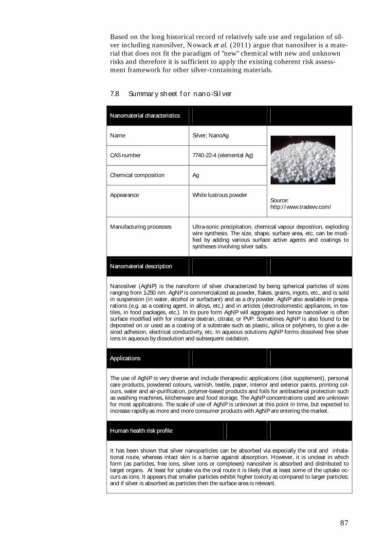
87
Based on the long historical record of relatively safe use and regulation of sil-ver including nanosilver, Nowack et al. (2011) argue that nanosilver is a mate-rial that does not fit the paradigm of "new" chemical with new and unknown risks and therefore it is sufficient to apply the existing coherent risk assess-ment framework for other silver-containing materials.
7.8 Summary sheet for nano-Silver
Nanomaterial characteristics
Name Silver; NanoAg
CAS number 7740-22-4 (elemental Ag)
Chemical composition Ag
Appearance White lustrous powder
Source: http://www.tradevv.com/
Manufacturing processes
Ultra-sonic precipitation, chemical vapour deposition, exploding wire synthesis. The size, shape, surface area, etc. can be modi-fied by adding various surface active agents and coatings to syntheses involving silver salts.
Nanomaterial description
Nanosilver (AgNP) is the nanoform of silver characterized by being spherical particles of sizes ranging from 1-250 nm. AgNP is commercialized as powder, flakes, grains, ingots, etc., and is sold in suspension (in water, alcohol or surfactant) and as a dry powder. AgNP also available in prepa-rations (e.g. as a coating agent, in alloys, etc.) and in articles (electrodomestic appliances, in tex-tiles, in food packages, etc.). In its pure form AgNP will aggregate and hence nanosilver is often surface modified with for instance dextran, citrate, or PVP. Sometimes AgNP is also found to be deposited on or used as a coating of a substrate such as plastic, silica or polymers, to give a de-sired adhesion, electrical conductivity, etc. In aqueous solutions AgNP forms dissolved free silver ions in aqueous by dissolution and subsequent oxidation.
Applications
The use of AgNP is very diverse and include therapeutic applications (diet supplement), personal care products, powdered colours, varnish, textile, paper, interior and exterior paints, printing col-ours, water and air-purification, polymer-based products and foils for antibacterial protection such as washing machines, kitchenware and food storage. The AgNP concentrations used are unknown for most applications. The scale of use of AgNP is unknown at this point in time, but expected to increase rapidly as more and more consumer products with AgNP are entering the market.
Human health risk profile
It has been shown that silver nanoparticles can be absorbed via especially the oral and inhala-tional route, whereas intact skin is a barrier against absorption. However, it is unclear in which form (as particles, free ions, silver ions or complexes) nanosilver is absorbed and distributed to target organs. At least for uptake via the oral route it is likely that at least some of the uptake oc-curs as ions. It appears that smaller particles exhibit higher toxicity as compared to larger particles; and if silver is absorbed as particles then the surface area is relevant.
88
Should silver uptake occur solely as ions, the database for silver could be applied to assess sys-temic silver nanoparticle toxicity. For that exercise, it would need to be considered whether and how the dramatically increased surface area and possibly increased solubility of silver nanoparti-cles would need to be taken into account.
Quite a few, mainly in vitro studies, have shown that the main mechanism of silver nanoparticle toxicity seems to be mediated by an increase in ROS production, stimulating inflammation and genotoxic events and apoptosis or necrosis. The concentration of the administered nanoparticles is able to influence the toxicity, specifically, at low levels of oxidative stress a protective response is initiated which progresses to a damaging response with increasing particle concentration, and therefore oxidant levels. It is thus relevant to consider that the toxicity of silver nanoparticles is thresholded.
No data are identified about consumer exposure to nano-silver. The most important exposure route for consumers is expected to be dermal exposure from textiles and cosmetics. However, from available data only minimal absorption is expected even on damaged skin, but further in vivo studies would result n more precise estimates.
Consumer exposure via inhalation can occur from products used in spray form. Based on available toxicological data, it is recommended to avoid exposure via inhalation, because health effects like reduced lung function and toxic effect on the liver are considered to be the critical effects from inhalation.
It is still unclear whether nano-silver is more toxic than bulk silver after oral exposure, especially after long term exposure. The toxicity of silver after oral exposure is low, however after long term exposure silver can accumulate in the skin and different organs, which can result in a bluish-gray colouring of the skin (agyria) or deposition in the eyes (agyrosis). The effect on other organs after long term exposure is still unknown.
For long term (life time) exposure a limit of 0.01 mg/L is set for silver in drinking water.
Even though the oral exposure is not expected to be the main exposure route for consumers, expo-sure from tooth paste can occur and might be close to the exposure from drinking water. High exposure could be expected from intake of food supplements containing nano- (colloidal) silver, which are available on the internet, but not approved for use in Europe.
Environmental risk profile
Silver is known to be an ecotoxic metal and test with silver nanoparticles (AgNP) do also reveal very low effect concentrations. Thus, for algae EC50-values as low as 4 μg/L has been found and also for crustaceans values far below 1 mg/L has been reported. This ranks AgNP as “Aquatic Chronic 1”. It is also important to note that at concentrations below 1 mg/L inhibition of nitrifying bacteria can occur and thus the function of wastewater treatment plants may be affected by the presence of AgNP. For ionic silver it is known that the speciation in aqueous media is determining for the bioavailability and toxicity. This is most likely also the case for elemental silver nanoparti-cles, but influence of speciation on uptake, depuration, and toxicity remain to be studied in this case.
The environmental concentration resulting from the use of AgNP in consumer products are at present uncertain, even though a number of different estimates have been proposed. It has been documented that even though silver nanoparticles are incorporated in textiles, they can be re-leased upon washing. Concentrations in the low ng/L range have been proposed and even such low concentrations may under very precautious assumptions9 constitute an environmental risk due to the high toxicity of silver.
It is debated today whether silver nanoparticles are in fact more toxic that their bulk counterpart, since effects in many cases can be ascribed to the ionic form of silver (Ag+). Some studies have documented a higher effect of AgNP, but it is the widespread and dispersive use of silver in con-sumer products that poses the greatest risk to the aquatic and terrestrial environment. Even if AgNPs are “only” as toxic as larger silver particles, silver is still a metal of high environmental con-cern.
9 Using the highest assessment factor of 1000 and that the bioavailability of silver is not reduced due to complexation.
89
8 Nanoclay
8.1 General characteristics
Nanoclays are minerals which have a high aspect ratio and with at least one dimension of the particle in the nanometer range. The most important factor is the aspect ratio of the clay particle. The clays having a platy structure and a thickness of less than one nanometer are the clays of choice. Silica is the dominant constituent of clays followed by alumina. Depending on the chemi-cal composition and nanoparticle morphology, nanoclays can be organised in several classes including bentonite with montmorillonite as the principle clay mineral consituent, kaolin and smectite.10:
Bentonite is the rock or ore that contains the clay mineral montmoril-lonite.
Smectite is a clay mineral group characterized as an expanding clay mineral which includes the clay minerals vermiculite, sauconite, sapo-nite, nontronite, hectorite and montmorillonite.
Montmorillonite is a clay mineral species with a 2:1 expanding crystal lattice whereas kaolinite is a 1:1 layer type.
Cloisite® Na+ is a highly refined, untreated montmorillonite sold un-der the trademark of Cloisite® nanoclay by Southern Clay Products.
The terms Montmorillonite, Smectite and Bentonite are sometimes used in-terchangeably.
Montmorillonite clays are the most commonly used nanoclays. Montmorillo-nite nano clays are unique clays having a platy structure with a unit thickness of one nanometer or less but the surface dimension is generally 300 to more than 600 nm. The aspect ratio is in the 1000:1 range (Suresh et al., 2010).
Organically-modified nanoclays (organoclays) constitute a class of hybrid or-ganic-inorganic nanomaterials with potential uses in polymer nanocomposites, as rheological modifiers, gas absorbents and drug delivery carriers.
8.2 Manufacturing processes
Clays are mined and purified and then milled into the desired size.
Because montmorillonite clay is hydrophilic, it is not compatible with most polymers and must be chemically modified to make its surface more hydro-phobic. The most widely used surface treatments are ammonium cations, which can be exchanged for existing cations already on the surface of the clay. A lot of research is going into surface modication of nanoclay (e.g. onuim ion modification) in order to make nanoclay dispersed evenly in polymers which
10 From: http://www.nanoclay.com/faqs.asp
90
are generally organophilic (Nanocor, 2010; Vejen-Henriksen et al., 2009; Briel, 2010).
8.3 Uses
8.3.1 Main applications
The organically modified platelets have been proven to reinforce thermoplas-tics by enhancing flexural and tensile modulus while lowering CLTE (The Coefficient of Linear Thermal Expansion). They are also effective at improv-ing gas barrier properties of thermoplastic systems. The surface char forma-tion and flame retardance of thermoplastic systems have also been improved by incorporating the nanoclay particles into the structure. Other unique appli-cation areas have been shown to include improvement of the properties of in-jection molded pieces for the automotive industry, of flexible and rigid pack-aging such as films, bottles, trays, and blister packs, and also of electronics plastics such as wire and cable coatings.
Nanoclay has also been shown to improve the mechanical properties of ce-ment and result in higher compressive strength and tensile strength of cement mortars compared to plain cement with the same water-binder ratio (Morsy et al., 2010).
The main application of nanoclay seems to be food packaging where they may also substitute materials based on petrochemical polymers. The platelet mor-phology of nanoclays forces gases to follow a complicated path through the polymer, which slows down their transmission. The nano-layer structure thereby increases the path of diffusion that penetrating molecules of gases or other substances must take and significantly improves the polymer's barrier properties. Examples of pacgaging materials include beer bottles (PEN 2010). Often the nanoclay is embedded in a polypropylene layer that is itself placed between external and internal polyethylene layers (Taylor, 2008). No con-sumer products have been identified in the database to entail nanoclay on the Danish market.
The nanoclay concentrations used in food packaging have not been identified and the scale of use is not known at this point in time.
Other reported uses of nanoclays include the use as synergist flame retardant to substitute the halogen-containing flame retardants. Organoclays are not flame retardant agents as such, but they show synergistic effects with a broad range of conventional flame retardants like aluminium hydroxide, magnesium hydroxide as well as halogen- and phosphor-based flame retardants. By add-ing low concentrations of organoclay to thermoplastics the amount of conven-tional flame retardants can be reduced significantly (Pirngadi and Richter, 2005).
8.3.2 Results from industry survey
The finest clay minerals (e.g the mineral smectite) are in reality nanomateri-als. Nanoclay is the main component of bentonite, which is defined as a clay material dominated by smectite. Bentonit is among other places extracted in Denmark (at Tåsinge) (Lindgreen, 2010).
In Denmark nanoclay is e.g. utilised for production of leca (insulation material based on clay), drilling mud, and for tightening of drillings holes etc. Nano-
91
clay is known to be used in other countries as additive for lacquers etc. and probably also plastic materials. From other countries also patents on the use of nanoclay in cement exist. (Lindgreen, 2010).
Use of nanoclay in cement has also been tried in Denmark, but only as an ex-periment that now has been stopped (Damtoft, 2010).
No information on import of nanoclay in finished products to Denmark could be obtained.
The extraction of nanoclay in Denmark is anticipated to exceed 30.000 tons/year (Dantonit, 2010).
Information about use in food packaging was not obtained during the survey.
8.4 Eco-toxicological profile
No ecotoxicological studies of nanoclay have been identified as of 30 Novem-ber 2010. Since clay is a naturally occurring material for which environmental organisms have adapted throughout evolution, the inherent toxicity is ex-pected to be low, however, issues related to small particle sizes may occur.
8.5 Toxicological profile
Today few studies are available and there is limited knowledge about the tox-icity of nanoclays and the chemical derivatives that may be generated during production and processing of polymer nanoclay composites. However, in general nanoclay is not considered to pose a major health risk although a pos-sible content of crystalline quartz may constitute a risk. Furthermore, func-tionalised nanoclays containing quaternary ammonium or phosphonium func-tional groups on the surface are described as potentially problematic, as am-monium and phosphonium ions in their pure form can cause asthma symp-toms (NFA, 2010).
8.5.1 In vivo studies
Li et al. (2010) investigated the acute toxicity of nanoclay fed to Sprague-Dawley rats and found LD50 to be greater than 5700 mg/kg bw for both male and female rats.
Warheit et al. (2010) compared the pulmonary and extrapulmonary systemic impacts in rats of intratracheally instilled Sepiolite nanoclay samples with quartz or ultrafine titanium dioxide particle-types at doses of 1 mg/kg or 5 mg/kg. Dedicated groups were evaluated by bronchoalveolar lavage, lung cell proliferation, macrophage functional assays and full body histopathology at selected times postexposure (pe). Bronchoalveolar lavage results demon-strated that quartz particles produced persistent, dose-dependent lung in-flammatory responses measured from 24 h through 3 months pe. Exposures to Sepiolite samples produced transient neutrophilic responses at 24-h pe; however, unlike the other particle-types, Sepiolite exposures produced macrophage-agglomerates or multinucleate giant cells at 1 week, 5 weeks and 3 months pe. Lung parenchymal cell proliferation rates were increased in rats exposed to quartz but not Sepiolite. Histopathological evaluation of lung tis-sues revealed that pulmonary exposures to Sepiolite nanoclay or quartz sam-ples produced inflammation in centriacinar regions at 24 hours pe but the ef-
92
fects decreased in severity over time for Sepiolite and increased for quartz-exposed rats. The quartz-induced lesions were progressive. In the Sepiolite nanoclay group, the finding of multinucleated giant cell accumulation associ-ated with minor collagen deposition in acinar regions was rarely observed. Full body histopathology studies were conducted at 24 h and 3 months post parti-cle exposures. Histopathological evaluations revealed minor particle accumu-lations in some mediastinal or thoracic lymph nodes. No extrapulmonary tar-get organ effects were observed in any of the particle-exposed groups at 3 months post exposure.
8.5.2 In vitro studies
Lordan et al. (2011) evaluated the cytotoxic effects induced by unmodified (Cloisite Na+ ®) and organically modified (Cloisite 93A®) nanoclays in the human hepatic HepG2 cell line. Following 24 h exposure the nanoclays sig-nificantly decreased cell viability. Cloisite Na+ induced intracellular reactive oxygen species (ROS) formation which coincided with increased cell mem-brane damage, whilst ROS generation did not play a role in Cloisite 93A-induced cell death. Neither of the nanoclays induced caspase-3/7 activation. Moreover, in the cell culture medium the nanoclays aggregated differently and this appeared to have an effect on their mechanisms of toxicity. Based on re-sults from the study the authors concluded that nanoclays are highly cytotoxic and as a result pose a possible risk to human health.
Li et al. (2010) evaluated the in vitro cytotoxicity and genotoxicity of exfoli-ated silicate nanoclay using the Comet assay in Chinese Hamster Ovary (CHO) cells, a micronucleus test and the Salmonella gene mutation assay on strain TA98, TA100, TA102, TA1535 and TA1537. The Comet assay showed no DNA damage after 24 h of incubation with NSP of 1000 g/mL. No significant micronucleus induction was observed in the CHO cells at the concentrations tested, and no of mutations were found in the five Salmonellea strains. Furthermore, cytotoxicity of the same material was assayed, showing a low cytotoxicity on CHO cells below 1000 µg/mL after 12 h incubation period and a dose-dependent effect after 24 h incubation.
Sharma et al. (2010) investigated the potential of filtered and unfiltered water suspensions of the natural clay mineral montmorillonite (Cloisite® Na+) and an organo-modified montmorillonite (Cloisite®30B) in the Salmo-nella/microsome assay at concentrations up to 141 g/ml of the crude clay, using the tester strains TA98 and TA100. Both clays did not induce muta-tions in this assay. Filtered and unfiltered Cloisite®Na+ suspensions in cul-ture medium did not induce DNA strand-breaks in Caco-2 cells after 24 h of exposure, as tested in the alkaline comet assay. However, both the filtered and the unfiltered samples of Cloisite®30B induced DNA strand-breaks in a con-centration-dependent manner and the two highest test concentrations pro-duced statistically significantly different results from those seen with control samples. When tested in the same concentration range as used in the Comet assay, none of the clays produced ROS in a cell-free test system (the DCFH-DA assay). Inductively coupled plasma mass-spectrometry indicated that clay particles were absent in the filtered samples, which was independently con-firmed by dynamic light-scattering measurements. Detection and identifica-tion of free quaternary ammonium modifier in the filtered sample was carried out by HPLC-Q-TOF/MS and revealed a total concentration of a mixture of quaternary ammonium analogues of 1.57 mu g/ml. These findings suggest that the genotoxicity of organo-modified montmorillonite was caused by the organo-modifier. The detected organo-modifier mixture was synthesized and
93
Comet assay results showed that the genotoxic potency of this synthesized organo-modifier was in the same order of magnitude at equimolar concentra-tions of organo-modifier in filtrated Cloisite (R) 30B suspensions, and could therefore at least partly explain the genotoxic effect of Cloisite (R) 30B.
8.5.3 Summary
The acute toxicity of nanoclay is low based on the results of a rat study. Tran-sient inflammatory reactions were observed in rats after intratracheal instilla-tion of Sepiolite nanoclay but no extrapulmonary target organ effects were observed following the exposure.
No consistent picture regarding cytotoxicity and genotoxicity of nanoclays can be drawn based on the available studies.
8.6 Exposure scenarios
There is so far not much information about exposure to nanoclays. The use of nanoclay embedded in polymers is in general not considered to give rise to a risk of exposure neither to humans nor in the environment during the use phase of the products, although some experiments indicate that nanoclay can be released from polymer nanocomposite (PNC) film (NFA, 2010). Nano-clays are typically used in concentrations up to 5% in polymer materials (En-vironmental Project No. 1206, 2007).
When used for other applications e.g. insulation materials or in conncetion with other construction work a potential for dermal and inhalational exposure should be considered.
After disposal of the products, nanoclay may be released, but since nothing is known today about release of nanomaterials during waste management opera-tions (e.g. recycling, landfilling, or incineration), exposure estimations cannot be made.
8.7 Risk profile
8.7.1 Environment
No ecotoxicological studies of nanoclay have been identified as of 30 Novem-ber 2010. Since clay is a naturally occurring material for which environmental organisms have adapted throughout evolution, the inherent risk may be ex-pected to be low, however, issues related to small particle sizes may occur.
8.7.2 Human health
Nanoclay has low acute toxicity in animal experiments and is normally not considered to constitute a significant health hazard except for the possible ef-fects on the lungs from inhaling dust. For assessing risk in relation to con-struction applications and dermal and inhalational exposure there are no spe-cific data available related to the nanoform of the clay, so it is uncertain to which extent nanoclay have another toxicological profile compared to clay particles in general. A possible content of crytalline quartz (up to 0.5 %) may add to the risk from inhalation of the material. Likewise a possible content of quaternary ammonium or phosphonium functional groups on the surface may
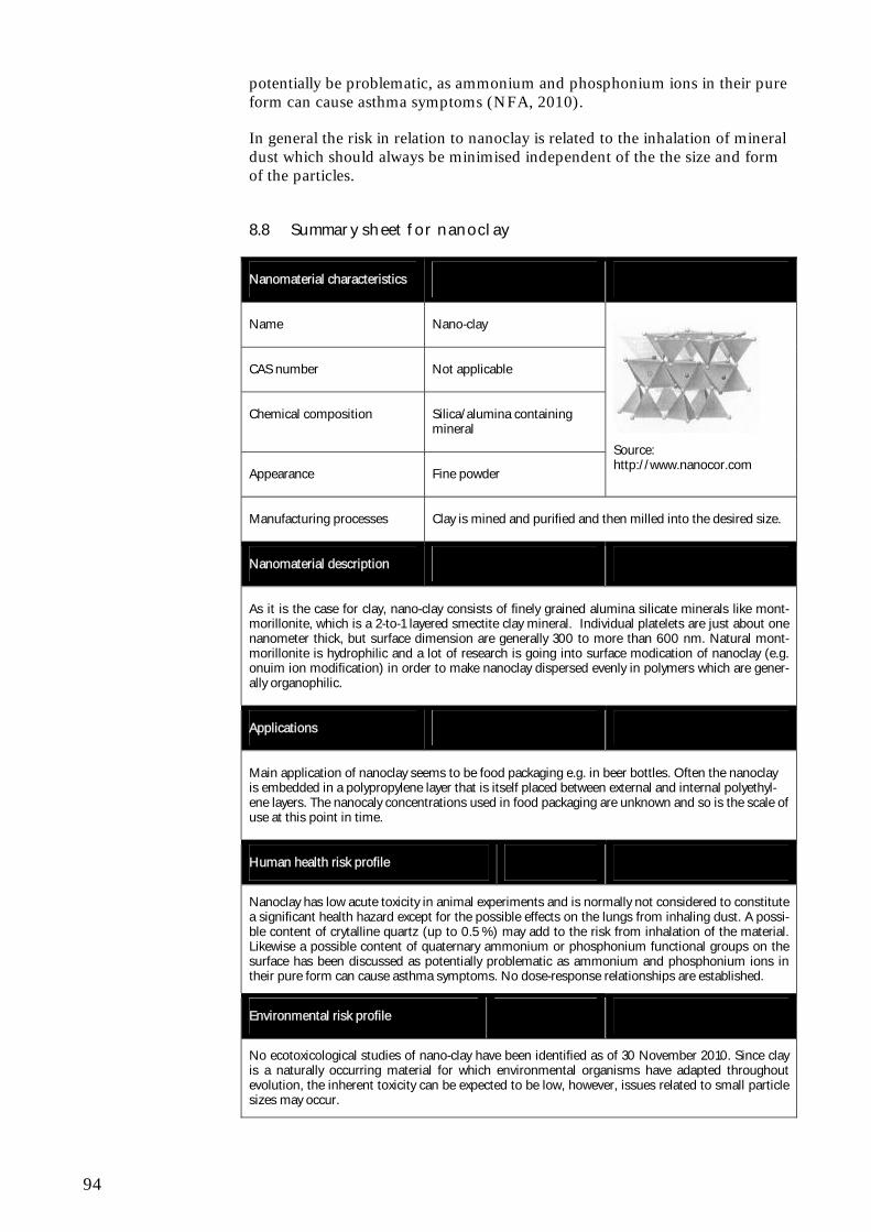
94
potentially be problematic, as ammonium and phosphonium ions in their pure form can cause asthma symptoms (NFA, 2010).
In general the risk in relation to nanoclay is related to the inhalation of mineral dust which should always be minimised independent of the the size and form of the particles.
8.8 Summary sheet for nanoclay
Nanomaterial characteristics
Name Nano-clay
CAS number Not applicable
Chemical composition Silica/alumina containing mineral
Appearance Fine powder
Source: http://www.nanocor.com
Manufacturing processes Clay is mined and purified and then milled into the desired size.
Nanomaterial description
As it is the case for clay, nano-clay consists of finely grained alumina silicate minerals like mont-morillonite, which is a 2-to-1 layered smectite clay mineral. Individual platelets are just about one nanometer thick, but surface dimension are generally 300 to more than 600 nm. Natural mont-morillonite is hydrophilic and a lot of research is going into surface modication of nanoclay (e.g. onuim ion modification) in order to make nanoclay dispersed evenly in polymers which are gener-ally organophilic.
Applications
Main application of nanoclay seems to be food packaging e.g. in beer bottles. Often the nanoclay is embedded in a polypropylene layer that is itself placed between external and internal polyethyl-ene layers. The nanocaly concentrations used in food packaging are unknown and so is the scale of use at this point in time.
Human health risk profile
Nanoclay has low acute toxicity in animal experiments and is normally not considered to constitute a significant health hazard except for the possible effects on the lungs from inhaling dust. A possi-ble content of crytalline quartz (up to 0.5 %) may add to the risk from inhalation of the material. Likewise a possible content of quaternary ammonium or phosphonium functional groups on the surface has been discussed as potentially problematic as ammonium and phosphonium ions in their pure form can cause asthma symptoms. No dose-response relationships are established.
Environmental risk profile
No ecotoxicological studies of nano-clay have been identified as of 30 November 2010. Since clay is a naturally occurring material for which environmental organisms have adapted throughout evolution, the inherent toxicity can be expected to be low, however, issues related to small particle sizes may occur.
95
9 Silicium dioxide SiO2
9.1 General characteristics
Silica or silicon dioxide (SiO2) is an abundant mineral commonly found in nature in the form of sand or quartz as well as in cell walls of diatoms. SiO2 has a number of distinct crystalline forms (rhombohedral, hexagonal, ortho-rhombic) in addition to amorphous forms e.g. α-quartz, β-quartz. Amorphous nanoSilica particles can be produced in all sizes e.g. one manufacturer has nanosilica particles available in the size of 9, 15, 30 and 55 nm with a specific surface area of 300, 200, 100 and 50 m2/g (Bayercoatings, 2010). The shape of nanosilica is normally spherical, but other forms exsist as well such as for instance springs and nanosilica can also vary in regard to their porousity. Nanosilica can furthermore be functionalized with for instance organogroups as well as other metals (mknano, 2010).
9.2 Manufacturing processes
Several methods can be used to produce nanoparticles. NanoSilica can be produced via the sol-gel method where silica monomers are allowed to con-dense to colloidal particles, which again can form aggregates and age. Another method is high-temperature hydrolysis in a hydrogen oxygen flame. The term pyrogenic silica refers to highly dispersed silicas formed from the gas phase at high temperature. The vapor-liquid-solid method can be used to produce sil-ica nanospring (Lidong et al., 2006). Silica nanoparticles are often used in formulation or as components of other nanoparticles as e.g. a surface silaniza-tion may result in less cytotoxic nanoparticles suited for bio-applications.
9.3 Uses
9.3.1 Main applications
Silica nanoparticles have numerous applications such as ingredients in ce-ment, paints, solid lubricants, shampoos, cosmetics, face creams and food (spices) (Wiesner, 2005; Environmental Project No. 1206, 2007; Winter et al., 2010; PEN, 2010). Bioelectrocemistry, curing, oil recovering applications and in formulation of other particles are furthermore potential applications (Sun et al., 2009; Makimura et al., 2010). Organo-silica has also been re-ported to be used as scratch resistant coatings (Boxall et al., 2008). No prod-ucts were identified using silica nanoparticles on the Danish marked, but a total of 14 products were identified that use the non-oxidized form of silica, namely silicon (Si). Of these products nine products were in the category Health and Fitness.
Very little information is available about the SiO2 concentrations used in the various products. Concentrations of 0.1% and 15% have been reported for paints, whereas organo-silica used in concentrations of 10% in scratch resis-tant coatings are reported. Very little is furthermore known about the scale of use, but one Danish company has been reported to handle 1-10 ton silica per year (Environmental Project No. 1206, 2007, Boxall et al., 2008).
96
9.3.2 Results from industry survey
Microsilica is a residual from manufacturing of ferrosilicium with a size of 1-10 nm. Microsilica may be used in concrete as well as in paint etc. The sur-vey on the use of microsilica has been focused on concrete assumed to be a dominant field of application.
In concrete, microsilica has been used since the 1980'es. Use of microsilica prevents alkali-silica reactions that may cause concrete to be torn apart. Mi-crosilica furthermore has a puzzulane effect and may increase the tightness and thereby the strength of concrete. Previously microsilica was widely used for manufacturing of concrete in Denmark. Today the use has been signifi-cantly reduced, primarily because of strong price increases due to heavy de-mand from the Middle East. (Eriksen, 2010). According to Environmental Project No. 1206 (2007), the commercially available silica nanoparticles (Mi-crosilica) with a grain size of 100-200 nm are used in the production of con-crete. In dry form the used silica nanoparticles are aggregated with an aggre-gate size above 100 nm. When dispersed in water the aggregates break up into primary particles smaller than 100 nm.
The content of microsilica used in concrete will correspond to 4-6 % (weight-based) of the amount of cement (Eriksen, 2010).
The present consumption of microsilica in concrete in Denmark may be roughly estimated to at least 3. - 4.000 tonnes/year (Jensen, 2010).
9.4 Eco-toxicological profile
For the following overview of the ecotoxicological profile of nano-scale silica (SiO2) it should be stressed that only a few ecotoxicological studies exist and only a part of these are aimed at the base-set organisms (fish, crustacean and algae) required for doing effects assessment according to REACH. Even fewer studies report the results in terms of the endpoints and values listed in Information Requirements of the REACH (e.g., LC50, EC50, NOEC, LOEC) (ECHA, 2008).
For zebrafish embryos (Danio rerio) significant incidences of jaw malforma-tions was noticed at the concentration of 250 mg/L, but other effects were not reported as result of the SiO2/AlO2 nanoparticles (Harper et al., 2008). Silica nanowires of 200 nm and 50 nm) with aspect ratios (i.e. ratio between length and diameter) greater than 1 has been found to cause zebrafish embryo de-formities, but particles with aspect ratio of 1 did not exhibit any toxic or tera-togenic activities at the same concentrations (Nelson et al., 2010).
A study by Fent et al. (2010) assessed the uptake and embryo toxicity of fluo-rescent silica nanoparticles in zebrafish (Danio rerio). Two different size fluo-rescent silica nanoparticles (~200nm and ~60nm) were used. Zebrafish em-bryos were exposed to concentrations of 0.0025-200 mg/L. No significant changes were observed in the survival time, hatching time, the morphology of embryos or larvae or development of zebrafish eggs.
For the freshwater crustacean, Daphnia magna, it was found that SiO2 concen-trations of 10 mg/L caused 70% mortality (Adams et al., 2006).
97
Growth inhibition tests using the algae Chlorella kessleri showed that the size of cells increased as a result of exposure with the 5 nm SiO2 particles and that these had a greater effect than the 26 and 78 nm particles (Fujiwara et al., 2008). Growth rate inhibition of the green algae Pseudokirchneriella subcapitata (72 h incubation) was observed after exposure to 12.5 nm and 27 nm silica NPs by van Hoecke et al. (2008). EC10, 72h were found to be 10.9 ± 4.4 mg/L and 15.0 ± 4.3 mg/L, and NOEC and LOEC were observed to be 4.6 and 10.0 mg/L for 12.5nm and 27 nm SiO2 NPs, respectively. Taking the specific surface area of the silica particles into account, van Hoecke et al. (2008) also expresse EC10-values as EC10, 72h in units of m2/l yielding values of 2.6 ± 1.0 and 2.0 ± 0.6 m2/L 12.5nm and 27 nm SiO2 NPs respectively. The toxicity of silica nanoparticles (10-20 nm) and the bulk particles (5-10μm) towards the green algae Scenedesmus obliquus revealed EC20 values of 388.1 mg/L (72 h) and 216.5 mg/L (96 h) (Wei et al. 2010).
No studies on the bioaccumulation of SiO2 have been found as of 22 October 2010. By definition metal oxide nanoparticles are not degradable and speci-ation of SiO2 (e.g. dissociation or complexation) is not likely to occur in aquatic media under normal conditions. Therefore, SiO2 particles can be re-garded as persistent in the sense that the particles will remain as SiO2-particles, however the sizes of the particles may change due to agglomeration and aggregation in aquatic media.
9.5 Toxicological profile
Most of the available toxicological research into bulk silica has focussed on coarse or fine crystalline silica particles of 0.5 to 10 m. Crystalline silica in the form of quartz is an IARC classified human carcinogen and is involved in the pathogenesis of silicosis. However, the mechanisms of crystalline silica toxicity at the cellular or molecular levels are still unclear. It is also not known if any single mechanism underlies the observed effects although severe in-flammation appears to be a common initiating step. The role of reactive oxy-gen species in the inflammatory, fibrogenic and carcinogenic activity of quartz is well established. Oxidative membrane and DNA damage are considered the most important mechanisms involved in the health effects of microsized crys-talline silica (Napierska et al., 2010). A large body of experimental work has shown that particle surface reactivity and the form of silica seem to govern the hazardous nature of crystalline silica and that cytotoxicity appears to be di-rectly related to the form of the particles and the extent of the exposed sur-face.
Amorphous silica and the naturally occurring amorphous silica such as diato-maceous earth are in general considered less harmful than its crystalline coun-terpart. With regard to the cytotoxic activity of different forms of amorphous silica, it has been shown not to depend on a crystalline silica component but rather on the surface charges and morphologic features of particles (Napier-ska et al., 2010).
For the silica nanoparticles (SNPs) the origin/synthesis of the particles plays an important role in determining the physico-chemical properties and conse-quently their potential interactions with biological systems. Relevant parame-ters are surface area, surface morphology, surface energy, dissolution layer properties, adsorption and aggregation properties (Napierska et al., 2010). Nanosilica occurs mainly in the amorphous form, and the potential hazard cannot be simply related to studies of micron-sized crystalline materials.
98
9.5.1 ADME studies
No studies investigating the absorption from the site of exposure into the blood have been identified. One in vivo study investigating the impact of size on tissue distribution and elimination of SNPs has been identified. In this study nanosilica (50, 100, and 200 nm diameter) injected intravenously into mice was observed in macrophages of the liver and spleen for four weeks after a single injection. The study also showed that the smaller particles were cleared via urine and bile more rapidly than larger particles (Cho et al., 2009).
Kumar et al. (2010) studied biodistribution of multimodal organically modi-fied SNPs intravenously administered to nude mice and found a greater ac-cumulation of nanoparticles in liver, spleen, and stomach than in kidney, heart, and lungs. Almost 100% of the injected nanoparticles were effectively cleared out of the animals over a period of 15 days via the hepatobiliary excre-tion. No signs of organs toxicity were observed.
In an in vitro study in human mesenchymal stem and 3T3-L1 cells the effect of surface charge on cellular uptake of mesoporous nanosilica (porous silicates with very big surface areas) was evaluated. The results showed that the uptake could be regulated by a threshold of positive surface charge and also imply that the modulation of surface charge is cell specific (Chung et al., 2007).
9.5.2 Short term toxicity
Inhalation Six in vivo studies, one involving inhalation and five involving intratracheal installation of a single dose of different forms of silica in rats and mice have been identified.
Sayes et al. (2010) exposed rats by inhalation to freshly generated, aerosolised amorphous silica NP for 1 or 3-days. Neither exposure period produced any significant pulmonary inflammatory, or adverse lung histopathological effects in rats exposed to very high particle numbers corresponding to a range of mass concentrations (1.8 or 86 mg/m3). It was also concluded that there was no genotoxic effects from the exposure.
In a study with intratracheal instillation of different sizes of nanoquartz and mined quartz particles in rats the theory that nanoparticles are more toxic than fine-sized particles of similar chemistry was contradicted. Well-characterized samples were tested for surface activity and hemolytic potential. In addition, groups of rats were instilled with either doses of 1 or 5 mg/kg of carbonyl iron (CI) or various alpha-quartz particle types in phosphate-buffered saline solu-tion and subsequently assessed using bronchoalveolar lavage fluid biomarkers, cell proliferation, and histopathological evaluation of lung tissue at 24 h, 1 week, 1 month, and 3 months postexposure. Exposures to the various alpha-quartz particles produced differential degrees of pulmonary inflammation and cytotoxicity, which were not always consistent with particle size but correlated with surface activity, particularly hemolytic potential. The results demonstrate that the pulmonary toxicities of alpha-quartz particles appear to correlate bet-ter with surface activity than particle size and surface area (Warheit et al., 2005).
Another study with intratracheal instillation of colloidal silica in rats revealed that the effect of fibrogenesis of nanosized SiO2 might be milder than that of microsized SiO2 in rats, potentially resulting from nanoparticles tending to be
99
diffused and easily translocated due to their ultrafine particle size compared to microsized particles (Chen et al., 2004).
Kaewamatawong et al. (2006) compared acute pulmonary toxicity from expo-sure to ultrafine colloidal silica particles (UFCSs) and fine colloidal silica par-ticles (FCSs) in mice intratracheally instilled with 3 mg of 14 nm UFCSs and 230 nm FCSs revealed for both sizes bronchiolar degeneration, necrosis, neu-trophilic inflammation, alveolar type II cell swelling and alveolar macrophage accumulation. UFCs induced extensive alveolar hemorrhage, a more severe bronchiolar epithelial cell necrosis and neutrophil influx in alveoli compared to FCSs. Electron microscopy demonstrated UFCSs and FCSs on bronchio-lar and alveolar wall surface as well as in the cytoplasm of alveolar epithelial cells, alveolar macrophages and neutrophils. Type I alveolar epithelial cell ero-sion with basement membrane damage in UFCSs treated animals was more severe than those in FCSs-treated animals. These findings suggest that UFCSs have greater ability to induce lung inflammation and tissue damages than FCSs.
Acute and subacute lung toxicity of low dose of UFCSs was further studied in mice intratracheally instilled with 0, 0.3, 3, 10, 30 or 100 g of UFCSs (Kae-wamatawong et al., 2006). Cellular and biochemical parameters in bronchoal-veolar lavage fluid (BALF), histological alteration and the body weight were determined at 3 days after instillation and in addition the time response was investigated. UFCSs induced moderate pulmonary inflammation and tran-sient injury on BALF indices. UFCSs-treated animals showed a significant increase of the apoptotic index in lung parenchyma at all observation times. Findings from the study suggest that instillation of a small dose of UFCSs causes transient acute moderate lung inflammation and tissue damage. Oxida-tive stress and apoptosis may underlie the lung tissue injury induction.
Cho et al. (2007) studied the pulmonary effects and inflammatory mecha-nisms of ultrafine amorphous silica particles (UFASs) intratracheally adminis-tered to A/J mice at doses of 0, 2, 10 and 50mg/kg (n=5 per group). Bron-choalveolar lavage fluid analysis, histopathological examination, quantitative real-time PCR and immunohistochemistry of the lung tissues were assessed. UFASs significantly increased the lung weights and total BAL cells following exposures. The histopathological examination revealed that UFASs induced transient but severe lung inflammation, with neutrophils, at an early stage and chronic granulomatous inflammation at the later stage. Cytokines (IL-1beta, IL-6, IL-8 and TNF-alpha) and chemokines (MCP-1 and MIP-2) were iden-tified as playing important roles in the inflammation induced by the intratra-cheal instillation of UFASs.
Dermal No in vivo dermal studies investigating short term toxicity have been identi-fied.
In a study designed to determine whether nanosilica has the potential to cause acute cutaneous toxicity, using cultured HaCaT keratinocytes (CHK) Park et al. (2010) evaluated cytotoxicity of two nanosilicas of particle size 7 and 10-20 nm. HaCaT cell viabilities were determined after exposure to the two nanosilicas at different concentrations for 48 h. Both particle sizes dose-dependently reduced cell viabilities (P < 0.001). No differences were found for the two particle sizes.
100
Oral No short term oral studies have been identified.
Intravenous, intraperitoneal, subcutaneous Hudson et al. (2008) studied the biocompatibility of mesoporous silicates and found that when particles were injected subcutaneously in rats, the amount of residual material decreased progressively over 3 months, with good biocom-patibility on histology at all time points. In contrast, intra-peritoneal and intra-venous injections in mice resulted in death or euthanasia. No toxicity was seen with subcutaneous injection of the same particles in mice. Microscopic analy-sis of the lung tissue of the mice indicated that death may be due to thrombo-sis. Although local tissue reaction to mesoporous silicates was benign, they caused severe systemic toxicity.
Nishimori et al. (2009) investigated the relationship between particle size and toxicity using silica particles with diameters of 70, 300 and 1000 nm (SP70, SP300, and SP1000) as a model material. Silica particles were administered intravenously to mice and to evaluate acute toxicity, histological analysis was performed for liver, spleen, kidney and lung. SP70-induced liver injury at 30 mg/kg body weight, while SP300 or 1000 had no effect even at 100 mg/kg. Administration of SP70 dose-dependently increased serum markers of liver injury, serum aminotransferase and inflammatory cytokines. Repeated ad-ministration of SP70 twice a week for 4 weeks, even at 10mg/kg, caused he-patic fibrosis. These findings indicate that nano-size materials may be hepato-toxic.
Park and Park (2009) investigated oxidative stress and proinflammatory re-sponses induced by silica nanoparticles in vivo and in vitro. A single treatment of SNPs (50 mg/kg) administered intravenously to mice led to the activation of peritoneal macrophages, increased cytokines, and increased level of nitric oxide released from the peritoneal macrophages. mRNA expressions of in-flammation-related genes were also elevated in the cultured peritoneal macro-phages harvested from the treated mice. Viability of splenocytes from the mice treated with silica nanoparticles was significantly decreased in the higher dose-treated groups (100 mg/kg, 200 mg/kg i.p.). However, cell proliferation without cytotoxicity was shown in the group treated with the low dose of 50 mg/kg i.p.
To elucidate the pro-inflammatory mechanism of silica nanoparticles in vivo, in vitro study using RAW 264.7 cell line which is derived from mouse perito-neal macrophage was carried out. Treatment of silica nanoparticles to the cul-tured RAW264.7 cells led to the reactive oxygen species (ROS) generation with a decreased intracellular GSH. In accordance with ROS generation, silica nanoparticles increased the level of nitric oxide released from the cultured macrophage cell line. These results suggested that silica nanoparticles gener-ate ROS and the generated ROS may trigger the pro-inflammatory responses both in vivo and in vitro (Park and Park, 2009).
9.5.3 Irritation and corrosion
Park et al. (2010) studied dermal irritation in a Draize test according to Korea Food and drug Administration guideline. Nanosilicas (7 and 10-20 nm) were applied to the shaved back of rabbits. No erythema or edema was observed after exposure for 24 or 72 hours and the results suggest that nanosilica does not induce acute cutaneous irritation.
101
In the same study Park et al. (2010) also carried out a skin irritation study us-ing a human skin equivalent model, EpiDerm. Models were treated with 500 g/ml of either nanosilica type for 5 and 18 h. Use of the model revealed no irritation potential at 500 g/ml nanosilica.
9.5.4 Skin and respiratory sensitization (in vitro and in vivo)
No studies addressing sensitisation in relation to nanosilica have been identi-fied.
9.5.5 Repeated dose toxicity, short term, sub-chronic and long term
Repeated dose: inhalation Two studies have investigated the pulmonary responses in rats after up to 4 weeks inhalation of Ludox colloidal silica. Warheit et al. (1991) exposed CD rats (nose-only) for 2 or 4 weeks at concentrations of 0, 10, 50, and 150 mg/m3 Ludox (dried SiO2). Additional groups of rats exposed for 4 weeks were given a 3-month recovery period. Following exposure and/or recovery, fluids and cells were recovered from the lungs by bronchoalveolar lavage (BAL) and measured for cellular and biochemical parameters. Results showed that exposures to 150 mg/m3 Ludox for 2 or 4 weeks produced pulmonary inflammation along with increases in BAL protein, LDH, and alkaline phos-phatase values (p less than 0.05) and reduced macrophage phagocytosis. Autoradiographic studies demonstrated that the labelling indices of terminal bronchiolar and lung parenchymal cells were generally increased in the 50 and 150 mg/m3 groups after 2 and 4 weeks of exposure but, with one exception, returned to normal levels following a 3-month post exposure period. No sig-nificant alterations in any measured parameters were detected in rats exposed to 10 mg/m3 Ludox at any time postexposure. The no-observable-effect level (NOEL) was determined at 10 mg/m3 which was consistent with results ob-tained by conventional toxicology methods.
Lee and Kelly (1992) exposed rats for 4 weeks at concentrations of 0, 10, 50, and 150 mg/m3 to Ludox colloidal silica. Rats were killed after 4 weeks of ex-posure and 10 days or 3 months after exposure. No effects were seen at the lowest concentration. After 4 weeks exposure, lung weights were increased significantly in the two highest dose groups but they were similar to the con-trols after 3 months. A dose dependent alveolar macrophage response, poly-morphonuclear leukocytic infiltration, and Type II pneumocyte hyperplasia in alveolar duct regions was reported. Lung-deposited nanosilica cleared rapidly from the lungs with half-times of approximately 40 and 50 days for the 50 and 150 mg/m3 groups, respectively. The lungs did not show fibrotic scar tis-sue formation or alveolar bronchiolarization.
Chen et al. (2008) investigated age-related differences in pulmonary and car-diovascular responses to SiO2 nanoparticle in a study with young, adult, and old rats exposed toair containing aerosol of manufactured SiO2 nanoparticles (24.1 mg/m3; 40 min/day) for four weeks. Inhalation of SiO2 nanoparticles under identical conditions caused pulmonary and cardiovascular alterations in old rats, and less change in young and adult rats, including pulmonary in-flammation, myocardial ischemic damage, atrio-ventricular blockage, and in-crease in fibrinogen concentration and blood viscosity. Old individuals were more sensitive to nanoparticle exposure than the young and adult rats. The risk of causing pulmonary damages was: old > young > adult and the risk of cardiovascular disorder was observed only in old age. The results suggest that
102
different ages may require different biomarkers for identifying pulmonary tox-icity during inhalation of nanoparticles.
Repeated dose: dermal No dermal in vivo studies with exposure to nanosilica have been identified.
Repeated dose: oral So et al. (2008) studied the effect of micro/nanoparticle silica fed to Balb/c and C57BL/6J mice for 10 weeks. The size of the nano and micron sized silica were approximately 30 nm and approximately 30 microm, respectively.Blood was tested biochemically and hematologically. There was no difference be-tween the groups in the tested items except ALT (Alanine Aminotransferase). The nano sized silica particle dieted group showed higher value of ALT than normal and micron sized silica dieted groups. H&E staining of the liver of the nano sized particle dieted group indicated some fatty liver pattern while the contents of Si in the livers of the groups were almost the same. From the re-sults, it was suggested that the nanosized silica particle had a toxic effect on the liver even though there was no significant difference in the health of mice fed amount of 140 g silica/kg mouse.
9.5.6 Mutagenicity/genotoxicity
Lison et al. (2009) investigated the genotoxic potential of SNP with the in vi-tro cytochalasin-B micronucleus (CBMN) test. This test was selected because it allows the detection of two types of chromosomal damage, i.e. clastogenic events or chromosome breaks and aneugenic events or chromosome loss. The CBMN assay was applied in A549 cells treated with different concentrations of 3 types of Stöber silica nanoparticles (16, 60 and 101 nm SNPs). A statisti-cally significant increase of micronuclei in binucleated cells was detected after treatment but only with the smallest SNPs, indicative of a size dependent genotoxic effect. However, results showed no dose-dependent effect.
Barnes et al. (2008) reported no significant genotoxicity of commercial colloi-dal and laboratory-synthesized silica nanoparticles tested using the single cell gel electrophoresis or Comet assay. Comet assays were performed on 3T3-L1 fibroblasts with 3, 6, and 24 h incubations and 4 or 40 μg/ml of silica nanoparticles. Results were independently validated in two separate laborato-ries.
In a review document discussing adaptations of the in vitro MN (micronu-cleus) assay for the genotioxicity assessment of nanomaterials, Gonzales et al. (2011) report on genotoxic effects of SiO2 nanoparticles investigated in two studies. Wang et al. (2007 as cited by Gonzales) demonstrated that crystalline 7.21 nm SiO2 nanoparticles induce MN. In contrast, no statistically significant induction of MN was found by amorphous SiO2 nanoparticles of 16, 60 and 104 nm. In this study, MN frequencies were expressed in function of mass dose, number of particles and surface area and this when taking into account both the nominal and cellular dose. They showed that there was a statistically significant positive correlation between the fold increase of MN and the parti-cle number or surface area, considering both nominal and cellular dose. No significant correlation was found between the fold increase of MN and mass dose.
Park et al. (2010) investigated the potential of four well-characterized amor-phous silica nanoparticles to induce chromosomal aberrations and gene muta-tions using the micronucleus and the plasmid lacZ gene mutation assay. The
103
80 (34) nm silica nanoparticles induced chromosomal aberrations in the mi-cronucleus assay using 3T3-L1 mouse fibroblasts and the 30 (34) and 80 (34) nm silica nanoparticles induced gene mutations in mouse embryonic fi-broblasts carrying the lacZ reporter gene. TEM imaging demonstrated that the majority of nanoparticles were localized in vacuoles and not in the nucleus of 3T3-L1 cells, indicating that the observed DNA damage was most likely a result of indirect mechanisms. The authors concluded that further studies are needed to reveal these mechanisms and to determine the biological relevance of the effects of these particular silica nanoparticles in vivo.
9.5.7 Carcinogenicity
No carcinogenicity studies involving SNPs have been identified.
9.5.8 Reproductive toxicity, developmental toxicity and teratogenicity
No reproductive toxicity, developmental toxicity and teratogenicity studies involving SNPs have been identified.
9.5.9 In vitro studies
Most studies carried out for mainly amorphous NSPs are in vitro studies in different cell types. As summarised by Napierska et al. (2019), most of these in vitro studies involving different SNPs documented the cytotoxic effects of these nanomaterials. The determinants of the observed cytotoxicity seem to be complex and vary with the particles used and cell type tested. Unfortu-nately, for many published studies, adequate material characterization is still missing.
Singh et al. (2009) also summarises results from in vitro studies: Silica has been shown to induce inflammatory and oxidative stress responses in vitro and also in vivo, but cytotoxicity is largely only observed at high concentra-tions. Additionally, silica nanoparticles have been shown to enter the cell nu-cleus where they could potentially bind to the DNA phosphate backbone. Since the silica nanoparticles can result in increased ROS (reactive oxygen species) levels and given that the hydroxyl radical is a highly reactive molecule the generation of OH close to the DNA could readily lead to the induction of DNA strand breaks and oxidised bases which could have important implica-tions in the development of cancer.
Singh et al. (2009) further summarises: Silica nanoparticles also have an im-pact on nuclear integrity by forming intranuclear protein aggregates that can lead to inhibition of replication, transcription, and cell proliferation. Addition-ally, they have been shown to reduce replication activity down to 67% and 60% after 6 and 24 h respectively and transcriptional activity down to 82% after 4 h, thus resulting in decreased cell proliferation after 24 h (88%) and 48 h (65%). This study underscores the importance of nanoparticles sizing since particles >200 nm fail to penetrate the nucleus, do not alter nuclear structure and function, and also do not interfere with gene expression.
There seems to be conflicting results as regards the genotoxicity of SNPs. On the one hand, there is limited evidence to suggest silica nanoparticles are genotoxic and some recent studies utilizing the comet assay have demon-strated that silica nanoparticles ranging in size from 20 to 400 nm do not exert significant genotoxicity (Singh et al., 2009). In contrast, one investigation based on the micronucleus assay found that these nanoparticles do indeed in-
104
duce chromosomal damage. Thus, to get a clear indication of genotoxic po-tential a battery of standardised tests that quantify different types of genetic aberrations are required to cover all potential forms of DNA damage that may be induced following exposure to nanoparticles.
9.5.10 Summary
For nano-silica particles there are relatively few studies of the toxicity available compared to what is available for titanium dioxide. Most studies are in vitro studies in different cell types. A few short-term and no chronic in vivo studies are available.
Results of available in vitro studies indicate that the particle surface area may play a crucial role in the toxicity of silica. The cytotoxic activity of silica parti-cles can be related to their surface interfacing with the biological milieu rather than to particle size or shape (Napierska et al., 2010). The effect related to other physico-chemical properties of SNPs are less well studied and no defi-nite conclusions can be made.
Results from in vivo studies indicate that the toxicity of SNPs can depend on not only the material itself but also the administration route, as was shown by Hudson et al. (2008). In this study subcutaneous injection presented good biocompatibility, whereas intraperitoneal and intravenous injection led to fatal outcomes.
Currently available data do not clarify whether amorphous SNPs - showing augmented cytotoxicity and presumably processing oxidative DNA damaging potential – are less or more harmful as compared with micron-sized silica (Napierska et al., 2010).
In most in vivo studies on acute or sub-acute health effects of SNPs the ani-mals have been exposed via inhalation or intratracheal instillation. Other ex-posure routes should therefore also be checked (e.g. blood, skin, gastrointesti-nal tract). In addition chronic studies are needed to supplement and verify the existing data.
Determining the association of results from in vitro and in vivo toxicity as-sessments is difficult; however, as concluded by Napierska et al. (2010) the common feature seems to be cytotoxicity and inflammatory response after exposure to SNPs.
Information is so far not sufficient to clearly identify and characterize the health hazards of SNPs related to the different physic-chemical characteris-tics.
9.6 Exposure scenarios
Apart from exposure through food containing silicium dioxide, consumers' direct exposure to nano-scale SiO2 is expected to be through the use of tooth-paste and cosmetic products. Oral and dermal contact are therefore the most likely routes of exposure unless the cosmetic products are applied as sprays. Dermal exposure to paints and inhalation of dust from grinding painted sur-faces are other potential exposures to NSPs.
Furthermore, it must be expected that the nanoscale fraction of microsilica in concrete (up to 6 % w/w) will give rise to both exposure of humans by inhala-
105
tion and of the environment. However, this is not a new situation, as mi-crosilica is a naturally occurring substance, which has been used in concrete since the 1980'es.
The widespread uses of SiO2 in consumer products like sunscreens, surface treatment and paints allow for emissions of SiO2 nanoparticles to the envi-ronment during and after application e.g. via wastewater or leaching of nanoparticles from treated surfaces.
9.7 Risk profile
9.7.1 Environment
In short-term ecotoxicity tests, silica nanoparticles has been found to exhibit the following effects in aquatic organisms: in zebrafish there was an incidence of jaw malformations and embryo deformities; in different algal species, cell coagulation, membrane damage, arrested growth and decrease in chlorophyll was found; an increased mortality was observed for Daphnia magna. For daphnids, SiO2 concentrations of 10 mg/L caused 70% mortality (Adams et al., 2006) and for algae EC10, 72h values in the range of 11-15 mg/L were found for two different sizes of SiO2 (van Hoecke et al., 2008). Not many studies have analyzed dose-response relationships, thus making it difficult to perform an effects assessment following the guidelines of ECHA (2008).
9.7.2 Human health
Acute toxicity from oral exposure is low. There is little information available with regard to toxicity from other exposure routes. In vitro studies give some indication of a genotoxic potential although the results from different studies were not consistent and therefore more standardized tets are required to pro-vide firm conclusions.With regard to the human health risk there is little in-formation available with regard to the specific amounts of nano SiO2 used in different product types and thereby the extent of the exposure.
Silica used in cosmetics and personal care products is in the amorphous form. In cosmetics for skin use, the silica is expected to present little, if any risk to people. For products that might be inhaled (such as a facial powder), silica particles are finely ground down and may be inhaled as such. However, as-suming that no crystalline silica is present, the exposure to silica and the re-lated risk is considered low even though the inhaled particles are associated with respiratory toxicity.
For products where silica is integrated in a matrix as in the case with sporting goods like tennis rackets and fishing rods, no risk is identified.
Conclusions from recent research into the "nanosilica hazard" states that, until now, the health effects of SNPs have mainly been studied in terms of expo-sure via the respiratory tract, after acute or sub-acute exposure. Therefore other exposure routes should be checked (e.g. blood, skin, gastrointestinal tract) in order to clarify if the SNP's have specific properties compared to their bulk counterparts. It is also stated that chronic studies are needed to supplement and verify the existing data. (Napierska et al., 2010)
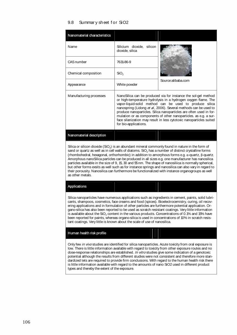
106
9.8 Summary sheet for SiO2
Nanomaterial characteristics
Name Silicium dioxide, silicon dioxide, silica
CAS number 7631-86-9
Chemical composition SiO2
Appearance White powder
Source:alibaba.com
Manufacturing processes
NanoSilica can be produced via for instance the sol-gel method or high-temperature hydrolysis in a hydrogen oxygen flame. The vapor-liquid-solid method can be used to produce silica nanospring (Lidong et al., 2006). Several methods can be used to produce nanoparticles. Silica nanoparticles are often used in for-mulation or as components of other nanoparticles. as e.g. a sur-face silanization may result in less cytotoxic nanoparticles suited for bio-applications.
Nanomaterial description
Silica or silicon dioxide (SiO2) is an abundant mineral commonly found in nature in the form of sand or quartz as well as in cell walls of diatoms. SiO2 has a number of distinct crystalline forms (rhombohedral, hexagonal, orthorhombic) in addition to amorphous forms e.g. α-quartz, β-quartz. Amorphous nanoSilica particles can be produced in all sizes e.g. one manufacturer has nanosilica particles available in the size of 9, 15, 30 and 55 nm. The shape of nanosiliica is normally spherical, but other forms exsits as well such as for instance springs and nanosilica can also vary in regard to their porousity. Nanosilica can furthermore be functionalized with instance organogroups as well as other metals.
Applications
Silica nanoparticles have numerous applications such as ingredients in cement, paints, solid lubri-cants, shampoos, cosmetics, face creams and food (spices). Bioelectrocemistry, curing, oil recov-ering applications and in formulation of other particles are furthermore potential application. Or-gano-silica has also been reported to be used as scratch resistant coatings. Very little information is available about the SiO2 content in the various products. Concentrations of 0.1% and 15% have been reported for paints, whereas organo-silica is used in concentrations of 10% in scratch resis-tant coatings. Very little is known about the scale of use of nanosilica.
Human health risk profile
Only few in vivo studies are identified for silica nanoparticles. Acute toxicity from oral exposure is low. There is little information available with regard to toxicity from other exposure routes and no dose-response relationships are established. In vitro studies give some indication of a genotoxic potential although the results from different studies were not consistent and therefore more stan-dardized tets are required to provide firm conclusions. With regard to the human health risk there is little information available with regard to the amounts of nano SiO2 used in different product types and thereby the extent of the exposure.
107
Environmental risk profile
In short-term ecotoxicity tests, silica nanoparticles has been found to exhibit the following effects in aquatic organisms: in zebrafish there was an incidence of jaw malformations and embryo de-formities; in different algal species, cell coagulation, membrane damage, arrested growth and de-crease in chlorophyll was found; an increased mortality was observed for Daphnia magna. For daphnids, SiO2 concentrations of 10 mg/L caused 70% mortality and for algae EC10, 72h values in the range of 11-15 mg/L were found for two different sizes of SiO2. These values indicate that nanosilica has low toxicity towards aquatic organisms. Only very few studies have analyzed dose-response relationships and estimated environmental concentrations are lacking. Therefore, no quantitative risk characterization can be made at this point in time.

108
109
10 Exposure and risk potential
Bulk versus nano?
A key question in relation to risk and safety assessment of nanomaterials, is to which extent the existing knowledge base about toxicity and risk related to the bulk counterparts can be used in the evaluation of the nanomaterials and sim-ply be scaled based on the size and knowledge about possible nano-specific behaviour and effects.
As discussed in the ENHRES study (Stone et al., 2010), and under the as-sumption that 'chemistry' is a key driver for toxicity, this would at least require that the chemistry should be similar for the bulk form and the nano-form of the material. This is, however, not always the case, e.g. for carbonbased nanomaterials like fullerenes, where specific surface modifications are intro-duced in order to obtain the required properties. In addition the impact of ag-glomeration and aggregation needs to be considered. For the more chemically inert particles like TiO2 and SiO2, the possibility of scaling toxicity and risk information from bulk to nano, seems to be more of an option, although it will require further investigations before firm conclusions can be made. However, for materials like TiO2, SiO2 and to some extent nanoclay, where naturally oc-curring, unmodified nanoscale particles have been part of some of the 'con-ventional' materials, the existing knowledge base is likely to cover some of the nanospecific properties and effects for these materials.
In relation to the present survey, it is important to note that all the reviewed studies are dealing with the core-particles, whereas most real-world uses of engineered nanoparticles require a surface functionalisation. How these func-tionalisations/coatings affect the bioavailability, uptake / distribution / metabo-lism / excretion, and toxicity nanoparticles is at present unknown.
Ecotoxicity and toxicity of nanomaterials?
The bulk counterparts of the seleted nanomaterials are generally not perceived as very hazardous chemicals, although some of them do have hazardous prop-erties that may constitute a risk in relation to certain exposure situations.
The overall picture in relation to the reviewed nanomaterials is that the range of toxicological and ecotoxicological studies available is not sufficient to allow firm conclusions with regard to the toxicity of the nanoparticles compared to theit bulk counterparts. This is also due to the fact that principles for physico-chemical characterization, which is a critical factor for any toxicity evaluation, still need to be further developed.
The issue of characterization in the media where the nanomaterials are tested, is a frequently discussed topic in the nano (eco)toxicological literature. In the newest studies, characterisation of the nanoparticles that are tested is most often carried out in terms of chemical composition, sizes, and surface area. However, it still remains a scientific challenge to characterize the test materials in the actual test media, and hence to control the exposure. Therefore more research is needed before a link between specific properties, or combinations of properties, and observed effects can be made.
110
While reviewing the literature on environmental effects of the selected nano-materials, a lack of studies addressing degradability and accumulation became evident – as also described by Stone et al. (2010). These two properties are as important as toxicity for determining the hazard profile of a material, and it is therefore recommended that more research efforts are directed towards bioac-cumulation and degradability of engineered nanomaterials.
In addition, the concentrations that are used in the ecotoxicological studies are mostly much higher than the concentrations expected to be environmentally realistic.
Another issue is that the majority of the reviewed studies have focussed on short-term effects of the nanomaterials whereas little information is available with regard to long-term exposures to lower concentrations aimed at chronic endpoints.
Furthermore, only part of the tests performed for environmental toxicity as-sessment are aimed at the base-set organisms and the endpoints required for doing effects assessment according to REACH as mentioned in the case of fullerenes.
For those substances that have been registered under REACH in 2010 (e.g. C60, Ag and TiO2) environmental data are now available for the test species in the base set for risk assessment in REACH (i.e., fish, crustacean, algae), al-though some of these data still need replication to improve quantification of exposure.
With regard to the evaluation of toxicological effects of the nanomaterials, an issue is that most studies are in vitro studies and only few studies are available in vivo. However, based on available studies there are indications that a com-mon mechanism of toxicity is that exposure to nanoparticles can generate an oxidative stress response and lead to pulmonary and cardiovascular inflamma-tion. Whether other mechanisms of toxicity are of importance needs to be de-termined. However, as not all nanoparticles induce toxicity, the mechanism behind the differences also needs clarification.
Main applications and exposure?
Although it may seem straight forward to determine the number of products containing nanomaterials, this is far from the case. Available databases con-taining information about nanomaterials are typically biased towards what is sold on the internet and not necessarily what is available on the market as a whole. Nanomaterials are used in a range of products and articles not defined as “nano-based” since the nanomaterials may only constitute part of the func-tionality of the product or article e.g. suntan lotions, car catalysts or laptop computers.
The results of the present survey indicate that titaniumdioxide, nanoclay and siliciumdioxide are the nanomaterials used in highest volumes in products that are also used by consumers, whereas use of cerium oxide and zero valent iron in product for consumers in Denmark has not been confirmed.
Nanosilver is likely to be found in many consumer products on the Danish market in addition to those that are commercially available on the internet and listed in the Woodrow Wilson database. It was not possible to quantify the
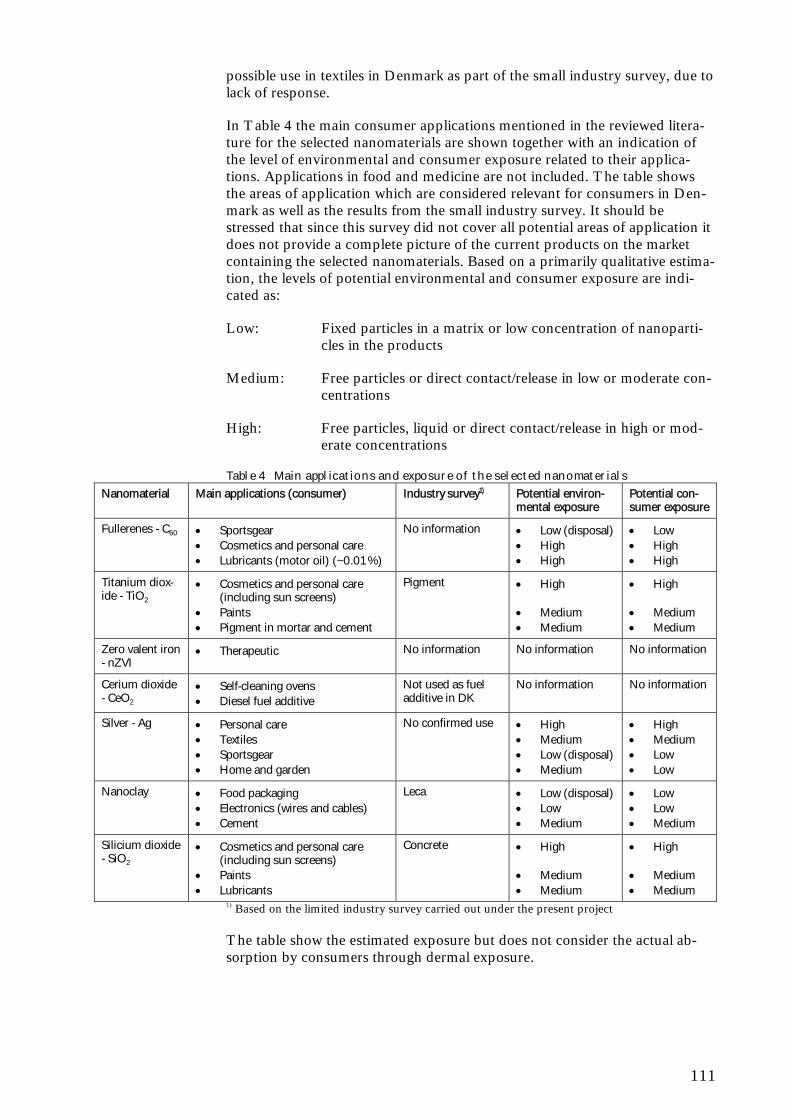
111
possible use in textiles in Denmark as part of the small industry survey, due to lack of response.
In Table 4 the main consumer applications mentioned in the reviewed litera-ture for the selected nanomaterials are shown together with an indication of the level of environmental and consumer exposure related to their applica-tions. Applications in food and medicine are not included. The table shows the areas of application which are considered relevant for consumers in Den-mark as well as the results from the small industry survey. It should be stressed that since this survey did not cover all potential areas of application it does not provide a complete picture of the current products on the market containing the selected nanomaterials. Based on a primarily qualitative estima-tion, the levels of potential environmental and consumer exposure are indi-cated as:
Low: Fixed particles in a matrix or low concentration of nanoparti-cles in the products
Medium: Free particles or direct contact/release in low or moderate con-centrations
High: Free particles, liquid or direct contact/release in high or mod-erate concentrations
Table 4 Main applications and exposure of the selected nanomaterials
Nanomaterial Main applications (consumer) Industry survey1) Potential environ-mental exposure
Potential con-sumer exposure
Fullerenes - C60 Sportsgear Cosmetics and personal care Lubricants (motor oil) (~0.01 %)
No information Low (disposal) High High
Low High High
Titanium diox-ide - TiO2
Cosmetics and personal care (including sun screens)
Paints Pigment in mortar and cement
Pigment High Medium Medium
High Medium Medium
Zero valent iron - nZVI
Therapeutic No information No information No information
Cerium dioxide - CeO2
Self-cleaning ovens Diesel fuel additive
Not used as fuel additive in DK
No information No information
Silver - Ag Personal care Textiles Sportsgear Home and garden
No confirmed use High Medium Low (disposal) Medium
High Medium Low Low
Nanoclay Food packaging Electronics (wires and cables) Cement
Leca Low (disposal) Low Medium
Low Low Medium
Silicium dioxide - SiO2
Cosmetics and personal care (including sun screens)
Paints Lubricants
Concrete High Medium Medium
High Medium Medium
1) Based on the limited industry survey carried out under the present project
The table show the estimated exposure but does not consider the actual ab-sorption by consumers through dermal exposure.
112
Risk?
The question regarding the possibility of scaling of the risk from the bulk sub-stances to the nanoform, is as mentioned not yet clarified for the many differ-ent nanomaterials and will require further research. The question is further complicated by the fact than many nanomaterials have surface modifications deliberately added to give them specific properties.
For the more chemically 'inert' particles, it is more likely that behavior and ef-fects can be scaled based on knowledge about the larger particles to the nano-scale materials.
With regard to human health, there are indications that some of the toxic ef-fects of nanosized TiO2 can be scaled based on surface area considerations from the toxicity of the micro-sized TiO2 (Stone et al., 2010). This may also be the case for SiO2 and to some extent nanoclay. For silver, more research is required, although products containing nanosilver particles are likely to have been commercially available for more than 100 years and are thereby included in some of the already existing toxicity test results. In addition there are some indications that the toxicity of silver is related the Ag+ ions but overall it is too early for firm scientific conclusions.
Fullerenes in contrast are more likely to be associated with specific nano-toxicological properties, because of the very different surface characteristics compared to the bulk material.
In order to determine whether the nanomaterials give rise to new risks com-pared to the bulk materials, it is necessary to have adequate risk assessment methodologies that are agreed and that information is available to allow com-parison of the results from different studies across different physico-chemical characteristics.
The future?
In order to answer the many questions regarding nanomaterials and risk more information and research is required in the future. Some of the gaps can be summarized as follows:
Characteristics sufficient for toxicity testing
Fate, behavior and kinetics of different nanoparticles
Agreement regarding risk assessment methodologies to comply with regulatory regimes
More information on chronic effects of nanomaterials
Effect of surface functionalisation on toxicity of the nanomaterials
114
ence of aqueous suspensions of aggregates of nano-C(60). Aquat Toxicol. 2008 Feb 18;86(3):379-87. Epub 2007 Dec 3.
Bayercoatings (2010): Dispercoll® S – Nano-silica dispersions for good initial wet strength and temperature resistance http://www.bayercoatings.de/bms/db-rsc/bms_rsc_cas.nsf/id/ADEN_Dispercoll_S(Accessed 28-11-2010)
Benn, T. M.; Westerhoff, P. (2008): Nanoparticle silver released into water from commercially available sock fabrics. Environ. Sci. Technol. 2008, 42 (11), 4133–4139.
Benn, T., Cavanagh, B., Hristovski, K., Posner, J.D., Westerhoff, P. (2010): The Release of Nanosilver from Consumer Products Used in the Home. J. Environ. Qual. 39:1875–1882.
Bermudez, E., Mangum, J. B., Wong, B. A., Asgharian, B., Hext, P. M., Warheit, D. B. and Everitt, J. I. (2004): "Pulmonary responses of mice, rats, and hamsters to subchronic inhalation of ultrafine titanium dioxide particles", Toxicol Sci, vol. 77, no. 2, pp. 347-357.
Blaser, S.A.,Scheringer, M.,MacLeod, M.,Hungerbühler, K. (2008): Estima-tion of cumulative aquatic exposure and risk due to silver: Contribution of nano-functionalized plastics and textiles. Science of The Total Environment. 390, 396-409
Boxall, A.B. A., Tiede, K., Chaudhry, Q. (2007): Engineered nanomaterials in soils and water: how do they behave and could they pose a risk to human health? Nanomedicine (Lond). 2007 Dec;2(6):919-27
Boxall, A.B. A., Chaudhry, Q., Sinclair, C., Jones, A., Aitken, R., Jefferson, B., Watts, C. (2008). Current And Future Predicted Environmental Exposure To Engineered Nanoparticles. York: Central Science Laboratory.
Braydich-Stolle, L., Hussain, S., Schlager, J.J. and Hofmann, M.C. (2005): "In vitro cytotoxicity of nanoparticles in mammalian germline stem cells", Toxicological Sciences, vol. 88, no.2, pp. 412–419
Braydich-Stolle LK, Lucas B, Schrand A, Murdock RC, Lee T, Schlager JJ, Hussain SM, Hofmann MC. (2010): Silver nanoparticles disrupt GDNF/Fyn kinase signaling in spermatogonial stem cells. Toxicol Sci. 2010 Aug;116(2):577-89.
Briel, B. (2010): Nanoclays – Counting on Consistency http://www.rockwoodadditives.com/nanoclay/pdfs/consistency.pdf
Bullard-Dillard, R., Creek, K.E., Scrivens, W.A. and Tour, J.M. (1996): "Tis-sue sites of uptake of 14C labelled C60", Bioorganic Chemistry, vol. 24, no. 4, pp. 376-385.
Cha, K., Hong, H. W., Choi, Y. G., Lee, M. J., Park, J. H., Chae, H. K., Ryu, G., and Myung, H. (2008): Comparison of acute responses of mice livers to short-term exposure to nano-sized or micro-sized silver particles, Biotech-nol.Lett., vol. 30, no. 11, pp. 1893-1899.
Chang, M., Kang, H. (2009): Remediation of pyrene-contaminated soil by synthesized nanoscale zero-valent iron particles. J. Environ. Sci. Health 44, 576–582
115
Charvin, P. Abanades, P., Bechea E., Lemont, F. and Flamant, G. (2009): Hydrogen production from mixed cerium oxides via three-step water-splitting cycles. Solid State Ionics, Volume 180, Issues 14-16, 25 June 2009, Pages 1003-1010
Chen, B.X., Wilson, S.R., Das, M., Coughlin, D.J. and Erlanger, B.F. (1998a): " Antigenicity of fullerenes: antibodies specific for fullerenes and their characteristics ", PNAS USA, vol. 95, no.18, pp.10809-10813.
Chen, H.H., Yu, C., Ueng, T.H., Chen, S., Chen, B.J., Huang, K.J. and Chiang, L.Y. (1998b): Acute and subacute toxicity study of water-soluble polyalkylsulfonated C60 in rats, Toxicol Pathol, vol. 26, no. 1, pp.143-151.
Chen, H. W., Su, S. F., Chien, C. T., Lin, W. H., Yu, S. L., Chou, C. C., Chen, J. J. and Yang, P. C. (2006): "Titanium dioxide nanoparticles induce emphysema-like lung injury in mice", FASEB Journal, vol. 20, no. 13, pp. 2393-2395.
Chen, X., Mao, S.S. (2007): Titanium Dioxide Nanomaterials: Synthesis, Properties, Modifications, and Applications Chem. Rev. 2007, 107, 2891-2959
Chen, C., Xing, G., Wang, J., Zhao, Y., Li, B., Tang, J., Jia, G., Wang, T., Sun, J., Xing, L., Yuan, H., Gao, Y., Meng, H., Chen, Z., Zhao. F, Chai, Z. and Fang, X. (2005): "Multihydroxylated [Gd@C82(OH)22]n nanoparticles: antineoplastic activity of high efficiency and low toxicity", Nano Letters, vol. 5, no.10, pp. 2050-2057.
Chen Z, Meng H, Xing G, Yuan H, Zhao F, Liu R, Chang X, Gao X, Wang T, Jia G, Ye C, Chai Z, Zhao Y. (2008): Age-related differences in pulmo-nary and cardiovascular responses to SiO2 nanoparticle inhalation: nanotoxic-ity has susceptible population. Environ Sci Technol. 2008 Dec 1;42(23):8985-92.
Cho WS, Choi M, Han BS, Cho M, Oh J, Park K, Kim SJ, Kim SH, Jeong J. (2007): Inflammatory mediators induced by intratracheal instillation of ul-trafine amorphous silica particles. Toxicol Lett. 2007 Dec 10;175(1-3):24-33. Epub 2007 Sep 29.
Cho, M., Cho, W.S., Choi, M., Kim, S.J., Han, B.S., Kim, S.., Kim, H.O., Sheen, Y.Y., and Jeong, J. (2009): The impact of size on tissue distribution and elimination by single intravenous injection of silica nanoparticles. Toxicol Lett, 189(3):177-183. PubMed abstract Internet address: http://www.ncbi.nlm.nih.gov/pubmed/19397964. Last accessed on December 20, 2010.
Choi, O. and Hu, 7. (2008): “Impact of Silver Nano-particles on Wastewater Treatment”, ACWA Annual Conference Presentation, Department of Civil and Environmental Engineering, University of Missouri, Columbia, USA.
Christensen FM, Johnston HJ, Stone V, Aitken RJ, Hankin S, Peters S, Aschberger K. (2010): Nano-silver - feasibility and challenges for human health risk assessment based on open literature. Nanotoxicology. 2010 Sep;4(3):284-95.
Chung, T.H., Wu, S.H., Yao, M., Lu, C.W., Lin, Y.S., Hung, Y., Mou, C.Y., Chen, Y.C., and Huang, D.M. (2007): The effect of surface charge on
116
the uptake and biological function of mesoporous silica nanoparticles in 3t3-l1 cells and human mesenchymal stem cells. Biomaterials, 28(19):2959-2966. Abstract from PubMed 17397919. PubMed abstract Internet address: http://www.ncbi.nlm.nih.gov/sites/entrez?orig_db=PubMed&db=pubmed&cmd=Search&TransSchema=title&term=17397919. Last accessed on Decem-ber 20, 2010.
Consumer Project No. 81 (2007): Stuer-Lauridsen, F., Kamper, A., Borling P., Petersen, G.I., Hansen, S.F., Baun, A. Mapping of products containing nanoparticles or based on nanotechnology, Danish EPA. Available: http://www2.mst.dk/common/Udgivramme/Frame.asp?pg=http://www2.mst.dk/Udgiv/publications/2007/978-87-7052-536-7/html/default_eng.htm
Cupi, D. (2010): Review on the Environmental Hazards and Human Health Effects of Silica Nanoparticles. Individual project, MSc Environmental Chemistry and Health, DTU.
Damtoft, J.S. (2010): Personal communication with Jesper Sand Damtoft, Ålborg Portland, Nov. 2010.
Danmarks Statistik (2010): http://www.statistikbanken.dk/statbank5a/default.asp?w=1280, , Nov. 2010.
Dantonit (2010): http://www.dantonit.dk/Default.aspx?ID=24
Das, M., Patil, S., Bhargava, N., Kang, J. F., Riedel, L. M., Seal, S. and Hickman, J. J. (2007): "Auto-catalytic ceria nanoparticles offer neuroprotection to adult rat spinal cord neurons", Biomaterials, vol. 28, no. 10, pp. 1918-1925.
Deshpande S., Patil S., Kuchibhatla S.V.N.T. and Seal S., (2005): Size dependency variation in lattice pa-rameter and valency states in nanocrystalline cerium oxide, Applied Physics Letters, 87, 133113, American Institute of Physics
Driscoll, K. E., Deyo, L. C., Carter, J. M., Howard, B. W., Hassenbein, D. G. and Bertram, T. A. (1997): "Effects of particle exposure and particle-elicited inflammatory cells on mutation in rat alveolar epithelial cells", Carcinogenesis, vol. 18, no.2, pp. 423-430.
ECHA (2008): Guidance on information requirements and chemical safety assessment Chapter R.7b: Endpoint specific guidance May 2008 (version 1.1), European Chemicals Agency.
Environmental Defense (ED) and Dupont (2007): Nanomaterial risk assess-ment worksheet: DuPont™ light stabilizer for use as a polymer additive. Washington DC: Environmental Defense - Dupont Nano Partnership. http://nanoriskframework.org/page.cfm?tagID=1326.
Environmental Project No. 1206 (2007): Tønning, K., Poulsen, M. Nanotechnology in the Danish industry - survey on production and applica-tion. Danish Ministry of the Environment Danish Environmental Protection Agency, 2007.
EOF (2010): Personal communication with Michael Mücke Jensen, Energi- og Olieforum, Danmark, Dec. 2010.
117
EPA (2007): Nanotechnology White Pater. Science Policy Council. United States Environmental Protection Agency, EPA 100/B-07/001 February 2007
Eriksen (2010): Personal communication with Kirsten Eriksen, COWI A/S, Dec. 2010.
Erlanger, B.F., Chen, B.X., Zhu, M. and Brus, L. (2001): "Binding of an anti-fullerene IgG monoclonal antibody to single wall carbon nanotubes", Nano Letters, vol. 1, no.9, pp. 465-467.
Espindola-Gonzalez A., Martinez-Hernandez A. L., Angeles-Chavez C., Castano V. M., Velasco-Santos C., (2010): Novel Crystalline SiO2 Nanoparticles via Annelids Bioprocessing of Agro-Industrial Wastes. Nanoscale Res Lett 5:1408–1417.
Everest (2010): http://www.everest.com.tw/_english/11_star_product/01_edit.aspx?ID=8], Dec. 2010
Farré, M., Péreza, S., Gajda-Schrantz, K., Osorio, V., Kantiani, L., Ginebreda A., Barceló, D. (2010): First determination of C-60 andC(70) fullerenes and N-methylfulleropyrrolidine C-60 on the suspended material of wastewater effluents by liquid chromatography hybrid quadrupole linear ion trap tandem mass spectrometry. Journal of Hydrology, 383 (1-2), 44 -51.
Federici, G., Shaw, B.J. and Handy, R.D. (2007): "Toxicity of titanium diox-ide nanoparticles to rainbow trout (Oncorhynchus mykiss): Gill injury, oxida-tive stress, and other physiological effects", Aquatic Toxicology, vol. 84, pp. 415-430.
Fent K., Weisbrod C.J., Wirth-Heller A., Pieles U., (2010): Assessment of uptake and toxicity of fluorescent silica nanoparticles in zebrafish (Danio re-rio) early life stages. Aquat Toxicol. 100(2):218-28.
Ferin, J., Oberdorster, G. and Penney, D. P. (1992): "Pulmonary retention of ultrafine and fine particles in rats", Am J Respir Cell Mol Biol, vol. 6, no.5, pp. 535-542.
Folkmann JK, Risom L, Jacobsen NR, Wallin H, Loft S, Møller P. (2009): Oxidatively damaged DNA in rats exposed by oral gavage to C60 fullerenes and single-walled carbon nanotubes. Environ Health Perspect. 2009 May;117(5):703-8. Epub 2008 Nov 12.
Franco, A., Hansen, S.F., Olsen, S.I., Butti, L. (2007): Limits and Prospects of the “Incremental Approach” and the European Legislation on the Man-agement of Risks related to Nanomaterials. Regulatory Toxicology and Phar-macology 48: 171-183.
Fujita, K., Morimoto, Y., Ogami, A., Myojyo, T., Tanaka, I., Shimada, M., Wang, W.N., Endoh, S., Uchida, K., Nakazato, T., Yamamoto, K., Fukui, H., Horie, M., Yoshida, Y., Iwahashi, H. and Nakanishi, J. (2009): "Gene ex-pression profiles in rat lung after inhalation exposure to C(60) fullerene parti-cles" Toxicology, vol. 258, no. 1, pp. 47-55.
Geranio, L., Heuberger, M., Nowack, B. (2010): The behaviour of silver nanotextiles during washing. Environ Sci Technol, 43, 8116-8118.
118
Gharbi, N., Pressac, M., Hadchouel, M., Szwarc, H., Wilson, S.R. and Moussa, F. (2005): "[60]Fullerene is a powerful antioxidant in vivo with no acute or subacute toxicity" Nano Letters, vol. 5, no. 12, pp. 2578-2585.
Gonzalez, L., Sanderson, B.J.S., Kirsch-Volders, (2011): Adaptations of the in vitro MN assay for the genotoxicity assessment of nanomaterials. Mutagenesis vol. 26 no. 1 pp. 185–191, 2011
Gottschalk, F.,Scholz, R.W.,Nowack, B. (2010a): Probabilistic material flow modeling for assessing the environmental exposure to compounds: Methodol-ogy and an application to engineered nano-tio2 particles. Environmental Modelling & Software. 25, 320-332.
Gottschalk, F.,Sonderer, T.,Scholz, R.W.,Nowack, B. (2010b): Possibilities and limitations of modeling environmental exposure to engineered nanomate-rials by probabilistic material flow analysis. Environmental Toxicology and Chemistry. 29, 1036-1048.
Grassian, V. H., Adamcakova-Dodd, A., Pettibone, J. M., O'shaughnessy, P. T. and Thorne, P. S. (2007): "Inflammatory response of mice to manufac-tured titanium dioxide nanoparticles: comparison of size effects through dif-ferent exposure routes", Nanotoxicol, vol. 1, no. 3, pp. 211-226
Grieger, K., Fjordbøge, A., Hartmann, N.B., Eriksson, E., Bjerg, P.L., Baun, A. (2010): Environmental benefits and risks of zero-valent iron nanoparticles (nZVI) for in situ remediation: risk mitigation or trade-off? Journal of Con-taminant Hydrology, 118: 165-183.
Griffitt, R.J., Luo, J., Gao, J., Bonzongo, J.C. and Barber, D.S. (2008): "Ef-fects of particle composition and species on toxicity of metallic nanomaterials in aquatic organisms", Environmental Toxicology and Chemistry, vol. 27, no. 9, pp. 1972-1978.
Gulbranson SH, Hud JA, Hansen RC. (2000): Argyria following the use of dietary supplements containing colloidal silver protein. Cutis 66:373_376.
Ha SW., Camalier C.E., Beck G.R. Jr., Lee JK., (2009): New method to pre-pare very stable and biocompatible fluorescent silica nanoparticles. Chem Commun (Camb). 28(20):2881–2883. doi:10.1039/b902195g.
Hansen, S.F., Michelson, E., Kamper, A., Borling, P., Stuer-Lauridsen, F. & Baun, A. (2008): Categorization framework to aid exposure assessment of nanomaterials in consumer products. Ecotoxicology 17 (5): 438-447.
Harper, S., Usenko, C., Hutchinson, J.E., Maddux, B.I.S., Tanguay, R.L. (2008): In vivo biodistribution and toxicity depends on nanomaterial compo-sition, size, surface functionalisation and routses of exposure. J. Experimental Nanoscience, vol. 3, no. 3, 195-206.Velzeboer, I., Hendriks, A.J., Ragas, A.M.J. and Van de Meent, D. 2008, "Aquatic ecotoxicity tests of some nano-materials", Environmental Toxicology and Chemistry, vol. 27, no. 9, pp. 1942-1947.
Hartmann, N.B., Von der Kammer, F., Hofmann, T., Baalousha, M., Ot-tofuelling, S., Baun, A. (2010): Algal testing of titanium dioxide nanoparti-cles—Testing considerations, inhibitory effects and modification of cadmium bioavailability. Toxicology 269 (2010) 190–197
119
Health and Safety Executive (2004): Health effects of particles produced for nanotechnologies HSE Hazard assessment document EH75/6 December. London: HSE.
Heinlaan, M., Ivask, A., Blinova, I., Dubourguier, H.C. and Kahru, A. (2008): "Toxicity of nanosized and bulk ZnO, CuO and TiO2 to bacteria Vi-brio fischeri and crustaceans Daphnia magna and Thamnocephalus platyu-rus", Chemosphere, vol. 71, no. 7, pp. 1308-1316.
Henry TB, Menn FM, Fleming JT, Wilgus J, Compton RN, Sayler GS. (2007): Attributing effects of aqueous C60 nano-aggregates to tetrahydrofu-ran decomposition products in larval zebrafish by assessment of gene expres-sion. Environ Health Perspect. 2007 Jul;115(7):1059-65.
Hill WR, Pillsbury DM. (1939): Argyria, the pharmacology of silver. Balti-more, MD, Williams and Wilkins.
Huczko, A., Lange, H. and Calko, E. (1999): "Fullerenes: experimental evi-dence for a null risk of skin irritation and allergy", Fullerene Sci Technol, vol. 7, no. 5, pp. 935-939.
Hudson SP, Padera RF, Langer R, Kohane DS. (2008): The biocompatibility of mesoporous silicates. Biomaterials. 2008 Oct;29(30):4045-55.
Hyun, J. S., Lee, B. S., Ryu, H. Y., Sung, J. H., Chung, K. H., and Yu, I. J. (2008): "Effects of repeated silver nanoparticles exposure on the histological structure and mucins of nasal respiratory mucosa in rats", Toxicol Lett, vol. 182, no. 1-3, pp. 24-28.
Imada, S., Yamanaka, N., Tamai, S. (2008): Water table depth affects Popu-lus alba fine root growth andwhole plant biomass. Funct. Ecol. 22, 1018–1026.
Inoue, K., Takano, H., Ohnuki, M., Yanagisawa, R., Sakurai, M., Shimada, A., Mizushima, K. and Yoshikawa, T. (2008): "Size effects of nanomaterials on lung inflammation and coagulatory disturbance", Int J Immunopathol Pharmacol, vol. 21, no. 1, pp. 197-206.
Intersport (2010): Personal communication with Intersport. Dec. 2010.
Jensen, I. (2010): Personal communication with Ib Jensen, Unicon, Nov. 2010.
Ji, J.H., Jung, J.H., Kim, S.S., Yoon, J.U., Park, J.D., Choi, B.S., Chung, Y.H., Kwon, I.H., Jeong, J., Han, B.S., Shin, J.H., Sung, J.H., Song, K.S. and Yu, I.J. (2007): "Twenty-eight-day inhalation toxicity study of silver nanoparticles in sprague-dawley rats", Inhalation Toxicology, vol. 19, no. 10, pp. 857-871.
Jin, C. Y., Zhu, B. S., Wang, X. F. and Lu, Q. H. (2008): "Cytotoxicity of titanium dioxide nanoparticles in mouse fibroblast cells", Chemical Research in Toxicology, vol. 21, no. 9, pp. 1871-1877.
Johansen A, Pedersen AL, Jensen KA, Karlson U, Hansen BM, Scott-Fordsmand JJ, Winding A. (2008): Effects of C60 fullerene nanoparticles on soil bacteria and protozoans. Environ Toxicol Chem. 2008 Sep;27(9):1895-903.
120
Kaegi R., Ulrich A., Sinnet B., Vonbank R., Wichser A., Zuleeg S., Simmler H., Brunner S., Vonmont H., Burkhardt M. and Boller M. (2008): Synthetic TiO2 nanoparticle emission from exterior facades into the aquatic environ-ment, Environ. Pollut., 2008, 156, 233–239.
Kaegi, R., Sinnet, B., Zuleeg, S., Hagendorfer, H., Mueller, E., Vonbank, R., Boller, M., Burkhardt, M. (2010): Release of silver nanoparticles from out-door facades. Environ Pollut, 158 (9), 2900-2905.
Kaewamatawong T., Shimada A., Okajima M., Inoue H., Morita T., Inoue K., Takano H., (2006): Acute and subacute pulmonary toxicity of low dose of ultrafine colloidal silica particles in mice after intratracheal instillation. Toxicol Pathol (34)7:958-965.
Kato S, Aoshima H, Saitoh Y, Miwa N. (2009): Biological safety of Lipo-Fullerene composed of squalane and fullerene-C60 upon mutagenesis, photo-cytotoxicity, and permeability into the human skin tissue. Basic Clin Pharma-col Toxicol. 104(6):483-7.
KemI (2009): The use of nanomaterials in Sweden 2008 - analysis and prog-nosis. Swedish Chemicals Agency. KemI PM 1/09.
Keenan CR, Goth-Goldstein R, Lucas D, Sedlak DL. (2009): Oxidative stress induced by zero-valent iron nanoparticles and Fe(II) in human bron-chial epithelial cells. Environ Sci Technol. 2009 Jun 15;43(12):4555-60.
Kim YS, Kim JS, Cho HS, Rha DS, Park JD, Choi BS, Lim R, Chang HK, Chung YH, Kwon IH, Jeong J, Han BS, Yu IJ. (2008): Twenty-eight-day oral toxicity, genotoxicity, and gender-related tissue distribution of silver nanopar-ticles in Sprague-Dawley rats. Inhal. Toxicol. 20: 575–583.
Kiss, B., Biro, T., Czifra, G., Toth, B. I., Kertesz, Z., Szikszai, Z., Kiss, A. Z., Juhasz, I., Zouboulis, C. C. and Hunyadi, J. (2008): "Investigation of mi-cronized titanium dioxide penetration in human skin xenografts and its effect on cellular functions of human skin-derived cells", Exp Dermatol, vol. 17, no. 8, pp. 659-667.
Komatsu, T., Tabata, M., Kubo-Irie, M., Shimizu, T., Suzuki, K.I., Nihei, Y. and Takeda K. (2008): "The effects of nanoparticles on mouse testis Leydig cells in vitro", Toxicol in Vitro, vol. 22, no. 8, pp. 1825–1831.
Kristensen, H.V., Vinding, K.F., Grieger, K.D., Hansen., S.F. (2009): Adopting Eco-Innovation in Danish Polymer Industry Working with Nanotechnology: Drivers, Barriers and Future Strategies, for the Nanotech-nology Law and Business 6(3):416-440.
Kumar R, Roy I, Ohulchanskky TY, Vathy LA, Bergey EJ, Sajjad M, Prasad PN. (2010): In vivo biodistribution and clearance studies using multimodal organically modified silica nanoparticles. ACS Nano. 2010 Feb 23;4(2):699-708.
Larese, F.F., D'Agostin, F., Crosera, M., Adami, G., Renzi, N., Bovenzi ,M. and Maina, G. (2009): "Human skin penetration of silver nanoparticles through intact and damaged skin", Toxicol, vol. 225, no. 1-2, pp.33-37.
121
K. P. Lee and D. P. Kelly (1992): The pulmonary response and clearance of Ludox colloidal silica after a 4-week inhalation exposure in rats. Fundamental and Applied Toxicology, Volume 19, Issue 3, October 1992, Pages 399-410
Lee, C., Kim, J.Y., Lee, W.I., Nelson, K.L., Yoon, J., Sedlak, D.L. (2008): Bactericidal effect of zero-valent iron nanoparticles on escherichia coli. Environ. Sci. Technol. 42, 4927–4933.
Li, X., Elliott, D.W., Zhang, W. (2006): Zero-valent iron nanoparticles for abatement of environmental pollutants: materials and engineering aspects. Crit. Rev. Solid State Mater. Sci. 31, 11–22.
Li, H., Zhou, Q., Wu, Y., Fu, J., Wang, T., Jiang, G. (2009): Effects of wa-terborne nano-iron on medaka (oryzias latipes): antioxidant enzymatic activ-ity, lipid peroxidation and histopathology. Ecotoxicol. Environ. Saf. 72, 684–692.
Li PR, Wei JC, Chiu YF, Su HL, Peng FC, Lin JJ. (2010): Evaluation on cy-totoxicity and genotoxicity of the exfoliated silicate nanoclay. ACS Appl Ma-ter Interfaces. 2010 Jun;2(6):1608-13.
Lide D.R., ed. (2009): CRC Handbook of Chemistry and Physics, 89th Edi-tion, CRC Press/Taylor and Fran-cis, Boca Raton, Florida
Lidong Wang, D Major, P Paga, D Zhang, M G Norton, D N McIlroy (2006): "High yield synthesis and lithography of silica-based nanospring mats". Nanotechnology 17: S298–S303
Lin, W., Huang, Y. W., Zhou, X. D. and Ma, Y. (2006): "Toxicity of cerium oxide nanoparticles in human lung cancer cells", Int J Toxicol, vol. 25, no.6, pp. 451-457.
Lindgreen, H. (2010): Personal communication with Holger Lindgreen, Geus, Nov. 2010.
Lison, D., Kirsch-Volders, M., Hoet, P.. Martens, J., Kirschhock, C. (2009): Physico-chemical determinants of toxicity : a rational approach towards safer nanostructured materials - S²NANO”. Final Report Phase 1. Brussels : Bel-gian Science Policy 2009 – 18 p. (Research Programme Science for a Sus-tainable Development)
Lovern SB, Klaper R. (2006): Daphnia magna mortality when exposed to titanium dioxide and fullerene (C60) nanoparticles. Environ Toxicol Chem.; 2006 Apr;25(4):1132-7.
Lovern SB, Strickler JR, Klaper R. (2007): Behavioral and physiological changes in Daphnia magna when exposed to nanoparticle suspensions (tita-nium dioxide, nano-C60, and C60HxC70Hx). Environ Sci Technol. 2007 Jun 15;41(12):4465-70.
Lowry, G. V. and Casman E. A. (2009): Nanomaterial Transport, Transfor-mation, and Fate ion the Environment: A Risk-based Perspective on Research Needs. In Risk, Uncertainty and Decision Analysis for Nanomaterials: Envi-ronmental Risks and Benefits and Emerging Consumer Products. Eds. Igor Linkov and Jefferey Stevens. Springer Verlag. pp. 125-139.
122
Luoma, S. N. (2008): Silver Nanotechnologies and the Environment Old Problems or New Challenges? PEN 15. Washington, DC: Project on Emerg-ing Nanotechnologies, Woodrow Wilson International Center for Scholars.
Makimura D., Metin C., Kabashima T., Matsuoka T., Nguyen Q. P., Miranda C.R. (2010): Combined modeling and experimental studies of hy-droxylated silica nanoparticles. J Mater Sci 45:5084–5088.
Mknano (2010): mkNANO. http://www.mknano.com/searchresultsproduct.asp (Accessed 28-11-2010)
Mori, T., Takada, H., Ito, S., Matsubayashi, K., Miwa, N. and Sawaguchi, T. (2006): "Preclinical studies on safety of fullerene upon acute oral administra-tion and evaluation for no mutagenesis", Toxicology, vol. 225, no. 1, pp. 48-54.
Morsy, M.S., Alsayed, S.H. and Aquel, M. (2010): Effect of Nano-clay on Mechanical Properties and Microstructure of Ordinary Portland Cement Mortar. International Journal of Civil & Environmental Engineering IJCEE-IJENS Vol: 10 No:01
Mueller, N.C., Nowack, B. (2008): Exposure modeling of engineered nanoparticles in the environment. Environmental Science & Technology. 42, 4447-4453.
Nano-C (2002): Masters of Flame: Industrial Production of Fullerenes Be-comes a Reality. Nano-C Inc., Westwood, MA.
Nanocor (2010): Nanocor. http://www.nanocor.com/
Nanowerk (2010): Nanomaterial Database Search. http://www.nanowerk.com/phpscripts/n_dbsearch.php (Accessed 5 Novem-ber, 2010).
Naota M, Shimada A, Morita T, Inoue K, Takano H. (2009): Translocation pathway of the intratracheally instilled C60 fullerene from the lung into the blood circulation in the mouse: possible association of diffusion and caveolae-mediated pinocytosis. Toxicol Pathol. 2009;37(4):456-62. Epub 2009 Apr 3.
Napierska, D., Thomassen, Leen CJ., Lison, Dominique, Martens, Johan A., Hoet, Peter H. (2010): The nanosilica hazard: another variable entity. Particle and Fibre Toxicology 7:39 http://www.particleandfibretoxicology.com/content/7/1/39
Nelson S.M., MahmoudT., Beaux II M., Shapiro P., McIlroy D.N., Stenkamp D.L., (2010): Toxic and teratogenic silica nanowires in developing vertebrate embryos. Nanomedicine 6(1): 93–102. doi:10.1016/j.nano.2009.05.003.
NFA (2010): Nanoplast - Nanoteknologiske materialer og produkter i plast-industrien. Baggrund. Det Nationale Forskningscenter for Arbejdsmiljø. http://www.arbejdsmiljoforskning.dk/da/projekter/nanoteknologiske-materialer-og-produkter-i-plastindustrien---nanoplast/baggrund
Nike (2010): Personal communication with Ms. Verena Janowski, Nike Den-mark. Dec. 2010.
123
Nishimori H, Kondoh M, Isoda K, Tsunoda S, Tsutsumi Y, Yagi K. (2009): Influence of 70 nm silica particles in mice with cisplatin or paraquat-induced toxicity. Pharmazie. 2009 Jun;64(6):395-7.
Nordberg, G.F. and L. Gerhardsson (1988): Silver. In: Handbook on Toxic-ity of Inorganic Compounds, H.G. Seiler and H. Sigel, eds. Marcel Dekker, Inc., New York, pp. 619-623.
Norwegian Labour Inspection Authority (2010): Survey on Production , Im-port and Use of Nanotechnological Products in Norway. Final Report, July 2010
Nowack B., Krug H., Height M. (2011): 120 Years of Nanosilver History: Implications for Policy Makers. Environ Sci Technol. 2011 Mar 2. [Epub ahead of print] NTP, 2009. Chemical information Review Document for Sil-ica flour. Micronised -quartz. Supporting Nomination for Toxicological Evaluation by the national Toxicology Program.
Oberdörster, E., Larkin, P., Rogers, J. (2006): Rapid environmental impact screening for engineered nanomaterials: a case study using microarray tech-nology. Project on Emerging Nanotechnologies, Woodrow Wilson Interna-tional Center for Scholars, June 2006, Washington D.C.
Park B, Martin P, Harris C, Guest R, Whittingham A, Jenkinson P, Handley J. (2007): Initial in vitro screening approach to investigate the potential health and environmental hazards of Enviroxtrade mark - a nanoparticulate cerium oxide diesel fuel additive. Part Fibre Toxicol. 2007 Dec 5;4:12.
Park, Barry, Donaldson, Kenneth, Duffin, Rodger, Tran, Lang, Kelly, Frank, Mudway, Ian, Morin, Jean-Paul, Guest, Robert, Jenkinson, Peter, Samaras, Zissis, Giannouli, Myrsini, Kouridis, Haris and Martin, Patricia (2008): Haz-ard and Risk Assessment of a Nanoparticulate Cerium Oxide-Based Diesel Fuel Additive—A Case. Study. Inhalation Toxicology, 20:6, 547 — 566
Park, E. J., Choi, J., Park, Y. K., and Park, K. (2008a): "Oxidative stress in-duced by cerium oxide nanoparticles in cultured BEAS-2B cells", Toxicol, vol. 245, no. 1-2, pp. 90-100.
Park EJ, Park K., (2009): Oxidative stress and pro-inflammatory responses induced by silica nanoparticles in vivo and in vitro. Toxicology Letters 184(1):18-25.
Park YH, Kim JN, Jeong SH, Choi JE, Lee SH, Choi BH, Lee JP, Sohn KH, Park KL, Kim MK, Son SW. (2010): Assessment of dermal toxicity of nanosilica using cultured keratinocytes, a human skin equivalent model and an in vivo model. Toxicology. 2010 Jan 12;267(1-3):178-81. Epub 2009 Oct 20.
Patil S, Sandberg A, Heckert E, Self W, Seal S. (2007): Protein adsorption and cellular uptake of cerium oxide nanoparticles as a function of zeta poten-tial. Biomaterials. 2007 Nov;28(31):4600-7. Epub 2007 Aug 1.
PEN (2011): The Project on Emerging Nanotechnologies. Consumer Prod-uct Inventory. Woodrow Wilson International Center for Scholars. Accessed January 2011: http://www.nanotechproject.org/inventories/consumer/
124
Phenrat T, Long TC, Lowry GV, Veronesi B. (2009): Partial oxidation ("ag-ing") and surface modification decrease the toxicity of nanosized zerovalent iron. Environ Sci Technol. 2009 Jan 1;43(1):195-200.
Pirngadi, P., Richter, S. (2005): New Developments in the Application of Nanoclays for HFFR Cable Compounds. Proceedings of the 54th IWCS/Focus Conference, 2005. http://ecadigitallibrary.com/pdf/IWCS05/IWCS0582_lke.pdf
Powell JJ, Faria N, Thomas-McKay E, Pele LC. (2010): Origin and fate of dietary nanoparticles and microparticles in the gastrointestinal tract. J Auto-immun. 2010 May;34(3):J226-33. Epub 2010 Jan 21.
Pronk, M. E. J.; Wijnhoven, S. W. P.; Bleeker, E. A. J.; Heugens, E. H. W.; Peijnenburg, W. J. G. M.; Luttik, R.; Hakkert, B. C. (2009): Nanomaterials under REACH: Nanosilver as a case study. National Institute for Public Health and the Environment, Bilthoven, the Netherlands
Prospect (2010): Global Nanomaterials Safety. Toxicological Review of Nano Cesium Oxide. Ecotoxicology Test Protocols for Representative Nanomateri-als in Support of the OECD Sponsorship Programme. 1 July 2010
Ramsden, C.S., Smith, T.J., Shaw, B.J., Handy, R.D. (2009): Dietary expo-sure to titanium dioxide nanoparticles in rainbow trout, (Oncorhynchus mykiss): no effect on growth, but subtle biochemical disturbances in the brain. Ecotoxicology 18:939–951.
Ratte, H. T. (1999): Bioaccumulation and toxicity of silver compounds: A review. Environmental Toxicology and Chemistry, 18: 89–108.
Regulation (EC) No 1272/2008 of the European Parliament and of the Council of 16 December 2008 on classification, labelling and packaging of substances and mixtures, amending and repealing Directives 67/548/EEC and 1999/45/EC, and amending Regulation (EC) No 1907/2006.
Regulation (EC) No 1907/2006 of the European Parliament and of the Council of 18 December 2006 concerning the Registration, Evaluation, Au-thorisation and Restriction of Chemicals (REACH), establishing a European Chemicals Agency, amending Directive 1999/45/EC and repealing Council Regulation (EEC) No 793/93 and Commission Regulation (EC) No 1488/94 as well as Council Directive 76/769/EEC and Commission Directives 91/155/EEC, 93/67/EEC, 93/105/EC and 2000/21/EC.
RIVM (2009): "Nanomaterials under REACH: Nanosilver as a case study", RIVM Report 601780003/2009
Roursgaard, M., Poulsen, S.S., Kepley, C.L., Hammer, M., Nielsen, G.D. and Larsen, S.T. (2008): "Polyhydroxylated C60 fullerene (fullerenol) at-tenuates neutrophilic lung inflammation in mice", Bas Clin Pharmacol Toxi-col, vol. 103, no. 4, pp. 386-388.
Saleh, N.B., Pfefferle, L.D. and Elimelech, M. (2008): “Aggregation kinetics of multiwalled carbon nanotubes in aquatic systems: measurements and envi-ronmental implications”, Environmental Science and Technology, vol. 42, pp. 7963-9.
125
Saleh, N., Kim, H., Phenrat, T., Lowry, G.V., Tilton, R.D., Matyjaszewski, K., Lowry, G.V., Tilton, R.D. (2008): Ionic strength and composition affect the mobility of surface-modified Fe0 nanoparticles in water-saturated sand columns. Environ. Sci. Technol. 42, 3349–3355.
Sayes, C.M., Fortner, J.D., Guo, W., Lyon, D.Y., Boyd, A.M., Ausman, K., Tao, Y.J., Sitharaman, B., Wilson, L.J., Hughes, J.B., West, J.L. and Colvin, V.L. (2004): "The differential cytotoxicity of water-soluble fullerenes", Nano Letters, vol. 4, no. 10, pp. 1881-1887.
Sayes, C.M., Marchione, A.A., Reed, K.L. and Warhei,t D.B. (2007): "Com-parative pulmonary toxicity assessments of C60 water suspensions in rats: few differences in fullerene toxicity in vivo in contrast to in vitro profiles", Nano Letters, vol. 7, no. 8, pp. 2399-2406.
Sayes CM, Reed KL, Glover KP, Swain KA, Ostraat ML, Donner EM, Warheit DB. (2010): Changing the dose metric for inhalation toxicity studies: Short-term study in rats with engineered aerosolized amorphous silica nanoparticles. Inhalation Toxicology, 22(4): 348-354 (March 2010).
SCENIHR (2005): Opinion on the appropiateness of existing methodologies to access the potential risks associated with engineered and adventitious prod-ucts of nanotechnologies. Brussels: European Commision, health and con-sumer protection directorate-general, Scientific Committee on Emerging and newly Identified Health Risks (SCENIHR)
Schreiner KM, Filley TR, Blanchette RA, Bowen BB, Bolskar RD, Hockaday WC, Masiello CA, Raebiger JW. (2009): White-rot basidiomycete-mediated decomposition of C60 fullerol. Environ Sci Technol. 2009 May 1;43(9):3162-8.
Schrick, B., Hydutsky, B.W., Blough, J.L., Mallouk, T.E. (2004): Delivery vehicles for zerovalent metal nanoparticles in soil and groundwater. Chem. Mater. 16, 2187–2193.
Scott-Fordsmand JJ, Krogh PH, Schaefer M, Johansen A. (2008): The toxic-ity testing of double-walled nanotubes-contaminated food to Eisenia veneta earthworms. Ecotoxicol Environ Saf. 2008 May 29.
Scown, T.M., van Aerle, R., Johnston, B.D., Cumberland, S., Lead, J.R., Owen, R. & Tyler, C.R. (2009): "High Doses of Intravenously Administered Titanium Dioxide Nanoparticles Accumulate in the Kidneys of Rainbow Trout but with no Observable Impairment of Renal Function", Toxicological Sciences, vol. 109, no. 2, pp. 372-380.
Sera, N., Tokiwa, H. and Miyata, N. (1996): "Mutagenicity of the fullerene C60-generated singlet oxygen dependent formation of lipid peroxides", Car-cinogenesis, vol. 17, no. 10, pp. 2163-2169.
Sharma AK, Schmidt B, Frandsen H, Jacobsen NR, Larsen EH, Binderup ML. (2010): Genotoxicity of unmodified and organo-modified montmorillo-nite. Mutat Res. 2010 Jul 19;700(1-2):18-25. Epub 2010 Apr 28.
Shinohara N, Matsumoto K, Endoh S, Maru J, Nakanishi (2009): J. In vitro and in vivo genotoxicity tests on fullerene C60 nanoparticles. Toxicol Lett. 2009 Dec 15;191(2-3):289-96. Epub 2009 Sep 20.
126
Schubert D, Dargusch R, Raitano J, Chan SW. (2006): Cerium and yttrium oxide nanoparticles are neuroprotective. Biochem Biophys Res Commun. 2006 Mar 31;342(1):86-91. Epub 2006 Feb 3.
Silver S. (2003): Bacterial silver resistance: Molecular biology and uses and misuses of silver compounds. FEMS Microbiol Rev 27:341_353.
Singh N, Manshian B, Jenkins GJ, Griffiths SM, Williams PM, Maffeis TG, Wright CJ, Doak SH. (2009): Nanogenotoxicology: The DNA damaging po-tential of engineered nanomaterials. Biomaterials. 2009 Aug;30(23-24):3891-914. Epub 2009 May 8.
Sitharaman, B., Wilson, L. J., Hughes, J. B., West, J. L., Colvin, V. L. (2004): The differential cytotoxicity of water-soluble fullerenes. Nano Lett 4(10): 1881-1887.
So SJ, Jang IS, Han CS. (2008): Effect of micro/nano silica particle feeding for mice. J Nanosci Nanotechnol. 2008 Oct;8(10):5367-71.
Stone, V., Hankin, S., Aitken, R., Aschberger, K., Baun, A., Christensen, F., Fernandes, T., Hansen, S.F., Hartmann, N.B., Hutchinson, G., Johnston, H., Micheletti, G., Peters, S., Ross, B., Sokull-Kluettgen, B., Stark, D., Tran, L. (2010): Engineered Nanoparticles: Review of Health and Environmental Safety (ENRHES). Available: http://nmi.jrc.ec.europa.eu/project/ENRHES.htm (Accessed February 1, 2010).
Suha W.H., Suslickb K.S., Stuckya G.D., Suh Y-H., (2009): Nanotechnol-ogy, nanotoxicology, and neuroscience. Prog Neurobiol. 87(3):133–170. doi:10.1016/j.pneurobio.2008.09.009.
Sun, H., Wang, L., Zhang, R., Sui, J., Xu, G. (2006): Treatment of ground-water polluted by arsenic compounds by zero valent iron. J. Hazard. Mater. 129, 297–303.
Sun W., Wang D., Zhai Z., Gao R., Jiao K., (2009): Direct Electrochemistry of Hemoglobin Immobilized in the Sodium Alginate and SiO2 Nanoparticles Bionanocomposite Film on a Carbon Ionic Liquid Electrode. J. Iran. Chem. Soc, 6(2): 412-419
Sung J.H., Ji J.H., Yoon J.U., Kim D.S., Song M.Y., Jeong J., Han B.S., Han J.H., Chung Y.H., Kim J., Kim T.S., Chang H.K., Lee E.J., Lee J.H., Yu I.J., (2008): Lung function changes in Sprague-Dawley rats after prolonged inha-lation exposure to silver nanoparticles. Inhal Toxicol 20:567-574.
Sung, J.H., Ji, J.H., Park, J.D., Yoon, J.U., Kim, D.S., Jeon, K.S., Song, M.Y., Jeong, J., Han, B.S., Han, J.H., Chung, Y.H., Chang, H.K., Lee, J.H., Cho, M.H., Kelman, B.J. and Yu, I.J. (2009): "Subchronic inhalation toxicity of silver nanoparticles", Toxicol Sciences, vol. 108, no.2, 452-461.
Suresh, R., Borkar, S.N., Sawant, V.A., Shende, V.S., Dimble, S.K. (2010): Nanoclay drug delivery System. International Journal of Pharmaceutical Sci-ences and Nanotechnology 3(2)
Tabata, Y., Murakami, Y. and Ikada, Y. (1997): "Photodynamic effect of polyethylene glycol-modified fullerene on tumour", Jap J Cancer Res, vol. 88, no. 11, pp. 1108-1116.
127
Takeara, H., Fujiwara, M., Arikawa, M., Diener, M.D., Alford, J.M. (2004): Experimental study of industrial scale fullerene production by combustion synthesis. Carbon 43 (2), 311–319.
Takenaka, S., Karg, E., Roth, C., Schulz, H., Ziesenis, A., Heinzmann, U., Schramel, P., and Heyder, J. (2001): "Pulmonary and systemic distribution of inhaled ultrafine silver particles in rats", Environ.Health Perspect., vol. 109 Suppl 4, pp. 547-551.
Taylor (2008): http://www.nanotechproject.org/process/assets/files/6704/taylor_gma_pen_packaging1.pdf
Terrones H., Terrones, M. (2003): The carbon nanocosmos: novel materials for the twenty-first century. Filosiphical transaction of the Royal Society, Vol. 361, p. 2789-2806.
Theogaraj, E., Riley, S., Hughes, L., Maier, M. and Kirkland, D. (2007): "An investigation of the photo-clastogenic potential of ultrafine titanium dioxide particles", Mut Res, vol. 634, no. 1-2, pp. 205-219.
Totsuka Y, Higuchi T, Imai T, Nishikawa A, Nohmi T, Kato T, Masuda S, Kinae N, Hiyoshi K, Ogo S, Kawanishi M, Yagi T, Ichinose T, Fukumori N, Watanabe M, Sugimura T, Wakabayashi K. (2009): Genotoxicity of nano/microparticles in in vitro micronuclei, in vivo comet and mutation assay systems. Part Fibre Toxicol. 2009 Sep 3;6:23.
Tratnyek and Johnson (2006): Nanotechnologies for environmental cleanup. NanoToday 1(2) 44-48.
Trop, M., Novak, M., Rodl, S., Hellbom, B., Kroell, W., Goessler, W. (2006): “Silver coated dressing Acticoat caused raised liver enzymes and ar-gyria-like symptoms in burn patient“, J Trauma, vol. 60, no. 3, pp. 648–652.
Tsuchiya, T., Oguri, I., Yamakoshi, Y.N. and Miyata, N. (1996): "Novel harmful effects of [60]fullerene on mouse embryos in vitro and in vivo", FEBS Letters, vol. 393, no. 1, pp. 139-145.
Van Hoecke K, Quik JT, Mankiewicz-Boczek J, De Schamphelaere KA, El-saesser A, Van der Meeren P, Barnes C, McKerr G, Howard CV, Van de Meent D, Rydzyński K, Dawson KA, Salvati A, Lesniak A, Lynch I, Silversmit G, De Samber B, Vincze L, Janssen CR. (2009): Environ Sci Technol., 43(12):4537-46.
Velzeboer, I., Hendriks, A.J., Ragas, A.M.J. and Van de Meent, D. (2008): "Aquatic ecotoxicity tests of some nanomaterials", Environmental Toxicology and Chemistry, vol. 27, no. 9, pp. 1942-1947.
Wadhera, A., and Fung, M. (2005): Systemic argyria associated with inges-tion of colloidal silver. Dermatology Online Journal, 11(1): 12.
Wakefield G., Wu X., Gardener M., Park B. and Anderson S. (2008): Envi-roxTM fuel-borne catalyst: Devel-oping and launching a nano-fuel additive, Technology Analysis & Strategic Management, Vol. 20, No. 1, pp. 127-136
128
Wang J., Zhang X., Chen Y., Sommerfield M. and Hu, Q. (2008): "Toxicity assessment of manufactured nanomaterials using the unicellular green alga Chlamydomonas reinhardtii.", Chemosphere, vol. 73, no. 7.
Wang, J., Zhou, G., Chen, C., Yu, H., Wang, T., Ma, Y., Jia, G., Gao, Y., Li, B., Sun, J., Li, Y., Jiao, F., Zhao, Y. and Chai, Z. (2007): "Acute toxicity and biodistribution of different sized titanium dioxide particles in mice after oral administration", Toxicology Letters, vol. 168, no. 2, pp. 176-185.
Wang, J., Chen, C., Liu, Y., Jiao, F., Li, W., Lao, F., Li, Y., Li, B., Ge, C., Zhou, G., Gao, Y., Zhao, Y. and Chai, Z. (2008a): "Potential neurological lesion after nasal instillation of TiO2 nanoparticles in the anatase and rutile crystal phases", Toxicology Letters, vol. 183, no. 1-3, pp. 72-80.
Wang, J., Liu, Y., Jiao, F., Lao, F., Li, W., Gu, Y., Li, Y., Ge, C., Zhou, G., Li, B., Zhao, Y., Chai, Z. and Chen, C. (2008b): "Time-dependent transloca-tion and potential impairment on central nervous system by intranasally in-stilled TiO2 nanoparticles", Toxicol, vol. 254, no. 1-2, pp. 82-90.
Warheit, D. B., Brock, W. J., Lee, K. P., Webb, T. R. and Reed, K. L. (2005): "Comparative pulmonary toxicity inhalation and instillation studies with different TiO2 particle formulations: impact of surface treatments on particle toxicity" Toxicol Sci, vol. 88, no.2, pp. 514-524.
Warheit, D. B., Webb, T. R., Reed, K. L., Frerichs, S. and Sayes, C. M. (2007): "Pulmonary toxicity study in rats with three forms of ultrafine-TiO2 particles: differential responses related to surface properties. Toxicol, vol. 230, no.1, pp. 90-104.
Wiesner, Mark R. (2005): “Science: Toward a green nanotechnology,” Daily Times, Monday, August 8, 2005, http://www.dailytimes.com.pk/default.asp?page=story_8-8-2005_pg6_7
Wijnhoven, S.W.P., W.J.G.M. Peijnenburg, C.A. Herberts, W.I. Hagens, A.G., Oomen, E.H.W. Heugens, B. Roszek, J. Bisschops, I. Gosens, D. Van De Meent, S. Dekkers, W.H. De Jong, M. van Zijverden, A.J.A.M. Sips, and R.E. Geertsma. (2009): Nano-silver: A review of available data and knowl-edge gaps in human and environmental risk assessment. Nanotoxicology, 3:109–138.
Winter M, Beer HD, Hornung V, Kramer U, Schins RPF, Forster I. (2010): Activation of the inflammasome by amorphous silica and TiO2 nanoparticles in murine dendritic cells. Nanotoxicology, Early Online, DOI: 10.3109/17435390.2010.506957.
Woodrow Wilson Inventory (2011): Project of Emerging Nanotechnologies at the Woodrow Wilson International Center for Scholars (2011). Nanotechnol-ogy Consumer Products Inventory. http://www.nanotechproject.org/inventories/consumer/analysis_draft/ (visited 22 March 2011)
Zhu X, Zhu L, Lang Y, Chen Y. (2008): Oxidative stress and growth inhibi-tion in the freshwater fish Carassius auratus induced by chronic exposure to sublethal fullerene aggregates. Environ Toxicol Chem. 2008 Sep;27(9):1979-85.
129
Zhu S, Oberdörster E, Haasch ML.(2006): Toxicity of an engineered nanoparticle (fullerene, C60) in two aquatic species, Daphnia and fathead minnow. Mar Environ Res. 2006 Jul;62 Suppl:S5-9. Epub 2006 Apr 22.
Zhu, X.S., Chang, Y. & Chen, Y.S. (2010): "Toxicity and bioaccumulation of TiO2 nanoparticle aggregates in Daphnia magna", Chemosphere, vol. 78, no. 3, pp. V-215.
Zhu, X.S., Zhu, L., Duan, Z.H., Qi, R.Q., Li, Y. and Lang, Y.P. (2008): "Comparative toxicity of several metal oxide nanoparticle aqueous suspen-sions to Zebrafish (Danio rerio) early developmental stage", Journal of Envi-ronmental Science and Health Part A-Toxic/hazardous Substances and Envi-ronmental Engineering, vol. 43, no. 3, pp. 278-284.
Yamago, S., Tokuyama, H., Nakamura, E., Kikuchi, K., Kananishi, S., Sueki, K., Nakahara, H., Enomoto, S. and Ambe F. (1995): "In vivo biological be-haviour of a water-miscible fullerene: 14C labelling, absorption, distribution, excretion and acute toxicity", Chem Biol, vol. 2, no.6, pp. 385-389.
Yin, J.J., Lao, F., Meng, J., Fu, P.P., Zhao, Y., Xing, G., Gao, X., Sun, B., Wang, P.C., Chen, C. and Liang, X.J. (2008): "Inhibition of tumor growth by endohedral metallofullerenol nanoparticles optimized as reactive oxygen spe-cies scavenger", Mol Pharmacol, vol. 74, no. 4, pp. 1132-1140.
Yong S Kim1,7, Moon Y Song1, Jung D Park2, Kyung S Song1, Hyeon R Ryu1, Yong H Chung3, Hee K Chang4, Ji H Lee8, Kyung H Oh5, Bruce J Kelman6, In K Hwang7 and Il J Yu1,8 (2010): Subchronic oral toxicity of silver nanoparticles. Particle and Fibre Toxicology 7:20. http://www.particleandfibretoxicology.com/content/7/1/20
Xia T, Kovochich M, Liong M, Mädler L, Gilbert B, Shi H, Yeh JI, Zink JI, Nel AE. (2008): Comparison of the mechanism of toxicity of zinc oxide and cerium oxide nanoparticles based on dissolution and oxidative stress proper-ties. ACS Nano. 2008 Oct 28;2(10):2121-34.
Xia XR, Monteiro-Riviere NA, Riviere JE. (2010): Skin penetration and ki-netics of pristine fullerenes (C60) topically exposed in industrial organic sol-vents. Toxicol Appl Pharmacol. 2010 Jan 1;242(1):29-37. Epub 2009 Sep 28.
Yeo, M.K. and Kang, M. (2008): "Effects of nanometer sized silver materials on biological toxicity during zebrafish embryogenesis", Bulletin of the Korean Chemical Society, vol. 29, no. 6, pp. 1179-1184.

130
Annex 1
131
Annex 1 Nano terminology and ac-ronyms
Nanoterminology
The terms explained below are primarily included to support the reader of this report. Several of these terms are currently under discussion and may be adjustded as part of the process of developing regulations and standards in the nanotechnology field.
Agglomerate: Collection of loosely bound particles or aggregates or mixtures of the two where the resulting external surface area is similar to the sum of the surface areas of the indi-vidual components. (Particles are held together by rela-tively weak forces, including Van der Waals forces, elec-trostatic forces and surface tension.)
Aggregate: Particle comprising strongly bonded or fused particles where the resulting external surface area may be signifi-cantly smaller than the sum of calculated surface areas of the individual components. (The forces holding an ag-gregate together are strong forces, e.g. covalent bonds.)
Coating: A covering that is applied to the surface of an object to alter or improve surface properties.
Derivatisation: Transforming a chemical compound into another com-pound (derivate) of similar structure.
Doping: Addition of controlled impurities to a semiconductor material to change its electrical properties.
Functionalisation: Addition of functional groups to the surface of the mate-rial in order to achieve specific surface properties.
Mesoporous: Possessing pores with at least one dimension between 2 nm to 50 nm.
132
Nanomaterial11: Material having one or more external dimensions in the nanoscale or which is nanostructured.
Nanoobject: Discrete piece of material with one or more external di-mensions in the nanoscale.
Nanoparticles: Nano-object with all three external dimensions in the nanoscale. (If the lengths of the longest and the shortest axes of the nano-object differ significantly (typically by more than three times) the terms nanorod or nanoplate are intended to be used instead of the term nanoparti-cle.)
Nanoscale: Size range from approximately 1 nm to 100 nm.
Nanostructured: Possessing a structure comprising contiguous elements with one or more dimensions in the nanoscale but ex-cluding any primary atomic or molecular structure.
Acronyms
ADME Absorption Distribution Metabolism Excretion
BAL BronchoAlveolar Lavage
BCF Bioconcentration Factor
CA Chromosomal Aberrations
CHL/IL Chinese Hamster Lung / Interleukin
CMC-Na Methylcellulose sodium
CNT Carbon NanoTubes
DNA Deoxyribonukleinsyre
DNEL Derived no Effect level
EC50 Effective Concentration 50%
FCM Food Contact Materials 11 A formally accepted definition of the term "nanomaterial"is not yet accepted. The European Commission has proposed the following definition to be used as the over-arching, broadly applicable reference term for any European Union communication or legislation addressing nanomaterials, but this may to subject to change in the fu-ture:
Nanomaterial: means a material that meets at least one of the following criteria: – consists of particles, with one or more external dimensions in the size range 1 nm
- 100 nm for more than 1 % of their number size distribution; – has internal or surface structures in one or more dimensions in the size range 1
nm – 100 nm; – has a specific surface area by volume greater than 60 m2/cm3, excluding materi-
als consisting of particles with a size lower than 1 nm.
Annex 1
133
FPG Formamido-pyrimidine-DNA-glycosylase
INEL Indicative Effect level
LC50 Lethal Concentration 50%
LD50 Lethal Dose 50%
LDH Lactate DeHydrogenase
LOAEC Lowest Observed Adverse Effect Concentration
LOEC Lowest Observed Effect Concentration
MMC Mitomycin C
MN Micronucleus
NOAEL No Observed Adverse Effect Level
NOAEC No Observed Adverse Effect Concentration
NOEC No Observed Effect Concentration
NOAEL No Observed Adverse Effect Level
NOEL No Observed Effect level
NP Nanoparticles
NSP Nano-Sized Particle
PEC Predicted Environmental Concentration
PNEC Predicted No-Effect Concentration
REACH Registration, Evaluation, Authorisation of Chemicals
ROS Reactive Oxygen Species
SNP Silica NanoParticle
TEM Transmission Electron Microscopy

134
135
Annex 2 Companies that commer-cialise nanotechnology and / or nano materials in Denmark
Companies that commercialize nanotechnology and/or nanomaterials in Denmark
Recently, the Nanowerk published an online Company & Labs directory with 4,196 links to labs, associations, networks and companies. This directory in-cludes only companies and labs that work with and/or commercialise nanotechnology and/or nanomaterials and does not include entities that only have “nano” in their name.
According to Nanowerk there are 18 companies in Denmark involved in vari-ous fields of technology commercially.
1. AQUAporin: The company is committed to revolutionizing water pu-rification through bionanotechnology with the development of a highly water-selective membrane.
2. Cantion: Provides nanomechanical products such as nanonanofluidic dispensers and nanomechanical cantilevers for label free detection and analysis of molecules.
3. Capres - Copenhagen Applied Research: CAPRES develops new technology for direct nano- and micro-scale electrical characterization of materials.
4. DME Danish Micro Engineering: Develops and manufactures Scan-ning Probe Microscopes.
5. Haldor Topsoe: Topsoe specialises in the production of heterogene-ous catalysts and the design of process plants based on catalytic proc-esses. Focus areas include the fertiliser industry, the chemical and pet-rochemical industries, and the energy sector (refineries and power plants). Topsoe has for many years dedicated large research efforts to nanoscience and nanotechnology. The efforts range from the devel-opment of novel preparation methods to theoretical and experimental investigations.
6. Image Metrology: Nanoscale image processing software and 3D visu-alization.
7. InMold Biosystems: InMold Biosystems proprietary technology offers a radical new approach to increase the functionality of plasticware. Biomolecules, like proteins, may be transferred to flat or structured surfaces in a pattern with nanometer sized details by micro contact
136
printing. 8. LiPlasome Pharma: The combination of a protected blood transport-
ing nanocarrier system and a tumor specific activation technology makes LiPlasome Pharma very competitive in a commercially attrac-tive and dynamic anticancer market, where drug delivery systems will gain increasing importance over the coming years.
9. NanoCover: NanoCover specialises in the production, distribution, sales and development of NanoCover, a series of surface treatment products. NanoCover is developed and produced using the newest and most advanced nanotechnologies.
10. Nanofiber: Develops protocols for growth and transfer of morphologi-cally and optically controlled organic nanofibers.
11. Nanon: Specializes in the nanoscale manipulation of 'difficult' poly-mers, such as silicone, with the goal of optimizing performance - with-out compromising the properties of the base material.
12. NanoWorld I/S: NanoWorld I/S specializes on research, test and dis-tribution of nano-based surface sealants. The company offers a wide range of nano sealing products for private and professional use.
13. NIL Technologies: The company sells stamps for nanoimprint lithog-raphy (NIL), provides imprint service, production by NIL, consul-tancy and enters into joint development of novel applications benefit-ing from nano-scale structures.
14. Noliac: Designs, develops and manufactures the total range of piezo-electric products - from powders to mono- and multilayer components and all the way to finished plug-and-play applications.
15. Proxeon: Proxeon develops state-of-the-art solutions to solve many of the scientific and technical challenges that are faced by those working in the field of proteomics. The company's core competencies are in the areas of mass spectrometry, protein analysis, hardware and soft-ware design for proteome analysis.
16. QuantumWise: QuantumWise produces software for atomic-scale modeling of nanoelectronic devices, wrapped in a platform with a graphical user interface and a scripting language interface. The com-pany is unique in offering a commercial code that can do transport calculations using first-principles methods, and at the same time the back-end provides state-of-the-art simulation capacity for large-scale systems, allowing for systems with over 1,000 atoms to be simulated on a single workstation.
17. SCF Technologies: SCF Technologies A/S develops and commercial-izes technologies, such as nanostructure materials, that are used to re-fine products, enhance processes and produce intelligent materials.
18. SunFlake: SunFlake is developing a new generation of solar cells based on a novel shape of semiconductor nanostructures (NanoFlakes). The nanostructures can eliminate the need for a lattice-matched substrate as well as the need for a clean solar grade substrate (Nanowerk 2010).