Embed Size (px)
Citation preview
Deutsche ForschungsgemeinschaftKennedyallee 40 · 53175 BonnPostanschrift: 53170 BonnTelefon: + 49 228 885-1Telefax: + 49 228 [email protected]
NanomaterialsCommission for the Investigationof Health Hazards of Chemical Compounds in the Work Area
Report
Nan
om
ater
ials

Nanomaterials
Report

Nanomaterials
Report
Commission for the Investigation of Health Hazards ofChemical Compounds in the Work Area
Bibliographic information published by the Deutsche NationalbibliothekThe Deutsche Nationalbibliothek lists this publication in the Deutsche Nationalbibliografie; detailed bibliographicdata are available on the Internet at <http://dnb.d-nb.de>.
Print ISBN: 978-3-527-33571-8© 2013 WILEY-VCH Verlag GmbH & Co. KGaA, Weinheim
All rights reserved (including those of translation into other languages). No part of this book may be reproducedin any form – by photoprinting, microfilm, or any other means – nor transmitted or translated into a machinelanguage without written permission from the publishers. Registered names, trademarks, etc. used in this book,even when not specifically marked as such, are not to be considered unprotected by law.
Cover Design: Tim Wübben, DFGTypesetting: primustype Hurler GmbH, NotzingenPrinting and Binding: Konrad Triltsch, Print und digitale Medien GmbH, 97199 Ochsenfurt-Hohestadt
Printed on FSC®-certified paper.Printed in the Federal Republic of Germany.
Deutsche ForschungsgemeinschaftGerman Research Foundation
Kennedyallee 40 · 53175 Bonn, Germany
Postal address: 53170 Bonn, Germany
Phone: +49 228 885-1
Fax: +49 228 885-2777
www.dfg.de
All books published by Wiley-VCH are carefully produced. Nevertheless, authors, editors, and publisher donot warrant the information contained in these books, including this book, to be free of errors. Readersare advised to keep in mind that statements, data, illustrations, procedural details or other items may inad-vertently be inaccurate.
5
Table of Contents
Preface . . . . . . . . . . . . . . . . . . . . . . . . . . . . . . . . . . . . . . . . . . . . . . . . . . . . 7
Members and Guests . . . . . . . . . . . . . . . . . . . . . . . . . . . . . . . . . . . . . . . . 8
1 Contributions . . . . . . . . . . . . . . . . . . . . . . . . . . . . . . . . . . . . . . . . . . . . 9
1 .1 Primary Particles – Agglomerates – Aggregates
Dirk Walter . . . . . . . . . . . . . . . . . . . . . . . . . . . . . . . . . . . . . . . . . . 9
1 .2 Exposure during Production and Handling of ManufacturedNanomaterials
Markus G. M. Berges . . . . . . . . . . . . . . . . . . . . . . . . . . . . . . . . . . . . 25
1 .3 Toxicokinetics of Inhaled Nanoparticles
Wolfgang G. Kreyling . . . . . . . . . . . . . . . . . . . . . . . . . . . . . . . . . . . 32
1 .4 Penetration of Nanoparticles through Intact andCompromised Skin
Gintautas Korinth and Hans Drexler . . . . . . . . . . . . . . . . . . . . . . . . 37
1 .5 Studies on the Inhalation Uptake and Effects of Nanomaterials
Robert Landsiedel . . . . . . . . . . . . . . . . . . . . . . . . . . . . . . . . . . . . . . 43
1 .6 Animal Studies on the Effect of Nanoparticles inOrgans other than the Lungs
Uwe Heinrich . . . . . . . . . . . . . . . . . . . . . . . . . . . . . . . . . . . . . . . . . 49
1 .7 Transport of Nanoparticles to the Brain:Concern for Neurotoxicity?
Andrea Hartwig . . . . . . . . . . . . . . . . . . . . . . . . . . . . . . . . . . . . . . . 53
1 .8 Genotoxicity of Nanoparticles
Roel Schins . . . . . . . . . . . . . . . . . . . . . . . . . . . . . . . . . . . . . . . . . . . 60
1 .9 Metal-based Nanoparticles with Special Emphasis to Copper
Andrea Hartwig . . . . . . . . . . . . . . . . . . . . . . . . . . . . . . . . . . . . . . . 65
1 .10 Common Denominators of Carbon Nanotubes
Jürgen Pauluhn . . . . . . . . . . . . . . . . . . . . . . . . . . . . . . . . . . . . . . . 68
1 .11 Epidemiological Data
Dirk Pallapies . . . . . . . . . . . . . . . . . . . . . . . . . . . . . . . . . . . . . . . . . 84

6 Table of Contents
2 Summary and Conclusions
Andrea Hartwig . . . . . . . . . . . . . . . . . . . . . . . . . . . . . . . . . . . . . . . . . . . . 91
2 .1 Characterization of Nanomaterials and Exposure Assessment . . 91
2 .2 Grouping of Nanomaterials . . . . . . . . . . . . . . . . . . . . . . . . . . . . . 92
2.3 Identification of Relevant Endpoints for Risk Assessmentand Threshold Value Setting under Realistic ExposureConditions . . . . . . . . . . . . . . . . . . . . . . . . . . . . . . . . . . . . . . . . . . 93
2.4 Research Needs . . . . . . . . . . . . . . . . . . . . . . . . . . . . . . . . . . . . . . 94
7
Preface
Production of nanomaterials has been constantly evolving over the last fewyears for manifold applications in electronic, optical and biomedical fields. Asa result, exposure towards nanoparticles in the workplace environment is in-creasing, while respective occupational exposure limits are lacking.
The Deutsche Forschungsgemeinschaft’s Commission for the Investigationof Health Hazards of Chemical Compounds in the Work Area (MAK commis-sion) recognized the importance of a scientifically based approach to the riskassessment of nanoparticles at the workplace and in 2009 established the ad-hoc working group “Nanoparticles”. Its task was to review the current databaseavailable for risk assessment for nanoparticles, to identify relevant endpoints oftoxicological concern and to define open questions for future research.
This publication contains overviews on the important toxicological aspects ofthe nanoparticles and a summary and conclusions of the discussions that tookplace during the meetings of the ad hoc working group “Nanoparticles” .
Prof. Dr. Andrea HartwigChair of the Commission for the Investigationof Health Hazards of Chemical Compounds in the Work Area
8
Members and Guests
Dr. Maren Beth-Hübner, HeidelbergProf. Dr. Paul Borm, Heerlen, NetherlandsProf. Dr. Hans Drexler, ErlangenProf. Dr. Helmut Greim, Freising-WeihenstephanProf. Dr. Ernst Hallier, GöttingenProf. Dr. Andrea Hartwig, Karlsruhe (Chair of the MAK-Commission)Prof. Dr. Uwe Heinrich, HannoverDr. Wolfgang Kreyling, MünchenProf. Dr. Hartwig Muhle, HannoverDr. Dirk Pallapies, BochumProf. Dr. Jürgen Pauluhn, WuppertalProf. Dr. Hans-Bernhard Richter-Reichhelm, BerlinDr. Roel Schins, DüsseldorfProf. Dr. Karl-Heinz Thielmann, HeidelbergDr. Dr. Dirk Walter, Gießen
Dr. Ute Bäumer, HannoverDr. Markus Berges, Sankt AugustinDr. Gintautas Korinth, ErlangenDr. Robert Landsiedel, Ludwigshafen
Scientific SecretariatDr. Heidrun Greim, Freising-WeihenstephanDr. Olga Krug, GöttingenDr. Ruth Lohmann, BerlinDr. Kyriakoula Ziegler-Skylakakis, Freising-Weihenstephan
9
1 Contributions
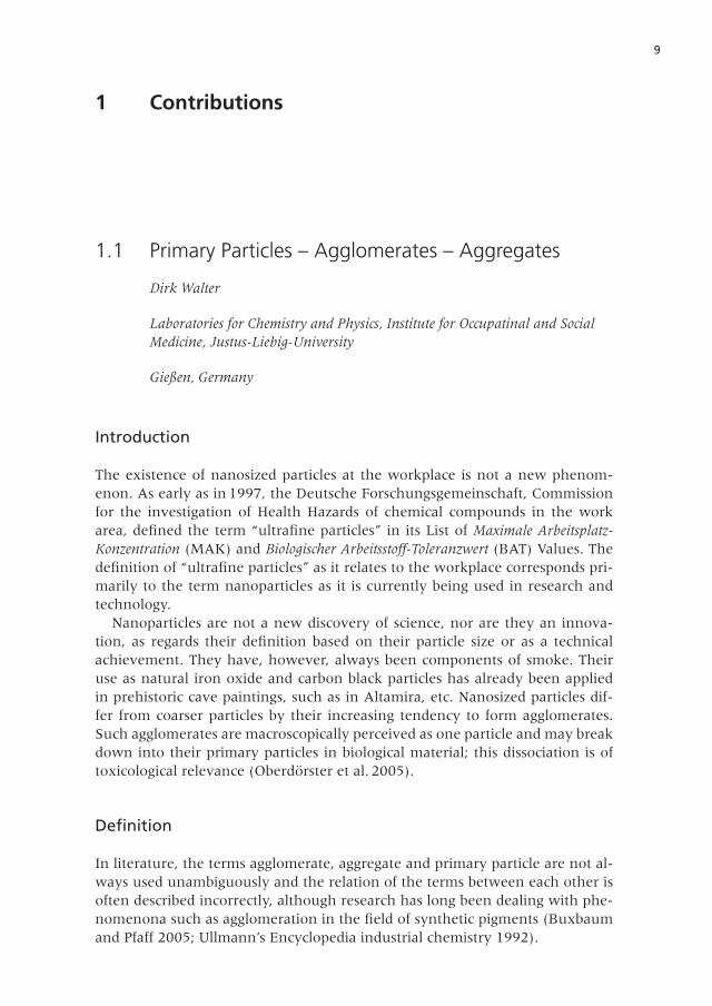
1.1 Primary Particles – Agglomerates – Aggregates
Dirk Walter
Laboratories for Chemistry and Physics, Institute for Occupatinal and SocialMedicine, Justus-Liebig-University
Gießen, Germany
Introduction
The existence of nanosized particles at the workplace is not a new phenom-enon . As early as in 1997, the Deutsche Forschungsgemeinschaft, Commissionfor the investigation of Health Hazards of chemical compounds in the workarea, defined the term “ultrafine particles” in its List of Maximale Arbeitsplatz-Konzentration (MAK) and Biologischer Arbeitsstoff-Toleranzwert (BAT) Values . Thedefinition of “ultrafine particles” as it relates to the workplace corresponds pri-marily to the term nanoparticles as it is currently being used in research andtechnology .
Nanoparticles are not a new discovery of science, nor are they an innova-tion, as regards their definition based on their particle size or as a technicalachievement. They have, however, always been components of smoke. Theiruse as natural iron oxide and carbon black particles has already been appliedin prehistoric cave paintings, such as in Altamira, etc. Nanosized particles dif-fer from coarser particles by their increasing tendency to form agglomerates .Such agglomerates are macroscopically perceived as one particle and may breakdown into their primary particles in biological material; this dissociation is oftoxicological relevance (Oberdörster et al. 2005).
Definition
In literature, the terms agglomerate, aggregate and primary particle are not al-ways used unambiguously and the relation of the terms between each other isoften described incorrectly, although research has long been dealing with phe-nomenona such as agglomeration in the field of synthetic pigments (Buxbaumand Pfaff 2005; Ullmann’s Encyclopedia industrial chemistry 1992) .
10 1 Contributions
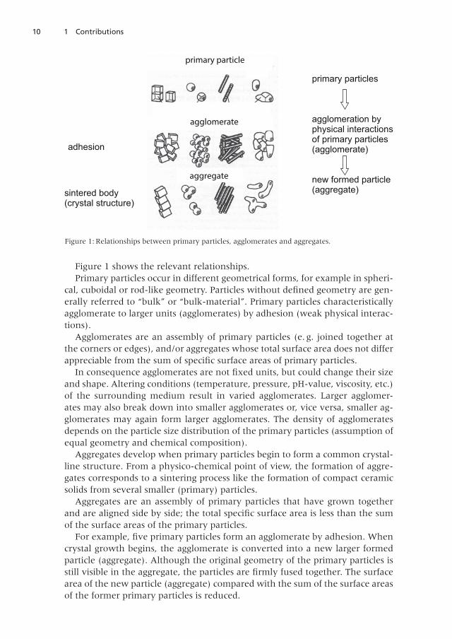
Figure 1 shows the relevant relationships.Primary particles occur in different geometrical forms, for example in spheri-
cal, cuboidal or rod-like geometry. Particles without defined geometry are gen-erally referred to “bulk” or “bulk-material” . Primary particles characteristicallyagglomerate to larger units (agglomerates) by adhesion (weak physical interac-tions) .
Agglomerates are an assembly of primary particles (e . g . joined together atthe corners or edges), and/or aggregates whose total surface area does not differappreciable from the sum of specific surface areas of primary particles.
In consequence agglomerates are not fixed units, but could change their sizeand shape. Altering conditions (temperature, pressure, pH-value, viscosity, etc.)of the surrounding medium result in varied agglomerates. Larger agglomer-ates may also break down into smaller agglomerates or, vice versa, smaller ag-glomerates may again form larger agglomerates . The density of agglomeratesdepends on the particle size distribution of the primary particles (assumption ofequal geometry and chemical composition).
Aggregates develop when primary particles begin to form a common crystal-line structure. From a physico-chemical point of view, the formation of aggre-gates corresponds to a sintering process like the formation of compact ceramicsolids from several smaller (primary) particles.
Aggregates are an assembly of primary particles that have grown togetherand are aligned side by side; the total specific surface area is less than the sumof the surface areas of the primary particles .
For example, five primary particles form an agglomerate by adhesion. Whencrystal growth begins, the agglomerate is converted into a new larger formedparticle (aggregate) . Although the original geometry of the primary particles isstill visible in the aggregate, the particles are firmly fused together. The surfacearea of the new particle (aggregate) compared with the sum of the surface areasof the former primary particles is reduced .
Agglomerate
Aggregate
e
Aggregate
Aglomerate
Aggregate
primary particle
agglomerate
aggregate
adhesion
sintered body(crystal structure)
primary particles
agglomeration byphysical interactionsof primary particles(agglomerate)
new formed particle(aggregate)
Figure 1: Relationships between primary particles, agglomerates and aggregates.
111.1 Primary Particles – Agglomerates – Aggregates
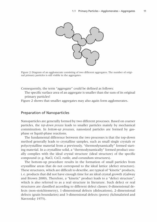
Consequently, the term “aggregate” could be defined as follows:The specific surface area of an aggregate is smaller than the sum of its originalprimary particles!
Figure 2 shows that smaller aggregates may also again form agglomerates .
Preparation of Nanoparticles
Nanoparticles are generally formed by two different processes . Based on coarserparticles, the top-down process leads to smaller particles mainly by mechanicalcomminution . In bottom-up processes, nanosized particles are formed by gas-phase or liquid-phase reactions.
The fundamental difference between the two processes is that the top-downmethod generally leads to crystalline samples, such as small single crystals orpolycrystalline material from a previously, “thermodynamically” formed start-ing material . In a crystalline solid, a “thermodynamically” formed product usu-ally complies with the ideal crystal structure (ideal structure) of the specificcompound (e . g . NaCl, CsCl, rutile, and corundum structures) .
The bottom-up procedure results in the formation of small particles fromcrystalline areas that do not correspond to the ideal lattice (defect structure) .These structures, which are difficult to describe, are typical of “kinetic” products,i. e. products that did not have enough time for an ideal crystal growth (Galwayand Brown 2000) . Therefore, a “kinetic” product leads to a “defect structure”,which is also referred to as a real structure in literature . Such defect or realstructures are classified according to different defect classes: 0-dimensional de-fects (non-stoichiometry), 1-dimensional defects (dislocations), 2-dimensionaldefects (grain boundaries) and 3-dimensional defects (pores) (Schmalzried andNavrotsky 1975).
Figure 2: Diagram of an agglomerate consisting of two different aggregates . The number of origi-nal primary particles is still visible in the aggregates.
12 1 Contributions
Defect Structures
Non-stoichiometry
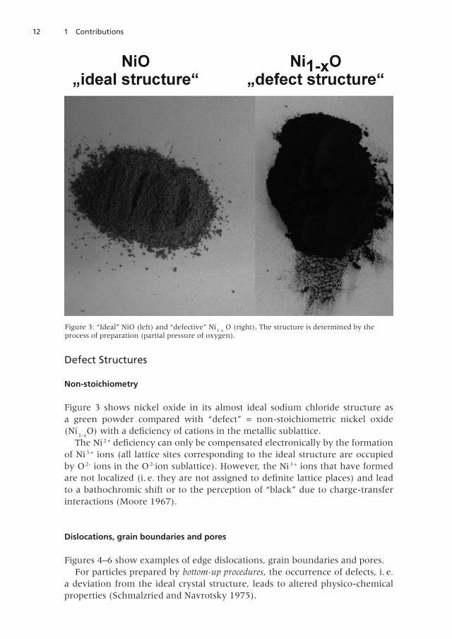
Figure 3 shows nickel oxide in its almost ideal sodium chloride structure asa green powder compared with “defect” = non-stoichiometric nickel oxide(Ni
1-xO) with a deficiency of cations in the metallic sublattice.
The Ni 2 + deficiency can only be compensated electronically by the formationof Ni 3 + ions (all lattice sites corresponding to the ideal structure are occupiedby O 2- ions in the O2-ion sublattice). However, the Ni3 + ions that have formedare not localized (i . e. they are not assigned to definite lattice places) and leadto a bathochromic shift or to the perception of “black” due to charge-transferinteractions (Moore 1967) .
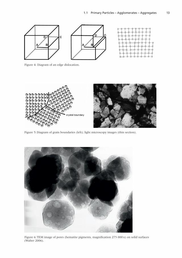
Dislocations, grain boundaries and pores
Figures 4–6 show examples of edge dislocations, grain boundaries and pores .For particles prepared by bottom-up procedures, the occurrence of defects, i . e.
a deviation from the ideal crystal structure, leads to altered physico-chemicalproperties (Schmalzried and Navrotsky 1975).
Figure 3: “Ideal” NiO (left) and “defective” Ni1-x
O (right) . The structure is determined by theprocess of preparation (partial pressure of oxygen) .
131.1 Primary Particles – Agglomerates – Aggregates
Figure 4: Diagram of an edge dislocation .
Figure 5: Diagram of grain boundaries (left); light microscopy images (thin section) .
Figure 6: TEM image of pores (hematite pigments, magnification 275 000x) on solid surfaces(Walter 2006) .
14 1 Contributions
Bottom-up processes may also form molecular structures resulting in clusters .Clusters are units consisting of several atoms or compounds whose physico-chemical properties depend on the size of the clusters . For example, gold clus-ters with fewer than 50 atoms have a molecular character, whereas clusterswith more than 50 atoms have a metallic character.
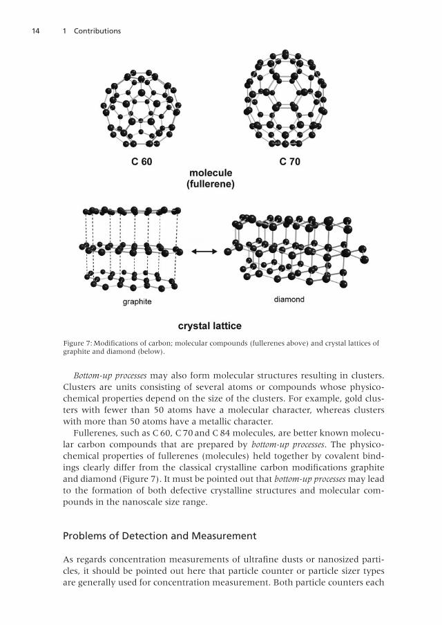
Fullerenes, such as C 60, C 70 and C 84 molecules, are better known molecu-lar carbon compounds that are prepared by bottom-up processes . The physico-chemical properties of fullerenes (molecules) held together by covalent bind-ings clearly differ from the classical crystalline carbon modifications graphiteand diamond (Figure 7) . It must be pointed out that bottom-up processes may leadto the formation of both defective crystalline structures and molecular com-pounds in the nanoscale size range .
Problems of Detection and Measurement
As regards concentration measurements of ultrafine dusts or nanosized parti-cles, it should be pointed out here that particle counter or particle sizer typesare generally used for concentration measurement . Both particle counters each
Figure 7: Modifications of carbon; molecular compounds (fullerenes above) and crystal lattices ofgraphite and diamond (below) .
151.1 Primary Particles – Agglomerates – Aggregates
have two disadvantages regarding toxicological aspects. First, they cannot dif-ferentiate between different substances (the difference between pollen, waterdroplets (= mist) and diesel soot, for example, is not detected) . Second, theydo not differentiate between primary particles and agglomerates or aggregates(Rödelsperger et al. 2009). This means that an agglomerate is recorded in thesame way as a primary particle, i. e. as an individual particle! Therefore, thedetected particles require further characterization.
Methods for the Characterization of Nanoparticles
What methods are actually suitable for characterizing nanoparticles? X-ray dif-fraction (XRD) is generally used to characterize solids. However, in the case ofparticles < 100 nm, XRD measurements are not very conclusive due to the ef-fects of scattering and are therefore not suitable for characterizing nano-dusts .An appropriate X-ray method seems to be the use of highly monochromaticsynchrotron radiation as applied for example at the DESY linear acceleratorin Hamburg, Germany. However, the time of measurement is available onlyin individual cases on request. Electron microscopy, in particular transmissionelectron microscopy (TEM) with subsequent elemental analysis (EDX), is an-other suitable method (Rödelsperger et al. 2003a). On account of the relativelysophisticated sample preparation, TEM analyses are only of limited suitabilityfor routine measurements . In addition to electron microscopy, thermal analysis(TG-FTR and TG-MS) has recently been used to investigate different agglomer-ate/aggregate constellations of nanoscale dusts (Eichholz et al . 2012) . Determi-nation of the specific surface by nitrogen adsorption according to the Brunauer,Emmett, and Teller (BET) adsorption is another method frequently used forcharacterization . The BET adsorption is based on a relationship between thenumber of adsorbed nitrogen molecules and the specific surface (Brunauer etal . 1938) .
Problem of Data on the Specific Surface
The following will explain why data published on the specific surface of dustsamples are hardly a characteristic parameter and must be reviewed critically.
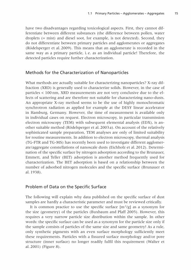
It is common practice to use the specific surface [m2/g] as a synonym forthe size (geometry) of the particles (Buxbaum and Pfaff 2005). However, thisrequires a very narrow particle size distribution within the sample. In otherwords: the specific surface can be used as a synonym for the particle size only ifthe sample consists of particles of the same size and same geometry! As a rule,only synthetic pigments with an even surface morphology sufficiently meetthese requirements. Particles with a fissured surface morphology and/or porestructure (inner surface) no longer readily fulfil this requirement (Walter etal . 2001) (Figure 8) .
16 1 Contributions
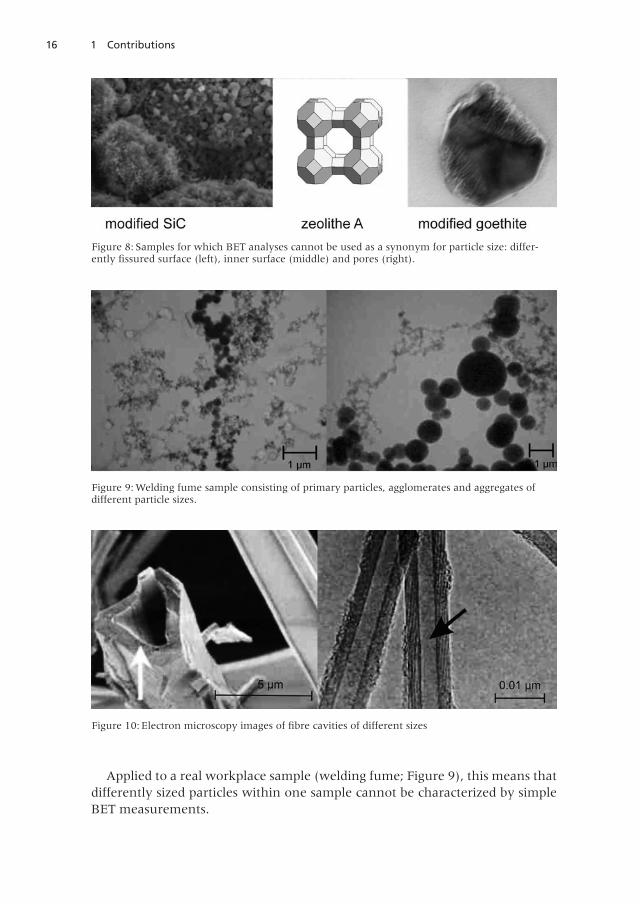
Applied to a real workplace sample (welding fume; Figure 9), this means thatdifferently sized particles within one sample cannot be characterized by simpleBET measurements .
Figure 8: Samples for which BET analyses cannot be used as a synonym for particle size: differ-ently fissured surface (left), inner surface (middle) and pores (right).
Figure 9: Welding fume sample consisting of primary particles, agglomerates and aggregates ofdifferent particle sizes .
Figure 10: Electron microscopy images of fibre cavities of different sizes
171.1 Primary Particles – Agglomerates – Aggregates
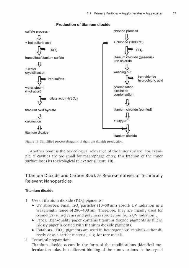
Another point is the toxicological relevance of the inner surface. For exam-ple, if cavities are too small for macrophage entry, this fraction of the innersurface loses its toxicological relevance (Figure 10).
Titanium Dioxide and Carbon Black as Representatives of TechnicallyRelevant Nanoparticles
Titanium dioxide
1 . Use of titanium dioxide (TiO2) pigments:
► UV absorber . Small TiO2
particles (10–50 nm) absorb UV radiation in awavelength range of 280–400 nm. Therefore, they are mainly used forcosmetics (sunscreens) and polymers (protection from UV radiation) .
► Paper. High-quality paper contains titanium dioxide pigments as fillers.Glossy paper is coated with titanium dioxide pigments .
► Catalysts . (TiO2) pigments are used in heterogeneous catalysis either di-
rectly or as a carrier material, e . g . for rare metals .2 . Technical preparation:
Titanium dioxide occurs in the form of the modifications (identical mo-lecular formulas, but different binding of the atoms or ions in the crystal
Figure 11: Simplified process diagrams of titanium dioxide production.
18 1 Contributions
lattice) rutile, anatase and brookite . Technically, two different processesare used to produce titanium dioxide: On the one hand, the “sulfate pro-cess”, in which titanium dioxide is produced via titanium sulfate startingfrom ilmenite and sulfuric acid, and, on the other hand, the “chloride pro-cess”, in which a solids mixture of titanium oxide and coke is convertedinto titanium chloride at high temperatures by means of chlorine . Subse-quently, titanium chloride reacts with oxygen to produce titanium dioxide.Ultimately, this means that the titanium dioxide sample may still carrytraces of sulfate/SO
2and chloride – incorporated in the crystal lattice and
attached to the surface, respectively – depending upon which of these di-vergent production processes is used.
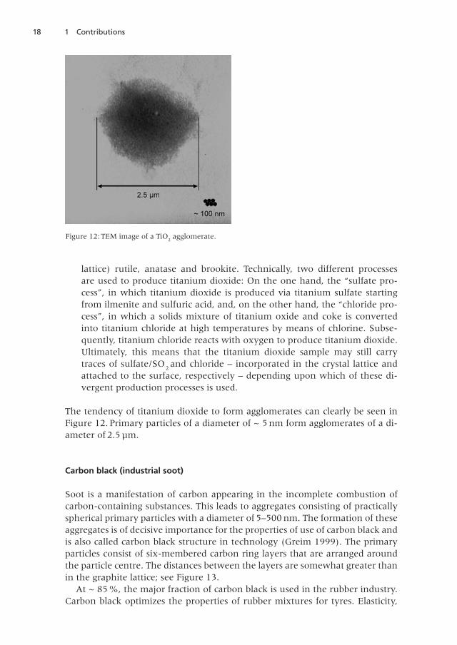
The tendency of titanium dioxide to form agglomerates can clearly be seen inFigure 12 . Primary particles of a diameter of ~ 5 nm form agglomerates of a di-ameter of 2 .5 µm .
Carbon black (industrial soot)
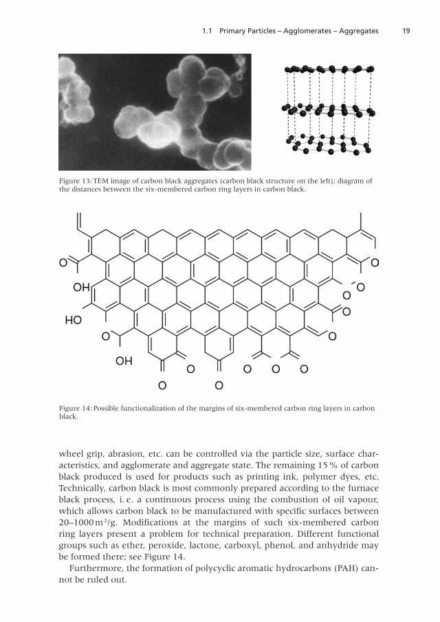
Soot is a manifestation of carbon appearing in the incomplete combustion ofcarbon-containing substances . This leads to aggregates consisting of practicallyspherical primary particles with a diameter of 5–500 nm . The formation of theseaggregates is of decisive importance for the properties of use of carbon black andis also called carbon black structure in technology (Greim 1999) . The primaryparticles consist of six-membered carbon ring layers that are arranged aroundthe particle centre . The distances between the layers are somewhat greater thanin the graphite lattice; see Figure 13 .
At ~ 85%, the major fraction of carbon black is used in the rubber industry .Carbon black optimizes the properties of rubber mixtures for tyres . Elasticity,
Figure 12: TEM image of a TiO2
agglomerate .
191.1 Primary Particles – Agglomerates – Aggregates
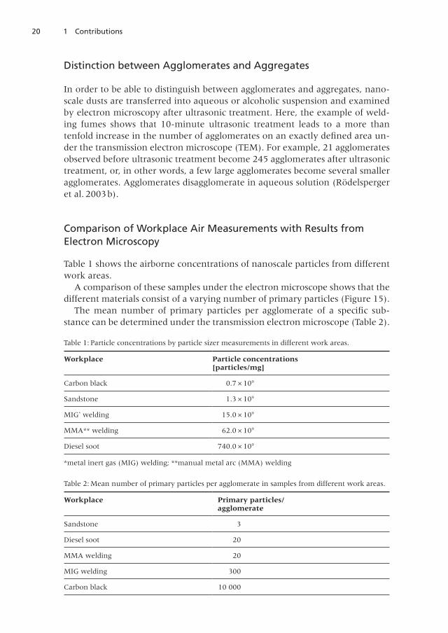
wheel grip, abrasion, etc. can be controlled via the particle size, surface char-acteristics, and agglomerate and aggregate state . The remaining 15% of carbonblack produced is used for products such as printing ink, polymer dyes, etc .Technically, carbon black is most commonly prepared according to the furnaceblack process, i. e. a continuous process using the combustion of oil vapour,which allows carbon black to be manufactured with specific surfaces between20–1000m 2/g. Modifications at the margins of such six-membered carbonring layers present a problem for technical preparation . Different functionalgroups such as ether, peroxide, lactone, carboxyl, phenol, and anhydride maybe formed there; see Figure 14 .
Furthermore, the formation of polycyclic aromatic hydrocarbons (PAH) can-not be ruled out .
Figure 13: TEM image of carbon black aggregates (carbon black structure on the left); diagram ofthe distances between the six-membered carbon ring layers in carbon black .
Figure 14: Possible functionalization of the margins of six-membered carbon ring layers in carbonblack .
20 1 Contributions
Distinction between Agglomerates and Aggregates
In order to be able to distinguish between agglomerates and aggregates, nano-scale dusts are transferred into aqueous or alcoholic suspension and examinedby electron microscopy after ultrasonic treatment . Here, the example of weld-ing fumes shows that 10-minute ultrasonic treatment leads to a more thantenfold increase in the number of agglomerates on an exactly defined area un-der the transmission electron microscope (TEM) . For example, 21 agglomeratesobserved before ultrasonic treatment become 245 agglomerates after ultrasonictreatment, or, in other words, a few large agglomerates become several smalleragglomerates. Agglomerates disagglomerate in aqueous solution (Rödelspergeret al . 2003b) .
Comparison of Workplace Air Measurements with Results fromElectron Microscopy
Table 1 shows the airborne concentrations of nanoscale particles from differentwork areas .
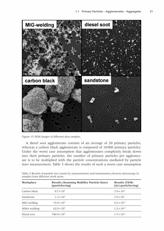
A comparison of these samples under the electron microscope shows that thedifferent materials consist of a varying number of primary particles (Figure 15).
The mean number of primary particles per agglomerate of a specific sub-stance can be determined under the transmission electron microscope (Table 2) .
Table 2: Mean number of primary particles per agglomerate in samples from different work areas .
Workplace Primary particles/agglomerate
Sandstone 3
Diesel soot 20
MMA welding 20
MIG welding 300
Carbon black 10 000
Table 1: Particle concentrations by particle sizer measurements in different work areas .
Workplace Particle concentrations[particles/mg]
Carbon black 0 .7 × 109
Sandstone 1 .3 × 109
MIG* welding 15 .0 × 109
MMA** welding 62 .0 × 109
Diesel soot 740 .0 × 109
*metal inert gas (MIG) welding; **manual metal arc (MMA) welding
211.1 Primary Particles – Agglomerates – Aggregates
A diesel soot agglomerate consists of an average of 20 primary particles,whereas a carbon black agglomerate is composed of 10000 primary particles .Under the worst case assumption that agglomerates completely break downinto their primary particles, the number of primary particles per agglomer-ate is to be multiplied with the particle concentrations mediated by particlesizer measurement . Table 3 shows the results of such a worst case assumption
Figure 15: SEM images of different dust samples .
Table 3: Results of particle size counts by measurements and transmission electron microscopy insamples from different work areas .
Workplace Results (Scanning Mobility Particle Sizer)[particles/mg]
Results (TEM)[(tr) particles/mg]
Carbon black 0 .7 × 109 7 .0 × 1012
Sandstone 1 .3 × 109 3 .9 × 109
MIG welding 15 .0 × 109 4 .5 × 1012
MMA welding 62,0 × 10⁹ 1 .2 × 1012
Diesel soot 740 .0 × 109 1 .5 × 1013
22 1 Contributions
(tr = maximum toxicologically relevant primary particle concentration). Thedifference between the measured particle concentration (particle sizer) and theprimary particle concentration determined under the electron microscope iscaused by the agglomeration behaviour of the ultrafine particles (Rödelspergeret al . 2009) .
Diversity of Primary Particles
Primary particles may be structured differently depending on the preparationprocess . A primary particle could consist of
1 . a molecule .
2 . an ideal crystal .
3. a real crystal with defects, although they have a homogeneous, intact surface.
4. a real crystal with defects and have a fissured, porous surface.
5. a real crystal with defects and have pores or an inner surface (zeolite).
231.1 Primary Particles – Agglomerates – Aggregates
Effect of Primary Particles
Primary particles may be poorly soluble and may correspond to a granular bio-persistent dust (GBD) and thus cause a “particle effect” (macrophages/reactiveoxygen species (ROS)) with a local effect (Figure 16). ROS can interfere withthe complete area of the surface .
They may also induce a particle effect with a systemic effect, for example byphagocytosis . Highly soluble primary particles lead to a local and/or systemicavailability of metal ions.
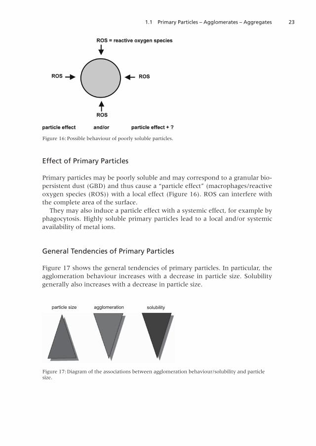
General Tendencies of Primary Particles
Figure 17 shows the general tendencies of primary particles . In particular, theagglomeration behaviour increases with a decrease in particle size. Solubilitygenerally also increases with a decrease in particle size .
Figure 16: Possible behaviour of poorly soluble particles.
Figure 17: Diagram of the associations between agglomeration behaviour/solubility and particlesize .
particle size agglomeration solubility
24 1 Contributions
References
Brunauer S, Emmett PH, Teller, E (1938) Adsorption of gases in multimolecularlayers . J Am Chem Soc 60: 309–319
Buxbaum G, Pfaff G (2005) Industrial inorganic pigments . Wiley-VCH Verlag,Weinheim
Eichholz S, Lerch M, Heck M, Walter D (2012) Various carbon dust particles –studies on thermal behaviour. J Therm Anal Calorim, DOI 101007/s 10973–011–2188-z
Galwey AK, Brown ME (2000) Thermal decomposition of ionic solids. Elsevier,Amsterdam
Greim H (Hrsg) (1999) Industrieruße (Carbon Black) in Form atembarer Stäube .Gesundheitsschädliche Arbeitsstoffe, Toxikologisch-arbeitsmedizinische Be-gründung von MAK-Werten 29. Lieferung. VCH, Weinheim List of MAKand BAT Values (2009) Wiley-VCH Verlag, Weinheim
Moore WJ (1967) Seven solid states. WA Benjamin, Inc., New YorkOberdörster G, Oberdörster E, Oberdörster J (2005) Nanotoxicology: An emerg-
ing discipline evolving from studies of ultrafine particles. Environ HealthPerspect 113: 823–839
Rödelsperger K, Podhorsky S, Brückel B, Dahmann D, Hartfiel GD, WoitowitzH-J (2003a) Measurements of granular ultrafine bio-durable particles forworkplace protection . Eur J Oncol 8: 103–112
Rödelsperger K, Podhorsky S, Brückel B, Dahmann D, Hartfiel GD, Woitow-itz H-J (2003b) Charakterisierung von Aerosolen ultrafeiner Teilchen fürden Arbeitsschutz, Abschlussbericht des Projektes F 1804 (Characterizationof ultrafine particle aerosols for occupational safety, final report of projectF 1804) (German), ISBN 3–88261–050–6 Dortmund-Berlin-Dresden
Rödelsperger K, Brückel B, Podhorsky S, Schneider J (2009) Charakterisierungultrafeiner Teilchen für den Arbeitsschutz – Teil 2, BAUA-Abschlussberichtdes Projektes F 2075 der Bundesanstalt für Arbeitsschutz und Arbeitsmedi-zin (Characterization of ultrafine particles for occupational safety, BAUAfinal report of project F 2075 of the Federal Institute for Occupational Safetyand Health) (German)
Schmalzried H, Navrotsky A (1975) Festkörperthermodynamik/Chemie desfesten Zustandes . Verlag Chemie, Weinheim
Ullmann’s Encyclopedia industrial chemistry VCH (1992), Vol . 20, WeinheimWalter D, Buxbaum G, Laqua W (2001) The mechanism of the thermal trans-
formation from goethite to hematite . J Therm Anal Cal 63: 733–748Walter D (2006) Characterization of synthetic hydrous hematite pigments .
Thermochim Acta 445: 195–199
251.2 Exposure during Production and Handling of Manufactured Nanomaterials
1.2 Exposure during Production and Handling ofManufactured Nanomaterials
Markus G. M. Berges
Institute for Occupational Safety and Health (IFA)
Sankt Augustin, Germany
In the traditional risk framework, risk management decisions concerning oc-cupational safety and health rely on site-specific risk assessment and informa-tion about the effectiveness of available measures to mitigate exposure. In itsturn, risk assessment builds on hazard and exposure assessments (Murashov etal. 2009). Though there is mounting evidence that some manufactured nano-materials may impose a health hazard to humans the target organs and end-points and the specific dose-response relationship are not clearly delineated. Inview of the uncertainty regarding the hazard of manufactured nanomaterialsthe assessment and control of the potential exposure of workers become crucialin occupational health and safety in order to minimize the risk of the workers .
The current method of assessing worker exposure to airborne particles in theworkplace involves the measurement of mass concentration of health-relatedfractions of particles in the worker’s breathing zone . The main exceptions to thismethodology are particle-number-based metrics for exposure for fibres and formicroorganisms (ISO/TR 12885, 2008; ISO 13794, 1999). However nanoparti-cles carry only very small masses and therefore generally contribute negligiblyto the integral mass concentration of the inhalable or respirable dust fraction .Furthermore, there is evidence that other metrics such as particle number con-centration or surface area may be better descriptors for the biological effects ofnanoparticles. The issue of exposure metrics has extensively been addressed byMaynard and Aitken (2007). Reflecting the state of the art they conclude thateffective approaches to measuring exposure to a wide range of manufacturednanomaterials/nanoobjects will require methods for measuring aerosol num-ber, surface area and mass concentration .
Back in 1998 several European OSH Institutes in collaboration with the DFG(the German Research Foundation) agreed on a convention (Möhlmann 1998,revised 2007, Riediger and Möhlmann 2001) for the measurement of ultrafineparticles in order to permit comparison of measurements . The core points ofthis convention were:► An ultrafine aerosol particle is a particle with a mobility equivalent diameter
of < 0 .1 µm► The particle number concentration should be measured in the range from
approximately 10 to 600 nm .► The entire particle size distribution should be measured if possible .► A concentration range of up to approximately 1 × 10⁸ particles/cm³ should
be covered.
26 1 Contributions
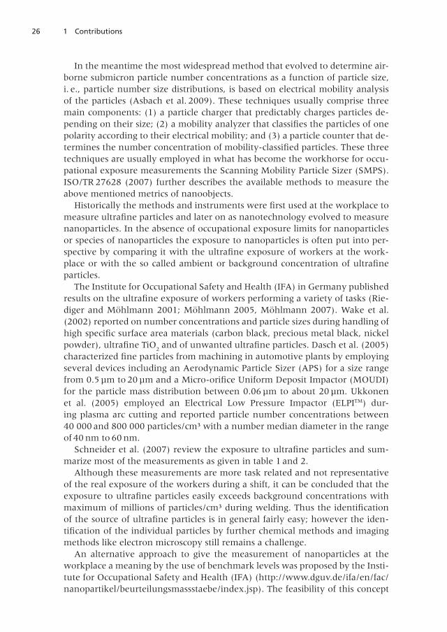
In the meantime the most widespread method that evolved to determine air-borne submicron particle number concentrations as a function of particle size,i . e ., particle number size distributions, is based on electrical mobility analysisof the particles (Asbach et al. 2009). These techniques usually comprise threemain components: (1) a particle charger that predictably charges particles de-pending on their size; (2) a mobility analyzer that classifies the particles of onepolarity according to their electrical mobility; and (3) a particle counter that de-termines the number concentration of mobility-classified particles. These threetechniques are usually employed in what has become the workhorse for occu-pational exposure measurements the Scanning Mobility Particle Sizer (SMPS) .ISO/TR 27628 (2007) further describes the available methods to measure theabove mentioned metrics of nanoobjects.
Historically the methods and instruments were first used at the workplace tomeasure ultrafine particles and later on as nanotechnology evolved to measurenanoparticles . In the absence of occupational exposure limits for nanoparticlesor species of nanoparticles the exposure to nanoparticles is often put into per-spective by comparing it with the ultrafine exposure of workers at the work-place or with the so called ambient or background concentration of ultrafineparticles .
The Institute for Occupational Safety and Health (IFA) in Germany publishedresults on the ultrafine exposure of workers performing a variety of tasks (Rie-diger and Möhlmann 2001; Möhlmann 2005, Möhlmann 2007). Wake et al.(2002) reported on number concentrations and particle sizes during handling ofhigh specific surface area materials (carbon black, precious metal black, nickelpowder), ultrafine TiO
2and of unwanted ultrafine particles. Dasch et al. (2005)
characterized fine particles from machining in automotive plants by employingseveral devices including an Aerodynamic Particle Sizer (APS) for a size rangefrom 0.5 µm to 20 µm and a Micro-orifice Uniform Deposit Impactor (MOUDI)for the particle mass distribution between 0 .06 µm to about 20 µm . Ukkonenet al . (2005) employed an Electrical Low Pressure Impactor (ELPITM) dur-ing plasma arc cutting and reported particle number concentrations between40 000 and 800 000 particles/cm³ with a number median diameter in the rangeof 40 nm to 60 nm .
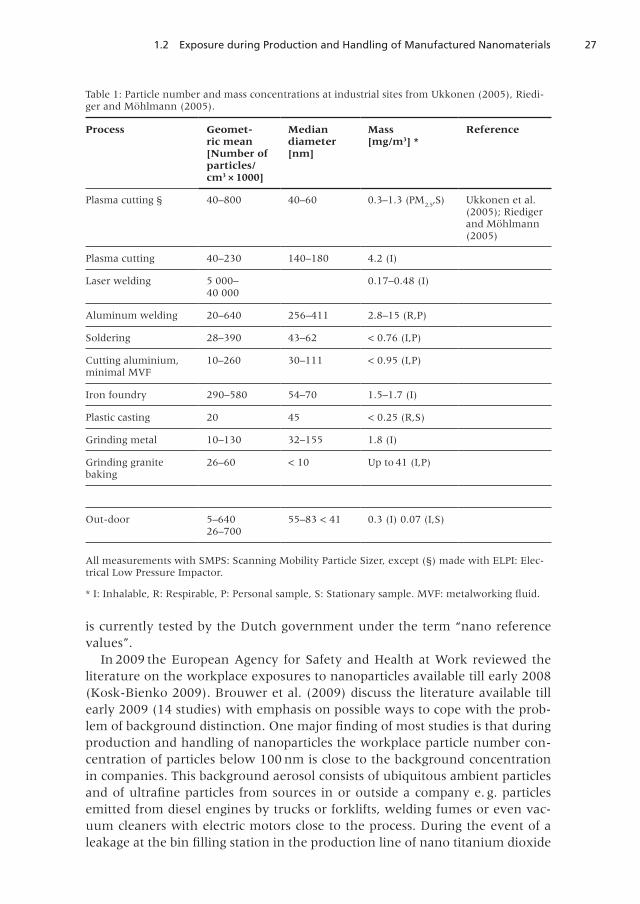
Schneider et al . (2007) review the exposure to ultrafine particles and sum-marize most of the measurements as given in table 1 and 2.
Although these measurements are more task related and not representativeof the real exposure of the workers during a shift, it can be concluded that theexposure to ultrafine particles easily exceeds background concentrations withmaximum of millions of particles/cm³ during welding. Thus the identificationof the source of ultrafine particles is in general fairly easy; however the iden-tification of the individual particles by further chemical methods and imagingmethods like electron microscopy still remains a challenge .
An alternative approach to give the measurement of nanoparticles at theworkplace a meaning by the use of benchmark levels was proposed by the Insti-tute for Occupational Safety and Health (IFA) (http://www.dguv.de/ifa/en/fac/nanopartikel/beurteilungsmassstaebe/index .jsp) . The feasibility of this concept
271.2 Exposure during Production and Handling of Manufactured Nanomaterials
is currently tested by the Dutch government under the term “nano referencevalues”.
In 2009 the European Agency for Safety and Health at Work reviewed theliterature on the workplace exposures to nanoparticles available till early 2008(Kosk-Bienko 2009). Brouwer et al. (2009) discuss the literature available tillearly 2009 (14 studies) with emphasis on possible ways to cope with the prob-lem of background distinction. One major finding of most studies is that duringproduction and handling of nanoparticles the workplace particle number con-centration of particles below 100 nm is close to the background concentrationin companies. This background aerosol consists of ubiquitous ambient particlesand of ultrafine particles from sources in or outside a company e. g. particlesemitted from diesel engines by trucks or forklifts, welding fumes or even vac-uum cleaners with electric motors close to the process. During the event of aleakage at the bin filling station in the production line of nano titanium dioxide
Table 1: Particle number and mass concentrations at industrial sites from Ukkonen (2005), Riedi-ger and Möhlmann (2005).
Process Geometric mean[Number ofparticles/cm3 × 1000]
Mediandiameter[nm]
Mass[mg/m3] *
Reference
Plasma cutting § 40–800 40–60 0 .3–1 .3 (PM2 .5
,S) Ukkonen et al .(2005); Riedigerand Möhlmann(2005)
Plasma cutting 40–230 140–180 4 .2 (I)
Laser welding 5 000–40 000
0 .17–0 .48 (I)
Aluminum welding 20–640 256–411 2.8–15 (R,P)
Soldering 28–390 43–62 < 0 .76 (I,P)
Cutting aluminium,minimal MVF
10–260 30–111 < 0 .95 (I,P)
Iron foundry 290–580 54–70 1 .5–1 .7 (I)
Plastic casting 20 45 < 0.25 (R,S)
Grinding metal 10–130 32–155 1 .8 (I)
Grinding granitebaking
26–60 < 10 Up to 41 (I,P)
Out-door 5–64026–700
55–83 < 41 0 .3 (I) 0 .07 (I,S)
All measurements with SMPS: Scanning Mobility Particle Sizer, except (§) made with ELPI: Elec-trical Low Pressure Impactor .
* I: Inhalable, R: Respirable, P: Personal sample, S: Stationary sample. MVF: metalworking fluid.
28 1 Contributions
up to 130000 particles/cm3 have been encountered. The chemical identity ofthe nanoparticles was verified by transmission electron microscopy. Anothercommon finding is that aggregates or agglomerates above 100 nm in size werequite often detected at the workplace and correlated with the operations.
High aspect ratio nanoparticles (HARN) such as carbon nanotubes (CNT) pre-sent an additional challenge to the employed SMPS (Scanning Mobility ParticleSizer) as they clearly are not spherical particles for which the SMPS has beendesigned in the first place. The manufactures usually calibrate these instrumentsonly with spherical particles . The SMPS response is in direct relation with theCNTs transport through its differential mobility analyzer (DMA). This is influ-enced by the peculiar shape of the CNT and their orientation in the electric fieldof the DMA . Kim et al . (2005) show exemplary CNTs correlations between CNTlengths and electrical mobility diameter . By using Multi-Walled Carbon Nano-tubes (MWCNTs) of approximately 15 nm diameter and irregular alignment,the electrical mobility diameter chosen by a DMA underestimates the geometri-
Table 2: Number concentrations and particles sizes from Wake et al . (2002) .
Materialor type ofindustry
Outside Workplace Outside Workplace Activity
Range/ Range/ Number Number
cm3 × 1000 cm3 × 1000 median,nm (GSD)
median,nm (GSD)
Carbon black 649–3836 3 .5–50 44 (3 .2) 51–400 (2 .4) Bagging
Nickel powder 3 .3–16 3 .7–212 23 (1 .9) 49 (3 .3) Bagging
Titaniumdioxide
10–58 4 .2–17 Bagging
Precious metal 19–62 23–71 Sieving
blacks
Zinc refining 20–23 12–24 503 (5 .3) 70 (2 .2) Sintering
Zinc refining 20–23 56 100 Casting
Plasma coating 2 .3–8 .0 2 .8–905 41 (2 .2) 587 (1 .3) Wire coating
Galvanizing 15–37 10–683 64 (2 .0) 99 (2 .1) Galvanizing
Steel foundry 13–72 118→ 500 46 (1 .9) 66 (2 .0) Fettling
Welding 10–19 117→ 500 53 (2 .1) 179 (2 .2) MIG „Metal-Inert-Gas”welding
Plastic welding 1 .2–5 .2 111–3766 31 (2 .0) 37 (1 .7) Welding
Hand solder-ing
2 .2–11 12→500 41 (2 .0) 72 (2 .3) Tinning
GSD: Geometric Standard Deviation
291.2 Exposure during Production and Handling of Manufactured Nanomaterials
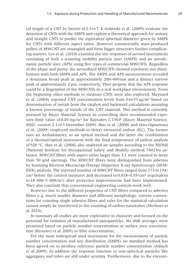
cal length of a CNT by factors of 2.5 to 7. 4. Golanski et al. (2009) evaluate thedetection of CNTs with the SMPS and explore a theoretical approach for unitaryand straight CNTs to predict the equivalent spherical diameter given by SMPSfor CNTs with different aspect ratios. However commercially mass-producedpellets of MWCNT are entangled and form bigger structures further complicat-ing matters . Lee et al . (2010) examine the size responses of aerosol instrumentsconsisting of both a scanning mobility particle sizer (SMPS) and an aerody-namic particle sizer (APS) using five types of commercial MWCNTs. Regardlessof the phase and purity, the aerosolized MWCNTs showed consistent size distri-butions with both SMPS and APS. The SMPS and APS measurements revealeda dominant broad peak at approximately 200–400 nm and a distinct narrowpeak at approximately 2 µm, respectively. They propose that the APS responsecould be a fingerprint of the MWCNTs in a real workplace environment. Fromthe beginning other methods to measure CNTs were also explored . Maynardet al. (2004) reported CNT concentration levels from 0 to 53 µg/m³ based ondetermination of metals from the catalyst and backward calculations assuminga known percentage of metals of the CNT material . This method is currentlypursued by Bayer Material Science in controlling their recommended expo-sure limit value of 0.05 mg/m³ for Baytubes C 150 P (Bayer Material Science,MSD, version 2.1 of 3 December 2009). Han et al. (2008) and Ono-Ogasawaraet al . (2009) employed methods to detect elemental carbon (EC) . The formeruses an Aethalometer, as an optical method and the latter the combinationof a thermal/optical instrument with the final temperature of carbon analysisof 920 °C . Han et al . (2008) also analyzed air samples according to the NIOSH(National Institute for Occupational Safety and Health) method 7402 for as-bestos. MWCNT fibres with aspect ratios larger than 3:1 were counted in morethan 50 grid openings. The MWCNT fibres were distinguished from asbestosby Scanning Electron Microscope/Energy Dispersive X-ray Spectroscopy (SEM/EDX) analysis. The reported number of MWCNT fibres ranged from 173 to 194/cm³ before the control measures and decreased to 0.018–0.05/cm³ (equivalentto 18 000–5 000/m³) after protective improvements had been implemented.They also conclude that conventional engineering controls work well.
However due to the different properties of CNT fibres compared to asbestosfibres e. g. much smaller diameter and different morphology, current conven-tions for counting single asbestos fibres and rules for the statistical calculationcannot simply be transferred to the counting of carbon nanotubes (Methner etal . 2010) .
In summary all studies are more explorative in character and focused on thepotential for emission of manufactured nanoparticles. No shift averages werepresented based on particle number concentration or surface area concentra-tion (Brouwer et al. 2009) or fibre concentration.
For the most widespread used instrument for the measurement of particlenumber concentration and size distribution (SMPS) no standard method hasbeen agreed on to produce reference particle number concentration (Asbachet al . 2009) . In addition the response function to non-spherical particles likeaggregates and tubes are still under scrutiny . Furthermore, due to the encoun-
30 1 Contributions
tered rather low concentrations in comparison to those of ultrafine particlesat the workplace additional analytical methods to determine the morphologyand chemical composition of the nanoparticles are necessary in order to copewith the problem of background distinction . The SMPS is still too bulky, com-plicated to use and too expensive for routine operation by small and mediumsized companies. Smaller handheld instruments can however be successfullyused to monitor the efficiency of engineering controls which up to now haveproven to effectively control the exposure of workers against nanoparticles atthe workplace .
References
Asbach C, Kaminski H, Fissan H, Monz C, Dahmann D, Mülhopt S, Paur HR,Kiesling HJ, Herrmann F, Voetz M, Kuhlbusch TAJ (2009) Comparision offour mobility particle sizers with different time resolution for stationarymeasurement. J Nanopart Res 11: 1593–1609
Brouwer D, van Duuren-Stuurman B, Berges M, Jankowska E, Bard D, MarkD (2009) From workplace air measurement results towards estimates ofexposure? Development of a strategy to assess exposure to manufacturednano-objects. J Nanopart Res 11: 1867–1881
Dasch J, D’Arcy J, Gundrum A, Sutherland J, Johnson J, Carlson D (2005)Characterization of fine particles from machining in automotive plants. JOccup Environ Hyg 2: 609–625
Golanski L, Bernard S, Guiot A, Poncelet O, Le Poche H, Tardif F (2009) Specificresponse of SMPS particle counter to CNT . J Physics: Conference Series 170
Han JH, Lee EJ, Lee JH, So KP, Lee YH, Bae GN, Lee S-B, Ji JH, Cho MH, Yu IJ(2008) Monitoring multiwalled carbon nanotube exposure in carbon nano-tube research facility . Inhal Toxicol 20: 741–749
ISO/TR 27628 (2007) Workplaces atmospheres – Ultrafine, nanoparticle andnano-structured aerosols – Inhalation exposure characterization and assess-ment, ISO copyright office, Case postale 56, CH-1211 Geneva 20
ISO/TR 12885 (2008) Nanotechnologies – Health and safety practices in occu-pational settings relevant to nanotechnologies, ISO copyright office, Casepostale 56, CH-1211 Geneva 20
ISO 13794 (2005) Ambient air – Determination of asbestos fibres – Indirecttransfer transmission electron microscopy method
Kim SH, Zachariah MR (2005) In-flight size classification of carbon nanotubesby gas phase electrophoresis . Nanotechnology 16: 2149–2152
Kosk-Bienko J (2009) Workplace exposure to nanoparticles . European Agencyfor Safety and Health at work (EU-OSHA)
Lee SB, Lee JH, Bae GN (2010) Size response of an SMPS-APS system to com-mercial multi-walled carbon nanotubes. J Nanopart Res 12: 501–512
Maynard AD, Baron PA, Foley M, Shvedova AA, Kisin ER, Castranova V (2004)Exposure to carbon nanotube material: aerosol release during the handling
311.2 Exposure during Production and Handling of Manufactured Nanomaterials
of unrefined single-walled carbon nanotube material. J Toxicol EnvironHealth A 67: 87–107
Maynard AD, Aitken RJ (2007) Assessing exposure to airborne nanomaterials;current abilities and future requirements. Nanotoxicology 1: 26–41
Methner M, Hodson L, Dames A, Geraci C (2010) Nanoparticle assessmenttechnique (NEAT) for the identification and measurement of potential in-halation exposure to engineered nanomaterials – Part B: Results from 12Field Studies . JOEH 7: 163–176
Möhlmann C (2005) Vorkommen ultrafeiner Aerosole an Arbeitsplätzen. Ge-fahrstoffe – Reinhalt. Luft 65: 469–471
Möhlmann C (2007) Ultrafeine (Aerosol-)Teilchen und deren Agglomerate undAggregate, Kennziffer 0425/5 38. Lfg. IV/07, 4 S. In: Messung von Gefahr-stoffen – BGIA-Arbeitsmappe (Hrsg) Berufsgenossenschaftliches Institut fürArbeitsschutz – BGIA . Erich Schmidt Verlag, Berlin 1989 – Loseblatt-Aus-gabe . ISBN: 978 3 503 02085 3
Möhlmann C (2007) Ultrafeine Aerosole am Arbeitsplatz, Kennzahl 120130. Lfg.IX/2007 . In: BGIA-Handbuch Sicherheit und Gesundheitsschutz am Arbe-itsplatz (Hrsg) Berufsgenossenschaftliches Institut für Arbeitsschutz – BGIA .Erich Schmidt Verlag, Berlin 1989 – Loseblatt-Ausgabe . ISBN: 978 3 50302085 3
Murashov V, Engel S, Savolainen K, Fullam B, Lee M, Kearns P (2009) Occupa-tional safety and health in nanotechnology and organisation for economiccooperation and development. J Nanopart Res 11: 1587–1591
Ono-Ogasawara M, Serita F, Takaya M (2009) Distinguishing nanomaterial par-ticles from background airborne particulate matter for quantitative expo-sure assessment . J Nanopart Res 11: 1651–1659
Riediger G; Möhlmann C ( 2001) Ultrafeine Aerosole an Arbeitsplätzen – Kon-ventionen und Beispiele aus der Praxis. Gefahrstoffe – Reinhalt Luft 61:429–434
Schneider T (2007) Evaluation and control of occupational health risks fromnanoparticles . Tema Nord 2007: 581, Nordic Council of Ministers, Copen-hagen
Ukkonen A, Lamminen E, Kasurinen H (2005) Measuring the size distributionand concentration of particles formed in plasma arc cutting . NOSA AerosolSymposium, Göteborg, Sweden
Wake D, Mark D, Northage C (2002) Ultrafine Aerosols at the workplace. AnnOccup Hyg 46 (Suppl 1): 235–238
32 1 Contributions
1.3 Toxicokinetics of Inhaled Nanoparticles
Wolfgang G. Kreyling
Helmholtz Center Munich – Research Center for Environmental Health; Insti-tute for Lung Biology and Disease, Focus Network: Nanoparticles and Health
Neuherberg/Munich, Germany
Nanoparticles are increasingly used in a wide range of applications in science,technology and medicine. Since they are produced for specific purposes whichcannot be met by larger particles and bulk material they are likely to be highlyreactive, in particular, with biological systems. On the other hand a large bodyof know-how in environmental sciences is available from adverse effects ofultrafine particles after inhalative exposure. Since nanoparticles elicit the sameresponses as ultrafine particles, adverse health effects cannot be excluded anda safe and sustainable development of new emerging nanoparticles is required.
Inhaled nanoparticles deposit preferentially in the alveolar region of the lungswith a maximum at 20 nm . Below that size, increasing fractions deposit in theairways of the head and thorax according to their increasing diffusivity with de-creasing size . Once deposited in the peripheral lungs nanoparticles are not onlysubject to phagocytosis by alveolar macrophages but also by epithelial cells. Itappears plausible that macrophages take up all nanoparticles deposited in theirimmediate vicinity while distant nanoparticles are not recognized. As a resulta major fraction (> 80%) of the nanoparticles in the peripheral rodent lungenter the epithelium and penetrate into interstitial spaces (Semmler-Behnkeet al. 2007). Hence, nanoparticles behave differently compared to micro-sizedparticles which are retained on the rodent epithelium leaving the lungs bymacrophage-mediated clearance at a rate of 2–3% per day . Surprisingly, nano-particles are cleared by the same clearance rate via this macrophage-mediatedtransport process indicating relocation of the nanoparticles from the intersti-tial spaces and epithelium to top of the epithelium . Therefore, macrophage-mediated clearance is the most prominent long-term clearance mechanism inthe peripheral lung of rodents . In fact, while the nanoparticles are retainedin the interstitium close to lymphatic drainage and blood vessels only rathersmall fractions are removed via these two pathways. Particle clearance fromthe human peripheral lungs and from those of dogs and monkeys differ fromthat in rodents: in the three large species even micron-sized particles enter theepithelium and penetrate into interstitial spaces and macrophage-mediatedclearance occurs at a rate which is one order of magnitude lower than that ofrodents (Kreyling and Scheuch 2000) . Yet, since micron-sized particles pen-etrate into interstitial spaces it appears plausible that nanoparticles also followthis pathway . Unfortunately for humans neither macrophage-mediated clear-ance kinetics data nor translocation data of nanoparticles into the circulationare available. Existing data suggest that translocation fractions of nanoparticlesmust be below 1% of the deposited nanoparticles according to the lower limit
331.3 Toxicokinetics of Inhaled Nanoparticles
of experimental detection as no accumulation was observed in any second-ary target organ (Möller et al. 2008). An important underlying mechanism ofthe special behaviour of nanoparticles may be selected binding of proteins andnanoparticles affecting the biokinetic fate of the nanoparticles . In other words,coating of nanoparticles with selected proteins can influence their uptake anddistribution and direct them to specific locations.
Cardio-vascular effects observed in epidemiological studies triggered the dis-cussion on enhanced translocation of ultrafine particles from the respiratoryepithelium towards the circulation and subsequent target organs, such as heart,liver, spleen and brain, eventually causing adverse effects on cardiac functionand blood coagulation, as well as on functions of the central nervous system.There is clear evidence that nanoparticles can cross body membranes and reachin the above mentioned secondary target organs and accumulate there.
To determine accumulated fractions in such organs the ultimate aim is toquantitatively balance the fractions of nanoparticles in all relevant organs andtissues of the body and include the remainder body and total excretion collectedbetween application and autopsy . Otherwise substantial uncertainty remains ifonly selected organs are analysed . Since these gross determinations of nano-particle contents in organs and tissues do not provide microscopic informationon the anatomical and cellular location of nanoparticles such studies are to becomplemented by electron microscopy analysis using elemental mapping tech-nology .
Based on quantitative biokinetic analysis after nanoparticle application tothe lungs of a rat model, small fractions of nanoparticles (iridium, carbon,gold, and preliminary results on titanium dioxide) were found in all secondaryorgans studied including the brain, heart and even in the foetus (Kreyling etal . 2002; Semmler et al . 2004; Semmler-Behnke et al . 2007 a, b, 2008; Kreylinget al. 2009). All nanoparticles were radio-labelled with the label firmly fixed tothe particle core . Fractions in each of the secondary target organs were usuallybelow 0 .5% of the administered dose to the lungs but depended strongly onparticle size in an inverse fashion. However, nanoparticle fractions in soft tissueand skeleton (without blood content) increased the totally translocated frac-tion to 5–10% of the administered dose. Also negatively ionic surface chargednanoparticles translocated more rapidly than positively charged nanoparticlesof the same size . In addition, strong differences of the totally translocated frac-tions between chain-aggregated/agglomerated iridium and carbon nanoparti-cles versus gold spheres of same size highlights the importance of nanoparticlematerial, morphological and/or surface properties . Furthermore, nanoparticleaccumulation in the rat brain results from both pathways: via the olfactory bulbversus circulation.
The inhalation study using 20 nm iridium nanoparticles was extended to fol-low the fate of the nanoparticles over six months after a single one-hour in-halation and yielded significant retention in secondary target organs such asliver, spleen, kidneys, heart and brain (Semmler et al. 2004; Semmler-Behnkeet al . 2007a) . In this study, we found evidence for considerable particle reloca-tion within the pulmonary tissue during the six-month period . Combining ex-
34 1 Contributions
haustive bronchoalveolar lavages (BAL) with our long-term biokinetics studieswe observed three days after inhalation nanoparticles were only to 10–20%accessible to BAL and more than 80% had already been relocated into the epi-thelial and interstitial tissue. Even more surprising, these interstitially retainednanoparticles were predominantly cleared by macrophage mediation back tothe luminal side of the epithelium and towards the mucociliary escalator ofthe bronchiole and bronchi to the larynx (from where they were swallowedand excreted) (Semmler-Behnke et al . 2007 a) . Translocation to the lymphat-ic drainage and to blood circulation remained to be rather low even thoughthe nanoparticles were retained rather closely to the lymphatic system and theblood vessels.
While these macroscopic studies do not provide insight into which cells orcell compartments the nanoparticles are retained or relocated within an or-gan, complementary microscopic studies are required to fill this gap. Recentlywe performed a microscopic study directly after and 24 hours after inhalationof 20 nm chain-aggregated/agglomerated titanium dioxide (TiO
2) nanoparticles
showing rapid penetration of nanoparticles into the epithelial cell layer as wellas translocation into interstitial spaces and the vascular endothelium (Kapp etal . 2004; Geiser et al . 2005 and 2008) . But at that time no macroscopic inhala-tion study was available using the same TiO
2nanoparticles since the radio-la-
belling technology of the aerosolized TiO2nanoparticles was not yet developed.
This has now been achieved and first results indicate that modest nanoparticleaccumulation in all secondary target organs occurs but that total 24-hour trans-location of these chain-aggregated/agglomerated TiO
2nanoparticles is signifi-
cantly lower by a factor of 5 from that of similar 20 nm sized iridium nanopar-ticles (Kreyling, personal communication) . The aerosolized TiO
2nanoparticles
used were generated by spark ignition and are currently carefully character-ized showing 20 nm chain-aggregates/agglomerates of polycrystalline primaryanatase TiO
2particles of 3–5 nm size . As a result they are different from com-
mercially available TiO2
nanoparticles. However, they are designed to challengethe nanoscale size to the lower limits and to study the biokinetics of inhaled20 nm nanoparticles. Commercially available TiO
2nanoparticle powders can-
not be dispersed to such small entities yet . On the other hand disagglomerationof micron-sized commercial TiO
2nanoparticle agglomerates has been shown
(Ferin et al . 1991) and cannot be excluded neither in nanotechnological pro-cesses leading to human exposure nor after incorporation of those nanomateri-als in the body .
These data suggest nanoparticle parameters such as material, size, morphol-ogy, hydrophilicity/lipophilicity, surface charge, surface ligands and their pos-sible exchange in various body fluids need to be considered. Unfortunately,quantitative biokinetic studies are not possible in human subjects. Existing dataonly confirm that translocated fractions to secondary target organs do not ex-ceed the fractions found in the rat model. However, precise fractions in humansare still lacking .
Currently no biokinetic data exist on carbon nanotubes or nanowires whilethere are a few data on carbon black nanoparticles yet, there is considerable
351.3 Toxicokinetics of Inhaled Nanoparticles
data on the toxicological effects of carbon black nanoparticles in vitro and invivo as a result of the scientific interest of the effects of ambient ultrafine aero-sol particles in air pollution (Donaldson et al . 2005, 2006, Poland et al . 2008) .However, there is growing evidence, that inhalation exposure to carbon nano-tubes at elevated doses induce oxidative stress and pro-inflammatory reactionsin mouse models (Shvedova et al. 2009).
Acute effects resulting directly from translocated nanoparticles in second-ary target organs of humans are likely to be rather low because of the esti-mated rather low accumulation fractions of nanoparticles tested so far. Chronicexposure will lead to cumulative accumulation of insoluble nanoparticles insome secondary target organs which may well mediate adverse health effectsincluding inflammatory diseases in those secondary target organs. In addition,beyond the direct effect of translocated nanoparticles it appears worthwhile toinvestigate the effects caused by mediators released from the lungs as the pri-mary organ of intake to blood as a result of the interaction of freshly inhalednanoparticles with lung tissues even after short term exposures.
Hence there are gaps of knowledge which need to be addressed in due coursefor a comprehensive risk assessment. The following is a limited list of issues:► Mechanisms determining the transport of nanoparticles through cell mem-
branes and biological membranes such as the air-blood barrier of the lungs,the epithelium of the intestine, the blood-brain-barrier, the placental barrier,etc. and the role of nanoparticle properties and appropriate metrics.
► Toxicological responses of cells and membranes to nanoparticles using welldefined and relevant nanoparticle doses.
► Repeated or chronic nanoparticle exposure studies with subsequent accumu-lation of nanoparticles in secondary target organs for dose estimates to plansubsequent toxicological testing .
► Mediator release in circulation after nanoparticle exposure to the lungs andtheir role in triggering adverse cardio-vascular effects .
► Dose-response studies in case of toxic outcomes in primary or secondary tar-get organs .
References
Donaldson K, Tran L, Jimenez LA, Duffin R, Newby DE, Mills N, MacneeW, Stone V (2005) Combustion-derived nanoparticles: A review of theirtoxicology following inhalation exposure. Part Fibre Toxicol 2: 10, doi10.1186/1743-8977-2-10
Donaldson K, Aitken R, Tran L, Stone V, Duffin R, Forrest G, Alexander A(2006) Carbon nanotubes: a review of their properties in relation to pulmo-nary toxicology and workplace safety. Toxicol Sci 92: 5–22
Ferin, J.; Oberdöster, G.; Soderholm, S. C.; Gelein, R. Pulmonary tissue access ofultrafine 450 particles. Journal of Aerosol Medicine 1991, 4, 57-68.
36 1 Contributions
Geiser M, Casaulta M, Kupferschmid B, Schulz H, Semmler-Behnke M, Krey-ling W (2008) The role of macrophages in the clearance of inhaled ultrafinetitanium dioxide particles . Am J Resp Cell Mol 38: 371–376
Geiser M, Rothen-Rutishauser B, Kapp N, Schurch S, Kreyling W, Schulz H,Semmler M, Im Hof V, Heyder J, Gehr P (2005) Ultrafine particles cross cel-lular membranes by nonphagocytic mechanisms in lungs and in culturedcells. Environ Health Perspect 113: 1555–1560
Kapp N, Kreyling W, Schulz H, Im Hof V, Gehr P, Semmler M, Geiser M (2004)Electron energy loss spectroscopy for analysis of inhaled ultrafine particlesin rat lungs . Microsc Res Tech 63: 298–305
Kreyling W, Scheuch G (2000) Clearance of particles deposited in the lungs . In:Heyder J, Gehr P (ed) Particle Lung Interactions, Marcel Dekker, New York,USA, 323–376
Kreyling WG, Semmler M, Erbe F, Mayer P, Takenaka S, Schulz H, OberdörsterG, Ziesenis A (2002) Translocation of ultrafine insoluble iridium particlesfrom lung epithelium to extrapulmonary organs is size dependent but verylow. J Toxicol Environ Health 65: 1513–1530
Möller W, Felten K et al. (2008) Deposition, retention, and translocation ofultrafine particles from the central airways and lung periphery. Am J RespCrit Care Medicine 177: 426–32
Poland CA, Duffin R, Kinloch I, Maynard A, Wallace WAH, Seaton A, Stone V,Brown S, MacNee W, Donaldson K (2008) Carbon nanotubes introducedinto the abdominal cavity of mice show asbestos-like pathogenicity in apilot study. Nat Nanotechnol advanced online publication, doi:101038/nnano .2008111
Semmler M, Seitz J, Erbe F, Mayer P, Heyder J, Oberdörster G, Kreyling WG(2004) Long-term clearance kinetics of inhaled ultrafine insoluble iridiumparticles from the rat lung, including transient translocation into secondaryorgans . Inhal Toxicol 16: 453–459
Semmler-Behnke M, Takenaka S, Fertsch S, Wenk A, Seitz J, Mayer P, Oberdor-ster G, Kreyling WG (2007 a) Efficient elimination of inhaled nanoparticlesfrom the alveolar region: evidence for interstitial uptake and subsequentreentrainment onto airways epithelium. Environ Health Perspect 115: 728–733
Semmler-Behnke M, Fertsch S, Schmid O, Wenk A, Kreyling WG (2007 b) Up-take of 1.4 mm versus 18 mm Gold particles by secondary target organs issize dependent in control and pregnants rats after intertracheal or intra-venous application. Proceedings of Euro Nanoforum – Nanotechnology inindustrial application: 102–104
Semmler-Behnke M, Kreyling W, Lipka J, Fertsch S, Wenk A, Takenaka S,Schmid G, Brandau W (2008) . Biodistribution of 1 .4- and 18-nm Gold Par-ticles in Rats. Small 4: 2108–2111
Shvedova AA, Kisin ER, Porter D, Schulte P, Kagan VE, Fadeel B, Castranova V(2009) Mechanisms of pulmonary toxicity and medical applications of car-bon nanotubes: Two faces of Janus? Pharmacological & Therapeutics 121:192–204
371.4 Penetration of Nanoparticles through Intact and Compromised Skin
1.4 Penetration of Nanoparticles through Intact andCompromised Skin
Gintautas Korinth and Hans Drexler
Institute and Outpatient Clinic of Occupational, Social and EnvironmentalMedicine of University of Erlangen-Nuremberg
Erlangen-Nuremberg, Germany
Depending on their physicochemical properties, nanoscale chemical com-pounds are used, for example, to protect the skin from external exposure oras transdermal transport systems for medications . Metallic nanoparticles areof most significance for dermal exposure. They are used among others in cos-metics, sunscreens, toiletries, textiles and wound dressing materials and arethus accessible to the human organism via the skin. Application in nanoparticle(NP) form results in a more effective epidermal distribution of a compound ora higher topical release rate . Although dermal absorption of NP, particularly oftitanium dioxide (TiO
2), has been intensively investigated since the 1990s, the
data are still insufficient. Since NP have a high readiness to agglomerate/ag-gregate, it cannot be ruled out that, even before exposure, large particles maybe formed that can barely be absorbed through the skin. Older studies provideonly little data on the particle size or form in the application phase, nor are suchdata declared in consumer products in most cases . Therefore, studies in whichparticles are characterized according to size, form and stability in the applicationphase are of special interest .
There are numerous studies on the dermal penetration of TiO2and zinc oxide
(ZnO) from cosmetic formulations . Both compounds are added to sunscreens asUV radiation filters. Reviews suggest that, after short-term exposure, TiO
2and
ZnO barely penetrate the skin beyond the stratum corneum in vitro as well asin vivo (Crosera et al . 2009; Newman et al . 2009) . Long-term dermal exposuremay, however, lead to a systemic bioavailability and accumulation of TiO
2in
various organs. Wu et al . (2009) demonstrated dermal absorption of nanoscaleTiO
2in two animal models (hairless mice and pigs) in vivo . After dermal expo-
sure (60 days for 3 hours daily) of hairless mice, the nanoscale TiO2
level wasconsiderably increased in various organs as compared with the control groupand the group exposed to normal-scale TiO
2. Moreover, histopathological inves-
tigations showed severe skin damage in all mouse groups exposed to TiO2
NPformulations; it was not observed in the mouse groups exposed to the vehicle(control group) and to normal-scale TiO
2 . Penetration of TiO
2into the epider-
mis, but not into the dermis, was observed in the porcine skin, which shouldbe more similar to the human skin . Additionally, it was demonstrated that thepenetration capacity of TiO
2into deeper epidermal layers of porcine skin de-
pends on the particle size, where smaller NP showing higher penetration (Wuet al . 2009) . Sadrieh et al . (2010) were not able to confirm a significant penetra-tion of nanoscale TiO
2through the intact skin using a comparable study design
38 1 Contributions
in mini pigs; their results thus do not significantly differ from the results of Wuet al . (2009) in pig skin . In a diffusion cell study using excised human skin,Mavon et al. (2007) also showed nanoscale (~20 nm) TiO
2in the dermis from
a sunscreen formulation . After a 5-hour exposure, ~5% of the applied TiO2
was recovered in the epidermis. The authors assumed, however, that there wasno relevant TiO
2penetration beyond the stratum corneum . In the absence of
a negative control and in consideration of data in porcine skin from literature,Mavon et al. (2007) explained their results by stating that the NP found weredeposits of TiO
2in deeper furrows or that the values were in the range of the
analytical detection limit .In a diffusion cell study with human epidermal membranes, Cross et al .
(2007) detected significantly higher NP levels in the receptor phase in treatedskin as compared to the control after 24-hour exposure to nanoscale ZnO insunscreens . Since it was not possible to detect ZnO by electron microscopy inthe lower stratum corneum or epidermis, Cross et al . (2007) interpreted thespectrometric evidence of Zn in the receptor phase, suggesting that a small frac-tion of Zn must have been present in solution and penetrated the skin barrieras metallic zinc . In a diffusion cell study by Gamer et al . (2006) in porcine skinwith nanoscale ZnO (< 160 nm) applied in an oil/water emulsion, the back-ground exposure was higher than the amount of ZnO in the stratum corneum,skin and receptor phase . This shows the importance of a physicochemical char-acterization of the particles and the necessity of negative controls.
A diffusion cell study with gold NP in excised rat skin also demonstratedthat penetration essentially depends on NP size (Sonavane et al. 2008). Dermalpenetration of small gold NP (15 nm) was many times higher than that of largergold NP (102 and 198 nm) . In that study, the dermal penetration kinetics wasassessed and the penetration rates of NP of different sizes were compared semi-quantitatively by calculating permeability coefficients.
Ryman-Rasmussen et al. (2006) investigated the dermal penetration ofquantum dot (QD) 565 (spherical form) and 655 (ellipsoid form) NP coatedwith polyethylene glycol (PEG), polyethylene glycol amine (NH
2) or carboxylic
acid (COOH) through porcine skin in the diffusion cell model . After 8-hour ex-posure, confocal laser scanning microscopy revealed that QD 565 NP penetratedinto the epidermis (PEG and COOH) or dermis (NH
2) depending on the coating .
QD 655 NP were recovered in the stratum corneum (COOH) or epidermis (PEGor NH
2). After 24-hour exposure, QD 655 NP coated with COOH also penetrat-
ed into the epidermis .The available studies are of limited reliability for assessing the risk of occu-
pational NP exposure. Since evidence of relevant background exposure of TiO2
(Tan et al . 1996; Wu et al . 2009) and ZnO (Cross et al . 2007; Gamer et al . 2006)NP was provided both in human skin and in animal studies, negative controlsare necessary . In older studies, the method of tape stripping was often used todetermine the dermal penetration of NP. However, it is difficult to interpret theresults of this method, since it is based on indirect detection and it is not clearhow many tape strips are required to remove the stratum corneum. Moreover,only in a few older studies the particles were characterized physicochemically
391.4 Penetration of Nanoparticles through Intact and Compromised Skin
in the applied matrix . Most of the older studies did not take the high readinessof NP to agglomerate/aggregate into account .
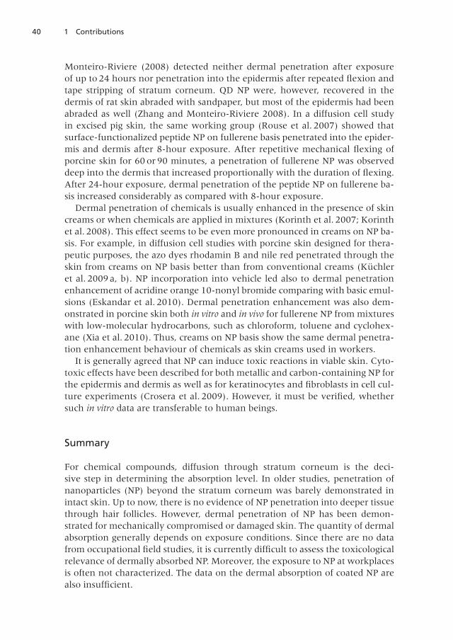
At industrial workplaces, up to 82% of workers may show visible damageof the epidermal barrier (for example Korinth et al . 2007) . Table 1 summa-rizes studies that allow a comparison to be made between the penetration ofNP through intact and compromised skin . In a diffusion cell study, Larese etal. (2009) investigated the penetration of silver (Ag) NP through intact andby scratch technique damaged human skin. Atomic absorption spectrometryshowed five times higher penetration of Ag through damaged than throughintact skin. However, this study was not reliable in differentiating Ag NP and el-emental silver. Transmission electron microscopy investigations showed Ag NPin stratum corneum and in the upper epidermal layers . Since deeper skin lay-ers were also damaged, such a model has limited reliability for the situation atworkplaces and in the environment. In their diffusion cell study in excised hu-man skin, Baroli et al . (2007) found that, a stratum corneum swelling by hydra-tion led to the formation of depots of iron and iron oxide NP. They also observedpenetration of iron-containing NP into the epidermis in experiments with intactskin. In a diffusion cell study in rat skin using two different QD NP, Zhang and
Table 1: Comparison of nanoparticle (NP) penetration through intact and compromised skin .
Nanoparticles
Particlecharacterization
Type ofstudy
Skin compromising technique
Skinpenetration
Reference
Silver coated withpolyvinylpyr-rolidone(Ø up to 25 nm)
controlleddiffusion cellstudy in hu-man skin
skin scratching 5-fold penetra-tion increase ofsilver throughdamaged skin
Larese etal . 2009
Fe2O
3
(magh-emite),iron
coated with or-ganic molecules(Ø < 24 nm)
controlleddiffusion cellstudy in hu-man skin
swelling of thestratum corneumcaused by 24-hourhydration
formation ofNP depots inhydrated stra-tum corneum
Baroli etal . 2007
Fullerene peptide coating(Ø ~ 4 nm)
controlleddiffusion cellstudy in por-cine skin
mechanical skinflexion
penetration en-hancement byskin flexion
Rouse etal . 2007
Quantumdots (QD)
Cd/Se core andZnS shell(Ø ≤ 18 nm)
controlleddiffusion cellstudy in ratskin
tape stripping,abrasion andmechanical skinflexion
penetration en-hancement inabraded skin
ZhangandMonteiro-Riviere2008
Berylliumoxide*
Ø ≤ 1 µm excised hu-man skin
mechanical skinflexion
penetration en-hancement byskin flexion
Tinkle etal . 2003
*Physicochemical characterization of beryllium oxide was not performed
40 1 Contributions
Monteiro-Riviere (2008) detected neither dermal penetration after exposureof up to 24 hours nor penetration into the epidermis after repeated flexion andtape stripping of stratum corneum. QD NP were, however, recovered in thedermis of rat skin abraded with sandpaper, but most of the epidermis had beenabraded as well (Zhang and Monteiro-Riviere 2008). In a diffusion cell studyin excised pig skin, the same working group (Rouse et al. 2007) showed thatsurface-functionalized peptide NP on fullerene basis penetrated into the epider-mis and dermis after 8-hour exposure. After repetitive mechanical flexing ofporcine skin for 60 or 90 minutes, a penetration of fullerene NP was observeddeep into the dermis that increased proportionally with the duration of flexing.After 24-hour exposure, dermal penetration of the peptide NP on fullerene ba-sis increased considerably as compared with 8-hour exposure .
Dermal penetration of chemicals is usually enhanced in the presence of skincreams or when chemicals are applied in mixtures (Korinth et al . 2007; Korinthet al. 2008). This effect seems to be even more pronounced in creams on NP ba-sis . For example, in diffusion cell studies with porcine skin designed for thera-peutic purposes, the azo dyes rhodamin B and nile red penetrated through theskin from creams on NP basis better than from conventional creams (Küchleret al. 2009 a, b). NP incorporation into vehicle led also to dermal penetrationenhancement of acridine orange 10-nonyl bromide comparing with basic emul-sions (Eskandar et al . 2010) . Dermal penetration enhancement was also dem-onstrated in porcine skin both in vitro and in vivo for fullerene NP from mixtureswith low-molecular hydrocarbons, such as chloroform, toluene and cyclohex-ane (Xia et al . 2010) . Thus, creams on NP basis show the same dermal penetra-tion enhancement behaviour of chemicals as skin creams used in workers.
It is generally agreed that NP can induce toxic reactions in viable skin. Cyto-toxic effects have been described for both metallic and carbon-containing NP forthe epidermis and dermis as well as for keratinocytes and fibroblasts in cell cul-ture experiments (Crosera et al. 2009). However, it must be verified, whethersuch in vitro data are transferable to human beings .
Summary
For chemical compounds, diffusion through stratum corneum is the deci-sive step in determining the absorption level. In older studies, penetration ofnanoparticles (NP) beyond the stratum corneum was barely demonstrated inintact skin. Up to now, there is no evidence of NP penetration into deeper tissuethrough hair follicles. However, dermal penetration of NP has been demon-strated for mechanically compromised or damaged skin. The quantity of dermalabsorption generally depends on exposure conditions . Since there are no datafrom occupational field studies, it is currently difficult to assess the toxicologicalrelevance of dermally absorbed NP. Moreover, the exposure to NP at workplacesis often not characterized . The data on the dermal absorption of coated NP arealso insufficient.
411.4 Penetration of Nanoparticles through Intact and Compromised Skin
Because of the background exposure to NP and the possible contamination,negative controls should be included in skin penetration studies. At present, theskin penetration of NP can be quantified only to a limited extent, since there areno validated analytical methods. Therefore, there is only little kinetic dermalpenetration data available.
In older studies, physicochemical characterization of the size and form of theparticles in the exposure phase was rarely performed . Due to the methodologi-cal problems, the dermal penetration data of NP is still insufficient. Further dataof well-designed studies and the development of new analytical methods arerequired for the quantification of dermal penetration of NP and for the risk as-sessment .
References
Baroli B, Ennas MG, Loffredo F, Isola M, Pinna R, López-Quintela MA (2007)Penetration of metallic nanoparticles in human full-thickness skin . J InvestDermatol 127: 1701–1712
Crosera M, Bovenzi M, Maina G, Adami G, Zanette C, Florio C, Larese Filon F(2009) Nanoparticle dermal absorption: a review of the literature . Int ArchOccup Environ Health 82: 1043–1055
Cross SE, Innes B, Roberts MS, Tsuzuki T, Robertson TA, McCormick P (2007)Human skin penetration of sunscreen nanoparticles: in vitro assessment ofa novel micronized zinc oxide formulation. Skin Pharmacol Physiol 20:148–154
Eskandar NG, Simovic S, Prestidge CA (2010) Mechanistic insight into the der-mal delivery from nanoparticle-coated submicron O/W emulsions. J PharmSci 99: 890–904
Gamer AO, Leibold E, van Ravenzwaay B (2006) The in vitro absorption ofmicrofine zinc oxide and titanium dioxide through porcine skin. Toxicol InVitro 20: 301–307
Korinth G, Weiss T, Penkert S, Schaller KH, Angerer J, Drexler H (2007) Percu-taneous absorption of aromatic amines in rubber industry workers: impactof impaired skin and skin barrier creams . Occup Environ Med 64: 366–372
Korinth G, Lüersen L, Schaller KH, Angerer J, Drexler H (2008) Enhancementof percutaneous penetration of aniline and o-toluidine in vitro using skinbarrier creams . Toxicol In Vitro 22: 812–818
Küchler S, Radowski MR, Blaschke T, Dathe M, Plendl J, Haag R, Schäfer-Ko-rting M, Kramer KD (2009) Nanoparticles for skin penetration enhance-ment – a comparison of a dendritic core-multishell-nanotransporter andsolid lipid nanoparticles . Eur J Pharm Biopharm 71: 243–250
Küchler S, Abdel-Mottaleb M, Lamprecht A, Radowski MR, Haag R, Schäfer-Korting M (2009) Influence of nanocarrier type and size on skin delivery ofhydrophilic agents . Int J Pharm 377: 169–172
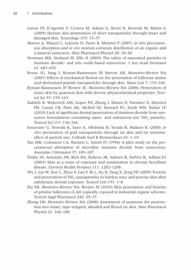
42 1 Contributions
Larese FF, D’Agostin F, Crosera M, Adami G, Renzi N, Bovenzi M, Maina G(2009) Human skin penetration of silver nanoparticles through intact anddamaged skin . Toxicology 255: 33–37
Mavon A, Miquel C, Lejeune O, Payre B, Moretto P (2007) In vitro percutane-ous absorption and in vivo stratum corneum distribution of an organic anda mineral sunscreen . Skin Pharmacol Physiol 20: 10–20
Newman MD, Stotland M, Ellis JI (2009) The safety of nanosized particles intitanium dioxide- and zinc oxide-based sunscreens . J Am Acad Dermatol61: 685–692
Rouse JG, Yang J, Ryman-Rasmussen JP, Barron AR, Monteiro-Riviere NA(2007) Effects of mechanical flexion on the penetration of fullerene aminoacid-derivatized peptide nanoparticles through skin. Nano Lett 7: 155–160
Ryman-Rasmussen JP, Riviere JE, Monteiro-Riviere NA (2006) Penetration ofintact skin by quantum dots with diverse physicochemical properties. Toxi-col Sci 91: 159–165
Sadrieh N, Wokovich AM, Gopee NV, Zheng J, Haines D, Parmiter D, SiitonenPH, Cozart CR, Patri AK, McNeil SE, Howard PC, Doub WH, Buhse LF(2010) Lack of significant dermal penetration of titanium dioxide from sun-screen formulations containing nano- and submicron-size TiO
2particles .
Toxicol Sci 115: 156–166Sonavane G, Tomoda K, Sano A, Ohshima H, Terada H, Makino K (2008) In
vitro permeation of gold nanoparticles through rat skin and rat intestine:effect of particle size . Colloids Surf B Biointerfaces 65: 1–10
Tan MH, Commens CA, Burnett L, Snitch PJ (1996) A pilot study on the per-cutaneous absorption of microfine titanium dioxide from sunscreens.Australas J Dermatol 37: 185–187
Tinkle SS, Antonini JM, Rich BA, Roberts JR, Salmen R, DePree K, Adkins EJ(2003) Skin as a route of exposure and sensitization in chronic berylliumdisease. Environ Health Perspect 111: 1202–1208
Wu J, Liu W, Xue C, Zhou S, Lan F, Bi L, Xu H, Yang X, Zeng FD (2009) Toxicityand penetration of TiO
2nanoparticles in hairless mice and porcine skin after
subchronic dermal exposure . Toxicol Lett 191: 1–8Xia XR, Monteiro-Riviere NA, Riviere JE (2010) Skin penetration and kinetics
of pristine fullerenes (C 60) topically exposed in industrial organic solvents.Toxicol Appl Pharmacol 242: 29–37
Zhang LW, Monteiro-Riviere NA (2008) Assessment of quantum dot penetra-tion into intact, tape-stripped, abraded and flexed rat skin. Skin PharmacolPhysiol 21: 166–180
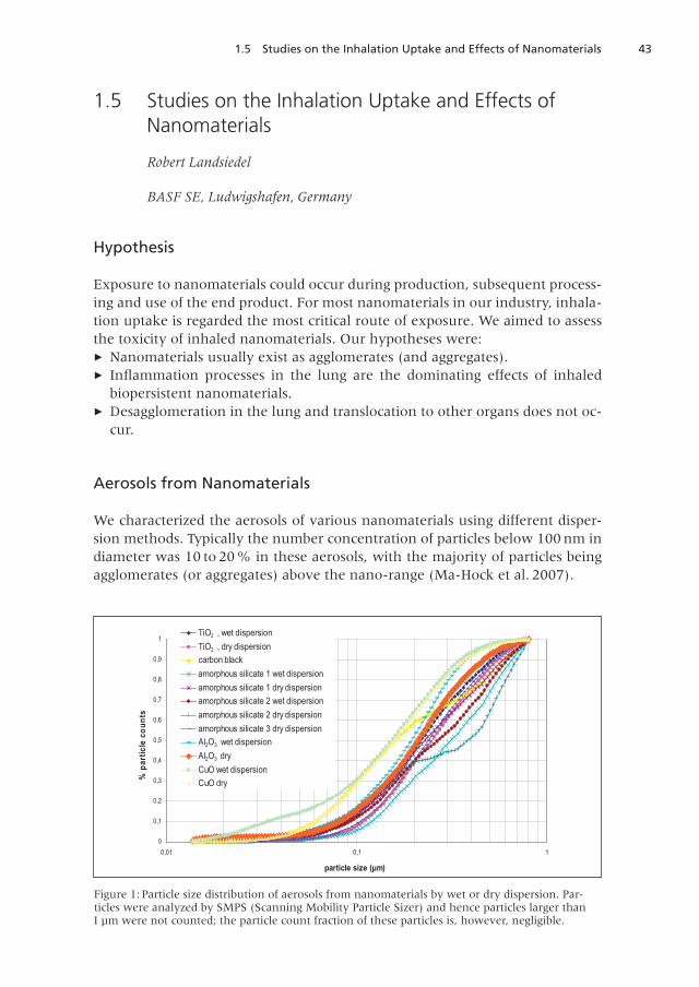
431.5 Studies on the Inhalation Uptake and Effects of Nanomaterials
1.5 Studies on the Inhalation Uptake and Effects ofNanomaterials
Robert Landsiedel
BASF SE, Ludwigshafen, Germany
Hypothesis
Exposure to nanomaterials could occur during production, subsequent process-ing and use of the end product . For most nanomaterials in our industry, inhala-tion uptake is regarded the most critical route of exposure . We aimed to assessthe toxicity of inhaled nanomaterials . Our hypotheses were:► Nanomaterials usually exist as agglomerates (and aggregates) .► Inflammation processes in the lung are the dominating effects of inhaled
biopersistent nanomaterials .► Desagglomeration in the lung and translocation to other organs does not oc-
cur .
Aerosols from Nanomaterials
We characterized the aerosols of various nanomaterials using different disper-sion methods . Typically the number concentration of particles below 100 nm indiameter was 10 to 20% in these aerosols, with the majority of particles beingagglomerates (or aggregates) above the nano-range (Ma-Hock et al. 2007).
Figure 1: Particle size distribution of aerosols from nanomaterials by wet or dry dispersion . Par-ticles were analyzed by SMPS (Scanning Mobility Particle Sizer) and hence particles larger than1 µm were not counted; the particle count fraction of these particles is, however, negligible.
0
0,1
0,2
0,3
0,4
0,5
0,6
0,7
0,8
0,9
1
0,01 0,1 1
particle size (µm)
%pa
rtic
leco
unts
TiO2 , wet dispersionTiO2 , dry dispersioncarbon blackamorphous silicate 1 wet dispersionamorphous silicate 1 dry dispersionamorphous silicate 2 wet dispersionamorphous silicate 2 dry dispersionamorphous silicate 3 dry dispersionAl2O3 wet dispersionAl2O3 dryCuO wet dispersionCuO dry
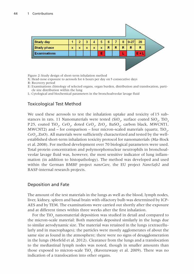
44 1 Contributions
Toxicological Test Method
We used these aerosols to test the inhalation uptake and toxicity of 15 sub-stances in rats . 11 Nanomaterials were tested (SiO
2, surface coated SiO
2, TiO
2
P 25, coated TiO2, CeO
2, doted CeO
2, ZrO
2, BaSO
4, carbon black, MWCNT1,
MWCNT2) and – for comparison – four micron-scaled materials (quartz, TiO2,
CeO2,ZnO). All materials were sufficiently characterized and tested by the well-
established short-term inhalation toxicity protocol for nanomaterials (Ma-Hocket al. 2008). For method development over 70 biological parameters were used.Total protein concentration and polymorphonuclear neutrophils in bronchoal-veolar lavage fluid was, however, the most sensitive indicator of lung inflam-mation (in addition to histopathology). The method was developed and usedwithin the German BMBF project nanoCare, the EU project NanoSafe2 andBASF-internal research projects .
Deposition and Fate
The amount of the test materials in the lungs as well as the blood, lymph nodes,liver, kidney, spleen and basal brain with olfactory bulb was determined by ICP-AES and by TEM . The examinations were carried out shortly after the exposureand at different times within three weeks after the first inhalation.
For the TiO2
nanomaterial deposition was studied in detail and compared tothe micron-scale material: Both materials deposited similarly in the lungs dueto similar aerodynamic size . The material was retained in the lungs (extracellu-larly and in macrophages); the particles were mostly agglomerates of about thesame size as found in the atmosphere; there were no signs of desagglomerationin the lungs (Morfeld et al . 2012) . Clearance from the lungs and a translocationto the mediastinal lymph nodes was noted, though in smaller amounts thanthose exposed to micron-scale TiO
2(Ravenzwaay et al. 2009). There was no
indication of a translocation into other organs .
Figure 2: Study design of short-term inhalation methodX: Head-nose exposure to aerosols for 6 hours per day on 5 consecutive daysR: Recovery periodE: Examinations (histology of selected organs, organ burden, distribution and translocation, parti-
cle size distribution within the lungL: Cytological and biochemical parameters in the bronchoalveolar lavage fluid
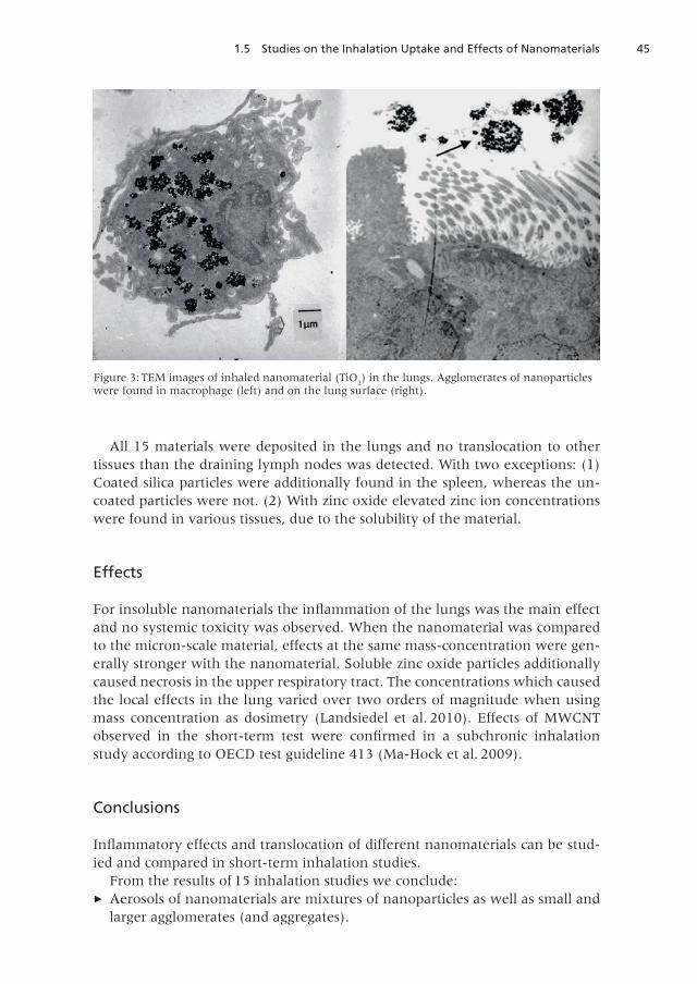
451.5 Studies on the Inhalation Uptake and Effects of Nanomaterials
All 15 materials were deposited in the lungs and no translocation to othertissues than the draining lymph nodes was detected . With two exceptions: (1)Coated silica particles were additionally found in the spleen, whereas the un-coated particles were not. (2) With zinc oxide elevated zinc ion concentrationswere found in various tissues, due to the solubility of the material.
Effects
For insoluble nanomaterials the inflammation of the lungs was the main effectand no systemic toxicity was observed. When the nanomaterial was comparedto the micron-scale material, effects at the same mass-concentration were gen-erally stronger with the nanomaterial . Soluble zinc oxide particles additionallycaused necrosis in the upper respiratory tract . The concentrations which causedthe local effects in the lung varied over two orders of magnitude when usingmass concentration as dosimetry (Landsiedel et al . 2010) . Effects of MWCNTobserved in the short-term test were confirmed in a subchronic inhalationstudy according to OECD test guideline 413 (Ma-Hock et al . 2009) .
Conclusions
Inflammatory effects and translocation of different nanomaterials can be stud-ied and compared in short-term inhalation studies .
From the results of 15 inhalation studies we conclude:► Aerosols of nanomaterials are mixtures of nanoparticles as well as small and
larger agglomerates (and aggregates) .
Figure 3: TEM images of inhaled nanomaterial (TiO2) in the lungs . Agglomerates of nanoparticles
were found in macrophage (left) and on the lung surface (right) .
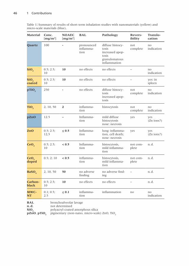
46 1 Contributions
Table 1: Summary of results of short-term inhalation studies with nanomaterials (yellow) andmicro-scale materials (blue) .
Material Conc.[mg/m3]
NOAEC[mg/m3]
BAL Pathology Reversibility
Translocation
Quartz 100 pronouncedinflamma-tion
diffuse histocy-tosisincreased apop-tosisgranulomatousinflammation
notcomplete
noindication
SiO2
0 .5; 2 .5;10
10 no effects no effects – noindication
SiO2
coated0 .5; 2 .5;10
10 no effects no effects – yes: inspleen
pTiO2
250 no effects diffuse histocy-tosisincreased apop-tosis
notcomplete
noindication
TiO2
2, 10, 50 2 inflamma-tion
histocytosis notcomplete
noindication
pZnO 12 .5 – Inflamma-tion
mild diffusehistocytosisnose: necrosis
yes yes(Zn ions?)
ZnO 0 .5; 2 .5;12,5
< 0.5 Inflamma-tion
lung: inflamma-tion, cell death;nose: necrosis
yes yes(Zn ions?)
CeO2
0 .5; 2 .5;10
< 0.5 Inflamma-tion
histocytosis,mild inflamma-tion
not com-plete
n .d .
CeO2
doped0 .5; 2; 10 < 0.5 inflamma-
tionhistocytosis,mild inflamma-tion
not com-plete
n .d .
BaSO4
2, 10, 50 50 no adversefinding
no adverse find-ing
– n .d .
Carbonblack
0 .5; 2 .5;10
10 no effects no effects – n .d .
MWCNT
0 .1; 0 .5;2 .5
< 0.1 inflamma-tion
inflammation no noindication
BAL bronchoalveolar lavagen. d. not determinedSiO
2polyacryl-coated amorphous silica
pZnO, pTiO2
pigmentary (non-nano, micro-scale) ZnO, TiO2
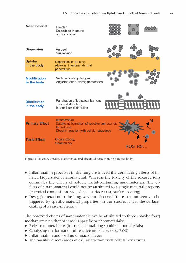
471.5 Studies on the Inhalation Uptake and Effects of Nanomaterials
► Inflammation processes in the lung are indeed the dominating effects of in-haled biopersistent nanomaterial . Whereas the toxicity of the released ionsdominates the effects of soluble metal-containing nanomaterials . The ef-fects of a nanomaterial could not be attributed to a single material property(chemical composition, size, shape, surface area, surface coating) .
► Desagglomeration in the lung was not observed. Translocation seems to betriggered by specific material properties (in our studies it was the surface-coating of a silica-material) .
The observed effects of nanomaterials can be attributed to three (maybe four)mechanisms; neither of those is specific to nanomaterials:► Release of metal ions (for metal containing soluble nanomaterials)► Catalysing the formation of reactive molecules (e. g. ROS)► Inflammation and loading of macrophages► and possibly direct (mechanical) interaction with cellular structures
Deposition in the lungAlveolar, intestinal, dermalpenetration
Uptakein the body
Organ toxicity,Genotoxicity
Toxic Effect
Dispersion AerosolSuspension
Nanomaterial PowderEmbedded in matrixor on surfaces
Penetration of biological barriersTissue distribution,Intracellular distribution
Distributionin the body
Modificationin the body
Surface coating changesAgglomeration, desagglomeration
InflammationCatalysing formation of reactive compoundsIon releaseDirect interaction with cellular structures
Primary Effect
ROS, RS, ...
M
Figure 4: Release, uptake, distribution and effects of nanomaterials in the body.
48 1 Contributions
These primary effects are modulated by the distribution and persistence ofthe nanomaterial in the lungs (or in other tissues) and compartments inside thecell, resulting in different effects of different nanomaterials . At this time it is notexactly known which material properties direct the toxic effects . There is, how-ever, no indication of a nano-specific toxicity per se. There are rather differenteffects and different potency of the different nanomaterials .
References
Landsiedel R, Ma-Hock L, Kroll A, Hahn D, Schnekenburger J, Wiench K,Wohlleben W (2010) Testing metal-oxide nanomaterials for human safety .Adv Mater 22: 1–27
Ma-Hock L, Gamer A, Landsiedel R, Leibold E, Frechen T, Sens B, Huber G, vanRavenzwaay B (2007) Generation and characterization of test atmosphereswith nanomaterials . Inhal Toxicol 19: 833–848
Ma-Hock L, Burkhardt S, Strauss V, Gamer AO, Wiench K, van RavenzwaayB, Landsiedel R (2009) Development of a short-term inhalation test in therat using nano-titanium dioxide as a model substance . Inhal Toxicol 21:102–118
Ma-Hock L, Treumann S, Strauss V, Brill S, Luizi F, Mertler M, Wiench K,Gamer AO, van Ravenzwaay B, Landsiedel R (2009) Inhalation toxicity ofmulti-wall carbon nanotubes in rats exposed for 3 months . Toxicol Sci 112:468–481
Morfeld P, Treumann S, Ma-Hock L, Bruch J, Landsiedel R (2012): Depositionbehaviour of inhaled nanostructured TiO
2in rats: number distribution of
particles by different lung lobes and fraction of particle diameters below100 nm . Inhal Toxicol 24: 939-993 .
Ravenzwaay B, Landsiedel R, Fabian E, Burkhardt S, Strauss V, Ma-Hock L(2009) Comparing fate and effects of three particles of different surfaceproperties: Nano-TiO
2, pigmentary TiO
2and quartz. Toxicol Lett 186: 152–
159
491.6 Animal Studies on the Effect of Nanoparticles in Organs other than the Lungs
1.6 Animal Studies on the Effect of Nanoparticles inOrgans other than the Lungs
Uwe Heinrich
Fraunhofer Institute for Toxicology and Experimental Medicine
Hanover, Germany
Apart from their different kinetics (distribution in the organism), nanometerand micrometer particles of the same material do not seem to differ qualitativelyregarding their toxic effects on the lungs . Based on the particle mass, which de-termines the dose, the inflammatory proliferative and tumour-inducing effectsof nanoparticles are severer than those of microparticles. The clearance half-lives of nanoparticles in the lungs are also longer than those of microparticles.
This statement does not apply to non-soluble or poorly soluble nanoparticles .Their toxicity originates from the physicochemical particle properties and fromthe surface activity of the particles. There is hardly any evidence of a possibletoxicity of such nanoparticles in organs other than the lungs . Numerous publi-cations on nanoparticles mainly deal with the question in which organs otherthan the lungs such nanoparticles can be detected after inhalation .
The particle effect of nanoparticles from metal and their inorganic com-pounds is also determined by the degree of the bioavailability of the metal sincethe metal ions may have an effect of their own. The metal nanoparticle is thus acarrier for the metal ion . A high cellular concentration of toxic metal ions mayform, particularly if the metal nanoparticle becomes bioavailable intracellularly.
Carbon nanotubes (CNT) are a group of nanomaterials that is not consideredhere. Because of their high biopersistence and possible occurrence in very dif-ferent lengths and diameters, some nanotubes may also have the potential toinduce mesotheliomas and lung tumours. No carcinogenicity tests or validatedscreening tests that can be used to rule out carcinogenicity are available for thisgroup of nanomaterials . Therefore, the effect of such nanotubes might be com-pared with an effect of granular biopersistent particles .
A special group of nanotubes retains the structure of a considerably entan-gled agglomerate and does not segregate into individual nanotubes even aftermechanical treatment and in suspension . If these nanotube agglomerates occurin a respirable size, they behave like granular biopersistent dusts, although theyhave a very low density and a great specific surface.
Publications of Effect Studies on Granular Biopersistent Nanodusts(GBND) and Metal Nanoparticles
The intranasal administration of carbon black nanoparticles in mice with pri-mary particle sizes of 14 nm (Printex 90; Degussa) and 95 nm (lampblack; De-gussa) and with BET surfaces (Brunauer, Emmett and Teller theory) of 300 m²/g
50 1 Contributions
or 20 m²/g in a dose of 62 .5 µg/naris once per week for 4 weeks showed a signif-icant increase in the mRNA expression of cytokines (IL-1beta and TNF-alpha)and chemokines (macrophage inflammatory protein CCL2, monocyte chemo-attractant protein CCL3 and the chemokine ligand CXCL9, which is related tothe Th 1 response) in the olfactory bulb only for Printex 90, but not for lamp-black. No evidence of this mRNA expression was provided in the hippocampusof the treated animals for any of the carbon black particles used . The moleculesexpressed here all play a role in neurogenic immune response and inflamma-tion (Tin-Tin-Win-Shwe et al . 2006) .
After i . p . injection into mice in doses of 6 .5 to 51 .8 mg, nano TiO2
(100 nm)was found in the liver, lung and kidney; no positive evidence was provided inthe heart or brain . Histopathological effects in the different organs were foundparticularly in the spleen (increased neutrophil concentration and inflamma-tion), in the liver (inflammation and fibrosis) and in the lung (increased occur-rence of neutrophil cells, thickened alveolar septa, TiO
2particles in pulmonary
vessels and thrombosis). Liver enzymes in the blood were also increased in thetwo highest dose groups; however, these dose groups also showed increasedmortality (Chen et al . 2009) .
Pregnant mice were subcutaneously treated with suspended nano TiO2
(par-ticle size: 25–0 nm; anatase) on days 3, 7, 10 and 14 after mating . The maleoffspring were examined when they were 4 days or 6 weeks old. Evidence ofTiO
2in the testes and brain of the offspring was provided with the electron mi-
croscope and the associated energy dispersive X-ray spectroscopy. Aggregatesof TiO
2nanoparticles (100–200 nm) were detected in Leydig cells, Sertoli cells
and spermatids of the testes of the offspring . A histopathological examination ofthe testicular morphology revealed clear changes in the 6-week-old offspring.Sperm production, sperm motility and the number of Sertoli cells were signifi-cantly reduced as compared with the controls . This describes an effect of treat-ment on spermatogenesis . TiO
2was also detected in brain cells of the offspring
(6 weeks). An increased number of cells with a positive reaction to apoptosismarkers (caspase-3) was found in the olfactory bulb, and obstruction of smallvessels and perivascular edema were described histopathologically (Takeda etal . 2009) .
Rats were exposed to MnO nanoparticles (primary particle size: 3–8 nm; ag-glomerate particle size: 30 nm) in a whole-body exposure inhalation systemfor 6 hours/day, 5 days/week, for up to 12 days . The particles had a solubil-ity of 1–1 .5% per day at a neutral pH . The exposure concentration was about500 µg/m³ . Both TNF-alpha gene expression and TNF-alpha protein expressionwere significantly increased in the olfactory bulb and in various other brain re-gions. The gene for the macrophage inflammatory protein-2, for the glial fibril-lary acidic protein and for the neuronal cell adhesion molecule was also signifi-cantly overexpressed in the olfactory bulb. Different approaches (comparativeinvestigations of MnCl
2with MnO; solubility tests of MnO using different pH
levels) used in these studies demonstrated that the main fraction of manganeseoxide reached the olfactory bulb in particle form (Elder et al . 2006) .
511.6 Animal Studies on the Effect of Nanoparticles in Organs other than the Lungs
In a study in pregnant mice, the gene expression was determined in the brainof the embryos and of the 1- and 2-day-old newborns after maternal subcu-taneous injection of nano-TiO
2(anatase) . Changes in the expression of genes
were found that were associated with encephalization, cell death, oxidativestress and the mitochondria during the perinatal period (Shimizu et al . 2009) .
In addition to toxic effects in the lung, histopathological changes in the liver(bile-duct hyperplasia) were also described after inhalation (28 and 90 days) ofsilver nanoparticles (metallic silver) in a particle size of 18 nm count mediandiameter (CMD). Apart from the lung, the liver had the highest silver concen-tration as compared with the kidney, brain and blood. The silver concentrationswere determined using an atomic absorption spectrophotometer . The solubil-ity of these metallic silver particles was not specified (Sung et al. 2008; Sung etal . 2009) .
Genotoxic effects in peripheral blood cells after i . p . injection of nano Al2O
3
(primary particle size 30 and 40 nm; agglomerate size in the suspending medium140–220 nm) were found in rats using the comet assay and micronucleus test .Al
2O
3with a particle size of 50–200 µm did not show these findings (Balasu-
bramanyam et al . 2009) .
References
Balasubramanyam A, Sailaja N, Mahboob M, Rahman MF, Hussain SM, GroverP (2009) In vivo genotoxicity assessment of aluminium oxide nanomaterialsin rat peripheral blood cells using the comet assay and micronucleus test .Mutagenesis 24: 245–251
Chen J, Dong X, Zhao J, Tang G (2009) In vivo acute toxicity of titanium diox-ide nanoparticles to mice after intraperitoneal injection . J Appl Toxicol 29:330–337
Elder A, Gelein R, Silva V, Feikert T, Opanashuk L, Carter J, Potter R, May-nard A, Ito Y, Finkelstein J, Oberdörster G (2006) Translocation of inhaledultrafine manganese oxide particles to the central nervous system. Envi-ron Health Perspect 114: 1172–1178. Erratum in: Environ Health Perspect.ug;114: 1178
Shimizu M, Tainaka H, Oba T, Mizuo K, Umezawa M, Takeda K (2009) Mater-nal exposure to nanoparticulate titanium dioxide during the prenatal pe-riod alters gene expression related to brain development in the mouse. PartFibre Toxicol 6: 1–8
Sung JH, Ji JH, Yoon JU, Kim DS, Song MY, Jeong J, Han BS, Han JH, ChungYH, Kim J, Kim TS, Chang HK, Lee EJ, Lee JH, Yu IJ (2008) Lung FunctionChanges in Sprague-Dawley rats after prolonged inhalation exposure to sil-ver nanoparticles. Inhal Toxicol 20: 567–574
Sung JH, Ji JH, Park JD, Yoon JU, Kim DS, Jeon KS, Song MY, Jeong J, HanBS, Han JH, Chung YH, Chang HK, Lee JH, Cho MH, Kelman BJ, Yu IJ(2009) Subchronic inhalation toxicity of silver nanoparticles. Toxicol Sci108: 452–461
52 1 Contributions
Takeda K, Suzuki K, Ishihara A, Kubo-Irie M, Fujimoto R, Tabata M, Oshio S,Nihei Y, Ihara T, Sugamata M (2009) Nanoparticles transferred from preg-nant mice to their offspring can damage the genital and cranial nerve sys-tems . J Health Sci 55: 95–102
Tin-Tin-Win-Shwe, Yamamoto S, Ahmed S, Kakeyama M, Kobayashi T, Fuji-maki H (2006) Brain cytokine and chemokine mRNA expression in miceinduced by intranasal instillation with ultrafine carbon black. Toxicol Lett163: 153–160
531.7 Transport of Nanoparticles to the Brain: Concern for Neurotoxicity?
1.7 Transport of Nanoparticles to the Brain:Concern for Neurotoxicity?
Andrea Hartwig
Karlsruhe Institute of Technology
Karlsruhe, Germany
There are several publications raising the question of whether there is concernfor neurotoxicity of nanoparticles, summarized for example by Oberdörster etal. (2009). One important prerequisite to induce oxidative stress or inflamma-tion in the central nervous system (CNS) would be a significant translocation ofnanoparticles to the brain upon realistic exposure conditions . As stated before byHeinrich (this volume) systemic availability of inhaled nanoparticles outside thelung upon realistic exposure conditions appears to be very limited; additionally,nanoparticles would have to cross the blood brain barrier, which has not beendemonstrated to occur in vivo yet. Nevertheless, one alternative way proposedfor the potential transfer of nanoparticles to the brain consists in the passagethrough sensory nerves. Thus, for nanoparticles of less than 0.1 µm diffusionappears to be the main mechanism of disposition during nasal breathing, andsignificant fractions are deposited in all parts of the respiratory tract, namely thenasal, the tracheobronchial and the alveolar region. Within the nasal region,the smallest nanoparticles may behave similar to smell molecules and have thepotential to be translocated along the sensory neuronal pathway to the olfac-tory bulb . Similarly, the nasopharyngeal and the tracheobronchial airways aresupplied with sensory nerves as well, whereas their presence in the alveolar re-gion of the lung is less clear (Oberdörster et al. 2009). Up to now, there are onlyfew studies available demonstrating olfactory nerve nanoparticle translocationto the brain (Table 1) .
As evident from Table 1, there is only one inhalation study demonstrat-ing an inflammatory response, determined by a pronounced change in TNF--expression, after realistic inhalation exposure to MnO nanoparticles in rats .Here, inhalation experiments with rats at 500 µg/m3 revealed higher concentra-tions of manganese in the olfactory bulb as compared to lung tissue; further-more, pronounced changes in TNF- gene and protein expression levels wereobserved specifically in the olfactory bulb as opposed to other brain regions(Elder et al. 2006). Even though further studies are needed, this example isinteresting since epidemiological evidence points towards a pronounced neuro-toxic potential especially of small manganese oxide particles generated predom-inantly during welding, which are likely to contain a large fraction of ultrafineparticles. Thus, a tenfold lower MAK value has been established in 2010 for par-ticles in the alveolar fraction (0.02 mg/m3) as compared to the inhalable fraction(0 .2 mg/m3) (Hartwig 2010) . These differences in neurotoxic potential can beexplained either by higher systemic bioavailability of smaller particles via bloodcirculation and translocation through the blood brain barrier or via sensory
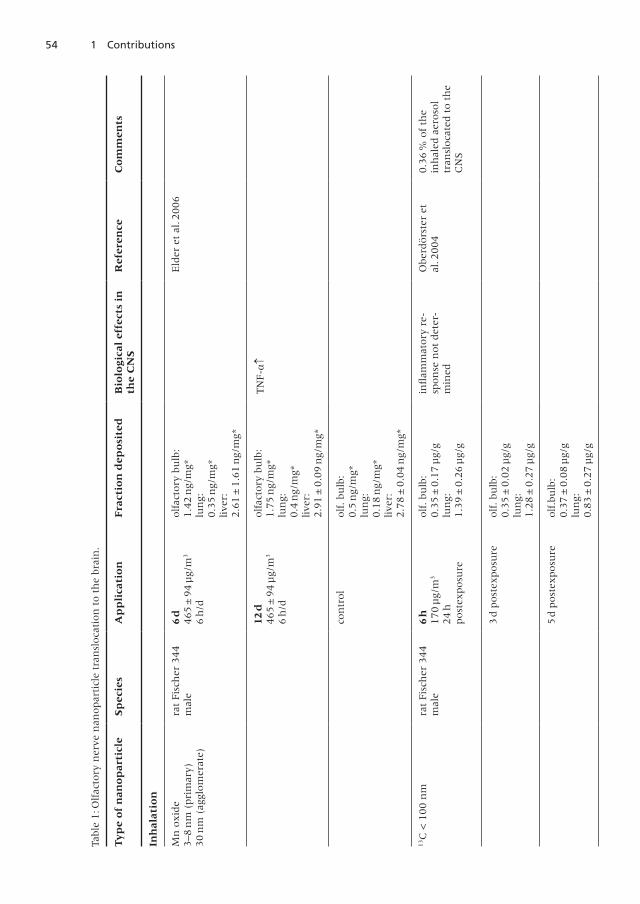
54 1 Contributions
Tabl
e1:O
lfac
tory
ner
ven
anopar
ticl
etr
ansl
oca
tion
toth
ebr
ain
.
Ty
pe
of
nan
op
arti
cle
Sp
ecie
sA
pp
lica
tio
nF
ract
ion
dep
osi
ted
Bio
logi
cal
effe
cts
inth
eC
NS
Ref
eren
ceC
om
men
ts
Inh
alat
ion
Mn
oxid
e3–8
nm
(pri
mar
y)30
nm
(agg
lom
erat
e)
rat
Fis
cher
344
mal
e6
d465
±94
µg/
m3
6h
/d
olf
acto
rybu
lb:
1 .4
2n
g/m
g*lu
ng:
0 .3
5n
g/m
g*live
r:2 .6
1±
1 .6
1n
g/m
g*
Eld
eret
al .2
006
12d
465
±9
4µ
g/m
3
6h
/d
olf
acto
rybu
lb:
1 .7
5n
g/m
g*lu
ng:
0 .4
ng/
mg*
live
r:2 .9
1±
0 .0
9n
g/m
g*
TN
F-↑
con
tro
lolf
.bu
lb:
0 .5
ng/
mg*
lun
g:0 .1
8n
g/m
g*live
r:2 .7
8±
0 .0
4n
g/m
g*
13C
<100
nm
rat
Fis
cher
344
mal
e6
h170
µg/
m3
24
hpost
exposu
re
olf
.bu
lb:
0 .3
5±
0 .1
7µ
g/g
lun
g:1 .3
9±
0 .2
6µ
g/g
infl
amm
atory
re-
spon
sen
ot
det
er-
min
ed
Obe
rdörs
ter
etal
.2004
0 .3
6%
of
the
inh
aled
aero
sol
tran
sloca
ted
toth
eC
NS
3d
post
exposu
reolf
.bu
lb:
0 .3
5±
0 .0
2µ
g/g
lun
g:1 .2
8±
0 .2
7µ
g/g
5d
post
exposu
reolf
.bu
lb:
0 .3
7±
0 .0
8µ
g/g
lun
g:0 .8
3±
0 .2
7µ
g/g
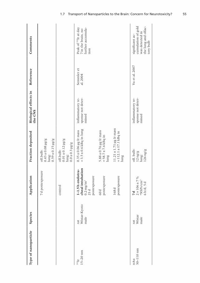
551.7 Transport of Nanoparticles to the Brain: Concern for Neurotoxicity?
Ty
pe
of
nan
op
arti
cle
Sp
ecie
sA
pp
lica
tio
nF
ract
ion
dep
osi
ted
Bio
logi
cal
effe
cts
inth
eC
NS
Ref
eren
ceC
om
men
ts
7d
post
exposu
reolf
.bu
lb:
0 .4
3±
0 .0
8µ
g/g
lun
g:0 .5
9±
0 .1
5µ
g/g
con
trol
olf
.bu
lb:
0 .0
1±
0 .1
2µ
g/g
lun
g:0 .0
±0 .3
µg/
g
19
2Ir
15–2
0n
mra
tW
ista
r-K
yoto
mal
e
1–1.
5h
end
otr
ach
eal
intu
bat
ion
0 .2
mg/
m3
21
dpost
exposu
re
0 .3
5±
0 .0
6µ
gIr
mas
s=
3.5
±0.6
kB
qin
lun
gin
flam
mat
ory
re-
spon
sen
ot
det
er-
min
ed
Sem
mle
ret
al .2
004
Pea
kof1
92Ir
atday
7in
the
brai
n,n
ofu
rth
erac
cum
ula
-ti
on
60
dpost
exposu
re5 .8
8±
0 .7
6µ
gIr
mas
s=
58.7
±7.6
kB
qin
lun
g
168
dpost
exposu
re11 .2
1±
1 .7
1µ
gIr
mas
s=
112.1
±17.1
kB
qin
lun
g
nA
u30–1
10
nm
rat
Wis
tar
mal
e
7d
2×
106
±7
%*N
SPs/
cm3
6h
/d,5
d
olf
.bu
lb:
12
ng/
glu
ng:
126
ng/
g
infl
amm
atory
re-
spon
sen
ot
det
er-
min
ed
Yu
etal
.2007
sign
ifica
nt
ac-
cum
ula
tion
of
gold
was
det
ecte
din
the
lun
gan
dolf
ac-
tory
bulb

56 1 Contributions
Ty
pe
of
nan
op
arti
cle
Sp
ecie
sA
pp
lica
tio
nF
ract
ion
dep
osi
ted
Bio
logi
cal
effe
cts
inth
eC
NS
Ref
eren
ceC
om
men
ts
21d
6h
/d,5
d/w
olf
.bu
lb:
13
ng/
glu
ng:
387
ng/
g
sign
ifica
nt
accu
mu
-la
tion
of
gold
was
det
ecte
din
the
lun
g,es
oph
agu
s,to
ngu
e,kid
ney
,ao
rta,
sple
en,
septu
m,h
eart
and
blood
con
trol
olf
.bu
lb:
4 .4
ng/
glu
ng:
5 .5
ng/
g
Inst
illa
tio
n
TiO
2
80
nm
Ru
til
mou
seC
D-
1(I
CR
)fe
mal
e
500
µg/
mou
sex
11
xev
ery
2d,x
5(1
0d)
1x
ever
y2
d,x
10
(20
d)
1x
ever
y2
d,x
15
(30
d)
con
tro
l
olf
.bu
lb:in
crea
segr
adu
ally
65
ng/
g*(c
on
trol)
to200
ng/
g*
Hip
poca
mpu
s:285
ng/
g(3
0d)*
TN
F-↑
IL-1
IL-6
:n
oin
-cr
ease
No
anti
oxid
ativ
ere
spon
ses
of
brai
n
Wan
get
al .
(200
8)
very
hig
hdose
(ato
taldose
of
7 .5
mg
of
nan
oTi
O2
inst
ille
din
tran
asal
lyin
tom
ice
wou
ldbe
equ
ival
ent
toin
-st
illin
g17 .5
gin
toth
en
ose
of
hu
man
s;O
berd
örs
ter
2009
b)
Co
25
nm
mou
seC
D-
1(I
CR
)fe
mal
e
1m
g/k
gbw
1x
ever
y2
d,x
348
hpo
stex
posu
re
olf
.bu
lb:
2 .8
µg/
g*br
ain
:17 .6
±1 .6
mg/
g
infl
amm
atory
re-
spon
sen
ot
det
er-
min
ed
Liu
etal
.(2
009)
con
trol
olf
.bu
lb:
2 .3
µg/
g*br
ain
:16 .7
±1 .7
mg/
g
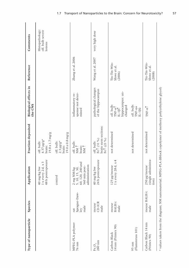
571.7 Transport of Nanoparticles to the Brain: Concern for Neurotoxicity?
Ty
pe
of
nan
op
arti
cle
Sp
ecie
sA
pp
lica
tio
nF
ract
ion
dep
osi
ted
Bio
logi
cal
effe
cts
inth
eC
NS
Ref
eren
ceC
om
men
ts
40
mg/
kg
bw1
xev
ery
2d,x
348
hpost
exposu
re
olf
.bu
lb:
4 .3
7µ
g/g*
brai
n:
19 .8
±1 .7
mg/
g
His
topat
holo
gy:
olf
.bu
lbse
vere
lesi
on
s
con
trol
olf
.bu
lb:
2 .3
µg/
g*br
ain
:19 .0
±0 .8
mg/
g
MPE
G-P
LA
poly
mer
76
nm
rat
Spra
gue-
Daw
-le
ym
ale
2m
gN
M/k
g5,10,1
5,30,
60,120
,240
and
360
min
post
-ad
min
istr
atio
n
olf
.bu
lb:
NM
↑br
ain
:N
M↑
infl
amm
atory
re-
spon
sen
ot
det
er-
min
ed
Zh
ang
etal
.2006
Fe 2
O3
280
nm
mou
seC
D-I
CR
mal
e
40
mg/
kg
bw24
hpo
stex
posu
reolf
.bu
lb:
Fe↑
(31
%)
brai
nst
emse
ctio
ns:
Fe↑
(21
%)
pat
holo
gica
lch
ange
sof
the
hip
poca
mpu
sW
ang
etal
.2007
very
hig
hdose
Car
bon
Bla
ck14
nm
(Pri
nte
x90)
mou
seB
ALB
/cm
ale
125
µg/
mou
se1
xev
ery
28
d,x
4n
ot
det
erm
ined
olf
.bu
lb:
TN
F-↑
IL-1↑
hip
poca
mpu
s:u
n-
chan
ged
Tin
-Tin
-Win
-Sh
we
etal
.(2
006)
95
nm
(Fla
mm
russ
101)
not
det
erm
ined
olf
.bu
lb:
TN
F-↓
IL-1↓
Car
bon
Bla
ck14
nm
(Pri
nte
x90)
mou
seB
ALB
/cm
ale
250
µg/
mou
se(s
ingl
ead
min
istr
a-ti
on
)
not
det
erm
ined
TN
F-↑
Tin
-Tin
-Win
-Sh
we
etal
.(2
008)
*va
lues
taken
from
the
dia
gram
;N
Mn
anom
ater
ial;
MPE
G-P
LA
dib
lock
copoly
mer
of
met
hoxy
poly
(eth
ylen
egl
ycol)
58 1 Contributions
nerve translocation from the nasal cavity. Within the brain cells, toxicity is mostlikely not a particle effect as such but rather due to the liberation of manganeseions leading to oxidative stress and related effects.
However, whether or not there will be significant transport of nanoparticlesthrough the sensory nerve system will have to be investigated on a case-by-case basis . Care must be taken to discriminate between hazard and risk, i . e .the potential to reach the brain (hazard) and the question whether the extentof translocation of the respective nanoparticles to the brain is sufficient to elicitadverse effects (risk). Thus, effects derived from experiments with bolus-typedelivery of nanoparticles such as nasal or tracheal instillation may provide valu-able information on hazard but not on risk; similarly, high dose experimentsare not representative for continuous low-dose exposure. Important for riskassessment appears to be not only the delivered dose per unit surface area ofthe respiratory tract, but also the dose rate, i . e . dose per unit time . One otherdecisive factor is the particle size, since only very small particles in the lowernanometer range will be able to traverse through sensory nerves; here, ag-glomeration and aggregation within the respiratory tract has to be taken intoaccount . Furthermore, the impact of the corona formation has been stressedduring the last years, also in the context of potential neurotoxic effects (Lynchand Dawson 2008) . Thus, nanoparticles in biological media attract proteins andlipids, depending on their size, surface area, charge, surface hydrophobicity andfurther characteristics on one side and extra- and intracellular fluids on theother side . The corona formation appears to be a dynamic process, leading toan equilibrium of association and dissociation of proteins and/or lipids. At theend, the composition of the corona will be one important factor with respectto translocation of nanoparticles to secondary organs and cell entry (Lynch etal . 2009) .
In summary, up to date there is no conclusive evidence that neurotoxic-ity may be a relevant endpoint for low-dose exposure towards nanoparticles,even though neurotoxic effects of specific types of particles cannot be excluded.Nevertheless, further research is needed providing data suitable for risk assess-ment of this important aspect of nanotoxicology .
References
Elder A, Gelein R, Silva V, Feikert T, Opanashuk L, Carter J, Potter R, MaynardA, Ito Y, Finkelstein J, and Oberdörster G (2006) Translocation of inhaledultrafine manganese oxide particles to the central nervous system. EnvironHealth Perspect 114: 1172–1178
Hartwig (2010) Aerosols . List of MAK and BAT Values, Maximum concentra-tions and biological tolerance values at the workplace, Commission for theInvestigation of Health Hazards of Chemical Compounds in the Work Area.Report 46, 179–187, Wiley-VCH, Weinheim
Liu Y, Gao Y, Zhang L, Wang T, Wang J, Jiao F, Li W, Liu Y, Li Y, Li B, Chai Z, WuG, Chen C (2009) Potential health impact on mice after nasal instillation
591.7 Transport of Nanoparticles to the Brain: Concern for Neurotoxicity?
of nano-sized copper particles and their translocation in mice . J NanosciNanotechnol 9: 6335–6343
Lynch I, Dawson KA (2008) Protein-nanoparticle interactions . Nanotoday 3:40–47
Lynch I, Salvati A, Dawson KA (2009) What does the cell see? Nature Nano-technol 4: 546–547
Oberdörster G, Sharp Z, Atudorei V, Elder A, Gelein R, Kreyling W, Cox C(2004) Translocation of inhaled ultrafine particles to the brain. Inhal Toxi-col 16: 437–445
Oberdörster G ., Elder A ., Rinderknecht A . (2009) Nanoparticles and the brain:cause for concern? J Nanosci Nanotechnol . 9: 4996–5007
Oberdörster G (2009b) Safety assessment for nanotechnology and nanomedi-cine: concepts of nanotoxicology . J Internal Med 267: 89–105
Semmler M, Seitz J, Erbe F, Mayer P, Heyder J, Oberdörster G, Kreyling WG(2004) Long-term clearance kinetics of inhaled ultrafine insoluble iridiumparticles from the rat lung, including transient translocation into secondaryorgans . Inhal Toxicol 16: 453–459
Tin-Tin-Win-Shwe, Yamamoto S, Ahmed S, Kakeyama M, Kobayashi T, Fuji-maki H (2006) Brain cytokine and chemokine mRNA expression in miceinduced by intranasal instillation with ultrafine carbon black. Tox Lett 163:153–160
Tin-Tin-Win-Shwe, Mitsushima D, Yamamoto S, Fukushima A, Funabashi T,Kobayashi T, Fujimaki H (2008) Changes in neurotransmitter levels andproinflammatory cytokine mRNA expressions in the mice olfactory bulbfollowing nanoparticle exposure . Toxicol Appl Pharmacol 226: 192–198
Wang B, Feng WY, Wang M, Shi JW, Zhang F, Ouyang H, Zhao YL, Chai ZF,Huang YY Xie YN, Wang HF, Wang J (2007) Transport of intranasally in-stilled fine Fe 203 particles into the brain: Micro-distribution, chemicalstates, and histopathological observation. Biol Trace Elem Res 118: 233–243
Wang J, Liu Y, Jiao F, Lao F, Li W, Gu Y, Li Y, Ge C, Zhou G, Li B, Zhao Y, Chai Z,Chen C (2008) Time-dependent translocation and potential impairment oncentral nervous system by intranasally instilled TiO2 nanoparticles. Toxicol-ogy 254: 82–90
Yu LE, Lanry Yung LY, Ong CN, Tan YL, Balasubramaniam KS, Hartono D, ShuiG, Wenk MR, Ong WY (2007) Translocation and effects of gold nanopar-ticles after inhalation exposure in rats . Nanotoxicology 1: 235–242
Zhang QZ, Zha LS, Zhang Y, Jiang WM, Lu W, Shi ZQ, Jiang XG, Fu SK (2006)The brain targeting efficiency following nasally applied MPEG-PLA nanopar-ticles in rats . J Drug Target 14: 281–290
60 1 Contributions
1.8 Genotoxicity of Nanoparticles
Roel Schins
Leibniz Research Institute for Environmental Medicine
Düsseldorf, Germany
Introduction
Current understanding about the mechanisms of genotoxicity of nanoparticlesis predominantly based on genotoxicity investigations performed with the par-ticulate carcinogen quartz as well as poorly soluble particles (PSP) of low toxic-ity such as carbon black, and titanium dioxide . Together with carcinogenicitystudies in rats, these investigations have lead to the introduction of the conceptsof primary versus secondary genotoxicity of inhalable particles. This particlegenotoxicity paradigm is of relevance in view of the observations that the tu-mourigenicity of PSP in rat lungs after chronic high exposures is associated withoverload and persistent inflammation (Greim et al. 2001; Knaapen et al. 2004;Schins and Knaapen 2007) .
Secondary genotoxicity refers to genetic damage that results from the oxida-tive DNA attack by reactive oxygen and nitrogen species (ROS/RNS) and possibleother mediators that are being generated during particle-elicited inflammationfrom recruited and activated phagocytes (macrophages, neutrophils). This sec-ondary genotoxicity is considered to involve a threshold; its value is set to bedetermined by the exposure concentration that will trigger inflammation andoverwhelm antioxidant and DNA damage repair capacities in the lung (Greim etal. 2001). Primary genotoxicity can be defined as the elucidation of genetic dam-age by particles in the absence of inflammation. This primary genotoxicity may befurther subdivided into direct and indirect pathways. Direct primary genotoxic-ity implies a direct physical interaction between nanoparticles and the genomicDNA. Indirect primary genotoxicity e. g. may result from the formation of ROS bynanoparticle-activated target cells, or from depletion of intracellular antioxidants(Schins and Knaapen 2007; Stone et al . 2009; Donaldson et al . 2010) .
Secondary Genotoxicity
Key studies which have lead to the above particle genotoxicity paradigm arethose by Driscoll and co-workers (1997). They investigated in vivo mutagenicity,in rat lungs 15 months after a single intratracheal instillation of different dosesof crystalline silica, carbon black or titanium dioxide. It was observed that mu-tagenesis (determined by HPRT assay (hypoxanthin-phosphoribosyl-transferaseassay) in the lung epithelial cells isolated from the lungs of the treated animals)was associated with the level of persisting inflammation (characterized as the %
611.8 Genotoxicity of Nanoparticles
of neutrophils in bronchoalveolar lavage fluid (BAL)) (Driscoll et al. 1997). Therole of particle-induced inflammation in driving genotoxicity in rat lungs sincethen has been supported by further investigations, e. g. in intratracheal instilla-tion studies in rats with quartz particles of different surface modification (nativeDQ 12 quartz, and DQ 12 coated with polyvinylpyridine-N-oxide or aluminiumlactate). The extent of neutrophilic inflammation measured in BAL from therats was associated with the level of DNA strand breakage measured by the invivo comet assay in the lung epithelial cells from these animals (Knaapen etal. 2002), as well as with the frequency of micronuclei in the epithelial cells(Albrecht et al . 2008) .
The role of inflammatory phagocytes (macrophages, neutrophils) in elicit-ing DNA damage and mutagenesis in neighbouring target cells is further sup-ported by numerous in vitro studies, that used co-culture models of inflamma-tory cells and various other cell types including lung epithelial cells (reviewedin Knaapen et al . 2006) . Although these in vitro studies indicate that the ROSgenerated by phagocytes are responsible for the genotoxic effects in the targetcells, the precise mechanism may be more complex . Exemplary, Johnston etal. (2000) observed contrasting in vivo mutagenesis for crystalline (fine) silicaversus amorphous silica (primary particle size 12 nm) in rat inhalation study.Despite the fact that a similar level of lung inflammation was triggered in therat lungs because of differently chosen exposure concentrations for both par-ticle types, only the crystalline silica caused increased HPRT mutations. Theauthors proposed that the strong apoptotic effects observed for the amorphoussilica as well as potential differences in proliferative effects may have led to theobserved differences. A further aspect which needs to be taken into account inthe Johnston et al . (2000) study is the much larger solubility, and hence lowerbiopersistence of the amorphous silica, when compared to the crystalline silica .Further investigations are required to determine the precise mechanisms thatdrive in vivo mutagenesis in the inflamed lung.
Is the Genotoxicity Paradigm of Inhalable Particles Applicable toNanoparticles?
Several observations provide support for the applicability of the particle geno-toxicity paradigm to nanoparticles, that is, to those that are poorly soluble:► The inflammatory potency of various poorly soluble particles in the lung
has been shown to be proportional to the surface area of these materials(Oberdörster et al. 2005; Stoeger et al. 2006; Monteiller et al. 2007). In theknowledge that nanoparticles have an increased surface area per unit masswhen compared to larger sized particles of the same chemical composition,this suggests that quantitative differences may exist in the threshold valuesfor secondary between fine and nanosize particles: For nanoparticles, in-flammation mediated genotoxic effect may occur at lower (cumulative) massdose. However, it should be noted that no in vivo genotoxicity study has beenpublished so far, that has demonstrated such quantitative difference.
62 1 Contributions
► There is an increasing number of in vitro studies where genotoxic and mu-tagenic effects of specific types of nanoparticles has been shown in variouscell types (reviewed by Landsiedel et al. 2009; several further studies suchas those discussed below). In a number of these studies it was observed thatnanosize particles were more potent in triggering genotoxicity than largersized particles of the same chemical composition . Since such in vitro studiesare performed in the absence of inflammatory cells, this indicates that prima-ry genotoxicity of specific nanoparticles cannot be ruled out per se. It shouldbe noted that among the various genotoxicity assays that are available toscreen for potential chemical carcinogens, only few have been used to con-siderable extent with nanoparticles, i. e. the Salmonella Reverse MutationAssay, the Micronucleus test and the alkaline comet assay (Stone et al . 2009) .
Potential Mechanisms of Primary Genotoxicity
On the basis of mechanistic in vitro genotoxicity studies with (nano)particles,several mechanisms of primary genotoxicity have been proposed.
Best described and discussed is a mechanism whereby the particles cause an(increased) formation of intracellular ROS. For instance, fine crystalline silicaparticles have been shown to cause oxidative DNA damage induction in humanlung epithelial cells at high treatment concentrations, in the absence of theirtranslocation into cell nuclei . This effect could be abolished by inhibition ofthe respiratory chain, which suggests that primary genotoxicity of quartz mayoccur, albeit in an indirect manner resulting from the oxidative attack of thegenomic DNA by mitochondria-derived reactive oxygen species (ROS) (Li etal . 2007) . More recently, Xu and Hei (2009) described in vitro mutagenic effectsof TiO
2nanoparticles and C 60, and showed that these effects occurred in an
indirect manner via the formation of peroxynitrite.Apart from ROS/RNS mediated mechanisms, genotoxicity may result from
various other mechanisms. These are however mostly under debate and not(yet) demonstrated in a concise manner . Potential mechanisms include a di-rect interaction of nanoparticles with the genomic DNA or interactions withstructural and functional components of the mitotic spindle apparatus . Suchmechanisms have been described and discussed for the particulate carcinogensasbestos as well as crystalline silica, and recently shown for some specific nano-particles, e. g. nanosilver (Arshani et al. 2009). Recent investigations with spe-cific carbon nanotubes suggest that a potential role for the negative charge ofthese materials in causing genotoxic effects via the disturbance of membranestability (Cveticanin et al. 2010). A further potential mechanism whereby nano-particles may cause genotoxic effects is through the inhibition of various DNArepair machineries . Yet another potential mechanism for nanoparticle genotox-icity was recently provided in a recent in vitro study by Bhabra et al . (2009) . Inthis study, it was revealed that chromium-cobalt alloy nanoparticles (~30 nm)can cause “bystander-type” DNA damaging effects in human fibroblasts acrossan in vitro cellular barrier model (BeWo placental cells) likely via an involve-
631.8 Genotoxicity of Nanoparticles
ment of ATP and intercellular signalling (GAP junctions/membrane channels) .Notably, in the study by Bhabra et al. (2009) the observed indirect intercellularDNA damaging effect was also observed with larger particles of the same chemi-cal composition, which suggests that the observed effect is not a nano-specificphenomenon .
Final Remarks
Although secondary genotoxicity is the most plausible mechanism for potentialgenotoxic effects of poorly soluble/insoluble nanoparticles, potential primarygenotoxicity cannot be entirely ruled out on the basis of current available invitro findings. However it is important to take into account the relevance of theapplied in vitro concentration in relation to the target cell type and mechanisminvestigated. Moreover, many of these studies have employed only limited gen-otoxicity measurements in relation to mechanistic investigations rather thanobeying to recommended genotoxicity testing approaches . Unlike recommen-dations in various genotoxicity guidelines, many investigations with nanopar-ticles have been performed without inclusion of appropriate (positive) controlsand/or were not carried out in appropriate dose-response range and in relationto cytotoxicity evaluation (Landsiedel et al. 2009; Stone et al. 2009; Donaldsonet al . 2010) .
References
Albrecht C, Knaapen AM, Cakmak G, Van Schooten FJ, Borm PJA, Schins RPF(2009) Intratracheal instillation of quartz in the rat model: Implications forthe role of neutrophils in the induction of genetic instability in the lung . JPhys Conf Ser 151: 012046
Asharani PV, Low Kah MG, Hande MP, Valiyaveetil S (2009) Cytotoxicity andgenotoxicity of silver nanoparticles in human cells. ACS Nano 3: 279–290
Bhabra G, Sood A, Fisher B, Cartwright L, Saunders M, Evans WH, SurprenantA, Lopez-Castejon G, Mann S, Davis SA, Hails LA, Ingham E, Verkade P,Lane J, Heesom K, Newson R (2009) Case CP. Nanoparticles can cause DNAdamage across a cellular barrier . Nat Nanotechnol 4: 876–83
Cveticanin J, Joksic G, Leskovac A, Petrovic S, Sobot AV, Neskovic O (2010)Using carbon nanotubes to induce micronuclei and double strand breaks ofthe DNA in human cells Nanotechnology . 21:015102
Driscoll KE, Deyo LC, Carter JM, Howard BW, Hassenbein DG, Bertram TA(1997) Effects of particle exposure and particle-elicited inflammatory cellson mutation in rat alveolar epithelial cells. Carcinogenesis 18: 423–30
Donaldson K, Poland C, Schins RPF (2010) Possible genotoxic mechanisms ofnanoparticles: criteria for improved test strategies. Nanotoxicol 4: 414–420
Duffin R, Tran L, Brown D, Stone V, Donaldson K (2007). Proinflammogeniceffects of low-toxicity and metal nanoparticles in vivo and in vitro: highlight-
64 1 Contributions
ing the role of particle surface area and surface reactivity. Inhal Toxicol 19:849–856
Greim H, Borm P, Schins R, Donaldson K, Driscoll K, Hartwig A, Kuempel E,Oberdörster G, Speit G (2001) Toxicity of fibers and particles. Report of theworkshop held in Munich, Germany, 26–27 October 2000 . Inhal Toxicol 13:737–754
Johnston CJ, Driscoll KE, Finkelstein JN, Baggs R, O’Reilly MA, Carter J, GeleinR, Oberdörster G (2000) Pulmonary chemokine and mutagenic responsesin rats after subchronic inhalation of amorphous and crystalline silica . Toxi-col Sci 56: 405–413
Knaapen AM, Borm PJA, Albrecht C, Schins RPF (2004) Inhaled particles andlung cancer, part A . Mechanisms . Int J Cancer 109: 799–809
Knaapen AM, Albrecht C, Becker A, Höhr D, Winzer A, Haenen GR, Borm PJ,Schins RP (2002) DNA damage in lung epithelial cells isolated from ratsexposed to quartz: role of surface reactivity and neutrophilic inflammation.Carcinogenesis 23: 1111–1120
Knaapen AM, Güngör N, Schins RPF, Borm PJA, Van Schooten FJ (2006) Neu-trophils and respiratory tract DNA damage and mutagenesis: a review. Mu-tagenesis 21: 225–236
Landsiedel R, Kapp MD, Schulz M, Wiench K, Oesch F (2009) Genotoxicity in-vestigations on nanomaterials: Methods, preparation and characterizationof test material, potential artifacts and limitations – Many questions, someanswers. Mutat Res 681: 241–258
Li H, Haberzettl P, Albrecht C, Höhr D, Knaapen AM, Borm PJA, Schins RPF(2007) Inhibition of the mitochondrial respiratory chain function abrogatesquartz induced DNA damage in lung epithelial cells. Mutat Res (Fund MolMech Mutagen) 617: 46–57
Monteiller C, Tran L, MacNee W, Faux S, Jones A, Miller B, Donaldson K(2007) The pro-inflammatory effects of low-toxicity low-solubility particles,nanoparticles and fine particles, on epithelial cells in vitro: the role of surfacearea. Occup Environ Med 64: 609–15
Oberdörster G, Oberdörster E, Oberdörster J (2005) Nanotoxicology: an emerg-ing discipline evolving from studies of ultrafine particles. Environ HealthPerspect 113: 823–839
Schins RP, Knaapen AM (2007) Genotoxicity of poorly soluble particles. InhalToxicol 19: 189–198
Stoeger T, Reinhard C, Takenaka S, Schroeppel A, Karg E, Ritter B, Heyder J,Schulz H (2006) Instillation of six different ultrafine carbon particles indi-cates a surface-area threshold dose for acute lung inflammation in mice.Environ Health Perspect 114: 328–333
Stone V, Johnston H, Schins RPF (2009) Development of in vitro systems forNanotoxicology – Methodological Considerations . Crit Rev Toxicol 39: 613–626
Xu A, Chai Y, Nohmi T, Hei TK (2009) Genotoxic responses to titanium dioxidenanoparticles and fullerene in gpt delta transgenic MEF cells . Part FibreToxicol 6: 3
651.9 Metal-based Nanoparticles with Special Emphasis to Copper
1.9 Metal-based Nanoparticles with Special Emphasis toCopper
Andrea Hartwig
Karlsruhe Institute of Technology
Karlsruhe, Germany
Many metal compounds have been shown to possess toxic and/or carcinogenicproperties . Among them are also essential trace elements such as iron or cop-per, which on conditions of disturbed homeostasis, can cause cellular overload.One aspect having frequently been discussed in recent years is the question asto whether metal-based nanoparticles exert higher toxicity when compared towater-soluble metal compounds or microscale particles of the same metal con-tent. This is of particular interest, since metal-based nanoparticles have mani-fold applications . For example, magnetic iron oxide nanoparticles are suitablefor various biomedical applications, such as magnetic resonance imaging (MRI)in medical diagnostics, and they are present in numerous consumer productssuch as cosmetics, textiles and inks .
Copper is one example for which a number of published data, mainly fromin vitro systems, exist . It has been shown that copper-based nanoparticles exerthigher cytotoxicity and stimulate more intense inflammatory responses thando copper chloride and microscale copper particles based on the same coppercontent (Cho et al . 2011, Midander et al . 2009; Karlsson et al . 2008; Karlssonet al . 2009) . Also, copper oxide was more toxic in cultured human laryngealepithelial cells as compared to amorphous silicon dioxide (SiO
2) and ferric oxide
(Fe2O
3) of the same particle size: this argues against a mere impact of particle
number and surface area, going along with a decreased antioxidative defence.The toxicity of the oxides was detected as decreased cell viability, generation ofreactive oxygen species and alteration of the activity of antioxidant enzymesand the level of oxidized glutathione (Fahmy and Cornier 2009). A possibleexplanation for the greater toxicity of copper oxide might be a higher extracel-lular solubilisation of this particle. However, measurements of copper releaseand comparative investigations with water-soluble copper compounds providedno conclusive evidence in this regard. Instead, the intracellular bioavailability ofcopper may be decisive. Most likely nanoparticles as well as microscale particlesare taken up by endocytosis and become deposited in the lysosomes . Here, dueto the acidic pH, they are dissolved, thereby delivering large amounts of copperions in close proximity to the nucleus. Even though this mechanism is the samefor all particles small enough to get phagocytosed, nanoparticles may get dis-solved more readily to give rise to higher concentrations of copper ions releasedintracellularly within a given time frame. Nevertheless, this hypothesis has stillto be confirmed experimentally. The induction of both inflammatory responsesand of oxidative DNA damage have been described as cellular effects mediatedby copper oxide nanoparticles (Ahamed et al . 2010, Cho et al . 2011, Fahmy and
66 1 Contributions
Cormier 2009, Studer et al. 2010). These results raise the question of whethersimilar effects occur in vivo under realistic exposure conditions . In support ofthe idea that Cu-containing nanoparticles are particularly toxic, they have beenshown to be most potent (in comparison with NiO and ZnO nanoparticles) withrespect to toxicity and inflammatory response in bronchoalveolar lavage fluid(BALF) cells after intratracheal instillation in rat lungs. Moreover, it was shownthat the role that soluble ions released from the metal oxide nanoparticles playin the inflammatory effects of these nanoparticles is variable and specific to themetal oxide (Cho et al . 2011) .
Furthermore, when comparing toxic effects related to oxidative stress in sub-cellular test systems, cell culture systems and after in vivo instillation, copper ex-erted the most pronounced activities of all nanomaterials tested (three differentparticles of TiO
2, Ag-, Cu-, Au-, C- and animated polystyrene-particles) . Also,
significant correlations between subcellular, cellular and in vivo systems on asurface-based dose and response metric were observed (Rushton et al. 2010).
Taken together, present experimental evidence suggests that metal-basednanomaterials need to be assessed on a case-by-case basis, since toxicity doesnot appear to be restricted to particle effects, but is also due to the intracellu-lar release of potentially toxic metal ions . It has to be further emphasized thatin vivo experiments have so far been performed via intratracheal instillation,which resembles a bolus-type delivery. This enables the ranking of hazard, butis not sufficient for risk estimation after chronic low dose inhalative exposure.
References
Ahamed M, Siddiqui MA, Akhtar MJ, Ahmad I, Pant AB, Alhadlaq HA (2010)Genotoxic potential of copper oxide nanoparticles in human lung epithelialcells. Bioch Biophys Res Commun 396: 578–583
Cho W-S, Duffin R, Poland CA, Duschil A, Oostingh GJ, Macnee W, Bradley M,Megson IL, Donaldson K (2011) Differential pro-inflammatory effects ofmetal oxide nanoparticles and their soluble ions in vitro and in vivo; zinc andcopper nanoparticles, but not their ions, recruit eosinophils to the lungs .Nanotoxicology DOI: 103109/174353902011 .552810
Fahmy B, Cormier SA (2009) Copper oxide nanoparticles induce oxidative stressand cytotoxicity in airway epithelial cells . Toxicol In Vitro 23: 1365–1371
Karlsson HL, Cronholm P, Gustafsson J, Möller L (2008) Copper oxide nanopar-ticles are highly toxic: a comparison between metal oxide nanoparticles andcarbon nanotubes. Chem Res Toxicol 21: 1726–1732
Karlsson HL, Gustafsson J, Cronholm P, Möller L (2009) Size-dependent toxic-ity of metal oxide particles – a comparison between nano- and micrometersize . Toxicol Lett 188: 112–118
Midander K, Cronholm P, Karlsson HL, Elinh K, Möller L, Leygraf C, WallinderIO (2010) Surface characteristics, copper release, and toxicity of nano- andmicrometer-sized copper and copper(II)oxide particles: a cross-disciplinarystudy . Small 5: 389–399
671.9 Metal-based Nanoparticles with Special Emphasis to Copper
Rushton EK, Jiang J, Leonard SS, Eberly S, Castranova V, Biswas P, Elder A,Han X, Gelein R, Finkelstein J, Oberdöster G (2010) Concept of assessingnanoparticle hazards considering nanoparticle dosemetric and chemical/biological response metrics. J Toxicol Environ Health 73: 445–461
Studer AM, Limbach LK, Duc LV, Krumeich F, Athanassiou EK, Gerber LC,Moch H, Stark WJ (2010) Nanoparticle cytotoxicity depends on intracellu-lar solubility: Comparison of stabilized copper metal and degradable copperoxide nanoparticles . Toxicol Lett 197: 169–174
68 1 Contributions
1.10 Common Denominators of Carbon Nanotubes
Jürgen Pauluhn
Bayer HealthCare – Dept. Inhalation Toxicology
Wuppertal, Germany
Nanotoxicology, a sub-specialty of particle toxicology, addresses the toxicol-ogy of nanoparticles (particles < 100 nm diameter at least in one dimension)which have been claimed to have some toxic effects that are unusual and notseen with larger micron-sized particles. Manufactured nanomaterials have at-tracted a great deal of attention due to their unique structural, chemical, andphysical characteristics. Along with the excitements over the prospects of nano-technology, there have been increasing concerns regarding the risks inhala-tion exposure to nanomaterials may pose (Donaldson et al . 2006; Stern andMcNeil 2008) . The pulmonary toxicity associated with these materials is likelyto be related to several interrelated variables, namely structural factors (shape,size), specific surface properties and surface area as well as the tendency to ag-glomerate into closely packed micron-sized structures of nanoparticles whichincrease the void-space volume of agglomerated particles. Thus, depending onthe nanomaterials addressed, “particles” may consist of micron-sized assembliesof nanoparticles which may or may not be disintegrated in the lining fluids ofthe lung. However, to disintegrate such packed, low density isometric or nano-tubular structures van der Waals binding forces favouring the attaching andself-assembly process must be overcome. It is subject to challenge whether theforces available in biological systems cause a post-deposition disintegration ofsuch agglomerates . In fact this has already been shown for nanostructured tita-nium dioxide, which is highly aggregated and agglomerated . Disagglomerationby lung surfactant has not been found (Maier et al . 2006) .
Isolated nanotubular structures have been generated when using high dis-persion energies and specialized media to disrupt adhesions mediated throughvan der Waals forces. Especially for nanotubes additional mechanical propertiesmay come into play, i . e ., whether they are smooth and thin enough to bendand to self-assemble into tangled, coil-like structures or whether the more rigidnanotubes have diameters large enough to prevent such processes to occur.More rigid/thick structures would then favour nanofibre formation.
These considerations demonstrate that nanomaterials are extremely diverse.To consider the diameter (of non-agglomerated substructures) alone as the keyunifying variable appears subject to challenge since other important proper-ties are not necessarily mirrored by this parameter . Apart from size, surfaceproperties, and low solubility their tendency to self-assemble into low densityagglomerated structures appears to be amongst the most critical variables be-cause much less particle mass is needed to exceed the volumetric overload limitfor the inhibition of macrophage-mediated clearance. Consequently, even formicron-sized, low-density agglomerates of NPs, on a mass-based metric, their
691.10 Common Denominators of Carbon Nanotubes
biopersistence in the lung is likely to be markedly higher than that of micron-sized high-density agglomerated particles .
Any unifying, most appropriate dose metric of NPs conferring both pulmo-nary biopersistence and toxicity has not been demonstrated yet . Hence, it istimely to analyse and to consider as to which extent current testing paradigmsneed to be modified in order to appropriately identify and rank the claimedunique hazards of nanomaterials. Some nanoparticle types seem to be able totranslocate from their site of initial deposition to distant, extrapulmonary sites .However, little appreciation has been given to link the mere presence of parti-cles at specific sites with associated adverse effects, including their toxicologicalsignificance. From the perspective of inhalation toxicology, it appears to be yetunresolved whether nanotoxicology has revolutionized particle toxicology andrejuvenated it or whether many of the reported characteristics that may confertoxicity are related to experimental shortcomings and non-inhalational testingstrategies (Maynard 2007) . The toxicokinetics (“partico-kinetics”), the distribu-tion and bio-persistence of inhaled nanoparticles within the body have receivedrelatively little attention so far.
Particles, Mode of Dosing, and Response
Poorly soluble particle toxicology is typically characterized by a series of changesleading eventually to lung injury. The extent of injury depends on the dose andmode of administration. The occurrence of artificial findings is highly depend-ent on the level of the control of dosing and particle shape/size. The administra-tion of particles to the lung by inhalation requires aerosolization by mechanicaldispersion. Extensive sonication (with ensuing increase of leachates into thesolute) or the use of unusual vehicle systems is not required by this method.Moreover, inhalation exposure resembles more closely the human exposurepatterns and minimizes artefacts occurring following bolus administration pro-tocols. Either methodology may elicit comparable changes; however, especiallyfor highly surface active, agglomerated hydrophobic materials the results ob-tained by the instillation of inhomogeneous particle suspensions may be highlycontingent upon the specific conditions chosen. These may be related to boththe instillation of a large bolus of vehicle containing surface active substancesand the instillation of structures commonly too large to gain access to the pul-monary region by physiological pathways . A destabilization of pulmonary sur-factant may occur due to high doses of surface active particles or by direct al-veolar C-fibre stimulation. Sustained surfactant dysfunction may lead to injuryof the alveolar gas exchange region which, secondarily, facilitates the transloca-tion of particles into the lymphatic system and/or the systemic circulation .
Inhalation/aspiration/instillation studies with Carbon Nanotubes (CNTs),which are carbon allotropes, have been reported (Warheit et al. 2004; Shvedovaet al. 2005, 2008). As evidenced by Baron et al. (2008), the degree and kindof aggregation of CNTs depends on the flexibility/rigidity of their nanotubularstructures . Thin hydrophobic Multiwalled Carbon Nanotubes (MWCNTs) were
70 1 Contributions
administered to rats either by single inhalation exposure (micronized dry dust)or by intratracheal instillation (suspension of micronized MWCNTs in saline) .Marked differences in retained particle sizes could be demonstrated approxi-mately one week after exposure/instillation, despite the fact that essentiallyidentical micronized MWCNTs were used . In rats exposed to MWCNTs by in-halation (single 6 hour exposure to 11 and 241 mg/m³ followed by a 3 monthspostexposure period) histopathology revealed an accumulation of enlarged and/or foamy macrophages with dark cytoplasmatic spots . At 241 mg/m³, the inten-sity of some findings (bronchiolo-alveolar hypercellularity, focal septal thick-ening and focally increased septal collagen) was minimally increased in somerats at the end of the 3 months postexposure period . Distinct time-dependentdifferences between recovery days 28 and 90 were not apparent. In contrast,granulomas, extensive inflammation and fibrosis have been described to occurfollowing the instillation of liquid suspensions of large CNT aggregates (Shve-dova et al. 2005). Concurrent with published evidence (Shvedova et al. 2008),the inhalation route is considered the most appropriate method to reveal thepotential hazards of these materials .
These examples demonstrate that results obtained with straightforward in-stillation techniques must be viewed critically and cautiously in the absenceof data revealing the shape and dose of the particles administered to the lungor retained within the lung . Extrapulmonary particle translocation cannot beconsidered as intrinsic particle property as long as the extent of acute deterio-ration and injury of the air-blood barrier is not appropriately addressed andcharacterized .
Extrapulmonary Translocation to Lung Associated Lymph Nodes
Common mechanisms associated with the clearance of particulate material inthe lung include the mucociliary escalator and phagocytosis by alveolar macro-phages . Additionally, the lymphatic system has long been recognized as anadditional pathway for alveolar particles clearance. The intra- and extrapul-monary lymphatic structures carry, store, and, in many instances, process afraction of the alveolar deposit, participating in this way in the lung defenseagainst inhaled particles .
The mammalian lung has two major lymphatic plexi, one forming a net-work within the connective tissue of the visceral pleura the other is forminga peribronchovascular network within the connective tissue surrounding theairways, arteries, and veins (Lauweryns and Baert 1977). The two plexi anasto-mose at the level of terminal bronchioles with valves toward the pleural plexuswhile the peribronchovascular lymphatics (bronchial associated lymphoid tis-sue BALT) have valves oriented toward the lung hilus (Miller 1947). Accord-ingly, granulomas may be preferentially occurring at intrapleural and/or BALTlocations .
Microgranulomas were observed in BALT following inhalation exposure andlung instillation at equivalent lung burdens of -quartz. Differences in the dis-
711.10 Common Denominators of Carbon Nanotubes
tribution of granulomatous lesions were described (Henderson et al . 1995) . Theauthors conclude that the instillation technique is not appropriate for studiesof the deposition patterns and clearance rates of particles or for characterizingthe site of induced toxicity within the respiratory tract . Accordingly, the clear-ance pathway favouring the formation of granuloma is not specific to the sizeof particles per se and its occurrence and extent need to be related to the degreeof pulmonary inflammation (and procedure of dosing). Interestingly, an asso-ciation of pulmonary inflammation and changes in BALT was also observed inrats exposed by inhalation to MWCNTs for 8 weeks (6 hours/day, 5 days/week)at 0 .4, 1 .5 and 6 mg/m³ . Despite focal collagen depositions at the bronchioloal-veolar junctions (Sirius red stained sections), granuloma did not occur follow-ing this extended inhalation exposure period (Pauluhn, 210a) .
Several studies in rodents have demonstrated that particles deposited in thelung, whether nano- or micron-sized, can translocate to the pulmonary inter-stitium (Brain 1986; Ferin and Feldstein 1978; Henderson et al . 1995) . Fromthere pulmonary lymph flows to the hilar lymph nodes and hence to the tra-cheobronchial and tracheal or paratracheal lymph nodes may also apply forinterstitionalized particles . The walls of the pulmonary blood capillaries seemto be much less permeable than the lymphatic walls, suggesting that the lym-phatics have a far more significant role in the translocation of particles than dothe blood capillaries. Several experimental studies have proved that particlescan be transported through the alveolar epithelium and that cell disruptionappears to be an important factor (Lauweryns and Baert 1977) . Theoreticalmodels addressing the dynamics of the compartmental pulmonary translocationof biopersistent particles during or after inhalation exposure are reviewed anddiscussed in detail elsewhere (Stöber et al. 1994; Stöber and McClellan 1997).
Ferin and Feldstein (1978) investigated the relationship between the amountsof micron-sized TiO
2in the lung at post-inhalation exposure/post-instillation
days 1 and 25 and in the hilar lymph nodes (LALNs) on day 25 . It was notedthat the absolute amount of particles cleared from the lung into the lymphnodes can be substantially enhanced (20-fold) when exceedingly high dosesare instilled . Thus, with increased particle load the amount of particles escapingthe clearance by alveolar macrophages increases and hence the lymph nodecontent is elevated. A precipitous increase in the lymphatic clearance occurredat 0 .7 mg TiO
2/lung suggesting that above this level the containment of the de-
posited particles within the alveoli is less effective. Such increase in transloca-tion was apparent following high dose intratracheal instillations but was absentfollowing inhalation .
The interstitial/lymphatic translocation of an initial particle lung load of 10 mgTiO
2/lung (day 1) was analyzed following acute instillation in rats . A similar
cumulative lung burden of the insoluble micron-sized ferric oxide magnetite(Fe
3O
4; BET: 10 .5 m²/g) was attained during a nose-only inhalation exposure
period of 4 weeks (6 hours/day, 5 days/week; Fe3O
4contains ≈70% Fe) . This
accumulated lung burden caused a ≈40-fold lower clearance of particles fromthe lung into the lymph nodes than TiO
2dosed intratracheally. This finding sup-
ports the notion that the repeated inhalation exposure to cumulative fractional
72 1 Contributions
doses over time cannot reliably be modeled by single high-dose, bolus instilla-tion techniques.
Increased translocation into the interstitium and lymph nodes is clearly as-sociated with increased lung burdens and reduced elimination of particles fromlung tissue of rats exposed at concentrations high enough to cause sustainedpulmonary inflammation. Tissue iron increased during the postexposure pe-riod in controls due to organ growth . Despite the marked iron accumulation inLALNs evidence of particle “break-through” into other extrapulmonary organs(testes, liver and spleen) was not apparent to any appreciable extent. Thesefindings suggest that particle-overloading conditions, defined as > 1 mg parti-cle mass per gram lung, are attained at exposure levels exceeding 20 mg/m³.The elimination half-time for alveolar clearance in the non-overloading state(rats) has been reported to be in the range of 50–65 days (Donaldson et al . 2008;Stöber and McClellan 1997; Yeh et al. 1996). Collectively, an association of in-creased particle translocation into LALNs and pulmonary inflammation couldbe demonstrated . Concurrent with the other poorly soluble particles, pulmo-nary inflammation was verified by a dose-dependent increased influx of neu-trophilic granulocytes in BAL .
Thus, intratracheal instillation techniques require not only a thoughtful se-lection of dose and vehicle system, but also a tight control of the dosing regimein regard to the size and shape of instilled particles . The non-uniformity ofintratracheal bolus instillations may result in heavy, localized deposits of largeparticles with ensuing localized inflammatory responses and increased perme-ability of the epithelium. Accordingly, the occurrence of unexpected findingsfollowing instillation techniques should be accompanied by data not only cat-egorizing the degree of pulmonary inflammation but also elucidating the kind,fate, and homogeneity of particles within the lung .
Extrapulmonary Translocation – Systemic Bioavailability
The empirical verification of the translocation of inhaled nanoparticles into thesystemic circulation is complicated by many experimental factors (for detailssee Donaldson et al. 2006; Oberdörster et al. 2002; Stern and McNeil 2008) andis also dependent on the localized deterioration of the air-blood barrier as inti-mated above. The available evidence supports the view that systemic transloca-tion does not occur to any appreciable extent at non-inflammatory exposurelevels (Wiebert et al. 2006; Geiser et al. 2008; Möller et al. 2008). In general,hepatic Kupfer cells are thought to play a major role in clearing the particulatematerials from the systemic circulation. Liver endothelial cells have also beenshown to be important scavengers for circulating macromolecular waste prod-ucts, including nanoparticulates (Moghimi et al . 1991; Ogawara et al . 1999) .Sinusoids in the liver lobule consist of highly fenestrated endothelial cells anda basal lamina is lacking leading to an open connection between the sinusoidallumen and the space of Disse with direct excess to the hepatocytes . Nanosized(50 nm) particles may be able to easily access hepatocytes directly, but 500 nm
731.10 Common Denominators of Carbon Nanotubes
sized particles may not (Ogawara et al . 1999) . The amount of particles distribut-ed to the liver, and to some extent also to the spleen and lung (after intravenousinjection) has been shown to be particle-size dependent . This might be due tothe difference in the capacity of the liver for the uptake of such microspheres.Overall, from a toxicological perspective, this published evidence suggests thatthe liver is the organ of choice to monitor empirically any “break-through”of particles from the alveolar-interstitial-lymphatic compartments into the sys-temic circulation .
Consistent with this hypothesis, the time-dependence of particle transloca-tion (measured by the analytical determination of metallic tracers rather thanthe particle per se) into the liver tissue was analyzed in rats exposed to na-no-sized MWCNTs . In contrast to micron-sized TiO
2or Fe
3O
4particles, even
at the high exposure concentration of 241 mg MWCNTs/m³ (nose only expo-sure of rats for 1 × 6 h), a translocation of MWCNTs into the draining LALNs,evidenced by the determination of the MWCNT-associated tracer metal cobalt,did not occur during a postexposure period of 3 months . Minimally increasedlevels of tracer metal were found in the liver shortly after exposure but notafter the 3 months postexposure period. This finding appears consistent withthe acute inflammatory response observed at this time point. Determinationsin other highly perfused organs were unobtrusive, except lung tissue whichshowed a concentration-dependent increase of the tracer metal with clearanceover time. As shown in subacute inhalation studies in rats with nanosized iso-metric AlOOH, increased aluminum in extrapulmonary organs, except LALNs,could not be revealed. The compartmentalization of two different nanosizedAlOOH particles (10 nm and 40 nm) with mass median aerodynamic diam-eters (MMADs) of 1 .7 and 0 .6 µm (actual exposure concentrations and GSD’sidentical) was more dependent on the aerodynamic diameter of agglomerated,micron-sized particles than on the geometric size of the primary particles . Theo-retically, due to the void space in agglomerated particles, equal-mass 10 nm and40 nm particles should have reciprocally increased displacement volumes (seebelow) . Nonetheless, based on actual exposure concentrations, the time-courseand magnitude of the associated pulmonary inflammatory responses were es-sentially similar, despite a marked difference in particle surface areas (for detailssee Pauluhn 2009a, b). Moreover, at identical particle exposure concentrations,lung burdens were substantially higher when the MMAD was ≈0 .6 µm as com-pared to ≈1 .7 µm . Therefore, if technically feasible, inhalation studies with anMMAD less than 1 µm should be targeted to maximize the retained pulmonarydose . From these studies it is concluded that the pulmonary toxicity of nano-sized, agglomerated AlOOH particles appears to be determined by the size ofagglomerated particles while their fate is more dependent on the primary parti-cle size . In regard to the toxicokinetics, the outcome is highly contingent uponthe total lung burden and especially whether overloading and non-overloadingconditions had been attained . These conclusions are coherent with publishedevidence (ILSI 2000; Pauluhn 2009a, b, 2010 a, b).
To summarize, the translocation of NPs to extrapulmonary organs has beenshown in research-scale studies (Geiser et al . 2005) . Signal-mediated transport
74 1 Contributions
via pores and non-receptor mediated uptake through “adhesive interactions”has been hypothesized (Rimai et al. 2000). Especially when focusing on thetranslocation of nanoparticles from pulmonary to extrapulmonary tissues, un-expected dispositional findings may incur either acute injury-related distur-bances of the air-blood barrier system or related to particle-specific, adsorptiveproperties. Thus, systemic bioavailability appears to be primarily driven by pul-monary inflammation rather than inherent particle properties. These conclu-sions call for a balanced empirical evaluation of toxicokinetics and toxicody-namic factors. Needles to say, particles clearance half-times at non-overloadingconditions are in the range of t 1/2 ≈50–60 days (Pauluhn 2009a, b, 2010 a,b) . Hence, postexposure periods must be long enough to allow for normal orabnormal clearance processes to occur and to be manifest . Therefore, postexpo-sure periods should not be shorter than one elimination t 1/2 .
Particle-Induced Acute Lung Injury and Definition of Dose
The major objective of hazard identification is to reveal the specific hazards as-sociated with inhalation exposure to NPs. Similarly, an additional objective isto rank the relative toxic potencies of different materials evaluated under stan-dardized, highly controlled testing conditions. However, this requires a basicunderstanding of mechanisms involved with lung injury as well as a unifyingdose metric . Accordingly, the choice of an appropriate measure of dose must bedefined by the nature of the pathogenesis process (i. e., defined according to themechanism of action) for the effect under consideration (Jarabek 1995) . Whenaccounting for the particle dose in the pool of cells accessible to bronchoalveolarlavage (BAL), it appears that the dose per BAL-cell is a better indicator of dosethan lung tissue levels.
It is generally conceived that the conceptually better alternative to particlemass or number as a measure of dose would be the surface area/activity. Sur-face area rather than mass accounts for the fact that biopersistent particles caninteract only by contact of their surface, determining an effective dose rate by acatalytic surface reaction rate that accumulates to an effective dose with increas-ing residence time in the target tissue (Stöber and McClellan 1997). However,a finite proportion of soluble fractions of particles may dissolve on contact withthe lung and so do not contribute to “surface area dose” (apart from the fact ofsurface area-dependent facilitated dissolution) . Along with these concerns andthe risks exposure to nanomaterials may pose to workers, issues regarding themost appropriate unifying metric of dose are still unresolved (Maynard 2007).However, from an experimental perspective as well as the robustness of inhala-tion exposure data, mass-based metrologies are considered to be most expedi-ent and least dependent on specialized equipment. Likewise, organ burdenscommonly use the mass as primary metric . Therefore, exposure data using thesame metric should be given preference for dosimetric comparisons.
It appears to be important to appreciate that the acute inflammatory pul-monary response at the site of initial deposition may initially be dependent on
751.10 Common Denominators of Carbon Nanotubes
the surface area/activity of the deposited particles which has already been sub-stantiated by the analysis of respiratory patterns with the non-irritant, surfaceactive, amorphous silica dry powder aerosol. On the other hand, concurrentwith published evidence (Morrow 1988; Stöber and McClellan 1997), the re-tention-related sustained inflammatory response is likely to be more dependenton the particle-volume rather than surface area (Pauluhn 2009a, b, 2010 a, b).In fact, this issue is complicated further as the surface area per se is not a uniquecharacteristic of a particle. For instance, the specific surface area (BET, N
2used
as adsorbent) of AlOOH 40 nm and AlOOH 10 nm after drying and degassing(100 ºC at 0.1 mbar for 16 hrs) was 46.3 and 159m²/g, respectively, while underother conditions of measurement (550 ºC for 3 hrs) the BETs reported were 100and 182m²/g, respectively. As long as the isotherms, actually available bindingsites of key proteins or peptides and competitive factors present in the alveo-lar lining fluids are yet ill-defined, surface area per se may be an incompletereflection of the potentially biologically active critical surface area. Hence, thebiologically active surface area may vary from one circumstance to another.
Another often unique feature of nanoparticles is the low density of NP-agglomerates . Micron-sized agglomerated arrangements of closely packed na-noparticles increase the void-space volume and must be added to the com-posite volume of phagocytized particles. Consequently, much less particlemass is needed to exceed the volumetric overload limit for the inhibition ofmacrophage-mediated clearance, which is estimated to start at ≈60 µm³ peralveolar macrophage (Morrow 1988). Loaded alveolar macrophages may beconsidered immobile at 10-times or thereabouts of volumetric loading. Whilethe specific density of primary particles is commonly well defined, equivalentdata for agglomerated particles are less defined. Accordingly, for lower thanunit density agglomerated particles that are not disintegrated upon deposition,the geometric diameter (dgeo) may become larger than the aerodynamic di-ameter (dae = dgeo × agglomerated density1/2) and both the particle volume(v = dgeo 3 ×π/6) and particle surface area (sa = dgeo 2 × π) increase in propor-tion to the geometric diameter. Based on Morrow’s hypothesis of the volumet-ric overload of alveolar macrophages, the particle volume rather than surfacearea appears to be the most critical metric . Hence, agglomerated nanoparticlespresent in micron-sized form may cause a volumetric overload of alveolar mac-rophages at lower exposure doses as compared to their micron-sized crystallinecounterparts .
The loose structures of thin-walled, tangled MWCNTs can be readily char-acterized in terms of their aerodynamic behaviour (daer) by cascade impactoranalyses . Otherwise, their tangled, coiled structures of self-assembled nano-tubes are somewhat difficult to describe. A quite similar type of self-assemblyprocess has been demonstrated for Single Walled CNTs (Baron et al . 2008) . Thecoiled assembly of thin-walled nanotubes appears to be conserved in alveolarmacrophages . Accordingly, for MWCNTs smooth and thin enough to bend andself-assemble, the metric of choice appears to be the mass-based aerodynamicproperties and the volume of coiled particle-like structures and not necessarilythe number concentration and length distribution of nanotubes (after vigorous
76 1 Contributions
dispersion in media with highly specialized properties to maximize the disag-gregating process . Harmonized methodologies to determine the bulk densitywhich, as a matter of fact, reflects the true agglomerate density by well-definedpyknometric procedures are urgently needed .
The impact of the residual catalyst cobalt (Co) on pulmonary toxicity wasexamined in identical MWCNTs, one of them was depleted of Co by washingwith diluted hydrochloric acid followed by extensive rinsing with water anddrying at 200 ºC . By using this procedure surface bound (or non-bound) Co wasremoved from the MWCNTs without affecting the matrix-bound catalyst. Themagnitude and time-course of the pulmonary inflammatory response (influxof PMNs) was indistinguishable . Hence, in regard to the sustained pulmonaryresponse to inhaled and retained MWCNTs, the structure per se appears to bemore important than surface activity or content of redox-active transitionalmetals .
Collectively, existing evidence from different types of isometric particles andself-assembling MWCNT structures appears to support a conclusion that evenwithin one single class of nanomaterials very diverse material properties existleading eventually to different biological responses. Particle-like structures oftangled, thin CNTs need to be distinguished from the more fibre-like thick-walled, rigid nanofibers. With regard to retention related long-term effects, theparticle displacement volume and associated protracted clearance appear to beimportant variables.
Hazard Identification of Nanoparticles
With few exceptions, most data generated so far in context with nanostruc-tured, engineered materials utilized single intratracheal or pharyngeal instil-lation techniques and high doses that raise the question about physiologicalrelevance. Information on biopersistence and disposition following inhalationexposure is scarce. Recent reviews have emphasized that the lack of definitiveinhalation studies that would avoid the potential for artifactual effects of largemats and agglomerates formed by CNTs during instillation procedures (Donald-son et al. 2006; Shvedova et al. 2008).
The stable, homogeneous, and day-by-day reproducible aerosolization ofnanoparticles in general and CNTs in particular for inhalation studies is im-mensely difficult and experimentally demanding. MWCNT have a strong ten-dency to bundle together and to form tangled, coiled assemblies . Due to thelimited dustiness of bulk material, micronization is therefore required to makeinhalation testing of MWCNTs possible .
For the MWCNT inhalation studies presented in this paper, the dustinessof the bulk material was increased by careful micronization using a Centrifu-gal Ball Mill S 100 . Grinding jars contained agate balls (at low concentrations:2 of 30 mm balls, at higher concentration: 50 of 10 mm balls) . The micronizationlasted 20 min at 500 revolutions/min. The characterization of the test specimensbefore and after micronization demonstrated that the physical characteristic of
771.10 Common Denominators of Carbon Nanotubes
the bulk material was not affected to any significant extent, with the exceptionof the mean length of tubes (after dispersion in specialized media) . Scanningelectron microscopy (SEM) of the bulk and micronized MWCNTs did not re-veal isolated tubes or granular break-off products in exposure atmospheres. Thelength of the tubes was determined after dispersion in ethanol using ultrasonictreatment (rt, 5 min, 35 kHz) followed by analysis with transmission electronmicroscopy (TEM) using AnalySIS pro version 3.2 to estimate length of tubes.The mean tube length in the bulk and micronized material was 1024 ± 628 nmand 309 ± 334 nm, respectively. Similarly, the d 50 (median of the particle sizedistribution) of the bulk and micronized material (median volume diametermeasure by laser diffraction, MS 2000 Hydro S) was 500 nm and 5 nm, respec-tively. BETs of micronized, nondispersed and dispersed MWCNTs were 257 and259m²/g, respectively. The bulk density of the bulk and micronized materialwas 0.156 and 0.109 mg/cm³, respectively. The ζ-potential (Malvern Zetasizer3000HSA) measured over a pH-range from 1–11 was not affected by microniza-tion . Cobalt concentrations were identical in the bulk and micronized materialwhich substantiate that no enrichment of Co-rich granular brake-off materialoccurred . Following dispersion into inhalation chambers, the cobalt concentra-tions were minimally reduced in filter samples from inhalation chambers (from0.46% to 0.38%) relative to the bulk product. The catalyst (Co) appears to bematrix rather than surface associated as demonstrated by XPS (X-ray ElectronSpectroscopy) (Pauluhn 2010 a) .
As already highlighted by Warheit et al . (2008), the most important issue innanotoxicology is to adequately characterize the nanomaterials prior to toxico-logical experimentation, after the dispersion in air and what type of structure iseventually retained in the lung. As detailed above, any additional (mechanical)procedure being applied to make such materials amenable to testing in inhala-tion bioassays must be utilized with due caution in order to maintain potential-ly critical factors of the test material, such as shape, surface properties, and stateof agglomeration. This needs to be accompanied by extensive analytical veri-fication to demonstrate that an analytically verified critical structural featurerather than an ill-characterized artefact is causative for the effects observed.CNT-related toxicity may be linked to both aerodynamic and geometric prop-erties which require complementary methods for meaningful determinations.Likewise, it must be demonstrated that procedure-specific, artifactual structuralentities or properties were not produced . In this context it is important to recallthat carbon nanotubes may differ appreciably from one manufacturer/produc-tion process to another. Specific surface fictionalizations may overrule struc-tural properties. Hence, the procedures used to convert the nanostructure intoany ‘testable’ form may affect the outcome of toxicological testing as well . Toexclude potential artefacts to occur, costly and elaborate analytical verificationshave to be applied.
Much research has focused on the attempt to create homogeneous disper-sions of nanoparticles (NPs) and CNTs in various carrier systems (Donaldsonet al. 2006). Their unique properties NPs may cause interferences especially instatic assay systems (Stern and McNeil 2008) . This interference could also lead
78 1 Contributions
to the adsorption of substrates present in the lining fluids of the lung, especiallywhen instilled as one single high-dose bolus . Undoubtedly, although elaborateand technically demanding, the advantage of inhalation studies is that artifac-tual carrier systems are not required and that the concentration and physicalproperties of inhaled particles can readily be analytically characterized and con-trolled at breathing zone level of exposed animals using established procedures.Some experimental examples are shown below .
Micron-sized crystalline quartz (DQ 12; BET: 3.2 m²/g), ultrafine carbon black(Printex® 90, CB; BET: ≈300 m²/g), and MWCNTs (BET: 253 m²/g) were com-pared at similar concentrations following a single 6-hour nose-only inhalationexposure. Rats of the control were nose-only exposed to air only. Pulmonaryresponses were examined by bronchoalveolar lavage (BAL) on postexposuredays 7, 28, and 90. For intraluminal changes the most accurate quantitationof pulmonary inflammation can be achieved by this method (Donaldson etal . 2006) . The precise composition of exudate probed by BAL depends on thecause of the injury at the site at which it occurs. However, the possibility ofsampling errors cannot be excluded due to the greater adherence of highly ac-tivated cells and more focal lesions may not necessarily be adequately accessedby the lavage procedure applied.
Inter alia, the yield of sampled cells can be affected by the age of rats, num-ber of lavage cycles, and whether excised lungs, whole lungs in situ or parts oflungs are lavaged. The average number of cells recovered per gram of lung fromrats using 12 washes was reported to be in the range of 4 .8–5 .7 × 106 cells/glung (Brain and Frank 1968a, b) . The methodological procedures used in thisstudy, i. e., using two lavage cycles each with a single volume of 5 ml/rat andcycle (20 ml/kg), provided a yield of 5.1 ± 1.4 × 10⁶ cells/g lung (air control,male Wistar rats). This demonstrates that a representative population of cellsand the maximum feasible number of cells was collected by the lavage methodused. It has been demonstrated that even twelve saline washings appeared tohave a negligible effect on the histologic appearance of the lung as viewed withthe light microscope and that it was nearly impossible to distinguish betweenwashed and unwashed lungs (Brain and Frank 1968a, b). The findings pre-sented by Pauluhn 2009a, b also challenge the current recommendation for theneed of absolute polymorphonuclear leukocyte cell (PMN) counts from BALanalyses (which means relative cytodifferentiation multiplied by total BAL cellcounts). Pauluhn has shown that, depending on the degree of activation of BALcells, approximately 20% of the total cells could be retrieved by lavage. Takinginto account the interlaboratory variability of lavage procedures, a similar value(≈14%) was reported by Rehn et al. (1992). This also means that total lung par-ticle burdens may not be equated to interstitialized particle burdens. Pauluhn’sdata appear to support the notion that lung burdens necessarily are mirroringthe non-lavaged cellular compartment and not the parenchymal compartment.Suffice it to say, especially in rats (Brown et al. 2005), this sustained sequestra-tion of overloaded/activated alveolar macrophages will eventually deterioratethe barrier function of the alveolar structures with associated long-term inflam-matory response .
791.10 Common Denominators of Carbon Nanotubes
Distinctive morphological features in the development of fibroblastic/myofi-broblastic foci at subepithelial mesenchymal levels may also occur subsequentto pulmonary injury and inflammation. These areas of active fibrogenesis playa crucial role in the progressive pulmonary fibrosis. Such changes require his-topathology for analysis . Accordingly, either assay could be the more sensi-tive under a given circumstance. Thus, the two approaches are complementaryand are used to best advantage in concert. However, due to its simplicity andexpedience, the primary focus in this comparative study was on conventionalendpoints in BAL that integrate the inflammatory response elicited by particlesdeposited in the pulmonary region (Driscoll et al. 1991; Pauluhn and Rosen-bruch 2003; Porter et al . 2004; Warheit et al . 1991; 2005) .
Selected pro-inflammatory cytokines and chemokines were compared withintegrating inflammatory endpoints. Time-profiles and particle-specific re-sponses differed appreciably between pro-inflammatory and inflammatoryendpoints. Therefore, although invaluable for mechanistic studies, pro-inflam-matory factors need to be interpreted with caution, especially in the absence ofback-up methodologies demonstrating the toxicological significance of changes.Furthermore, this comparison demonstrates that a postexposure period limitedto 1 month only does not clearly reveal the self-perpetuating and self-amplify-ing type of pulmonary inflammation following DQ 12 exposure.
The information provided following acute inhalation exposure/intratrachealinstillation appears to provide little, if any, relevance to occupational exposure.Poorly soluble particles may elicit a surface area-dependent destabilization ofthe air-blood barrier with ensuing inflammatory response. However, with thesetypes of poorly soluble materials, acute responses at high concentrations similarto those emerging from acute inhalation/instillation studies are unlikely to oc-cur in the workplace environment. Due to the initial overloading-dependentinhibition of clearance, the analysis of time-course-related changes is an inter-esting academic exercise, an elaborate but mandatory exercise. However, theassessment of workplace hazards appears to require regimens similar to thosepresent at the workplace . This can only be accomplished by repeated inhalationexposure studies (Pauluhn 2010 a, b) .
Neutrophils in BAL have been shown to be amongst the most sensitive end-points to probe for isometric particle- and CNT-induced changes . Based on thisendpoint the thin-walled tangled MWCNTs examined followed the anticipatedparadigm of low-density particles . After 2-months of inhalation exposure (6hours/day, 5-times/week) rats elaborated statistically significant increases inBAL-PMN counts at 0.4 mg/m³ and above while the exposure to 0.1 mg/m³did not cause an increased recruitment of cells or neutrophils . Likewise, histo-pathology was unobtrusive at this exposure level. So far, mesothelial changeshave been shown to occur only using rigid CNTs and bolus pharyngeal aspira-tion techniques.
80 1 Contributions
Conclusions
The comparison of inhalation studies with micron-sized isometric particles, ag-glomerated isometric nanoparticles, and nanostructured MWCNTs demonstratesimilar basic principles, viz. that the relative biopersistence and inflammogenic-ity are the driving factors of pulmonary toxicity. Due to the cumulative nature ofdose and effects toxicology tests must be devised to clearly distinguish these twoaspects . This means, a highly biopersistent low-toxic CNT may lead to higherlung burdens (and associated inflammation) as compared to highly-toxic nano-structures with negligible biopersistence. Moreover, it has to be appreciated thatmost of the particle-related oxidative burden originates from the increased re-cruitment of inflammatory, activated PMNs rather than particle surfaces per se.As exemplified for high surface area MWCNTs, some types of pulmonary toxic-ity may already occur shortly after single inhalation exposures . Therefore, thedesign of toxicity studies must envisage two modes of actions, namely the acutetoxicity due to the putative adsorptive depletion of essential homeostatic fac-tors involved in surfactant homeostasis and secondary responses. Any changeof these factors may increase extravascular leakage, associated with a dysfunc-tion of the air-blood barrier. On the other hand, volumetric particle overloadmay trigger retention-related responses which then affect the biopersistenceand long-term sequelae. A state-of-the-art test design should clearly disentan-gle these two mechanisms by postexposure periods of adequate duration whichmeans at least 1–2 multiples of the clearance half-times (t 1/2 ≈60 days) at non-overload and mild-overload conditions. Empirical data appear to suggest thatnew testing paradigms need not be invented for the evaluation and assessmentof inhalation toxicity of nanoparticles. Repeated inhalation studies still appearto be incumbent on inhalation toxicologists to demonstrate safe exposure lev-els. However, new methods may be required to better characterize the bulkdensity and particle volume of aggregated or self-assembled materials with lessthan unity density .
In regard to CNTs multiple variables may account for toxicity. This toxicityis clearly cumulative dose, structure (fibrillar, fibre-like, coiled and tangled as-semblies), and/or surface functionalization related (the latter may have signifi-cant impacts on biopersistence) . Impurities of transitional or rare earth metals,whether they are biosoluble/leachable from the CNT matrix, may be impor-tant modulating factors . So far, the structural complexity as well as the lack ofmechanism-of-toxicity based characterization methods can neither define criti-cal structural entities to define toxicity nor have toxicity test procedures beenable to show robust common denominators of toxicity from different materials .This issue appears to be complicated further as the CNT structure per se has tobe altered to make the structure dispersible in air or liquids. This also means,new structural entities can possibly be generated by the specific procedure ap-plied . These considerations demonstrate that common denominators of riskcannot be defined yet.
811.10 Common Denominators of Carbon Nanotubes
References
Baron PA, Deye GJ, Chen BT, Schwegler-Berry DE, Shvedova AA, Castranova V(2008) Aerosolization of single-walled carbon nanotubes for an inhalationstudy . Inhal Toxicol 20: 751–760 .
Brain JD (1986) Toxicological aspects of alterations of pulmonary macrophagefunction. Ann Rev Pharmacol Toxicol 26: 547–565
Brain JD, Frank NR (1968a) The relation of age to the numbers of lung freecells, lung weight, and body weight in rats . J Gerontol 23: 58–62
Brain JD, Frank NR (1968b) Recovery of free cells from rat lungs by repeatedwashings . J Appl Physiol 25: 63–69
Brown JS, Wilson WE, Grant LD (2005) Dosimetric comparisons of particledeposition and retention in rats and humans . Inhal Toxicol 17: 355–385 .
Donaldson K, Aitken R, Tran L, Stone V, Duffin R, Forrest G, Alexander A(2006) Carbon nanotubes: a review of their properties in relation to pulmo-nary toxicity and workplace safety . Toxicol Sci 92: 5–22
Driscoll KE, Lindenschmidt RC, Maurer JK, Perkins L, Perkins M, Higgins J(1991) Pulmonary response to inhaled silica or titanium dioxide . ToxicolAppl Pharmacol 111: 201–210
Ferin J, Feldstein ML (1978) Pulmonary clearance and hilar lymph node con-tent in rats after particle exposure. Environ Res 16: 342–352
Geiser M, Rothen-Rutishauser B, Kapp N, Schürch S, Kreyling W, Schulz H,Semmler M, Im Hof V, Heyder J, Gehr P (2005) Ultrafine particles cross cel-lular membranes by nonphagocytic mechanisms in lungs and in culturedcells. Environ Health Perspect 113: 1555–1560.
Geiser M, Casaulta M, Kupferschmid B, Schulz H, Semmler-Behnke M, Krey-ling, W (2008) The role of macrophages in the clearance of inhaled ultrafinetitanium dioxide particles. Am J Respir Cell Mol Biol 38: 371–376.
Henderson RF, Driscoll KE, Harkema JR, Lindenschmidt RC, Chang IY, MaplesKR, Barr EB (1995) A comparison of the inflammatory response of the lungto inhaled versus instilled particles in F 344 rats. Fundam Appl Toxicol 24:183–197
ILSI (International Life Science Institute) (2000) The relevance of the rat lungresponse to particle overload for human risk assessment: A workshop con-sensus Report – ILSI Risk Science Institute Workshop Participants. InhalToxicol 12: 1–17
Jarabek AM (1995) Consideration of temporal toxicity challenges current de-fault assumptions . Inhal Toxicol 7: 927–946
Lauweryns JM, Baert JH (1977) Alveolar clearance and the role of the pulmo-nary lymphatics. Am Rev Respir Dis 115: 625–683
Maier M, Hannebauer B, Holldorff H, Albers P (2006) Does lung surfactant pro-mote disaggregation of nanostructured titanium dioxide? J Occup EnvironMed 48: 1314–1320
Maynard AD (2007) Nanotechnology: The next big thing, or much ado aboutnothing? Ann Occup Hyg 51: 1–12
82 1 Contributions
Miller WS (1947) Lymphatics . In: Miller WS (2nd ed) The Lung , Ed . Thomas CC,Springfield, IL, USA: 89–118
Moghimi SM, Porter CJH, Muir IS, Illum L, Davis SS (1991) Non-phagocyticuptake of intravenously injected microspheres in the rat spleen. Influenceof particle size and hydrophilic coating. Biochem Biophys Res Commun177: 861–866
Möller W, Felten K, Sommerer K, Scheuch G, Meyer G, Meyer P, HäussingerK, Kreyling WG (2008) Deposition, retention, and translocation of ultrafineparticles from the central airways and lung periphery . Am J Respir Crit CareMed 177: 426–432 .
Morrow PE (1988) Possible mechanisms to explain dust overloading of thelungs . Fundam Appl Toxicol 10: 369–84
Oberdörster G ( 2002) Toxicology of ultrafine particles: In vivo studies . PhilosTrans R Soc Lond A 358: 2719–2740
Ogawara K-I, Yoshida M, Higaki K, Kimura T, Shiraishi K, Nishikawa M,Takakura, Y, Hashida M (1999) Hepatic uptake of polystyrene microspheresin rats: Effect of particle size on intrahepatic distribution. J Controlled Re-lease 59: 15–22
Pauluhn J, Rosenbruch M (2003) Inhalation toxicity of propineb. Part I: resultsof subacute inhalation exposure studies in rats . Inhal Toxicol 5: 411–434
Pauluhn J (2009a) Pulmonary toxicity and fate of agglomerated 10 nm and40nm aluminum oxyhydroxides (AlOOH) following 4-week inhalation ex-posure of rats: toxic effects are determined by agglomerated, not primaryparticle size . Toxicol Sci 109: 152–167
Pauluhn J (2009b) Retrospective analysis of 4-week inhalation studies in ratswith focus on fate and pulmonary toxicity of two nanosized aluminum oxy-hydroxides (boehmite) and pigment-grade iron oxide (magnetite): The keymetric of dose is particle mass and not particle surface area . Toxicology 259:140–148
Pauluhn, J. (2010a). Subchronic 13-week Inahlation Exposure of Rats to Multi-walled Carbon Nanotubes: Toxic Effects at determined by Density of Ag-glomerate Structure, not fibrillar Structures. Toxicological Sciences 113:226–242 .
Pauluhn, J . (2010 b) . Multi-walled Carbon Nanotubes (Baytubes®): Approachfor Derivation of Occupational Exposure L Regulatory Pharmacology andToxicology 57: 78–89 .
Porter DW, Hubbs AF, Mercer R, Robinson VA, Ramsey D, McLaurin J, Khan A,Battelli L, Brumbaugh K, Teass A, Castranova V (2004) Progression of lunginflammation and damage in rats after cessation of silica inhalation. ToxicolSci 79: 370–380
Rehn B, Bruch J, ZouT, Hobusch G (1992) Recovery of rat alveolar macro-phages by bronchoalveolar lavage under normal and activated conditions.Environ Health Persp 97: 11–16
Rimai DS, Quesnel DJ, Busnaia AA (2000) The adhesion of dry particles in thenanometer to micrometer range . Colloids Surf A Physicochem Eng Aspects165: 3–10
831.10 Common Denominators of Carbon Nanotubes
Shvedova AA, Kisin ER, Mercer R, Murray AR, Johnson VJ, Potapovich AI, Ty-urina YY, Gorelik O, Arepalli S, Schwegler-Berry D, Hubbs AF, Antonini J,Evans DE, Ku BK, Ramsey D, Maynard A, Kagan VE, Castranova V, BaronP (2005) Unusual inflammatory and fibrogenic pulmonary responses to sin-gle-walled carbon nanotubes in mice . Am J Physiol Lung Cell Mol Physiol289: L 698–708
Shvedova AA, Kisin E, Murray AR, Johnson VJ, Gorelik O, Arepalli S, HubbsAF, Mercer RR, Keohavong P, Sussman N, Jin J, Yin J, Stone S, Chen BT,Deye G, Maynard A, Castranova V, Baron PA, Kagan VE (2008) Inhalationvs. aspiration of single-walled carbon nanotubes in C 57BL/6 mice: inflam-mation, fibrosis, oxidative stress, and mutagenesis. Am J Physiol Lung CellMol Physiol 295: L 552–565
Stern ST, McNeil SE (2008) Nanotechnology safety concerns revisited. ToxicolSci 101: 4–21
Stöber W, Morrow PE, Koch W, Morawietz G (1994) Alveolar clearance and re-tention of inhaled insoluble particles in rats simulated by a model inferringmacrophage particle load distributions . J Aerosol Sci 25: 975–1002
Stöber W, McClellan RO (1997) Pulmonary retention and clearance of inhaledbiopersistent aerosol particles: Data-reducing interpolation models andmodels of physiologically based systems. A review of recent progress andremaining problems. Crit Rev Toxicol 27: 539–598
Warheit DB, Carakostas MC, Hartsky MA, Hansen JF (1991) Development ofa short-term inhalation bioassay to assess pulmonary toxicity of inhaledparticles: comparisons of pulmonary responses to carbonyl iron and silica .Toxicol Appl Pharmacol 107: 350–368
Warheit DB, Laurence BR, Reed KL, Roach DH, Reynolds GA, Webb TR (2004)Comparative pulmonary toxicity assessment of single-wall carbon nano-tubes in rats . Toxicol Sci 77: 117–125
Warheit DB, Brock WJ, Lee KP, Webb TR, Reed KL (2005) Comparative pulmo-nary toxicity inhalation and instillation studies with different TiO
2particle
formulations: impact of surface treatments on particle toxicity . Toxicol Sci88: 514–524
Warheit BD (2008) How meaningful are the results of nanotoxicology stud-ies in the absence of adequate material characterization? Toxicol Sci 101:183–185
Wiebert P, Sanchez-Crespo A, Falk R, Philipson K, Lundin A, Larsson S, MöllerW, Kreyling WG, Svartengren M (2006) No significant translocation of in-haled 35-nm carbon particles to the circulation in humans . Inhal Toxicol18: 741–747
Yeh HC, Cuddihy RG, Phalen RF, Chang IY (1996) Comparisons of calculatedrespiratory tract deposition of particles based on the proposed NCRP modeland the new ICRP66 model. Aerosol Sci Technol 25: 134–140
84 1 Contributions
1.11 Epidemiological Data
Dirk Pallapies
Institute for Prevention and Occupational Medicine of the German AccidentInsurance
Bochum, Germany
Epidemiological Studies
A comprehensive review by Schulte et al . (2009) did not report any epidemio-logical studies that have specifically recorded health effects caused by syntheticnanomaterials. The studies available on exposures involving nanoparticles in anoccupational setting and in the environment – welding fumes and ultrafine car-bon or titanium dioxide particles, on the one hand, and air pollution in general,on the other hand – demonstrate which end points also need to be consideredin view of “nano”-induced effects. However, they are not suitable for a specificassessment of dose-response relationships, as will be explained in detail below .
As described by Schulte et al. (2009), the target organs that need to be inves-tigated are primarily the lungs – with regard to both functional and malignantdiseases – and the cardiovascular system as well as neurological effects. Sincethe number of persons regularly exposed to synthetic nanomaterials is relative-ly small and, in many cases, the exposure itself took place only recently and fora short period, it cannot be expected that epidemiological studies with conclu-sive results on potential (mainly carcinogenic) effects resulting from prolongedexposure will be available in the next few years.
In welding, nanoparticles are only one component of exposure, whereaslarger particles and especially those components that are dependent on thewelding material used, such as metals, account for considerable parallel expo-sure. This makes it impossible to assess the specific effects of nanoparticles.
WHO International Agency for Research on Cancer Monograph WorkingGroup IARC (Baan et al. 2006) as well as the MAK Commission (Greim 1999)considered the epidemiological evidence of the carcinogenicity of carbon blackto be inadequate. At the same time, it was pointed out that no data were avail-able that would allow the characterization or quantification of exposure to pri-mary ultrafine particles. In a review, Valberg et al. (2006) also concluded thatno consistently increased lung cancer risks or dose-response relationships couldbe derived from the major studies with high carbon black exposures.
In 2006, titanium dioxide was evaluated by IARC (Baan et al. 2006) with thesame result: inadequate evidence on the basis of epidemiological data. It wasexplained that none of the epidemiological studies had been designed to assessthe potential effect of the particle size (fine or ultrafine) or of the coating mate-rials . The MAK Commission also assessed the data on titanium dioxide (Hartwig2009) as follows:
851.11 Epidemiological Data
The available epidemiological studies provide no reliable evidence of an in-creased lung cancer risk after prolonged exposure to dust from fine titaniumdioxide. However, in view of the methodological problems of some of thesestudies, it is currently not possible to make a final statement as to the carcino-genicity of titanium dioxide in humans .
Therefore, no reliable conclusions about the effect of nanoparticles can bedrawn from these studies .
Studies from the Environmental Sector
Several studies that also deal with the effect of ultrafine dusts are available fromthe environmental sector.
Exposure assessment is an essential problem in these studies, too . The work-ing group around Peters (Peters et al . 2005), for example, generally used thetotal number of all particles as an indicator of exposure to ultrafine dust andthus nanoparticles. They argued that ultrafine particles account for the largestfraction of particles in numerical terms. Moreover, measurements of exposuresin environmental studies are not person-related in most cases, but are to reflectthe concentrations at a certain place at which certain “events” such as myocar-dial infarctions or deaths were recorded at very different times after exposuremeasurement. In the above-mentioned study by Peters et al. (2005), no associa-tion was found between the total particle count and myocardial infarctions thathad occurred from a few hours up to 5 days after exposure . In 2005, Peters et al .concluded that there was little evidence of a relationship between exposure toultrafine particles and the exacerbation of cardiovascular diseases but referredto the following studies by Wichmann et al . (2000) and Pekkanen et al . (2002) .
In the study by Wichmann et al . (2000), which tried to characterize expo-sures to ultrafine particles in more detail by means of a wide range of estimatesand model calculations, borderline significant associations were found betweenexposure to ultrafine particles (and also larger ones and SO
2, for example; the
latter was interpreted as an artefact by the authors) and mortality especially inthe period up to 4 or 5 days after exposure . (Typically, in these time-series stud-ies, various periods ranging between 0 and 5 days after a specific measurementare analyzed and the significant results then highlighted.) One problem of thesestudies is the collinearity of various exposures and the effects can therefore notbe assigned to a clearly defined single exposure – even with optimum adjust-ment .
In the study by Pekkanen et al. (2002), the number of ultrafine particleswas recorded with an electric aerosol spectrometer. A significant associationbetween concentrations of ultrafine particles (and also other particles and ofNO
2and CO) and ST-segment depressions in electrocardiograms (ECGs) during
exercise tests of persons with coronary heart disease who lived within a 5-kilo-meter radius of the monitoring site was observed 2 days, but not 0, 1 or 3 daysafter exposure .
86 1 Contributions
Lanki et al. (2008) found no association between exposure to ultrafine parti-cles and ST-segment depressions in a small study group after a few hours .
De Hartog et al. (2003) investigated cardiac and respiratory symptoms ofpersons with coronary heart disease and revealed an association with ultrafineparticles only for the symptom “avoidance of activities”.
Another study by von Klot et al. (2005) reported a slight, but borderlinesignificant association (RR 1.026; 95% CI 1.005–1.048) between the total par-ticle count, as well as particulate matter < 10 µm (PM
10), CO, NO
2and O
3,
and hospital admissions for cardiac reasons – this time on the day of exposuremeasurement .
The same study group (Lanki et al . 2006) described associations between to-tal particle count and acute myocardial infarction on the same day, but theywere not significant and were only observed in 3 of the 5 study centres, mostconsistently one day after exposure for a subgroup of fatal cases aged below 75years .
Another publication by this group (Forastiere et al . 2005) dealt with out-of-hospital coronary deaths in Rome and described a significant association for thetotal number of particles on the same day (as well as for PM
10and CO); how-
ever, no association was found for mortality observed on the following threedays .
Kettunen et al. (2007) reported a just not significant association between theconcentration of ultrafine particles and fatal stroke on the following day, butonly for the season from May to September .
Penttinen et al. (2001) observed weak inverse, but mainly non-significantassociations between ultrafine particle concentrations in Helsinki and the PEFR(peak expiratory flow rate) among a group of 54 adult asthmatics. The studygroup seems to have been essentially identical with that of 57 asthmatics ofanother publication by the same authors (Penttinen et al . 2001), in which theyalso established an effect of ultrafine particles, although they could not defi-nitely separate it from effects of NO, NO
2or CO .
McCreanor et al. (2007) carried out a crossover study among 60 mild tomoderate asthmatics in London, with probably the best exposure assessmentto date . Volunteers walked along Oxford Street (considerable exposure mainlyto diesel engine emissions) for 2 hours or through Hyde Park . During this time,exposure was recorded via sampling devices installed on a pushcart accompa-nying them. Lung function and immunological parameters of the volunteerswere recorded . An about 5 % higher reduction in lung function parameters anda small decrease in the pH of the exhaled breath condensate were observed afterwalking on Oxford Street. The authors found a borderline significant associa-tion with exposure to ultrafine particles, but also with PM
2 .5, elemental carbon
and NO2. No clinically relevant symptoms occurred.
Rückerl et al. (2006) and Delfino et al. (2008) also published studies in whichbiomarkers of inflammation, antioxidative activity and coagulation were inves-tigated and occasional associations were described .
In summary, it must be pointed out that no consistent effects have been ob-served in environmental epidemiological studies to date, particularly as regards
871.11 Epidemiological Data
the time of occurrence after exposure. In most cases, individual measurementdata of exposure to ultrafine particles (and to other air pollutants) is missing;moreover, some studies use only the total number of particles as an index ofultrafine particles. Since the concentrations of many air pollutants are collinear,it is unclear which parameter is most probably causally related to the endpointeven if there are signs of effects on health. Other possible relevant confounders,especially such as noise or socio-economic status, have only been taken intoaccount insufficiently or not at all.
Therefore, these studies can hardly be used for the assessment of the effectsof nanoparticles .
Clinical Studies
In addition, clinical studies with short-term exposure to nanoparticles are avail-able .
Pietropaoli et al . (2004) found no effects on lung function or signs of in-flammatory effects on the airways either in healthy or asthmatic volunteersafter 2-hour exposure to ultrafine carbon particles at concentrations of 10 or25 µg/m3. After exposure of 16 healthy volunteers to a level of 50 µg/m3 whileengaging in physical exercise on a bicycle ergometer for 15 min every half hour,mild reversible effects of borderline significance (p = 0.04 as compared with air)were observed: reduction in FEF
25–75(forced expiratory flow) of 4.34 ± 1.78%
and in CO diffusing capacity of 1 .76 ± 0 .66 ml/min/mm Hg . There were no con-sistent differences in symptoms, induced sputum or exhaled NO, nor were thereany signs of inflammatory effects on the airways.
Reactive hyperaemia of the forearm and venous nitrate and nitrite levelswere determined by means of venous occlusion plethysmography among theabove-mentioned 16 healthy volunteers 3.5, 21 and 45h after exposure (Shahet al. 2008). As compared with air, absence of reactive hyperaemia was only ob-served 3.5 hours after ultrafine particle exposure (9.31 ± 3.41 vs. 1.09 ± 2.55 ml/min/100 ml; p = 0.03), venous nitrate and nitrite levels decreased slightly afterultrafine particle exposure whereas there were no significant differences forblood pressure or other parameters .
In another publication based on the same clinical study (Frampton etal . 2006), alterations in leukocyte distribution and expression of adhesion mol-ecules associated with exposure to the particles were only described after physi-cal exercise . As also assessed by the authors, the differences after exposure toultrafine carbon particles were so small that adverse health effects were notexpected .
Beckett et al . (2005) compared the effects of 2-hour exposure to ultrafinezinc oxide particles (500 µg/m3) and fine zinc oxide particles of the same massand air among 12 healthy adults. No acute systemic effects on various respira-tory, haematological or cardiovascular endpoints were observed.
Whereas the above studies used artificially generated nanoparticles of a cer-tain substance, Gong Jr . et al . (2008) exposed 17 healthy and 14 asthmatic
88 1 Contributions
volunteers for 2 hours to concentrated ultrafine particles collected in a Los An-geles suburb with substantial motor vehicle pollution. According to the authors,inhaled ultrafine particle counts (mean 143000/cm3 with a range of 39000–312000/cm3) were 7 to 8 times higher than ambient levels. Although a numberof lung function parameters, symptoms, exhaled NO concentrations, ECG andinflammatory markers in peripheral blood and sputum were evaluated, onlyvery sporadic associations with exposure to particles were found. The describedeffects such as a 0 .5% decrease in arterial oxygen saturation or a 2% decreasein FEV
1(forced expired volume) on the morning after exposure are clinically
insignificant at least on an individual level and might also be incidental find-ings .
Seen as a whole, clinical studies have up to now not provided definite evi-dence of effects specifically mediated by nanoparticles.
Occupational Medical Case Report
Song et al . (2009) reported the development of progressive lung fibrosis inseven female Chinese printers. The authors related clinical symptoms to na-noparticle exposure, although they occurred after massive exposure to a paste(an ivory-coloured, soft polyacrylic ester mixture), which was sprayed underpressure to coat polystyrene surfaces heated up to 100 °C in a windowless roomof about 70m2 – with smoke development. The workplace conditions are verylikely responsible for the lung damage and symptoms observed in the femaleworkers, but the information available does not allow any conclusion with re-gard to a potential contribution of nanoparticles to the observed health effects.
References
Baan R, Straif K, Grosse Y, Secretan B, El Ghissassi F, Cogliano V (2006) Carci-nogenicity of carbon black, titanium dioxide, and talc, WHO InternationalAgency for Research on Cancer Monograph Working Group. Lancet Oncol7(4): 295–296
Beckett WS, Chalupa DF, Pauly-Brown A, Speers DM, Stewart JC, FramptonMW, Utell MJ, Huang LS, Cox C, Zareba W, Oberdörster G (2005) Compar-ing inhaled ultrafine versus fine zinc oxide particles in healthy adults: ahuman inhalation study. Am J Respir Crit Care Med 15, 171: 1129–1135
Delfino RJ, Staimer N, Tjoa T, Polidori A, Arhami M, Gillen DL, Kleinman MT,Vaziri ND, Longhurst J, Zaldivar F, Sioutas C (2008) Circulating biomarkersof inflammation, antioxidant activity, and platelet activation are associatedwith primary combustion aerosols in subjects with coronary artery disease .Environ Health Perspect 116: 898–906
Forastiere F, Stafoggia M, Picciotto S, Bellander T, D’Ippoliti D, Lanki T, von KlotS, Nyberg F, Paatero P, Peters A, Pekkanen J, Sunyer J, Perucci CA (2005) A
891.11 Epidemiological Data
case-crossover analysis of out-of-hospital coronary deaths and air pollutionin Rome, Italy. Am J Respir Crit Care Med 172: 1549–1555
Frampton MW, Stewart JC, Oberdörster G, Morrow PE, Chalupa D, PietropaoliAP, Frasier LM, Speers DM, Cox C, Huang LS, Utell MJ (2006) Inhalation ofultrafine particles alters blood leukocyte expression of adhesion moleculesin humans. Environ Health Perspect 114(1): 51–58
Gong H Jr, Linn WS, Clark KW, Anderson KR, Sioutas C, Alexis NE, CascioWE, Devlin RB (2008) Exposures of healthy and asthmatic volunteers toconcentrated ambient ultrafine particles in Los Angeles. Inhal Toxicol 20(6):533–545
Greim H (1999) Industrieruße (Carbon black) in Form atembarer Stäube,Deutsche Forschungsgemeinschaft, Toxikologisch-Arbeitsmedizinische Be-gründungen von MAK-Werten, 29. Lieferung, Wiley-VCH Verlag, Wein-heim, 1–45
Hartwig A (2009) Titaniumdioxid (einatembare Fraktion), Deutsche For-schungsgemeinschaft, Toxikologisch-Arbeitsmedizinische Begründungenvon MAK-Werten, 47. Lieferung, Wiley-VCH Verlag, Weinheim,1–61
de Hartog JJ, Hoek G, Peters A, Timonen KL, Ibald-Mulli A, Brunekreef B,Heinrich J, Tiittanen P, van Wijnen JH, Kreyling W, Kulmala M, Pekkanen J(2003) Effects of fine and ultrafine particles on cardiorespiratory symptomsin elderly subjects with coronary heart disease: the ULTRA study. Am J Epi-demiol 157(7): 613–623
Kettunen J, Lanki T, Tiittanen P, Aalto PP, Koskentalo T, Kulmala M, SalomaaV, Pekkanen J (2007) Associations of fine and ultrafine particulate air pol-lution with stroke mortality in an area of low air pollution levels. Stroke38: 918–922
von Klot S, Peters A, Aalto P, Bellander T, Berglind N, D’Ippoliti D, Elosua R,Hörmann A, Kulmala M, Lanki T, Löwel H, Pekkanen J, Picciotto S, SunyerJ, Forastiere F (2005) Health Effects of Particles on Susceptible Subpopu-lations (HEAPSS) Study Group . Ambient air pollution is associated withincreased risk of hospital cardiac readmissions of myocardial infarction sur-vivors in five European cities. Circulation 112: 3073–3079
Lanki T, Hoek G, Timonen KL, Peters A, Tiittanen P, Vanninen E, Pekkanen J(2008) Hourly variation in fine particle exposure is associated with tran-siently increased risk of ST segment depression Occup Environ Med 65:782–786
Lanki T, Pekkanen J, Aalto P, Elosua R, Berglind N, D’Ippoliti D, Kulmala M,Nyberg F, Peters A, Picciotto S, Salomaa V, Sunyer J, Tiittanen P, von KlotS, Forastiere F (2006) Associations of traffic related air pollutants with hos-pitalisation for first acute myocardial infarction: the HEAPSS study. OccupEnviron Med 63: 844–851
McCreanor J, Cullinan P, Nieuwenhuijsen MJ, Stewart-Evans J, Malliarou E,Jarup L, Harrington R, Svartengren M, Han IK, Ohman-Strickland P, ChungKF, Zhang J (2007) Respiratory effects of exposure to diesel traffic in per-sons with asthma . N Engl J Med 357: 2348–2358

90 1 Contributions
Pekkanen J, Peters A, Hoek G, Tiittanen P, Brunekreef B, de Hartog J, HeinrichJ, Ibald-Mulli A, Kreyling WG, Lanki T, Timonen KL, Vanninen E (2002)Particulate air pollution and risk of ST-segment depression during repeatedsubmaximal exercise tests among subjects with coronary heart disease: theExposure and Risk Assessment for Fine and Ultrafine Particles in AmbientAir (ULTRA) study. Circulation 106: 933–938
Penttinen P, Timonen KL, Tiittanen P, Mirme A, Ruuskanen J, Pekkanen J(2001) Number concentration and size of particles in urban air: effects onspirometric lung function in adult asthmatic subjects. Environ Health Per-spect 109: 319–323
Penttinen P, Timonen KL, Tiittanen P, Mirme A, Ruuskanen J, Pekkanen J(2001) Ultrafine particles in urban air and respiratory health among adultasthmatics . Eur Respir J 17: 428–435
Peters A, von Klot S, Heier M, Trentinaglia I, Cyrys J, Hörmann A, HauptmannM, Wichmann HE, Löwel H (2005) Particulate air pollution and nonfatalcardiac events. Part I. Air pollution, personal activities, and onset of myo-cardial infarction in a case-crossover study. Res Rep Health Eff Inst 124:1–66; discussion 67–82, 141–148
Pietropaoli AP, Frampton MW, Hyde RW, Morrow PE, Oberdörster G, Cox C,Speers DM, Frasier LM, Chalupa DC, Huang LS, Utell MJ (2004) Pulmonaryfunction, diffusing capacity, and inflammation in healthy and asthmaticsubjects exposed to ultrafine particles. Inhal Toxicol 16: 59–72
Rückerl R, Ibald-Mulli A, Koenig W, Schneider A, Woelke G, Cyrys J, HeinrichJ, Marder V, Frampton M, Wichmann HE, Peters A (2006) Air pollution andmarkers of inflammation and coagulation in patients with coronary heartdisease. Am J Respir Crit Care Med 173: 432–441
Schulte PA, Schubauer-Berigan MK, Mayweather C, Geraci CL, Zumwalde R,McKernan JL (2009) Issues in the development of epidemiologic studiesof workers exposed to engineered nanoparticles . J Occup Environ Med 51:323–335
Shah AP, Pietropaoli AP, Frasier LM, Speers DM, Chalupa DC, Delehanty JM,Huang LS, Utell MJ, Frampton MW (2008) Effect of inhaled carbon ultra-fine particles on reactive hyperemia in healthy human subjects . EnvironHealth Perspect 116: 375–380
Song Y, Li X, Du X . Exposure to nanoparticles is related to pleural effusion, pul-monary fibrosis and granuloma (2009) Eur Respir J 34: 559–567
Valberg PA, Long CM, Sax SN . Integrating studies on carcinogenic risk of carbonblack: epidemiology, animal exposures, and mechanism of action (2006) JOccup Environ Med 48: 1291–1307
Wichmann HE, Spix C, Tuch T, Wölke G, Peters A, Heinrich J, Kreyling WG,Heyder J (2000) Daily mortality and fine and ultrafine particles in Erfurt,Germany part I: role of particle number and particle mass. Res Rep HealthEff Inst 98: 5–94
91
2 Summary and Conclusions
Andrea Hartwig
Karlsruhe Institute of Technology
Karlsruhe, Germany
Since nanomaterials are increasingly used in industrial and consumer products,a science-based risk assessment is highly required. The aim of the MAK workinggroup was to summarize current scientific evidence needed for risk assessmentof nanomaterials with respect to workplace exposure . Within this task, theprincipal issue was the identification of critical reactions of toxicological con-cern and whether or not there are modes of action unique for nanomaterialsas compared to particles in the microscale range. In general, current evidencesuggests that nanoparticles evoke the same toxic reactions, i. e. inflammationand oxidative stress, as do their counterparts in the micrometer alveolar frac-tion. Nevertheless, there are pronounced quantitative differences in the extentof the toxic reactions at a given dose, including those related to carcinogenicityunder conditions of particle overload (for details see Gebel (2011)), and perhapsalso qualitative differences in translocation to different targets in vivo . Since thegoal was risk assessment and not hazard identification, emphasis was put onavailable data allowing the establishment of dose-response relationships withrespect to exposure and biological effects, i . e . toxicokinetic and toxicodynamicinteractions, as a basis for the setting of occupational exposure limits .
2.1 Characterization of Nanomaterials and ExposureAssessment
One indispensable prerequisite for the risk assessment concerns the assessmentof the extent of exposure towards nanomaterials. This provides a major chal-lenge in several aspects. Nanoparticles differ from larger particles in their ten-dency to form agglomerates and aggregates. Even though agglomerates appearmacroscopically as one particle, they may break down in primary particles inbiological systems. In contrast, particles within aggregates are firmly fused to-gether, providing a surface area smaller than the sum of the primary particles.This raises several questions. First, what is the biologically and thus toxicologi-cally relevant measure? Even though there is no definite answer to this ques-tion, relevant parameters are size, shape, surface area and surface properties.Furthermore, the so called protein corona generated in vivo appears to be ofmajor importance for the transport and translocation of the respective nanopar-ticles . The second aspect concerns the measurement of particles within expo-sure assessment . In the case of microscale and larger particles the mass is usu-ally given as mg/m3. However, since nanoparticles exhibit a far higher particleto mass ratio or surface area to mass ratio, the measurement based on mg/m3
92 2 Summary and Conclusions
may not be appropriate. The background exposure towards ultrafine particlesmust also be taken into account . Indoor and outdoor measurements withoutadditional occupational exposure revealed several thousand ultrafine particlesper cm3; but these values can be exceeded more than 1000-fold in occupationalsettings such as welding .
2.2 Grouping of Nanomaterials
When assessing the potential toxicity of nanoparticles, attention has to be givento the chemical properties of the particles and to potential effects provokedby them as such . Accordingly, a distinction should be made between differenttypes of nanomaterials as outlined below .
In some cases, nanoparticles are applied as delivery systems, e. g . for drugs orbioactive food ingredients. They are designed to target drugs to specific organsor to increase the bioavailability of bioactive food ingredients that may have ahealth impact. Here the potential benefit or risk depends on the delivered com-pound and the concentrations achieved in vivo. These issues should be evalu-ated on case-by-case basis .
Another relevant group is granular biopersistent particles (GBP) with no orlittle intrinsic chemical toxicity (inert particles). Relevant toxic effects are in-flammation, oxidative stress and secondary genotoxicity at the lung as the tar-get organ after inhalation . Here, differences between microscale particles andnanoscale particles depend on particle size and further particle characteristicsand will be discussed in more detail below .
One further group of nanoparticles with manifold application potentials aremetal-based particles. Examples are so-called ”quantum dots“ as well as metal-lic particles or metal oxides which may exert metal toxicity in addition to par-ticle effects . Metal containing quantum dots are frequently used as semiconduc-tors. Even though they are coated nanoparticles, they may release toxic metalions in biological systems, depending on the chemical and physical propertiesof the surrounding shell . Therefore, risk assessment should be performed againon a case-by-case basis, taking resulting metal ion concentrations in affectedorgans into consideration. In addition to quantum dots, metal-based nanoparticleshave manifold applications, such as magnetic iron oxide nanoparticles in medi-cal diagnostics, and they are further present in numerous consumer productssuch as cosmetics, textiles and inks . Toxicity depends largely on the actual com-pound, i . e. type of metal, metal species and surface characteristics; relevantendpoints are again oxidative stress and inflammation as well as metal-specificcellular interactions . Interestingly, some nanoparticles, as shown in the caseof copper, appear to be more toxic and stimulate more intense inflammatoryresponses than do their water soluble or microscale particle counterparts, basedon the same metal content . This appears not to be due to increased extracellu-lar solubility, but may be explained by higher intracellular bioavailability afterendocytosis and lysosomal dissolution of the particles, although this still has tobe confirmed experimentally. Nevertheless, most studies have been performed
932.3 Identification of Relevant Endpoints for Risk Assessment
in cell culture systems, and studies in experimental animals are needed to elu-cidate the relevance under in vivo conditions . Also, dose-response relationshipsneed to be established to identify no-observed-adverse-effect levels (NOAELs).
Finally, another group of nanomaterials is fibre-like nanomaterials, e. g .nanotubes. They may exert fibre-like toxicity, depending on shape and chemi-cal and physical characteristics. Risk assessment should be performed takingthe dimensions and biopersistence of the respective materials into account. Thisrequires a case-by-case evaluation.
2.3 Identification of Relevant Endpoints for RiskAssessment and Threshold Value Setting underRealistic Exposure Conditions
Uptake, Toxicokinetics and Relevant Target Organs
One indispensable requirement for risk assessment consists of the identificationof relevant routes of exposure, especially with respect to bioavailability, and ofrelevant target organs under realistic exposure conditions.
At workplaces, important exposure scenarios are inhalation and dermal contact .With respect to the latter, nanoparticles have been shown to be taken up throughdamaged skin; whereas, there is currently no evidence for considerable uptake ofnanoparticles through the intact skin . There is a need for further research in this re-gard, which must specifically take background nanoparticle exposure into account.
Considering inhalative exposure, one important question concerns the majortarget organs . In the case of microscale GBP, the lung is the critical target organand lung tumors have been observed in experimental animals upon high expo-sure conditions. The proposed mechanism is chronic inflammation due to particleoverload as well as secondary genotoxicity, and a MAK value of 0.3 mg/m3 hasbeen established recently in order to protect against chronic inflammation. Also,GBP have been assigned to be MAK Category 4 for carcinogens. This raises thequestion of whether there are qualitative and/or quantitative differences betweenmicroscale and nanoscale biopersistent particles . Potential differences could relateto differences in uptake/passage through biological membranes, in toxicokinetics,in receptor interactions and in additional target organs. Even though these aspectsare being investigated, there is no conclusive evidence at present regarding real-istic exposure conditions . With respect to target organs other than lung, the car-diovascular system and the brain have been discussed. There are some indicationsfrom environmental exposure towards ultrafine particles that point to an increasein cardiovascular diseases. However, due to mixed exposure in the environment,the data are not suitable for risk assessment towards specific nanoparticles atthe workplace. Also, even though there is some evidence for the distribution ofnanoparticles in organs other than the lung after instillation, the fraction of parti-cles found in other organs is rather small, usually lower than 1% . Also, data from
94 2 Summary and Conclusions
bolus type applications such as intratracheal instillation are of limited value forrisk assessment, and data on inhalative exposure towards low concentrations arestill missing. Moreover nanoparticles may extent neurotoxic effects. In this regardthey are not thought to pass the blood-brain barrier after systemic distribution inrelevant amounts, but they may rather translocate through neuronal axons anddendrites to the olfactory bulb . This has been demonstrated in the case of MnOnanoparticles, and may account for the higher neurotoxicity of small Mn particles(alveolar fraction) as compared to the inhalable fraction of manganese dusts.
Genotoxicity
Finally, one important question relates to the genotoxicity of nanoparticlesin the lung. As stated above, in the case of microscale GPB without intrinsicchemical toxicity, genotoxicity is thought to be secondary as a consequence ofchronic inflammation. The same applies for nanoscale GPB; however, in cellculture systems there is some evidence for additional primary genotoxicity inthe absence of inflammatory cells, depending on particle size. Possible reasonsare direct interactions with DNA and/or interactions with the mitotic spindle .However, the relevance of this indication for the in vivo situation under lowexposure conditions remains at present unclear .
2.4 Research Needs
In conclusion, risk assessment of nanomaterials requires the same informationas needed for microscale materials, and also requires comprehensive charac-terization of the particles under investigation, including agglomeration/aggre-gation in relevant biological fluids. At present there are increasing numbersof publications dealing with distinct aspects of the toxicity of nanomaterials .However, only during the very last years has attention been given to a care-ful characterization of the particles and also to the role of the protein coronaaround the particles, which appears to be critical for translocation and distri-bution. Since it seems to be neither feasible nor desirable to investigate everysingle particle type, common criteria for risk assessment should be defined forgroups of nanoparticles, including comparison with microscale particles of thesame material . To establish science-based occupational exposure limits such asMAK values, especially dose-response experiments under low exposure condi-tions are needed to identify relevant target organs and critical cellular reactionsincluding primary and secondary genotoxicity, and to derive no observed effectlevels (NOELs) and NOAELs after inhalation at realistic exposure levels.
References
Gebel T (2011) Arch Toxicol DOI 10 .1007/s00204-012-0835-1