Embed Size (px)
Citation preview
lable at ScienceDirect
Polymer 50 (2009) 3661–3669
Contents lists avai
Polymer
journal homepage: www.elsevier .com/locate/polymer
Nanofibrous chitosan non-wovens for filtration applications
Keyur Desai a, Kevin Kit a,*, Jiajie Li b, P. Michael Davidson b, Svetlana Zivanovic b, Harry Meyer c
a Department of Material Science and Engineering, University of Tennessee, Knoxville, TN 37996, USAb Department of Food Science and Technology, University of Tennessee, Knoxville, TN 37996, USAc HTML Share user facility, Oak Ridge National Laboratory, Oak Ridge, TN 37831, USA
a r t i c l e i n f o
Article history:Received 8 April 2009Received in revised form12 May 2009Accepted 19 May 2009Available online 6 June 2009
Keywords:ChitosanFiltrationNanofibers
* Corresponding author. Tel.: þ1 865 974 7055; faxE-mail address: [email protected] (K. Kit).
0032-3861/$ – see front matter � 2009 Elsevier Ltd.doi:10.1016/j.polymer.2009.05.058
a b s t r a c t
Chitosan containing nanofibrous filter media has the advantage of filtering material based on both its sizeand functionality. They can be potentially applicable in a wide variety of filtration applications rangingfrom water purification media to air filter media. We have fabricated nanofibrous filter media by elec-trospinning of chitosan/PEO blend solutions onto a spunbonded non-woven polypropylene substrate.Filter media with varying fiber diameter and filter basis weight were obtained. Heavy metal binding,anti-microbial and physical filtrations efficiencies of these chitosan based filter media were studied andcorrelated with the surface chemistry and physical characteristics of these nanofibrous filter media.Filtration efficiency of the nanofiber mats was strongly related to the size of the fibers and its surfacechitosan content. Hexavalent chromium binding capacities up to 35 mg chromium/g chitosan wereexhibited by chitosan based nanofibrous filter media along with a 2–3 log reduction in Escherichia colibacteria cfu.
� 2009 Elsevier Ltd. All rights reserved.
1. Introduction
There is an enormous requirement for cleaner air and wateraround the world which has sparked immense interest in thedevelopment of high efficiency filters. Fibrous media in the form ofnon-wovens have been widely used for filtration applications. Non-woven filters are made of randomly laid micron-sized fibers whichprovide a physical, sized-based separation mechanism for thefiltration of air and water borne contaminants [1]. Non-wovennanofibrous filter media (nanofiber is defined as having diameter<0.5 mm by non-wovens industry [2]) would offer a unique advan-tage as they have high specific surface area, good interconnectivity ofpores, and ease of incorporation of specific functionality on thesurface effectively filtering out contaminants by both physical andchemical mechanisms. A number of companies are developingnanofibrous filter media such as Donaldson Company (Ultra-Weband Fibra-Web), Finetex Mats�, Amisol EA Air Filters [2].
Chitosan, a polycation, is a non-toxic, biodegradable poly-saccharide which can derived from naturally occurring chitin. Chitinis the second most abundant polysaccharide found in the exoskel-eton of crustaceans, crab and shrimp shells, insects and fungalmycelia [3,4]. Chitinous biopolymers have also been found in wastesof mushrooms such as Agaricus bisporus (most consumed variety of
: þ1 865 974 4115.
All rights reserved.
mushroom in the US) [5]. Based on the mushroom waste generatedannually, mushrooms could yield up to 1000 metric tons of crudefungal chitin. Chitosan is a copolymer of N-acetyl-D-glucosamine andD-glucosamine, and the D-glucosamine content is dependent on thedegree of deacetylation (DDA) of chitin to chitosan. Chitosan hasa pKa near 6.5 and in slightly acidic solutions, some of the aminegroups become protonated [6]. The fraction of repeat units which arepositively charged is a function of the degree of deacetylation andsolution pH. A higher degree of deacetylation would lead to a largernumber of positively charged groups on the chitosan backbone.Sorlier et al. [6] have studied the effect of pH and degree of deace-tylation (DDA) on chitosan solution pKa and found that for varyingDDA from 5% to 75%, pKa varies between 6.3 and 7.2. Chitosan hasseveral unique properties such as the ability to chelate ions fromsolution and to inhibit the growth of a wide variety of fungi, yeastsand bacteria [7]. The antibacterial properties of chitosan are due tothe interaction between the positively charged amine groups on thechitosan backbone and negatively charged components in themicrobial cell membranes. Binding between chitosan and cell wallcomponents alters the barrier properties and prevents entry ofnutrients or causes leakage of intracellular components [8], both ofwhich lead to death of the cell. The factors that affect the anti-microbial effectiveness of chitosan are similar to those that affect itsmetal binding capacity like degree of deacetylation, pH, molecularweight, crystallinity, and microbial buffer solution temperature [9].
Electrospinning is a process by which sub-micron sized polymerfibers can be produced using an electrostatically driven jet of

K. Desai et al. / Polymer 50 (2009) 3661–36693662
polymer solution [10]. The fibers are collected as a non-woven mat.Electrospun nanofiber mats offer a distinctly high surface area tomass ratio (typically ranging from 40 to 100 m2/g, compared to 0.05–10 m2/g for micron sized spunbonded or melt blown non-wovens)which can be beneficial in a variety of applications. Using electro-spinning to fabricate nanofibrous filter media, the fiber diameter,filter thickness and porosity can be controlled. The total availableactive surface area of nanofibrous filter media is directly propor-tional to mass of the fiber mat and inversely proportional to fiberdiameter. The present use of nanofibrous filter media is limited topre-filtration, due to its small pore size and lack of self-supportingmechanical strength [1]. Nanofibrous filter media have been fabri-cated and tested by electrospinning a layer of synthetic polymerfibers of nylon-6, polyvinylidene fluoride, and polyacrylonitrile ontomelt blown or spunbonded non-woven substrates for varied appli-cations such as for aerosol particulate filtering [11,12], high efficiencyparticulate air filters (HEPA) [13], antimicrobial air filter [14,15],coalescence oil filter [16] and catalytic filters for recycling andreusing highly specific catalysts like enzymes [12]. All these studiesshowed that addition of the nanofibrous layer increased the filtrationefficiency with slight increase in pressure drop. The improvement inperformance is due to physical characteristics of the nanofibrouslayer i.e. small fiber size and small filter pore size. Chitosan basednanofibrous filter media would offer a unique advantage of usingboth physical and chemical mechanisms to effectively neutralizetoxic pollutants from air and liquid media, delivering the nextgeneration of non-toxic, environmentally benign filter media madefrom naturally occurring biodegradable materials.
We have previously reported the fabrication of chitosan/PEOnanofibers made using 90% chitosan in blend solution with fiberdiameters as low as 80 nm [17]. Fiber formation improved withaddition of PEO and beadless nanofibers were obtained by additionof 25% PEO in blend solutions. These nanofibers, when tested fortheir metal binding efficiencies, showed high binding capacities upto 16 mg chromium/g chitosan [17]. In the current work, nano-fibrous filter media have been fabricated by electrospinning chi-tosan:PEO (90:10) blend solutions onto a spunbondedpolypropylene (PP) substrate, and nanofibrous filter media withvarying fiber diameter and basis weight (thickness of nanofiberlayer) were obtained. To characterize the filtration efficiencies ofthese nanofibrous filter media, a dynamic filtration unit was set upwhich could simulate a real-time filtration environment and theCr(VI) bonding, anti-microbial and physical (polystyrene bead andaerosol) filtration efficiencies of the nanofibrous filter media weretested. This is the first comprehensive study which demonstratesthe applicability of chitosan based nanofibrous filter media for airand water filtration.
2. Materials and methods
2.1. Materials
Chitosan with two different molecular weights was used. Chi-tosan of molecular weight Mw¼ 1400 kDa (HMW) with varyingdegree of deactylation (DDA) i.e. 80%, 70%, and 67% DDA was usedas-received from Primex Inc. Chitosan of lower molecular weightMw¼ 100 kDa (LMW) and 83% DDA was used as-received fromSigma. Polyethylene oxide (Mw¼ 900 kDa) was used as-receivedfrom Scientific Polymer Inc. The solvent for making electrospinningsolutions, acetic acid (AA) was purchased from Sigma.
2.2. Fabrication of nanofibrous filter media
The nanofibrous filter media was fabricated by electrospinningthe chitosan solutions directly onto a 36.5 gsm (g/m2) spunbonded
polypropylene (PP) mat obtained from The University of TennesseeNon-wovens Research Lab (UTNRL). The electrospinning apparatusconsisted of a metered flow pump (Harvard Apparatus Pump II),and a high DC voltage supply (Gamma High Voltage Research, Inc.Model HV ES 30P/DAM). The solution was ejected through a syringe(Popper & Sons,7935) using a feed rate of 0.08 ml/min, an appliedvoltage of 30 kV, and tip-target distance of 10 cm. Electrospinningsolutions were prepared by dissolving the required polymers onwt% basis in the solvent and stirring the solutions for 24 h to makea well mixed homogenous solution. Circular discs of 47 mmdiameter were cut from these composite fibrous media for char-acterization of metal binding, anti-microbial filtration and poly-styrene particle filtration efficiencies. For measuring aerosolfiltration efficiency, a 178 mm square mat was cut.
2.3. Filter media structural characterization
The nanofibrous filter media were characterized using fieldemission scanning electron microscope (FESEM, LEO 1525) to studythe fiber morphology. The SEM samples were sputter coated withgold to prevent charging during SEM imaging. Image processingsoftware ImageJ (NIH) was used to measure the fiber diameter fromthe SEM micrographs. For each sample, fiber diameter wasmeasured at 60 different points. The basis weight (g/m2 or gsm) ofthe nanofibrous filter media was measured using ASTM D 3776-96.It is measured by dividing the weight of a filter mat by its area. Theporosity of the nanofibrous filter media was measured using a PMIcapillary flow porometer (Porous Materials Inc.). A wetting liquidGalwick� (Porous Materials Inc.) was used as a wetting liquid tospontaneously fill the pores in the nanofibrous membrane. Themaximum pore size was detected as gas flow began througha wetted sample at bubble point pressure. The complete poreanalysis was difficult to achieve as the nanofibrous mat delami-nated from the spunbond PP substrate even at low pressure. The airpermeability, which is another measure of the porosity of thenanofibrous membrane, was measured according to ASTM D737-96using a Textest FX 3300.
2.4. Surface chemistry
The surface chemistry of the electrospun fibers was character-ized using Thermo Scientific K-alpha X-ray photoelectron spectro-photometer (XPS) at the Shared Research Equipment (SHaRE) UserFacility at the Oak Ridge National Laboratory. Monochromatic Al K-a X-rays (1480 eV) were used. A spot size of 400 microns(maximum allowable) was used to scan the surface of the samplesso as to account for surface variations and obtain an averagedsignal. A surface scan of the sample identified the chemical moie-ties on the sample surface, and a high resolution scan for ‘‘C’’, ‘‘N’’and ‘‘O’’ was performed to identify the elemental peaks with variedchitosan % in blend fiber, chitosan DDA, fiber diameter, chitosanmolecular weight, and after metal binding of blend fibers (averageof 30 scans). XPS data was analyzed using Thermo Avantage V 3.74software to calculate atom% of various elements found on electro-spun fiber sample surface. Peak fitting was done on the highresolution elemental scans (average of 10 scans) to obtain surfacechemistry information. To correct for the surface charging effect,the C1s electron binding energy was shifted to characteristic value285.0 eV obtained from literature [18] and the flood gun was turnedon.
2.5. Metal binding
The metal binding properties of the electrospun fiber mats weremeasured using the NIOSH manual of analytical methods (NMAM)

K. Desai et al. / Polymer 50 (2009) 3661–3669 3663
[19]. Chromium solution (5 mg/l) was made by diluting the stan-dard 1 mg/ml K2CrO4 solution purchased from Sigma. The set-upconsisted of a filtration flask, filtration funnel and a fritted glassfilter support of 47 mm diameter. The filtration unit was used as-received from Millipore (Millipore 47 mm All-Glass Vacuum FilterHolder, XX15-04700). The composite fiber membranes fabricatedas mentioned earlier were placed on top of the filter support andthe assembly was clamped. Chromium solution (100 ml, 5 mg/l)was passed through the filter membrane for ten consecutive passes.A slight vacuum of w1 mm Hg was applied to maintain a filtrationtime of 2 min. After each pass of 100 ml chromium solution, 1 ml ofsolution was removed from the sample and analyzed for chromiumcontent. The latter was added to 7 ml of 0.5 N sulphuric acid(H2SO4). Diphenylcarbazide solution (0.5 ml) was added to abovesolution (as an indicator) and volume was adjusted to 25 ml byadding 0.5 N H2SO4. The chromium ion absorbance was measuredat 540 nm using a Shimadzu UV–vis spectrophotometer (UV2102PC, Shimadzu). Before each experiment, the spectrophotometerwas calibrated and standard curves obtained by measuring absor-bance for solutions prepared with known chromium concentra-tions (0–0.2 mg/L). For the nanofibrous filter mats, metal bindingwas determined by reading the chromium concentration atmeasured absorbance from the standard curves and then calcu-lating the metal binding capacity on weight basis i.e. mg chromium/g chitosan. Measurements were done in triplicates.
2.6. Anti-microbial
The antimicrobial properties of the electrospun fiber mats weredetermined using Escherichia coli K-12, as the test microorganism.E. coli K-12 was grown in Brain Heart Infusion (BHI; Difco) broth for48 h at 35 �C. Anti-microbial properties were tested under bothstatic and dynamic conditions. For static testing, fiber mats ofknown weight were submerged into culture tubes containing 9 mlsterile phosphate buffer (0.05 M, pH¼ 7.08) inoculated with ca.106 CFU/ml bacteria, and mixed by vortexing and incubating for 6 hat 25 �C. Phosphate buffer with the same E. coli K-12 inoculum butwith no fiber was used as positive control and phosphate bufferwith fiber but no inoculum as the negative control. The survival ofE. coli K-12 was determined using the pour-plate method onTrypticase Soy Agar (TSA) medium [20]. All measurements wereperformed with 3 replications. The reduction in E. coli count wasreported as log reduction which is defined as:
log reduction [ log ðinitial bacteria concentrationÞL log ðfinal bacteria concentrationÞ:
The dynamic anti-microbial filtration properties were testedusing the same Millipore unit described above. It was found thatwhen 100 ml of 7 log concentration of E. coli K-12 test microor-ganism was passed through the filter membrane once it wasoverwhelmed by the high initial concentration of bacteria, and flowwas completely blocked. Further tests were conducted by usinga lower concentration of 4 log of E. coli K-12 test microorganism.The anti-microbial efficacy of the filter membrane was determinedin the same manner as explained for the static test.
2.7. Polystyrene latex beads
The liquid filtration efficiency of the nanofibrous filter mediawas assessed by passing 10 ml of 200 ppm 3 mm diameter poly-styrene latex beads (obtained from Sigma) using the Milliporefiltration unit. The concentration of polystyrene latex beads inwater was measured using a Shimadzu UV–vis spectrophotometer(UV2102 PC, Shimadzu) at 490 nm wavelength. Stock solutions of
varying ppm of PS latex beads were prepared and a master curve ofconcentration vs. absorbance was obtained using the UV–vis. Theconcentration of the filtrate was interpolated from the knownabsorbance value obtained by UV–vis measurements of the filtratesolution. Measurements were done in replicates of three.
2.8. Aerosol filtration
The aerosol filtration efficiency was measured using a TSI Corp.model 8130 automated filtration testing unit at UTNRL. NaClaerosol particles of 0.26 mm mean diameter, 0.075 mm countmedian diameter and concentration of 15–20 mg/m3 were used.The penetration and pressure drop across the 178 mm squarenanofibrous filter media was measured.
2.9. Statistical analysis
Various data collected for structural and filtration performanceof nanofibrous filter were analyzed using the one-way AnovaTukey–Kramer test for statistical difference in means betweendifferent sample groups using the JMP 6.0 statistical analysissoftware.
3. Results and discussion
3.1. Fabrication of nanofibrous filter media
The goal of this research is to develop chitosan based nano-fibrous filtration media which possess enhanced filtration effi-ciencies owing to the positive charge on filter fiber surface and sizeeffect of nanofibers. As reported earlier, we were able to fabricatechitosan/PEO blend nanofibers in different size ranges and differentblend ratios [17]. Nanofibers with higher chitosan % in blendsolution (90%), higher molecular weight (HMW chitosan) andhigher degree of deacetylation (80% DDA) exhibited the highestmetal binding efficiencies for chitosan/PEO blend fibers [17].
A nanofibrous filter media comprising of a top layer of chitosanblend nanofibers electrospun on a spunbonded non-woven PP fibersubstrate was fabricated. Spunbonded PP was used to providemechanical and structural support to the thin layer of electrospunnanofibers. Initially melt-blown non-woven PP mats were chosenas the substrate as melt-blown mats have finer fibers and lowerpore size compared to spunbonded non-wovens. However, it wasnot possible to electrospin a continuous layer of chitosan fibers onmelt-blown PP webs, possibly due to the dense nature of the PPmat. Electrospinning of chitosan blend solutions on spunbonded PPsubstrates led to successful fabrication of chitosan based nano-fibrous filter media. Filter media of HMW chitosan/PEO blendswere fabricated with 90% chitosan in blend solution, while varyingthe electrospun mat density (achieved by spinning for differenttimes), fiber diameter, and chitosan DDA.
To obtain varying fiber diameter HMW chitosan: PEO blends thestrength of the acid solution was varied and a non-ionic surfactantBrij-35 (polyoxyethyleneglycol dodecyl ether) was used. Ourcollaborators at University of Massachusetts, Amherst, Kriegel et al.[21] have shown that increasing strength of acid reduces solutionsurface tension with an increase in solution viscosity and additionof 2 mM brij-35 leads to increase in solution viscosity with slightincrease in solution conductivity and surface tension. Thicker fibersare formed by spinning 1.33 wt% HMW chitosan: PEO (90:10)blends with increasing strength of acetic acid from 75% to 90% andaddition of 2 mM brij-35 as shown in Fig. 1. Fiber mats of differentbasis weight (0.5, 1 and 1.5 gsm) were fabricated by spinning fordifferent times (1–4 h).

Fig. 1. SEM images and fiber diameter of 1.33 wt% HMW chitosan:PEO (90:10) fibers spun on spunbonded PP (a) using 75% acetic acid (fiber diameter¼ 88 (�24) nm), (b) using 90%acetic acid (fiber diameter¼ 114 (�37) nm), (c) using 75% acetic acidþ 2 mM brij-35 (fiber diameter¼ 132 (�34) nm).
K. Desai et al. / Polymer 50 (2009) 3661–36693664
3.2. Surface chemistry of nanofibrous filter layer
X-ray photoelectron spectroscopy (XPS) analysis of surface ofelectrospun fiber mats was performed to determine the chitosansurface composition of the fiber blends. XPS scans of pure 80%DDAHMW chitosan films solvent cast from 1% HCl, and pure PEO solventcast from water served as references (Table 1). Atomic % of the threemain elemental peaks i.e. carbon, nitrogen and oxygen obtainedfrom the XPS analysis of blend fibers was compared to those thatcan be derived theoretically knowing the chemical structure of therepeating unit of the individual polymers to calculate the weightfraction of chitosan present on fiber surface. It can be seen from thedata in Table 1 that the surface elemental composition as obtainedfrom XPS for the pure polymers is in close agreement with thoseobtained theoretically from the structure of their repeat units. Theconcentration of C1s peak is higher than the theoretical value andthis could be due to surface contamination (similar results havebeen obtained for surface analysis of chitosan and PEO in literature[22–24]). Fig. 2 shows the ‘‘C’’, ‘‘N’’, and ‘‘O’’ elemental scans for thepure chitosan film. The elemental scans are in agreement withthose obtained by Matienzo and Winnacker [22]. Peak fitting of thecarbon curve shows presence of four different signals. The structureof chitosan is very complex, and Matienzo and Winnacker havecorrelated each of the four different carbon signals with structure ofglucosamine and N-acetyl glucosamine units that make up chito-san. The carbons at C2, C6 from glucosamine along with the C2, C6,and C8 from N-acetyl glucosamine make up one carbon environ-ment (30.08%, 285.0 eV) consisting of C–C or C–H linkages. Thecarbons at C3, C4 and C5 of both glucosamine and N-acetylglucosamine make up a second carbon environment (51.37%,286.66 eV) consisting of C–OH, C–O linkages. The carbons at C1 ‘‘O–C–O’’ makes up the third environment (16.32%, 288.25 eV) whereasthe carbons at C7 (H2N–C]O) in the N-acetyl glucosamine makesup the fourth environment (2.2%, 289.43 eV). The nitrogen curvealso shows two peaks corresponding to the 58.48% protonatedamine (400.3 eV) and 41.52% unprotonated amine (398.3 eV) [22].The oxygen curve shows presence of three regions contributed bythe carbonyl groups, ether linkage and hydroxyl groups present inchitosan. The elemental scans for PEO (Fig. 2) also show expectedpeaks. The C1s peak from PEO shows three different regions at
Table 1Surface atomic composition of pure polymers obtained from their structure and XPSanalysis.
Sample Atom% ‘‘C/N’’ ratio
C1s N1s O1s Cl2p
80% DDA HMWchitosan
Theoretical 56.14 8.77 35.08 6.4From XPS (film) 61.11 5.6 28.18 5.11 10.92
Pure PEO Theoretical 66.67 33.33 N
From XPS (film) 66.77 32.39 0.11 N
283.4, 284.97 and 287.22 eV. The peak at 284.97 and 287.22 eV canbe attributed to the C–C, and C–O linkages present in PEO respec-tively. In insulating samples a small peak is seen at lower bindingenergies (w283 eV), and this is not attributed to any chemicalgroup in the structure but to charging effects on the sample surface[25]. The O1s peak in PEO shows a single peak at 531.0 eV [24].
Fig. 3 shows a plot of the surface nitrogen composition (atom%)as obtained from XPS vs. the chitosan concentration (wt%) insolution. It can be seen that with decreasing chitosan content theatom% N (or surface content of chitosan) is decreasing. Also it canbe seen that blend solutions made using higher molecular weight(HMW) chitosan have higher surface nitrogen content than lowmolecular weight chitosan blends for same blend ratios. As chito-san % in blend solution decreases, the elemental ‘‘C’’ peak of theblend fibers starts to lose its characteristic shape and evolves in toa broad peak which begins to narrow as concentration approachesthat of pure PEO (which has a narrower C1s peak, Fig. 4) in blendsolution. The N1s peak for pure chitosan shows two peaks but forthe blend samples the protonated peak is not evident and the sizeof the protonated peak is decreasing (Fig. 4).
3.3. Metal binding properties of nanofibrous filter media
3.3.1. Effect of fiber diameter and gsmOur earlier work on Cr(VI) binding using chitosan/PEO blend
fibers showed that binding decreased with increasing % PEO inblend solution and decreasing molecular weight of PEO [17]. XPSanalysis of the blend fibers supports this result as we see thatsurface chitosan content decreases with increasing PEO in blendfibers and decreasing molecular weight of chitosan. As these chi-tosan/PEO blend fibers are intended for filtration applications, theCr (VI) binding efficiencies of our nanofibrous mats were deter-mined in simulated flow conditions as opposed to the static studiesreported previously [17].
The dynamic metal binding properties of chitosan blend fibermats were measured using the procedure described earlier. The pHof the prepared K2CrO4 solution was 7.3 and at that pH K2CrO4
dissociates into CrO4�2 [26]. K2CrO4 (100 ml, 5 mg/l) solution was
passed through chitosan nanofibrous filter media ten consecutivetimes and samples were taken after each pass to determine reduc-tion in solution chromium concentration. It was seen that with eachadditional pass, the amount of binding increased. Therefore, for alltests on mg chromium bound per gram chitosan fiber are reportedafter the 5th and 10th passes.
All data showed herewith is for chitosan fibers made using HMWchitosan of 80% DDA except when otherwise noted. Fig. 5 shows theCr (VI) binding capacity of HMW chitosan: PEO (90:10) blend fibersas a function of fiber diameter using 0.5 and 1 gsm chitosan nano-fibers. It can be seen that with increasing fiber diameter, bindingcapacity decreases or remains statistically unchanged. It should be

Fig. 2. High resolution ‘‘C’’, ‘‘N’’ and ‘‘O’’ elemental scans of pure chitosan(left) and high resolution ‘‘C’’ and ‘‘O’’ elemental scans of pure PEO (right).
K. Desai et al. / Polymer 50 (2009) 3661–3669 3665
noted that the binding efficiencies of these nanofiber mats are muchgreater than that of a 93 mm solution-cast film of similar composi-tion (0.44 mg/g chitosan) [17].
We have developed a model to predict Cr (VI) binding capacityof chitosan blend fibers [27] with known fiber diameter, % chitosanin blend solution, and chitosan % DDA. The model predicts that thebinding capacity should vary by 30% over the range of diametersstudied (88–131 nm) which is well within the standard deviation of
the obtained results. It is observed that with increasing gsm forchitosan/PEO blend fibers (Fig. 5), a slight decrease in bindingcapacity is observed. The % chromium bound does not change withincreasing fiber mat gsm (% chromium bound after 10 passes for0.5 gsm web¼ 5.67%, 1 gsm web¼ 6.77% and 1.5 gsm web¼ 5.4%)which indicates that the binding efficiency of the fibers is constantirrespective of the basis weight of the mat. SEM images of driedHMW chitosan: PEO (90:10) blend nanofibrous filter after binding

Fig. 3. Surface nitrogen (atom%) vs. chitosan wt% in blend solution.
Fig. 5. Dynamic Cr(VI) binding capacity of 80% DDA 1.33 wt% HMWchitosan:PEO(90:10) blend fibers of varying fiber diameter and gsm(error bars represent standarddeviation (n¼ 3), letters indicate significant difference at p< 0.05).
K. Desai et al. / Polymer 50 (2009) 3661–36693666
studies (Fig. 6) revealed a distinctly visible nanofibrous structurecovered by a layer of polymer film. This film could be the redepo-sition of partially dissolved polymer which upon drying formsa film-like structure on top of the chitosan nanofibers. The XPSsurface analysis of this film-like layer shows that the film hassimilar nitrogen content as pure chitosan film (atom% N of filmlayer¼ 4.83%, atom% N of pure chitosan film¼ 5.6%) which suggeststhat the film is rich in chitosan. It is hypothesized that during thebinding experiments in aqueous medium, surface PEO dissolves inthe water, while surface chitosan only partially dissolves or swellsin the water. This swelling of chitosan could expose additional NH3
þ
sites for metal binding. In fact, our measured chromium bindingefficiencies were 3 times higher than the maximum metal bindingefficiencies predicted by our model [27]. Upon drying of this wetnanofibrous filter media, formation of a chitosan-rich polymer filmwould then appear on top of the electrospun fiber. This surfaceswelling effect may negate the expected effect of fiber diameter onbinding properties as has been seen in Fig. 5. Although this swellingbehavior may increase the initial binding capacity of the mats, theformation of a film-like layer during use will likely limit the extentto which these mats can be regenerated and reused.
Fig. 4. C1s (left) and N1s (right) elemental scans of chitosan/P
3.3.2. Effect of %DDAFig. 7 shows the binding capacity of HMW chitosan: PEO (90:10)
nanofibrous filter media with increasing % DDA. As has been seen inearlier static tests [17], binding capacity increased with increased %DDA even in the dynamic filtration studies. For the 67% and 70%DDA chitosan blend fibrous media there is no increase in bindingbetween 5 and 10 passes as can be seen in the 80% DDA sample. Thebinding capacity of fibrous media fabricated with lower % DDAchitosan may saturate before or during pass #5 compared to the80% DDA chitosan fibrous media.
Comparing metal binding results between the dynamic filtra-tion tests obtained after 20 min of contact between fibers and metalsolution and those discussed previously [17] wherein the contacttime was 3 h, it can be seen that higher binding capacity per gramof chitosan fiber is observed in the dynamic filtration tests. Thiscould be because of lower weight of fiber mats used in the dynamictests (wt of filter mats used in dynamic studies is w0.7–2.5 g,weight of mats used previously >3 g [17]). The thickness of the
EO blend fibers with decreasing % chitosan in blend fiber.

Fig. 6. SEM images of 1.33 wt% HMW chitosan:PEO (90:10) nanofibrous filter mediaafter Cr (VI) binding.
K. Desai et al. / Polymer 50 (2009) 3661–3669 3667
nanofiber layer is w3 microns. In the literature, it has been reportedthat the binding experiment reaches equilibrium after 12 h andafter 20 min only 36% of available chromium has been bound bychitosan (i.e. 37.5% of maximum chromium ions that can be boundhave been bound) [26]. Our studies show that after 20 min onlyw7%–10% of chitosan is bound whereas the binding efficiency after3 h testing is w15%–30% [17]. The binding capacity after 20 min indynamic filtration tests is approximately 40% of the maximumbinding capacity (i.e. binding capacity after 3 h) which is similar toliterature [26].
3.4. Anti-microbial properties of nanofibrous filter media
The anti-microbial properties of chitosan/PEO blend fibers weremeasured using the procedure outlined earlier. Chitosan fibersexhibit anti-microbial properties due to the positively charged NH3
þ
on the surface which can bind to the negatively charged componentsof the bacterial cell wall and inhibit the growth of the cell andeventually kill the microorganism. Effect of chitosan content inelectrospun chitosan/PEO blend nanofibers, molecular weight ofchitosan, and degree of deacetylation of chitosan were studied onanti-microbial performance of chitosan/PEO blend nanofibers. Inanti-microbial studies, the reduction in microbial activity is normally
Fig. 7. Dynamic Cr(VI) binding capacity of HMWchitosan:PEO (90:10) blend fibers ofvarying chitosan % DDA (error bars represent standard deviation (n¼ 3), letters indi-cate significant difference at p< 0.05).
reported on log basis. However, since all previous analysis has beenbased on weight of chitosan, we have also plotted data based onreduction in bacteria cfu (colony forming unit) divided by weight ofthe electrospun mat (Fig. 8). Increasing % PEO in blend fiber anddecreasing molecular weight of chitosan leads to reduction in anti-microbial properties. We see a 2.5–3.0 log reduction indicatinga bacteriostatic effect, this value is similar to ones reported for 35 mmthick films of chitosan:PEO blends with similar blend ratios andsimilar MW chitosan, but the mass of chitosan in films was up to 10times higher than in the fibers [28]. The effect of molecular weight onanti-bacterial activity of chitosan is not fully understood. Somegroups have suggested there is a threshold molecular weightw220 kDa until which the anti-microbial activity increases withincreasing chitosan molecular weight. However, upon exceeding thisthreshold molecular weight the anti-microbial activity decreasesbecause it is believed the molecules pack more densely leading toincreased inter and intra-molecular hydrogen and a decrease in thenumber of available protonated amine sites [29]. Fig. 9 shows a plotof effect of increasing chitosan % DDA for 1.33 wt% HMW chito-san:PEO (90:10) blend fibers. Although one would expect an increasein anti-microbial activity with increasing % DDA because of theincrease of number of available protonated amine sites, results fromFig. 9 show the contrary. The slight decrease in anti-microbialactivity with increasing % DDA could be because fibers formed at 80%DDA have larger fiber diameter (118 nm) compared to the fibersformed using 70% and 67% DDA (62 and 45 nm respectively). Thisincrease in fiber diameter would lead to greater reduction in numberof available protonated –NH3
þ amine sites than would be increased byincreasing % DDA. The number of available protonated –NH3
þsites at
respective fiber diameters and % DDA for the 80% DDA and 70% DDAchitosan as calculated by our model are 2.15Eþ19 and 3.52Eþ19which means the finer 70% DDA chitosan fibers have higher numberof protonated amine sites which could result in better anti-microbialactivity [27]. During all the anti-microbial tests, positive controls (nofibers present) as well as pure PEO electrospun mats were found tohave 0 log reduction.
The anti-microbial properties of chitosan: PEO nanofibermembranes were studied using the dynamic filtration set-up. After1 pass of 104 cfu/ml of E. coli K-12, less than 0.5 log reduction inbacteria was observed for all samples tested (varying fiber diam-eter, nanofiber gsm and chitosan DDA) for HMW chitosan:PEO
Fig. 8. Effect of % chitosan in blend fiber and molecular weight of chitosan in blendfiber on the # cfu/g chitosan in chitosan/PEO blend fibers of 80% DDA (HMW) (errorbars represent standard deviation (n¼ 3), letters indicate significant difference atp< 0.05, n¼ 3).

Fig. 9. Effect of chitosan % DDA on the of # cfu/g chitosan in chitosan/PEO (90:10)blend fibers (error bars represent standard deviation (n¼ 3), letters indicate significantdifference at p< 0.05, n¼ 3).
Fig. 11. SEM images of 1 gsm HMW chitosan:PEO nanofibrous filter media before andafter passing 10 ml of 200 ppm 3 mm PS beads (top), PS bead removal efficiency ofvarying fiber diameter and fiber gsm HMW chitosan:PEO nanofibrous filter media(bottom).
K. Desai et al. / Polymer 50 (2009) 3661–36693668
(90:10) blend fibers after w2 min of contact of fiber with bacterialsolution. To understand the kinetics of the anti-microbial activity ofchitosan, a time dependant test wherein bacterial survival aftercontact times between 2 min and 6 h were measured for 1 gsm 80%DDA HMW chitosan:PEO (90:10) blend fibers soaked in 107 cfu/mlbacteria solution. As seen in Fig. 10, there was <1 log reduction upto 2 h in bacteria and increased activity (>2 log reduction) onlyoccurred after 4 h. Therefore, whatever reduction in bacteria wasseen in the dynamic filtration test was probably due to the sizeeffect of the nanofiber (which can trap the approximately 0.5micron sized E. coli bacteria) and not any metabolic mechanism.
3.5. Physical filtration properties of chitosan nanofibrous filtermedia
The applicability of chitosan based nanofibrous filter media toeffectively filter out heavy metal ions and micro-organism frompollutant water streams based on the polycationic nature of chi-tosan has been discussed so far.
The particle filtration efficiency of chitosan based nanofibrousfilter media was characterized by passing 10 ml of 3 micron sized200 ppm polystyrene beads through filter media using filtration
Fig. 10. Log reduction in E. coli test micro-organism after soaking 1 gsm HMW chito-san/PEO (90:10) nanofibrous filter media for different times in bacteria solution (right)(error bars represent standard deviation (n¼ 3)).
set-up. Fig. 11 shows the filtration efficiency of 80% DDA HMWchitosan/PEO (90:10) blend nanofibrous filter media of varyingfiber gsm and fiber diameter. With increasing fiber diameter, the PSbead filtration efficiency decreased. This is likely due to highermaximum pore size observed with increasing fiber diameter alongwith increase in air permeability (measured max. pore size of 1 gsm65 nm diameter fiber¼ 1.95 mm, measured max. pore size of 1 gsm110 nm diameter fiber¼ 2.5 mm). Fig. 11 also shows the SEM imagesof HMW chitosan/PEO (90:10) blend nanofibrous filter mediabefore and after passing PS beads. It can be seen that the fiber matsappear to be torn after filtration. The mechanical integrity of themat has been affected by the pressure exerted by the appliedvacuum (w2 mm Hg) on the filter membrane during the
Fig. 12. Aerosol filtration efficiency and maximum pore size of 1 gsm HMW chito-san:PEO (90:10) nanofibrous filter media (numbers in parenthesis are the air perme-ability values in cfm for nanofiber mats at different fiber diameters, error barsrepresent standard deviation (n¼ 3)).
K. Desai et al. / Polymer 50 (2009) 3661–3669 3669
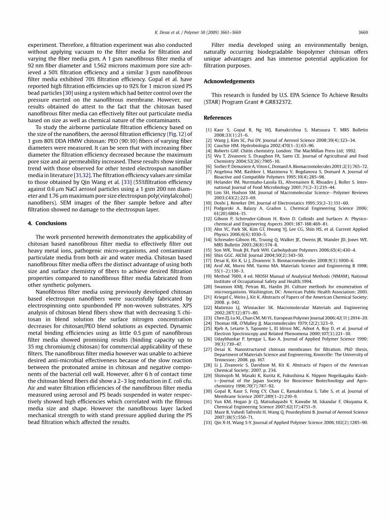
experiment. Therefore, a filtration experiment was also conductedwithout applying vacuum to the filter media for filtration andvarying the fiber media gsm. A 1 gsm nanofibrous filter media of92 nm fiber diameter and 1.562 microns maximum pore size ach-ieved a 50% filtration efficiency and a similar 3 gsm nanofibrousfilter media exhibited 70% filtration efficiency. Gopal et al. havereported high filtration efficiencies up to 92% for 1 micron sized PSbead particles [30] using a system which had better control over thepressure exerted on the nanofibrous membrane. However, ourresults obtained do attest to the fact that the chitosan basednanofibrous filter media can effectively filter out particulate mediabased on size as well as chemical nature of the contaminants.
To study the airborne particulate filtration efficiency based onthe size of the nanofibers, the aerosol filtration efficiency (Fig. 12) of1 gsm 80% DDA HMW chitosan: PEO (90:10) fibers of varying fiberdiameters were measured. It can be seen that with increasing fiberdiameter the filtration efficiency decreased because the maximumpore size and air permeability increased. These results show similartrend with those observed for other tested electrospun nanofibermedia in literature [31,32]. The filtration efficiency values are similarto those obtained by Qin Wang et al. [33] (55%filtration efficiencyagainst 0.6 mm NaCl aerosol particles using a 1 gsm 200 nm diam-eter and 1.76 mm maximum pore size electrospun poly(vinylalcohol)nanofibers). SEM images of the fiber sample before and afterfiltration showed no damage to the electrospun layer.
4. Conclusions
The work presented herewith demonstrates the applicability ofchitosan based nanofibrous filter media to effectively filter outheavy metal ions, pathogenic micro-organisms, and contaminantparticulate media from both air and water media. Chitosan basednanofibrous filter media offers the distinct advantage of using bothsize and surface chemistry of fibers to achieve desired filtrationproperties compared to nanofibrous filter media fabricated fromother synthetic polymers.
Nanofibrous filter media using previously developed chitosanbased electrospun nanofibers were successfully fabricated byelectrospinning onto spunbonded PP non-woven substrates. XPSanalysis of chitosan blend fibers show that with decreasing % chi-tosan in blend solution the surface nitrogen concentrationdecreases for chitosan/PEO blend solutions as expected. Dynamicmetal binding efficiencies using as little 0.5 gsm of nanofibrousfilter media showed promising results (binding capacity up to35 mg chromium/g chitosan) for commercial applicability of thesefilters. The nanofibrous filter media however was unable to achievedesired anti-microbial effectiveness because of the slow reactionbetween the protonated amine in chitosan and negative compo-nents of the bacterial cell wall. However, after 6 h of contact timethe chitosan blend fibers did show a 2–3 log reduction in E. coli cfu.Air and water filtration efficiencies of the nanofibrous filter mediameasured using aerosol and PS beads suspended in water respec-tively showed high efficiencies which correlated with the fibrousmedia size and shape. However the nanofibrous layer lackedmechanical strength to with stand pressure applied during the PSbead filtration which affected the results.
Filter media developed using an environmentally benign,naturally occurring biodegradable biopolymer chitosan offersunique advantages and has immense potential application forfiltration purposes.
Acknowledgements
This research is funded by U.S. EPA Science To Achieve Results(STAR) Program Grant # GR832372.
References
[1] Kaur S, Gopal R, Ng WJ, Ramakrishna S, Matsuura T. MRS Bulletin2008;33(1):21–6.
[2] Wang J, Kim SC, Pui DY. Journal of Aerosol Science 2008;39(4):323–34.[3] Cauchie HM. Hydrobiologia 2002;470(1–3):63–96.[4] Roberts GAF. Chitin chemistry. London: The MacMillan Press Ltd; 1992.[5] Wu T, Zivanovic S, Draughon FA, Sams CE. Journal of Agricultural and Food
Chemistry 2004;52(26):7905–10.[6] Sorlier P, Denuziere A, Viton C, Domard A. Biomacromolecules 2001;2(3):765–72.[7] Angelova NM, Rashkov I, Maximova V, Bogdanova S, Domard A. Journal of
Bioactive and Compatible Polymers 1995;10(4):285–98.[8] Helander IM, Nurmiaho-Lassila EL, Ahvenainen R, Rhoades J, Roller S. Inter-
national Journal of Food Microbiology 2001;71(2–3):235–44.[9] Lim SH, Hudson SM. Journal of Macromolecular SciencedPolymer Reviews
2003;C43(2):223–69.[10] Doshi J, Reneker DH. Journal of Electrostatics 1995;35(2–3):151–60.[11] Podgorski A, Balazy A, Gradon L. Chemical Engineering Science 2006;
61(20):6804–15.[12] Gibson P, Schreuder-Gibson H, Rivin D. Colloids and Surfaces A: Physico-
chemical and Engineering Aspects 2001;187-188:469–81.[13] Ahn YC, Park SK, Kim GT, Hwang YJ, Lee CG, Shin HS, et al. Current Applied
Physics 2006;6(6):1030–5.[14] Schreuder-Gibson HL, Truong Q, Walker JE, Owens JR, Wander JD, Jones WE.
MRS Bulletin 2003;28(8):574–8.[15] Son WK, Youk JH, Park WH. Carbohydrate Polymers 2006;65(4):430–4.[16] Shin GGC. AIChE Journal 2004;50(2):343–50.[17] Desai K, Kit K, Li J, Zivanovic S. Biomacromolecules 2008;9(3):1000–6.[18] Arof AK, Morni NM, Yarmo MA. Materials Science and Engineering B 1998;
55(1–2):130–3.[19] Method 7600, 4 ed. NIOSH Manual of Analytical Methods (NMAM), National
Institute of Occupational Safety and Health;1994.[20] Swanson KMJ, Petran RL, Hanlin JH. Culture methods for enumeration of
microorganisms. Washington, DC: American Public Health Association; 2001.[21] Kriegel C, Weiss J, Kit K. Abstracts of Papers of the American Chemical Society;
2008. p. 042.[22] Matienzo LJ, Winnacker SK. Macromolecular Materials and Engineering
2002;287(12):871–80.[23] Chen ZJ, Lu XL, Chan CM, Mi YL. European Polymer Journal 2006;42(11):2914–20.[24] Thomas HR, O’Malley JJ. Macromolecules 1979;12(2):323–9.[25] Rjeb A, Letarte S, Tajounte L, El Idrissi MC, Adnot A, Roy D, et al. Journal of
Electron Spectroscopy and Related Phenomena 2000;107(3):221–30.[26] Udaybhaskar P, Iyengar L, Rao A. Journal of Applied Polymer Science 1990;
39(3):739–47.[27] Desai K. Nanostructured chitosan membranes for filtration. PhD thesis,
Department of Materials Science and Engineering, Knoxville: The University ofTennessee; 2008. pp. 167.
[28] Li J, Zivanovic S, Davidson M, Kit K. Abstracts of Papers of the AmericanChemical Society; 2007. p. 234.
[29] Shimojoh M, Masaki K, Kurita K, Fukushima K. Nippon Nogeikagaku Kaish-idJournal of the Japan Society for Bioscience Biotechnology and Agro-chemistry 1996;70(7):787–92.
[30] Gopal R, Kaur S, Feng CY, Chan C, Ramakrishna S, Tabe S, et al. Journal ofMembrane Science 2007;289(1–2):210–9.
[31] Yun KM, Hogan Jr CJ, Matsubayashi Y, Kawabe M, Iskandar F, Okuyama K.Chemical Engineering Science 2007;62(17):4751–9.
[32] Maze B, Vahedi Tafreshi H, Wang Q, Pourdeyhimi B. Journal of Aerosol Science2007;38(5):550–71.
[33] Qin X-H, Wang S-Y. Journal of Applied Polymer Science 2006;102(2):1285–90.


![MORPHOLOGICAL, MECHANICAL AND BIOLOGICAL ...applications [3-5]. In this regards, chitosan nanofibrous webs are used for biomedical and tissue engineering application [1, 6, 7]. But](https://img.pdfslide.us/doc/110x75/60e25a39b57276029e607d1a/morphological-mechanical-and-biological-applications-3-5-in-this-regards.jpg)