Embed Size (px)
Citation preview
RSC Advances
REVIEW
Ope
n A
cces
s A
rtic
le. P
ublis
hed
on 2
4 Ju
ly 2
020.
Dow
nloa
ded
on 2
/23/
2022
5:5
4:22
PM
. T
his
artic
le is
lice
nsed
und
er a
Cre
ativ
e C
omm
ons
Attr
ibut
ion-
Non
Com
mer
cial
3.0
Unp
orte
d L
icen
ce.
View Article OnlineView Journal | View Issue
Nanocarriers for
aIndustrial Research Laboratory, Departm
Technology and Science (BITS), Pilani, Pil
E-mail: [email protected] Development, Slayback Phar
500072, IndiacBirla Institute of Technology & Science (BIT
† Authors contributed equally.
Cite this: RSC Adv., 2020, 10, 27835
Received 5th June 2020Accepted 7th July 2020
DOI: 10.1039/d0ra04971a
rsc.li/rsc-advances
This journal is © The Royal Society o
ocular drug delivery: currentstatus and translational opportunity
Srividya Gorantla,†a Vamshi Krishna Rapalli,a Tejashree Waghule, a
Prem Prakash Singh,b Sunil Kumar Dubey, a Ranendra N. Sahaac
and Gautam Singhvi †*a
Ocular diseases have a significant effect on vision and quality of life. Drug delivery to ocular tissues is
a challenge to formulation scientists. The major barriers to delivering drugs to the anterior and posterior
segments include physiological barriers (nasolacrimal drainage, blinking), anatomical barriers (static and
dynamic), efflux pumps and metabolic barriers. The static barriers comprise the different layers of the
cornea, sclera, and blood–aqueous barriers whereas dynamic barriers involve conjunctival blood flow,
lymphatic clearance and tear drainage. The tight junctions of the blood–retinal barrier (BRB) restrict
systemically administered drugs from entering the retina. Nanocarriers have been found to be effective
at overcoming the issues associated with conventional ophthalmic dosage forms. Various nanocarriers,
including nanodispersion systems, nanomicelles, lipidic nanocarriers, polymeric nanoparticles, liposomes,
niosomes, and dendrimers, have been investigated for improved permeation and effective targeted drug
delivery to various ophthalmic sites. In this review, various nanomedicines and their application for
ophthalmic delivery of therapeutics are discussed. Additionally, scale-up and clinical status are also
addressed to understand the current scenario for ophthalmic drug delivery.
1. Introduction
As per a World Health Organization (WHO) report, every veseconds someone in the world goes blind and every minute a childloses their sight.1 The International Classication of Diseases (IDC-11) (2018) states that approximately 1.3 billion people live withsome form of vision impairment globally.2 These ocular diseasesaffect the vision and quality of life of patients. Considerableachievements have been made in the supervision of oculardiseases. In the last decade, extensive research has been done atthe preclinical and clinical level for the development of thera-peutics for various ocular diseases, including glaucoma, uveitis,age-related macular degeneration (AMD), cataracts, and diabeticretinopathy. Recent advances in the treatment of ophthalmicdiseases at the clinical level include anti-vascular endothelialgrowth factor drugs, gene therapy, laser surgery on the eye, andocular sealants. To deliver these therapeutics, various drug deliverysystems, such as eye drops (solutions, suspensions, emulsions), insitu gels, ocular inserts, contact lenses, punctum plugs, intraocularinjections, and implants, have been explored for effective ocular
ent of Pharmacy, Birla Institute of
ani Campus, Rajasthan, India, 333031.
c.in
ma India LLP, Hyderabad, Telangana,
S), Pilani, Dubai Campus, UAE
f Chemistry 2020
drug delivery.3–5 The unique structural features of the eye and thephysiological ocular barriers are major challenges for effectivedelivery at the disease site.6–10 Recent advances in bioadhesive insitu gelling systems and nanotechnology-based drug deliverysystems are gaining substantial attention for overcoming the drugdelivery challenges. Nanocarrier-based therapeutic deliverysystems have been developed to promote sustained and targeteddrug delivery to both the anterior and posterior segments of theeye.11,12 However, translation of nanotechnology-based drugdelivery systems from bench to bedside are associated with scaleup and quality control challenges.13
In this review, we focus on the anatomical and physiologicalbarriers to ocular drug delivery. Further, we discuss the limitationsof conventional formulations and other routes of drug delivery.Overcoming the limitations of current therapies, advanced nano-carriers have been shown to be effective in treating ocular diseases.Various nanomedicines and their ndings are compiled tounderstand the impact of nanocarriers in the treatment ofophthalmic diseases. Moreover, this review addresses the currentchallenges in the translation of nanomedicine, including the large-scale production and quality control aspects of nanomedicine.
2. Anatomical and physiologicalbarriers to ocular drug delivery
The eye can be broadly divided into the anterior and posteriorsegments. The anterior segment includes the cornea,
RSC Adv., 2020, 10, 27835–27855 | 27835
RSC Advances Review
Ope
n A
cces
s A
rtic
le. P
ublis
hed
on 2
4 Ju
ly 2
020.
Dow
nloa
ded
on 2
/23/
2022
5:5
4:22
PM
. T
his
artic
le is
lice
nsed
und
er a
Cre
ativ
e C
omm
ons
Attr
ibut
ion-
Non
Com
mer
cial
3.0
Unp
orte
d L
icen
ce.
View Article Online
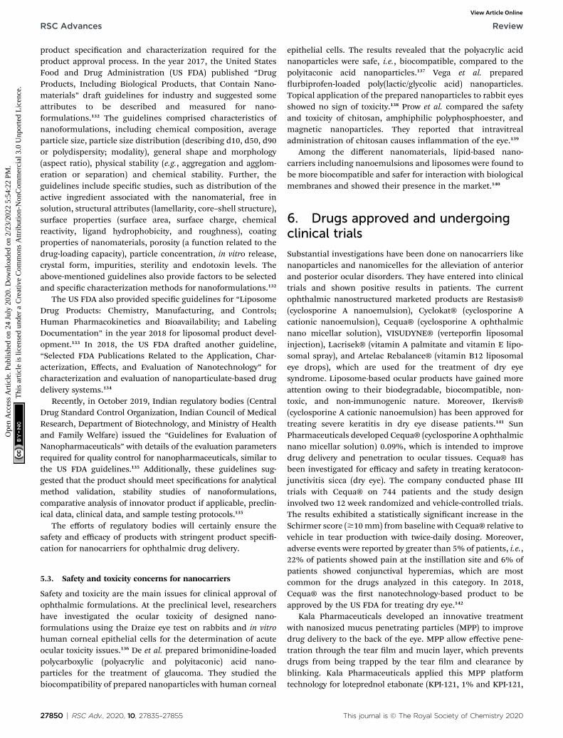
conjunctiva, iris, ciliary body, lens, and aqueous humour, whilethe posterior segment includes the sclera, choroid, retina, andvitreous body. The anterior and posterior segments of the eyeare affected by several vision-threatening diseases.14 To treat eyediseases, topical administration is the preferred non-invasivetechnique. However, 90% of currently available conventionalophthalmic formulations are eye drops, which are principallyadministered into the conjunctival cul-de-sac and exhibit poorocular bioavailability15 because various anatomical and physi-ological constraints impede drug delivery to both the anteriorand posterior regions of the eye.16 These include physiologicalbarriers (nasolacrimal drainage, lacrimation rate, blinking),anatomical barriers (static and dynamic), efflux pumps, andmetabolism in ocular tissues.14 Fig. 1 shows the anatomy of theeye and the physiological barriers to ocular drug delivery.17
The tear lm also acts as a barrier and prevents drugabsorption on topical application. The tear lm is composed ofan outer lipid layer, a middle aqueous layer, and an innermucus layer. The tear lm can act as a barrier for administereddrugs owing to the high tear turnover rate of lacrimal uid anda gel-like mucus layer. Under physiological conditions, the tearow is �1.2 mL min�1, renewing the tear lm for every 5minutes, but when the eye is irritated by reex stimulation,lachrymation increases to �300 mL min�1. The drug is thusdiluted and easily washed away by the tear lm. Moreover,mucin present in tear lm forms a hydrophilic layer on the
Fig. 1 Schematic representation of the anatomy of the eye and physioldiffusional barriers whereas green colour indicates routes of eliminatadministration (1). The conjunctival and scleral route allows some hydrosystemic administration, small compounds diffuse from the iris blood vsegment are removed via aqueous humor outflow (4) or diffuse across thand the retinal capillary endothelium act as major barriers to systemicallyof these, for effective drug delivery intravitreal injections are used (7). Dru(8) or by diffusion into the anterior chamber (9). Reproduced with perm
27836 | RSC Adv., 2020, 10, 27835–27855
glycocalyx of the ocular surface and protects the eye from celldebris and foreign substances, acting as another barrier toadministered drugs.18
The anterior segment's static barriers (corneal epithelium,stroma, and blood–aqueous barrier) and dynamic barriers (suchas conjunctival blood, lymph ow, and lachrymation) limit drugentry into the anterior chamber of the eye.19 The human corneacomprises ve layers, i.e. epithelium, Bowman's membrane,stroma, Descemet's membrane, and endothelium, which eachhave varying polarity. The cornea epithelium comprises 5–6layers of close-packed cells with tight junctions that prevent theentry of microbes and drugs. The posterior and anteriorchambers of the eye are lled with clear transparent uid(aqueous humour). The aqueous humour is produced by theepithelium of the ciliary body and provides nutrition to thecornea. The aqueous humour ows from the posterior chamberacross the pupil into the anterior chamber.20
However, drug delivery to the posterior segment is limited bythe static barriers (sclera, choroid, Bruch's membrane, andblood–retinal barrier) and the dynamic barriers (choroidal bloodand lymph ow).19 The sclera is the outermost layer of the eyewith irregularly arranged collagen bers, which prevent the entryof foreign substances to the posterior ocular tissues. Therefore,drugs with high lipophilicity and a highmolecular radius can notpermeate through the aqueous scleral pores. Additionally, thethickness of the sclera varies from 1 mm at the posterior pole to
ogical barriers to ocular drug delivery (red colour indicates the ocularion). The cornea is the main route for drug penetration on topicalphilic drugs, which further diffuse into the ciliary body (2). Followingessels into the anterior segment (3). Further, the drugs in the anteriore iris surface via venous blood flow (5). The retinal pigment epitheliumadministered drugs reaching the retina and vitreous humour (6). Insteadgs are removed from the vitreous humour via the blood–retinal barrierission from ref. 17. Copyright 2005, Elsevier.
This journal is © The Royal Society of Chemistry 2020
Fig. 2 Schematic representation of the routes of administration for ocular drug delivery.
Review RSC Advances
Ope
n A
cces
s A
rtic
le. P
ublis
hed
on 2
4 Ju
ly 2
020.
Dow
nloa
ded
on 2
/23/
2022
5:5
4:22
PM
. T
his
artic
le is
lice
nsed
und
er a
Cre
ativ
e C
omm
ons
Attr
ibut
ion-
Non
Com
mer
cial
3.0
Unp
orte
d L
icen
ce.
View Article Online
25 to 250 nm in the equatorial region in the posterior region,which exhibits low drug permeability. The choroid eliminatesadministered drugs before they reach Bruch's membrane. Theaccumulation of cell debris at Bruch's membrane prevents theexchange of nutrients and drugs. The posterior segment consistsof tight junctions of the blood–retinal barrier (BRB) with theinner retinal vascular endothelium and outer retinal pigmentepithelium. These restrict the penetration of administered drugsinto the intraocular chambers.14,16
The conjunctiva is composed of multilayered epithelium andstroma; there are fewer intercellular spaces in the conjunctivaare than in the corneal epithelium. Thus, the cornea andconjunctiva act as the rate-limiting step for hydrophilic drugs.The conjunctival stroma consists of blood capillaries andlymphatics, which leads to drug loss into the systemic circula-tion. Moreover, efflux proteins prevent the entry of adminis-tered antiviral and anti-glaucoma drugs. In contrast, metabolic
Table 1 Routes of administration, benefits, and challenges for ocular de
Route Benets
Topical eye drop High patient compliance, self-administrable and non-invasive
Oral/systemic Patient compliance
Intravitreal Direct delivery to posterior region(vitreous and retina), sustains druglevels, evades BRB
Intracameral Provides higher drug levels in theanterior chamber, eliminates theuse of topical drops, reducescorneal and systemic side effectsseen with topical steroid therapy
Subconjunctival Delivery to the anterior andposterior segment, a site for depotformulations
This journal is © The Royal Society of Chemistry 2020
enzymes prevent the entry of xenobiotics.18 The extent to whichthe above-mentioned barriers inuence drug bioavailability isdependent on the route of administration.
3. Benefits and limitations of oculardrug administration routes
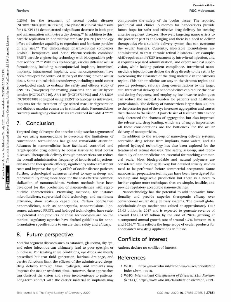
There are multiple routes of drug delivery to the eye: systemicadministration (oral, parenteral), topical administration, andocular injections (subconjunctival, periocular, and intra-vitreal).21 Fig. 2 shows the routes of administration for oculardrug delivery.21
3.1. Systemic administration
Parenteral and oral dosing are considered under systemicadministration methods for ocular drug delivery. The eye has
livery systems
Challenges References
Higher tear dilution and turnoverrate, cornea acts as a barrier, effluxpumps, bioavailability (BA) < 5%
23
Blood–aqueous barrier (BAB), BRB,high dosing causes toxicity, BA < 2%
23
Retinal detachment, haemorrhage,cataracts, endophthalmitis,intraocular damage, patientcompliance
23
TASS (toxic anterior segmentsyndrome), TECCDS (toxicendothelial cell destructionsyndrome)
35
Conjunctival and choroidalcirculation, trans scleral diffusionof the drug
36
RSC Adv., 2020, 10, 27835–27855 | 27837
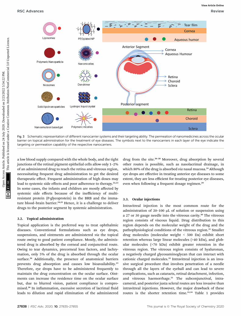
Fig. 3 Schematic representation of different nanocarrier systems and their targeting ability. The permeation of nanomedicines across the ocularbarrier on topical administration for the treatment of eye diseases. The symbols next to the nanocarriers in each layer of the eye indicate thetargeting or permeation capability of the respective nanocarriers.
RSC Advances Review
Ope
n A
cces
s A
rtic
le. P
ublis
hed
on 2
4 Ju
ly 2
020.
Dow
nloa
ded
on 2
/23/
2022
5:5
4:22
PM
. T
his
artic
le is
lice
nsed
und
er a
Cre
ativ
e C
omm
ons
Attr
ibut
ion-
Non
Com
mer
cial
3.0
Unp
orte
d L
icen
ce.
View Article Online
a low blood supply compared with the whole body, and the tightjunctions of the retinal pigment epithelial cells allow only 1–2%of an administered drug to reach the retina and vitreous region,necessitating frequent drug administration to get the desiredtherapeutic effect. Frequent administration of high doses maylead to systemic side effects and poor adherence to therapy.22,23
In some cases, the infants and children are mostly affected bysystemic side effects because of the inefficiency of multi-resistant protein (P-glycoprotein) in the BRB and the imma-ture blood–brain barrier.24,25 Hence, it is a challenge to deliverdrugs to the posterior segment by systemic administration.
3.2. Topical administration
Topical application is the preferred way to treat ophthalmicdiseases. Conventional formulations such as eye drops,suspensions, and ointments are administered via the topicalroute owing to good patient compliance. Mostly, the adminis-tered drug is absorbed by the corneal and conjunctival route.Owing to tear dynamics, precorneal loss factors, and lachry-mation, only 5% of the drug is absorbed through the ocularsurface.26 Additionally, the presence of anatomical barriersprevents drug absorption and causes low bioavailability.15
Therefore, eye drops have to be administered frequently tomaintain the drug concentration on the ocular surface. Oint-ments can increase the residence time on the ocular surfacebut, due to blurred vision, patient compliance is compro-mised.26 In inammation, excessive secretion of lacrimal uidleads to dilution and rapid elimination of the administered
27838 | RSC Adv., 2020, 10, 27835–27855
drug from the site.26–28 Moreover, drug absorption by severalother routes is possible, such as nasolacrimal drainage, inwhich 80% of the drug is absorbed via nasal mucosa.24 Althougheye drops are effective in treating anterior eye diseases to someextent, they are less efficient for treating posterior eye diseases,even when following a frequent dosage regimen.29
3.3. Ocular injections
Intravitreal injection is the most common route for theadministration of 20–100 mL of solution or suspension usinga 27 or 30 gauge needle into the vitreous cavity.30 The vitreousregion consists of viscous liquid. Drug distribution to thisregion depends on the molecular weight of the drug and thepathophysiological conditions of the vitreous region.31 Smallerdrug molecules (molecular weight < 500 Da) exhibit shortretention whereas large linear molecules (>40 kDa), and glob-ular molecules (>70 kDa) exhibit greater retention in thevitreous region. The vitreous region consists of hyaluronan,a negatively charged glycosaminoglycan that can interact withcationic charged molecules.32 Intravitreal injection is an inva-sive surgical procedure that involves penetration of a needlethrough all the layers of the eyeball and can lead to severecomplications, such as cataracts, retinal detachment, infection,and vitreous haemorrhage.33 The subconjunctival, intra-cameral, and posterior juxta scleral routes are less invasive thanintravitreal injections. However, the major drawback of theseroutes is the shorter retention time.33,34 Table 1 provides
This journal is © The Royal Society of Chemistry 2020
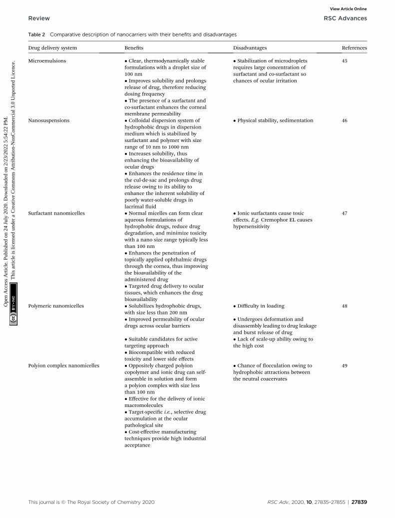
Table 2 Comparative description of nanocarriers with their benefits and disadvantages
Drug delivery system Benets Disadvantages References
Microemulsions � Clear, thermodynamically stableformulations with a droplet size of100 nm
� Stabilization of microdropletsrequires large concentration ofsurfactant and co-surfactant sochances of ocular irritation
45
� Improves solubility and prolongsrelease of drug, therefore reducingdosing frequency� The presence of a surfactant andco-surfactant enhances the cornealmembrane permeability
Nanosuspensions � Colloidal dispersion system ofhydrophobic drugs in dispersionmedium which is stabilized bysurfactant and polymer with sizerange of 10 nm to 1000 nm
� Physical stability, sedimentation 46
� Increases solubility, thusenhancing the bioavailability ofocular drugs� Enhances the residence time inthe cul-de-sac and prolongs drugrelease owing to its ability toenhance the inherent solubility ofpoorly water-soluble drugs inlacrimal uid
Surfactant nanomicelles � Normal micelles can form clearaqueous formulations ofhydrophobic drugs, reduce drugdegradation, and minimize toxicitywith a nano size range typically lessthan 100 nm
� Ionic surfactants cause toxiceffects. E.g. Cremophor EL causeshypersensitivity
47
� Enhances the penetration oftopically applied ophthalmic drugsthrough the cornea, thus improvingthe bioavailability of theadministered drug� Targeted drug delivery to oculartissues, which enhances the drugbioavailability
Polymeric nanomicelles � Solubilizes hydrophobic drugs,with size less than 200 nm
� Difficulty in loading 48
� Improved permeability of oculardrugs across ocular barriers
� Undergoes deformation anddisassembly leading to drug leakageand burst release of drug
� Suitable candidates for activetargeting approach
� Lack of scale-up ability owing tothe high cost
� Biocompatible with reducedtoxicity and lower side effects
Polyion complex nanomicelles � Oppositely charged polyioncopolymer and ionic drug can self-assemble in solution and forma polyion complex with size lessthan 100 nm
� Chance of occulation owing tohydrophobic attractions betweenthe neutral coacervates
49
� Effective for the delivery of ionicmacromolecules� Target-specic i.e., selective drugaccumulation at the ocularpathological site� Cost-effective manufacturingtechniques provide high industrialacceptance
This journal is © The Royal Society of Chemistry 2020 RSC Adv., 2020, 10, 27835–27855 | 27839
Review RSC Advances
Ope
n A
cces
s A
rtic
le. P
ublis
hed
on 2
4 Ju
ly 2
020.
Dow
nloa
ded
on 2
/23/
2022
5:5
4:22
PM
. T
his
artic
le is
lice
nsed
und
er a
Cre
ativ
e C
omm
ons
Attr
ibut
ion-
Non
Com
mer
cial
3.0
Unp
orte
d L
icen
ce.
View Article Online
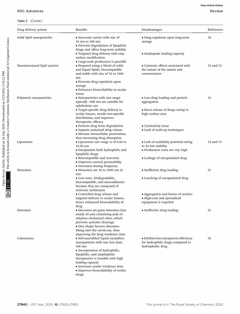
Table 2 (Contd. )
Drug delivery system Benets Disadvantages References
Solid lipid nanoparticles � Non-toxic carrier with size of10 nm to 500 nm
� Drug expulsion upon long-termstorage
50
� Prevents degradation of lipophilicdrugs and offers long-term stability� Targeted drug delivery with easysurface modication
� Inadequate loading capacity
� Large-scale production is possibleNanostructured lipid carriers � Prepared using a blend of solid
and liquid lipids, biocompatibleand stable with size of 50 to 1000nm
� Cytotoxic effects associated withthe nature of the matrix andconcentration
51 and 52
� Prevents drug expulsion uponstorage� Enhances bioavailability to oculartissue
Polymeric nanoparticles � Nanoparticles with size rangetypically <400 nm are suitable forophthalmic use
� Low drug loading and particleaggregation
53
� Target-specic drug delivery toocular tissues, avoids non-specicdistribution, and improvestherapeutic efficacy
� Burst release of drugs owing tohigh surface area
� Protects drug from degradation � Cytotoxicity issue� Imparts sustained drug release � Lack of scale-up techniques� Elevates intracellular penetration,thus increasing drug absorption
Liposomes � Liposomes size range is of 0.08 to10.00 mm
� Lack of scalability potential owingto its low stability
54 and 55
� Encapsulate both hydrophilic andlipophilic drugs
� Production costs are very high
� Biocompatible and non-toxic � Leakage of encapsulated drug� Improves corneal permeability� Decreases dosing frequency
Niosomes � Niosomes are 10 to 1000 nm insize
� Inefficient drug loading 21
� Less toxic, biodegradable,biocompatible, and mucoadhesivebecause they are composed ofnonionic surfactants
� Leaching of encapsulated drug
� Controlled drug release andtargeted delivery to ocular tissues,hence enhanced bioavailability ofdrug
� Aggregation and fusion of vesicles� High-cost and specializedequipment is required
Discomes � Discomes are giant niosomes (sizenearly 20 mm) containing poly-24ethylene cholesteryl ether, whichprevents systemic drainage
� Ineffective drug loading 21
� Disc shape favours discomestting into the cul-de-sac, thusimproving the drug residence time
Cubosomes � Self-assembled liquid crystallinenanoparticles with size less than500 nm
� Exhibits low entrapment efficiencyfor hydrophilic drugs compared tohydrophobic drug
56
� Incorporation of hydrophilic,lipophilic, and amphiphilictherapeutics is feasible with highloading capacity� Increases ocular residence time� Improves bioavailability of oculardrugs
27840 | RSC Adv., 2020, 10, 27835–27855 This journal is © The Royal Society of Chemistry 2020
RSC Advances Review
Ope
n A
cces
s A
rtic
le. P
ublis
hed
on 2
4 Ju
ly 2
020.
Dow
nloa
ded
on 2
/23/
2022
5:5
4:22
PM
. T
his
artic
le is
lice
nsed
und
er a
Cre
ativ
e C
omm
ons
Attr
ibut
ion-
Non
Com
mer
cial
3.0
Unp
orte
d L
icen
ce.
View Article Online
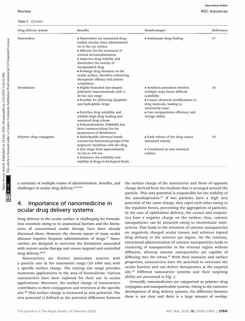
Table 2 (Contd. )
Drug delivery system Benets Disadvantages References
Nanowafers � Nanowafers are nanosized drug-loaded circular discs administeredon to the eye surface
� Inadequate drug loading 57
� Effective for the treatment ofcorneal neovascularization� Improves drug stability anddiminishes the toxicity ofencapsulated drug� Prolongs drug duration on theocular surface, therefore enhancingtherapeutic efficacy and patientcompliance
Dendrimers � Highly branched star-shapedpolymeric macromolecule with 5–20 nm size range
� Synthesis procedure involvesmultiple steps hence difficultscalability
58
� Feasible for delivering lipophilicand hydrophilic drugs
� Causes chemical modications todrug molecule, leading tocytotoxicity issue
� Enriches drug solubility andexhibits high drug loading andsustained drug release
� Low encapsulation efficiency andstorage ability
� Polyamidoamine (PAMAM) hasbeen commercialized for thepreparation of dendrimers
Polymer–drug conjugates � Hydrolysable chemical bondsconnect the functional groups of thepolymeric backbone with the drug
� Early release of the drug causesunwanted toxicity
59
� Size range from approximately10 nm to 100 nm
� Considered as new chemicalentities
� Enhances the solubility andstability of drugs in biological uids
Review RSC Advances
Ope
n A
cces
s A
rtic
le. P
ublis
hed
on 2
4 Ju
ly 2
020.
Dow
nloa
ded
on 2
/23/
2022
5:5
4:22
PM
. T
his
artic
le is
lice
nsed
und
er a
Cre
ativ
e C
omm
ons
Attr
ibut
ion-
Non
Com
mer
cial
3.0
Unp
orte
d L
icen
ce.
View Article Online
a summary of multiple routes of administration, benets, andchallenges in ocular drug delivery.23,35,36
4. Importance of nanomedicine inocular drug delivery systems
Drug delivery to the ocular surface is challenging for formula-tion scientists owing to its anatomical barriers and the limita-tions of conventional ocular therapy have been alreadydiscussed above. However, the chronic nature of many oculardiseases requires frequent administration of drugs.37 Nano-carriers are designed to overcome the limitations associatedwith current ocular therapy and ensure targeted and controlleddrug delivery.38,39
Nanocarriers are distinct particulate systems witha particle size in the nanometer range (10–1000 nm) witha specic surface charge. The varying size range providesnumerous applications in the area of biomedicine. Variousnanocarriers have been explored for their use in ocularapplications. Moreover, the surface charge of nanocarrierscontributes to their conjugation and retention at the specicsite.40 This surface charge is measured as zeta potential. Thezeta potential is dened as the potential difference between
This journal is © The Royal Society of Chemistry 2020
the surface charge of the nanocarrier and those of oppositecharge derived from the medium that is arranged around theparticle. This zeta potential is responsible for the stability ofthe nanodispersion.41 If two particles have a high zetapotential of the same charge, they repel each other owing tothe repulsive forces, preventing the aggregation of particles.In the case of ophthalmic delivery, the cornea and conjunc-tiva have a negative charge on the surface; thus, cationicnanoparticles can be attracted owing to electrostatic inter-actions. This leads to the retention of cationic nanoparticleson negatively charged ocular tissues and achieves topicaldrug delivery to the anterior eye region. On the contrary,intravitreal administration of cationic nanoparticles leads toclustering of nanoparticles in the vitreous region withoutdiffusion, whereas anionic nanoparticles are capable ofdiffusing into the retina.41 With their nanosize and surfaceproperties, nanocarriers have the potential to overcome theocular barriers and can deliver therapeutics at the targetedsite.41 Different nanocarrier systems and their targetingability are presented in Fig. 3.
Generally, nanomedicines are categorized as polymer–drugconjugates and nanoparticulate systems. Owing to the extensivedevelopment of drug delivery systems, the difference betweenthese is not clear and there is a large amount of overlap.
RSC Adv., 2020, 10, 27835–27855 | 27841
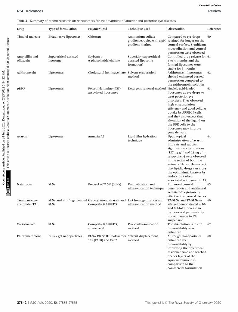
Table 3 Summary of recent research on nanocarriers for the treatment of anterior and posterior eye diseases
Drug Type of formulation Polymer/lipid Technique used Observation Reference
Timolol maleate Bioadhesive liposomes Chitosan Ammonium sulfategradient coupled with a pH-gradient method
Compared to eye drops,retained for longer on thecorneal surface. Signicantmucoadhesion and cornealpermeation were observed
60
Ampicillin andooxacin
Supercritical-assistedliposome
Soybean L-a phosphatidylcholine
SuperLip (supercritical-assisted liposomeformation)
Controlled drug release for3 to 4 months and theformed liposomes werestable for 3 months
61
Azithromycin Liposomes Cholesterol hemisuccinate Solvent evaporationmethod
Azithromycin liposomesshowed enhanced cornealpermeation compared tothe azithromycin solution
62
pDNA Liposomes Polyethylenimine (PEI)-associated liposomes
Detergent removal method Nucleic acid-loadedliposomes as eye drops totreat posterior eyedisorders. They observedhigh encapsulationefficiency and good cellularuptake by ARPE-19 cells,and they also expect thatalteration of the ligand onthe RPE cells to theliposomes may improvegene delivery
63
Avastin Liposomes Annexin A5 Lipid lm hydrationtechnique
Upon topicaladministration of avastininto rats and rabbits,signicant concentrations(127 ng g�1 and 18 ng g�1,respectively) were observedin the retina of both theanimals. Hence, they expectthat lipidic drugs can crossthe ophthalmic barriers byendocytosis whenassociated with annexin A5
64
Natamycin SLNs Precirol ATO 5® (SLNs) Emulsication andultrasonication technique
Enhanced cornealpenetration and antifungalactivity. No cytotoxicityeffect on the corneal tissues
65
Triamcinoloneacetonide (TA)
SLNs and in situ gel loadedSLNs
Glyceryl monostearate andCompritol® 888ATO
Hot homogenization andultrasonication method
TA-SLNs and TA-SLNs-insitu gel demonstrated a 10-and 9.3-fold increase intranscorneal permeabilityin comparison to TAsuspension
66
Voriconazole SLNs Compritol® 888ATO,stearic acid
Probe ultrasonicationmethod
The dissolution rate andbioavailability wereenhanced
67
Fluorometholone In situ gel nanoparticles PLGA RG 503H, Poloxamer188 (P188) and P407
Solvent displacementmethod
In situ gel nanoparticlesenhanced thebioavailability byimproving the precornealresidence time and reacheddeeper layers of theaqueous humour incomparison to thecommercial formulation
68
27842 | RSC Adv., 2020, 10, 27835–27855 This journal is © The Royal Society of Chemistry 2020
RSC Advances Review
Ope
n A
cces
s A
rtic
le. P
ublis
hed
on 2
4 Ju
ly 2
020.
Dow
nloa
ded
on 2
/23/
2022
5:5
4:22
PM
. T
his
artic
le is
lice
nsed
und
er a
Cre
ativ
e C
omm
ons
Attr
ibut
ion-
Non
Com
mer
cial
3.0
Unp
orte
d L
icen
ce.
View Article Online
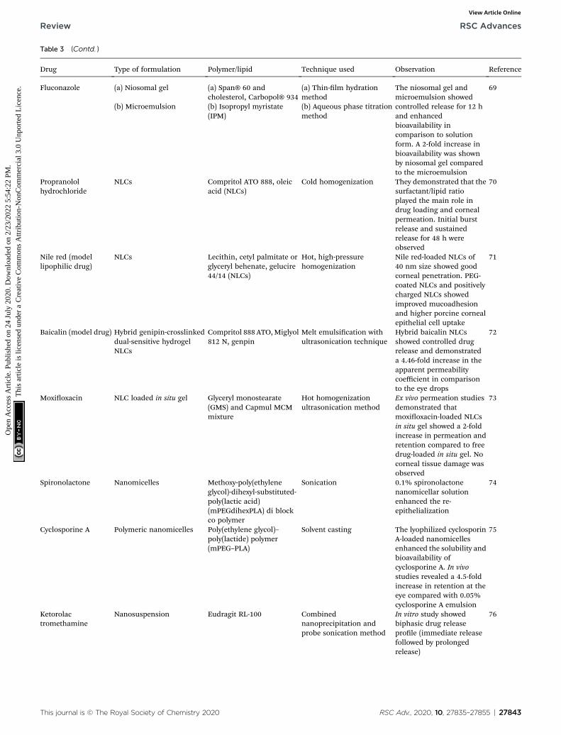
Table 3 (Contd. )
Drug Type of formulation Polymer/lipid Technique used Observation Reference
Fluconazole (a) Niosomal gel (a) Span® 60 andcholesterol, Carbopol® 934
(a) Thin-lm hydrationmethod
The niosomal gel andmicroemulsion showedcontrolled release for 12 hand enhancedbioavailability incomparison to solutionform. A 2-fold increase inbioavailability was shownby niosomal gel comparedto the microemulsion
69
(b) Microemulsion (b) Isopropyl myristate(IPM)
(b) Aqueous phase titrationmethod
Propranololhydrochloride
NLCs Compritol ATO 888, oleicacid (NLCs)
Cold homogenization They demonstrated that thesurfactant/lipid ratioplayed the main role indrug loading and cornealpermeation. Initial burstrelease and sustainedrelease for 48 h wereobserved
70
Nile red (modellipophilic drug)
NLCs Lecithin, cetyl palmitate orglyceryl behenate, gelucire44/14 (NLCs)
Hot, high-pressurehomogenization
Nile red-loaded NLCs of40 nm size showed goodcorneal penetration. PEG-coated NLCs and positivelycharged NLCs showedimproved mucoadhesionand higher porcine cornealepithelial cell uptake
71
Baicalin (model drug) Hybrid genipin-crosslinkeddual-sensitive hydrogelNLCs
Compritol 888 ATO,Miglyol812 N, genpin
Melt emulsication withultrasonication technique
Hybrid baicalin NLCsshowed controlled drugrelease and demonstrateda 4.46-fold increase in theapparent permeabilitycoefficient in comparisonto the eye drops
72
Moxioxacin NLC loaded in situ gel Glyceryl monostearate(GMS) and Capmul MCMmixture
Hot homogenizationultrasonication method
Ex vivo permeation studiesdemonstrated thatmoxioxacin-loaded NLCsin situ gel showed a 2-foldincrease in permeation andretention compared to freedrug-loaded in situ gel. Nocorneal tissue damage wasobserved
73
Spironolactone Nanomicelles Methoxy-poly(ethyleneglycol)-dihexyl-substituted-poly(lactic acid)(mPEGdihexPLA) di blockco polymer
Sonication 0.1% spironolactonenanomicellar solutionenhanced the re-epithelialization
74
Cyclosporine A Polymeric nanomicelles Poly(ethylene glycol)–poly(lactide) polymer(mPEG–PLA)
Solvent casting The lyophilized cyclosporinA-loaded nanomicellesenhanced the solubility andbioavailability ofcyclosporine A. In vivostudies revealed a 4.5-foldincrease in retention at theeye compared with 0.05%cyclosporine A emulsion
75
Ketorolactromethamine
Nanosuspension Eudragit RL-100 Combinednanoprecipitation andprobe sonication method
In vitro study showedbiphasic drug releaseprole (immediate releasefollowed by prolongedrelease)
76
This journal is © The Royal Society of Chemistry 2020 RSC Adv., 2020, 10, 27835–27855 | 27843
Review RSC Advances
Ope
n A
cces
s A
rtic
le. P
ublis
hed
on 2
4 Ju
ly 2
020.
Dow
nloa
ded
on 2
/23/
2022
5:5
4:22
PM
. T
his
artic
le is
lice
nsed
und
er a
Cre
ativ
e C
omm
ons
Attr
ibut
ion-
Non
Com
mer
cial
3.0
Unp
orte
d L
icen
ce.
View Article Online
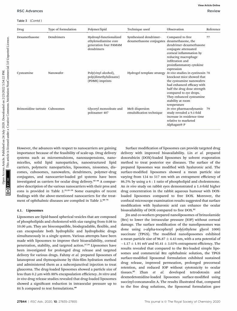
Table 3 (Contd. )
Drug Type of formulation Polymer/lipid Technique used Observation Reference
Dexamethasone Dendrimers Hydroxyl-functionalizedethylenediamine coregeneration four PAMAMdendrimers
Synthesized dendrimer–dexamethasone conjugates
Compared to freedexamethasone, thedendrimer–dexamethasoneconjugate attenuatedcorneal inammation byreducing macrophageinltration andproinammatory cytokineexpression
77
Cysteamine Nanowafer Poly(vinyl alcohol),poly(dimethylsiloxane)(PDMS) imprints
Hydrogel template strategy In vivo studies in cystinosinknockout mice showed thatthe cysteamine nanowafershad enhanced efficacy withhalf the drug dose strengthcompared to eye drops.They enhanced cysteaminestability at roomtemperature
78
Brimonidine tartrate Cubosomes Glyceryl monooleate andpoloxamer 407
Melt dispersionemulsication technique
In vivo pharmacodynamicstudy revealed a 9.1-foldincrease in residence timerelative to marketedAlphagan® P
79
RSC Advances Review
Ope
n A
cces
s A
rtic
le. P
ublis
hed
on 2
4 Ju
ly 2
020.
Dow
nloa
ded
on 2
/23/
2022
5:5
4:22
PM
. T
his
artic
le is
lice
nsed
und
er a
Cre
ativ
e C
omm
ons
Attr
ibut
ion-
Non
Com
mer
cial
3.0
Unp
orte
d L
icen
ce.
View Article Online
However, the advances with respect to nanocarriers are gainingimportance because of the feasibility of scale-up. Drug deliverysystems such as microemulsions, nanosuspensions, nano-micelles, solid lipid nanoparticles, nanostructured lipidcarriers, polymeric nanoparticles, liposomes, niosomes, dis-comes, cubosomes, nanowafers, dendrimers, polymer–drugconjugates, and nanocarrier-loaded gel systems have beeninvestigated as carriers for ocular drug delivery.42–44 A compar-ative description of the various nanocarriers with their pros andcons is provided in Table 2.21,45–59 Some examples of recentndings with the above-mentioned nanocarriers for the treat-ment of ophthalmic diseases are compiled in Table 3.60–79
4.1. Liposomes
Liposomes are lipid-based spherical vesicles that are composedof phospholipids and cholesterol with size ranging from 0.08 to10.00 mm. They are biocompatible, biodegradable, exible, andcan encapsulate both hydrophilic and hydrophobic drugssimultaneously in a single system. Various attempts have beenmade with liposomes to improve their bioavailability, cornealpenetration, stability, and targeted action.27,54 Liposomes havebeen investigated for prolonged drug release and targeteddelivery for various drugs. Fahmy et al. prepared liposomes oflatanoprost and thymoquinone by thin-lm hydration methodand administered them as a subconjunctival injection to treatglaucoma. The drug-loaded liposomes showed a particle size ofless than 0.2 mm with 88% encapsulation efficiency. In vitro andin vivo drug release studies revealed that drug-loaded liposomesshowed a signicant reduction in intraocular pressure up to84 h compared to test formulations.80
27844 | RSC Adv., 2020, 10, 27835–27855
Surface modication of liposomes can provide targeted drugdelivery with improved bioavailability. Lin et al. prepareddoxorubicin (DOX)-loaded liposomes by solvent evaporationmethod to treat posterior eye diseases. The surface of theprepared liposomes was modied with hyaluronic acid. Thesurface-modied liposomes showed a mean particle sizevarying from 134 to 517 nm with an entrapment efficiency of86.5% by using a 6 : 1 ratio of phospholipid and cholestenone.An in vivo study on rabbit eyes demonstrated a 1.4-fold higherdrug concentration in the rabbit aqueous humour with DOX-loaded liposomes compared to free DOX. Moreover, theconfocal microscope examination results suggested that surfacemodication with hyaluronic acid can enhance the ocularbioavailability of DOX compared to free DOX.81
Jin and co-workers prepared nanoliposomes of brinzolamide(Brz) to lower the intraocular pressure (IOP) without cornealdamage. The surface modication of the nanoliposomes wasdone using D-alpha-tocopheryl poly(ethylene glycol 1000)succinate (TPGS). The modied nanoliposomes exhibiteda mean particle size of 96.87 � 4.43 nm, with a zeta potential of�1.17 � 1.91 mV and 95.41 � 3.03% entrapment efficiency. Theresults revealed that compared to the Brz-loaded simple lipo-somes and commercial Brz ophthalmic solution, the TPGSsurface-modied liposomal formulation exhibited sustaineddrug release, improved permeation, prolonged precornealretention, and reduced IOP without cytotoxicity to oculartissues.82 Zhan et al. developed tetrodotoxin anddexmedetomidine-loaded liposomes surface-modied usingsuccinyl-concanavalin A. The results illustrated that, comparedto the free drug solutions, the liposomal formulation gave
This journal is © The Royal Society of Chemistry 2020
Review RSC Advances
Ope
n A
cces
s A
rtic
le. P
ublis
hed
on 2
4 Ju
ly 2
020.
Dow
nloa
ded
on 2
/23/
2022
5:5
4:22
PM
. T
his
artic
le is
lice
nsed
und
er a
Cre
ativ
e C
omm
ons
Attr
ibut
ion-
Non
Com
mer
cial
3.0
Unp
orte
d L
icen
ce.
View Article Online
sustained drug release behavior and prolonged liposomalpersistence on the corneal surface. Therefore, such modiedliposomes can provide a longer period of extreme analgesiaunder topical anesthesia.83
4.2. Solid lipid nanoparticles and nanostructured lipidcarriers
Solid lipid nanoparticles (SLNs) are colloidal carrier systemsmade up of lipids dispersed in an aqueous surfactant systemwith a particle size of 10 nm to 500 nm. They are most suitablefor the delivery of hydrophobic drugs.84 SLNs have been shownto have improved retinal permeation and sustained drug releasefor a longer duration at the ocular site. They can reduce thetoxicity related to the repetitive administration of a high dose.Ahmed et al. prepared etoposide-loaded SLNs by melt emulsi-cation and ultrasonication technique for intravitreal admin-istration to treat retinal diseases. The particle size of theprepared SLNs was found to be 239.43 � 2.35 nm with 80.96 �2.21% entrapment efficiency. The prepared formulationexhibited biphasic drug release, i.e. initial burst release andsustained release for 7 days in the vitreous region. Histopath-ological studies revealed reduced toxicity in the retinal region.84
Li et al. prepared anionic (TET-NP) and cationic SLNs of tet-randrine (TET-CNP) by emulsion evaporation–solidicationmethod to treat glaucoma and retinopathy. The prepared TET-NP and TET-CNP showed particle sizes of 18.77 � 1.23 nmand 15.29 � 1.34 nm with zeta potentials of �8.71 � 1.23 mVand 5.11 � 1.03 mV, respectively. Flow cytometry and confocalmicroscopy analysis demonstrated that the negatively chargednanoparticles were efficiently internalized by cells and showedsignicantly higher cellular uptake than cationic nanoparticleson human lens epithelial cells (SRA 01/04). This study showedthat anionic SLNs diffused faster into the vitreous region andprovided greater penetration in the inner retinal layerscompared to cationic SLNs.85
SLN-based nanocarriers exhibit limitations such as poorloading capacity, drug expulsion by lipid crystallization, andconversion of alpha to beta conrmation upon storage. Nano-structured lipid carriers (NLCs) have been investigated as next-generation lipid nanocarriers that can provide improved drugloading and stability. NLCs are developed with a combination ofsolid and liquid lipids in a nanocarrier system. NLCs have anasymmetric structure, which prevents drug expulsion andresults in comparatively slow drug release. NLCs are an idealdrug delivery system for the posterior region of the eye owing totheir lipid character, efficient drug-loading capacity and goodpermanence.86–88 Lakhani et al. prepared an NLC formulation ofcurcumin using an organic solvent-free hot-melt emulsicationtechnique. The entrapment efficiency of curcumin was found tobe 96 � 1.6%. The curcumin NLCs were further tested fortranscorneal permeation and toxicity across excised rabbitcorneas. The results indicated that the prepared curcuminNLCs were safe and showed a 2.5-fold increase in cornealpermeability compared to a propylene glycol-based curcuminsuspension. Thus, NLCs have the potential to treat variousanterior segment diseases.89
This journal is © The Royal Society of Chemistry 2020
Puglia et al. prepared stable NLCs of palmitoylethanolamide(PEA) to treat diabetic retinopathy using two different methods.In one method, the NLCs were prepared using a combination ofhigh-shear homogenization (HSH) and ultrasonication (HSH/US), while in the other method only HSH was used. Theresults showed that the particles prepared using the combina-tion technique had good physical stability with enhancedentrapment efficiency (from 20.6% to 82.3%), and drug loading(from 0.08% to 0.32%) compared to HSH alone. In vivo phar-macokinetic studies with PEA-NLC onmale Sprague-Dawley ratsrevealed that the PEA-loaded NLCs reached the retinal tissue ontopical administration and notably inhibited the retinal tumornecrosis factor-a levels in streptozotocin-induced diabetic ratscompared to PEA as a suspension.90 Rathod et al. prepared non-steroidal anti-inammatory drug-loaded NLCs using dynasan114 as the solid lipid and miglyol 840 as the liquid lipid for thetreatment of postoperative ocular inammation. In in vitro drugrelease studies, the NLCs exhibited sustained drug release up to12 h.91
The NLC surface can be modied with a suitable polymer tomake it bioadhesive and enhance its retention at the diseasesite. Selvaraj et al. developed chitosan-coated itraconazole-loaded NLCs using a high-pressure homogenization method.The prepared NLCs showed an average particle size of 86.75 nm,zeta potential of +17.2 mV, and entrapment efficiency of 98% �1.02%. The results revealed that the chitosan coating resulted inexcellent mucoadhesion and promoted longer retention on theocular surface by interacting with the negatively chargedmucous membrane of the eye. The prepared NLCs concomi-tantly inhibited the vascular endothelial growth factors (VEGF-165) and showed an anti-neovascularization effect on vascularendothelial growth factor-induced diabetic retinopathy rats.92
The incorporation of hydrophilic polyethylene glycol (PEG) andPEG derivatives on NLCs can increase their dispersion stabilityand uptake by cells. Patil et al. developed PEGylated NLCs ofnatamycin for ocular application. Their in vivo biodistributionstudy revealed that PEGylated NLCs exhibited two-fold higherconcentrations in the cornea and iris ciliary body compared tothe marketed suspension formulation (Natacyn®). Thus, NLCscould be a potential alternative to conventional suspension forthe treatment of fungal keratitis.93
Salamouni et al. carried out a comparative study with oculardelivery of brimonidine via SLNs, NLCs, and conventional eyedrops. In their ex vivo permeation study, the NLCs showeda 1.27-fold increase in permeability coefficient in comparison toSLNs because the liquid lipid possesses a stronger affinitytowards the cell membrane compared to the solid lipid. More-over, NLCs sustained the drug release and lowered the intra-ocular pressure in comparison to SLNs and eye drops.94
4.3. Polymeric nanoparticles
Polymeric nanoparticles are made up of natural and syntheticpolymers and are categorized as nanospheres and nanocapsuleswith sizes ranging from 10 to 1000 nm. They provide theadvantages of increased bioavailability, adherence and resi-dence time.56,68,95–97 Li et al. prepared betaxolol hydrochloride-
RSC Adv., 2020, 10, 27835–27855 | 27845
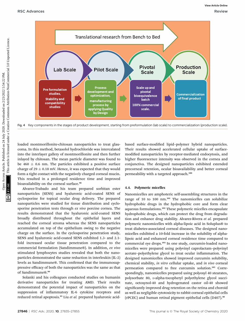
Fig. 4 Key components in the stages of product development, starting from preformulation (lab scale) to commercialization (production scale).
RSC Advances Review
Ope
n A
cces
s A
rtic
le. P
ublis
hed
on 2
4 Ju
ly 2
020.
Dow
nloa
ded
on 2
/23/
2022
5:5
4:22
PM
. T
his
artic
le is
lice
nsed
und
er a
Cre
ativ
e C
omm
ons
Attr
ibut
ion-
Non
Com
mer
cial
3.0
Unp
orte
d L
icen
ce.
View Article Online
loaded montmorillonite-chitosan nanoparticles to treat glau-coma. In this method, betaxolol hydrochloride was intercalatedinto the interlayer gallery of montmorillonite and then furtherinlayed by chitosan. The mean particle diameter was found tobe 460 � 0.6 nm. The particles exhibited a positive surfacecharge of 29 � 0.18 mV. Hence, it was expected that they wouldform a tight contact with the negatively charged corneal mucin.This resulted in a prolonged residence time and improvedbioavailability on the corneal surface.98
Alvarez-Trabado and his team prepared sorbitan esternanoparticles (SENS) and hyaluronic acid-coated SENS ofcyclosporine for topical ocular drug delivery. The preparednanoparticles were studied for tissue distribution and cyclo-sporine penetration tests through ex vivo porcine cornea. Theresults demonstrated that the hyaluronic acid-coated SENSbroadly distributed throughout the epithelial layers andreached the corneal stroma whereas the SENS nanoparticlesaccumulated on top of the epithelium owing to the negativecharge on the surface. In the cyclosporine penetration study,SENS and hyaluronic acid-coated SENS exhibited 1.3- and 2.1-fold increased ocular tissue penetration compared to thecommercial formulation (Sandimmune®). In addition, ex vivostimulated lymphocytes studies revealed that both the nano-particles demonstrated the same reduction in interleukin (IL-2)levels as Sandimmune®. This conrmed that the immunosup-pressive efficacy of both the nanoparticles was the same as thatof Sandimmune®.99
Solanki and his colleagues conducted studies on humaninderivative nanoparticles for treating AMD. Their resultsdemonstrated the potential impact of nanoparticles on thesuppression of inammatory IL-6 cytokine receptors andreduced retinal apoptosis.95 Liu et al. prepared hyaluronic acid-
27846 | RSC Adv., 2020, 10, 27835–27855
based surface-modied lipid–polymer hybrid nanoparticles.Their results showed accelerated cellular uptake of surface-modied nanoparticles by receptor-mediated endocytosis, andhigher uorescence intensity was observed in the cornea andconjunctiva. The designed nanoparticles exhibited extendedprecorneal retention, ocular bioavailability and better cornealpermeability with a targeted approach.100
4.4. Polymeric micelles
Nanomicelles are amphoteric self-assembling structures in therange of 10 to 100 nm.101 The nanomicelles can solubilizehydrophobic drugs in the hydrophobic core and form clearaqueous formulations.102 These polymeric micelles encapsulatehydrophobic drugs, which can protect the drug from degrada-tion and enhance drug stability. Alvarez-Rivera et al. preparedpolymeric nanomicelles with alpha-lipoic acid in Soluplus® totreat diabetes-associated corneal diseases. The designed nano-micelles exhibited a 10-fold increase in the solubility of alpha-lipoic acid and enhanced corneal residence time compared tocommercial eye drops.103 In one study, curcumin-loaded nano-micelles were prepared using polyvinyl caprolactam–polyvinylacetate–polyethylene glycol to treat ocular inammation. Thedesigned nanomicelles showed improved curcumin solubility,chemical stability, in vitro cellular uptake, and in vivo cornealpermeation compared to free curcumin solution.102 Corre-spondingly, nanomicelles prepared using polyoxyl 40 stearates,polysorbate 80, D-alpha-tocopheryl polyethylene glycol succi-nate, octoxynol-40 and hydrogenated castor oil-40 showedsignicantly improved drug retention on the retina and choroidas well as negligible cytotoxicity to rabbit corneal epithelial cells(rPCEC) and human retinal pigment epithelial cells (D407).48
This journal is © The Royal Society of Chemistry 2020
Review RSC Advances
Ope
n A
cces
s A
rtic
le. P
ublis
hed
on 2
4 Ju
ly 2
020.
Dow
nloa
ded
on 2
/23/
2022
5:5
4:22
PM
. T
his
artic
le is
lice
nsed
und
er a
Cre
ativ
e C
omm
ons
Attr
ibut
ion-
Non
Com
mer
cial
3.0
Unp
orte
d L
icen
ce.
View Article Online
4.5. Nanocarrier-loaded gels
Nanocarrier-loaded in situ gels have gained attention for theirtargeted delivery with stimuli-responsive behavior. Theophthalmic in situ gel consists of environmentally responsivepolymers that change structurally in response to small changes inspecic circumstances like temperature, pH, and ionic strengthin the environment. Additionally, the loading of nanoparticlesinto the gel improves the burst release problems observed withnanoparticles and gives prolonged drug release.26,104 Studies haverevealed that temperature-sensitive polymers like Pluronic (PF-127 & PF-68), poloxamer 407 and poloxamer 188, Carbopol934P, sodium alginate, and hydroxypropyl methylcellulose(HPMC) K4M provide prolonged drug release, increased ocularbioavailability and sustained drug release for about 8 h.26
Morsi et al. investigated nanoemulsion-based ion-sensitivein situ gels for the delivery of acetazolamide. The stability ofnanoemulsions depends on the surfactant and co-surfactantconcentration. The nanoemulsion formulation was preparedby mixing oil (peanut oil), surfactants (Tween 80 and Cremo-phor EL), and Transcutol P as a co-surfactant and then inte-grating into the gellan gum alone and in combination withxanthan gum, HPMC or Carbopol. The results showed that thecombination of gellan/xanthan and gellan/HPMC gave goodstability and enhanced therapeutic efficacy of acetazolamidecompared to commercial eye drops and oral tablets.105 Patel andteam members prepared cationic nanoemulsion-based in situophthalmic gel of loteprednol etabonate (LE), which demon-strated a 2.54-fold increase in bioavailability compared to themarketed formulation.106 Phua et al. reported a 12-foldincreased residence time with a liposome-loaded hydrogelcompared to free liposomes.107 Thus, the delivery of therapeu-tics using nanocarriers can provide improved permeation andprolonged retention at ocular sites owing to its nanoscale.
Further, surfacemodication of nanocarriers can improve theirtargeting ability and mucoadhesion at the ocular surface, whichcan improve both therapeutic efficacy and patient compliance.
5. Current challenges in thetranslation of ophthalmicnanomedicine
Nanomedicine has to cross many hurdles to reach clinical trials.The current obstacles in the translation of nanomedicine concern-ing nanopharmaceutical design include large-scale production toGood Manufacturing Practice standards and quality control assaysfor characterization.13 Fig. 4 shows the key components in the stagesof product development, starting from preformulation (lab scale) tocommercialization (production scale). The marketed productshould be within acceptable limits for safety, efficacy, stability, andpatient acceptance. Themethod used during formulation should bewithin the standards and it should be reproducible.
5.1. Scale-up of nanomedicine
Translation from laboratory scale to commercial scale is chal-lenging in the case of nanomedicine.108 The success of these
This journal is © The Royal Society of Chemistry 2020
formulations depends on their scalability and reproducibility. Ascalable formulation should always be robust at the lab, pilot,and industrial levels. Advances in scale-up technology andQuality by Design (QbD) concepts have led to signicant prog-ress and smoothed the process of commercialization ofnanomedicine.109
Recent advances in formulation development have led to theentry of many nanoformulations, such as liposomes, solid lipidnanoparticles, nanostructured lipid particles, nanoparticles,micelles, and nano in situ gels, for preclinical and clinicaltesting. However, efforts are still being made to bring theseadvanced nanomedicine formulations to the market.
Nanocrystals have been explored well for commercialization.Currently, 15 nanocrystal based products are available in themarket for different routes of administration, among whichIlevro® (nepafenac ophthalmic suspension, 0.3%) is availablefor ophthalmic administration.110 During the production ofnanocrystals, the size, shape, and other process parameters canbe controlled.111 Two main methodologies are used for thepreparation of nanocrystals, bottom-up (precipitation process)and top-down (including wet-bead milling, nanospray dryer,and high-pressure homogenization). In a study, nanocrystals ofsteroid drugs uorometholone and dexamethasone wereprepared by using nano Spray Dryer B-90 technology (Buchi®).The authors observed that the particle size changed dependingon the mesh aperture size. The results revealed that with meshaperture sizes of 4.0, 5.5, and 7.0 mm, the average particle sizesfor uorometholone nanocrystals were found to be 620 � 268,795 � 285, and 856 � 344 nm and for dexamethasone nano-crystals were found to be 833 � 402, 1118 � 573, and 1344 �857 nm, respectively.112
Nanoparticles can be crystalline or amorphous, dependingon the manufacturing technique and material employed. Theproduction of nanoparticles is a challenging task in terms ofreproducibility of size and polydispersity index. Critical processparameters involved in preparation techniques greatly inu-ence the particle characteristics. Galindo-Rodriguez et al.prepared nanoparticles using three different methods:emulsion-based, salting out, and nanoprecipitation methods.The particle size ranges changed with the different methodsand variations in the physicochemical properties of the aqueousand organic phases. Particle size range of 123–710 nm, 108–715 nm and 147–245 nm were observed with salting out,emulsication-diffusion, and nanoprecipitation methods,respectively.113
Colombo et al. conducted a study to investigate the essentialscale-up parameters for nanocapsule production. Theyemployed the emulsication-diffusion method for the prepa-ration of nanocapsules and assessed at pilot scale by increasingthe laboratory batch volume by 33-fold, i.e. from 60 mL to 2 L.They observed that increasing the impeller speed and durationof agitation led to a slight decrease in emulsion size.111
Nanoparticle preparation from micro/nanoemulsions usinglow-energy approaches, such as phase inversion temperature,phase inversion composition, and emulsion inversion pointmethods, has led to scale-up issues including variation inphysicochemical properties and also requires a large amount of
RSC Adv., 2020, 10, 27835–27855 | 27847
RSC Advances Review
Ope
n A
cces
s A
rtic
le. P
ublis
hed
on 2
4 Ju
ly 2
020.
Dow
nloa
ded
on 2
/23/
2022
5:5
4:22
PM
. T
his
artic
le is
lice
nsed
und
er a
Cre
ativ
e C
omm
ons
Attr
ibut
ion-
Non
Com
mer
cial
3.0
Unp
orte
d L
icen
ce.
View Article Online
surfactant. During the scale-up of solid lipid nanoparticles bya hot-melt extrusion process, the process parameters such asfeed rate, extruder diameter, and heat transfer play an impor-tant role as the change in the feed rate may change the resi-dence time of the material in the barrel.114 High-energyapproaches, such as ultrasonication, high-pressure homogeni-zation, and microuidization, have been explored to control thecharacteristics and reproducibility of nanoparticles. The majorproblem observed with sonication and homogenization is therecoalescence of new droplets, leading to the formation ofthermodynamically unstable formulations.115,116 In one study,a pilot-scale batch of nanoparticles was prepared by ionotropicgelation method. It was observed that increasing the homoge-nization speed from 500 to 900 rpm contributed to an increasein the heat and kinetic energy of the molecule, which in turn ledto agglomeration of the emulsion.117 This showed the hugedifference between lab-scale and industrial-scale equipment.Formulation scientists need to optimize all the processparameters extensively in the scale-up of nanocarrier prepara-tion using homogenizers.
For achieving adequate particle size, scalability, and repro-ducibility, microuidization has become a potential approachfor commercial applications. Microuidic technology is anoptimum technique to support, speed up, and favour clinicaltranslation of nanomedicine by reducing the batch to batchvariability. This technique involves the rapid mixing of lipidsand the aqueous phase in a micronized chamber (chip) withinmilliseconds. The accurate mixing of uids in the microchannelfacilitates precise control over the physicochemical propertiesof the nanoparticles. The nanoparticles are prepared by nano-precipitation, and self-assembly with continuous ow gave thesame quality over time for the produced nanoparticles, avoidingthe batch to batch variability. This is the most important featurefor industrialization from the lab scale.118 Ali et al. prepareda hydrocortisone nanosuspension using microuidic nano-precipitation and wet milling. The particle size range of 295 �32 nm (simple mixing) and 300 nm (with 105 minutes mixing)was observed with y-junction microuidic and wet millingtechnique, respectively. The results revealed that both thenanosuspensions showed sustained action and enhanced 1.8-fold bioavailability compared to the commercial solution.Microuidic nanoprecipitation is advantageous over millingowing to the low energy of the process. The microuidic tech-nique reduces both the production time and the economic costsinvolved in nanoparticle production.119 Thus, by controllinga few parameters, such as the ow rate and organic phase ratio,uniform-sized nanoparticles can be produced on a large scale.Gdowski et al. synthesized a nanolipomer formulation usinga high-ow microuidic system and observed batch to batchuniformity upon increasing to pilot-scale production.120
Other advanced technologies such as BUONAPART-E (betterscale-up and optimization of nanoparticles and nanostructureproduction using electrical discharges) can be used to synthe-size high-purity metallic nanoparticles. This technique utilizesthe arc and spark discharge method. It is a stable and low-costprocess with a high production rate and energy efficiency. This
27848 | RSC Adv., 2020, 10, 27835–27855
technique can be further scaled up with multiple parallel elec-trodes set to increase the nanoparticle production.121
The supercritical solvent technique has also been exploredfor scale-up of nanocarrier preparation. Jung et al. preparednanoparticles at three different scales (0.5 L, 4 L, and 50 L) byusing a supercritical anti-solvent process. They observed nochange in particle size distribution and no loss of residualsolvent and percent yield.122 Pham et al. determined the scale-up ability of liposomes and niosomes by using a syringepump at the lab scale and then membrane contractors at pilotscale. They prepared 30 mL of liposomes and 20 mL of nio-somes by using a syringe pump at a laboratory scale to pilot-scale production of 750 mL of liposomes and 1000 mL nio-somes using a membrane contactor. They reported that repro-ducible results were observed concerning size and entrapmentefficiency.123
Recent advances in nanocarrier development for oculardelivery include particle replication in non-wetting template(PRINT) technology and hydrogel template method.124,125 InPRINT technology, the particles are produced by manufacturingroll-to-roll with the required particular size, shape, andmodulus. It includes three steps, fabrication of micro moldswith a precise cavity, molding of suitable material into thecavities, and separation of the formed particles. The continuousproduction of particles with the desired size, shape, and surfacecan be achieved by altering the composition of the materialmatrix and post functionalization.124 This technique can beutilized with a large number of biocompatible polymers andtherapeutic agents, including peptide molecules, nucleic acids,proteins, and antibodies.124 The hydrogel template method canbe widely used for nano- and micro-size particle development.This template was utilized for the production of a nanowafercontaining a nano-reservoir as an ultra-thin lens.125 In a study,a silicon-based wafer template was fabricated using e-beamlithography and polyvinyl alcohol was used to prepare thetemplate with different arrays of wells. The drug solution wasloaded into the arrays to form nanowafers. The particle size anddrug loading can be controlled by using these drug reservoirs,and these are utilized as a lens by placing on the ocularsurface.126
The success of nanomedicine is governed by the reproduc-ibility of the physicochemical characteristics at the industrialscale. Recent advances in technology such as microuidic, high-pressure homogenization, PRINT, and hydrogel templatemethods have provided hope for commercialization.127 Further,guideline progression and clinical acceptance of nanocarriersfrom regulatory agencies will accelerate the development ofnanomedicine for ophthalmic therapy.
5.2. Quality control of nanomedicine
The quality and cost-effective production are importantconsiderations in pharmaceutical product development. Forquality, the stability and the manufacturing process are themain aspects for nanoformulations. The challenges in nano-particle preparation include batch to batch reproducibility,dispersion stability and the safety of the nanomaterials. A small
This journal is © The Royal Society of Chemistry 2020
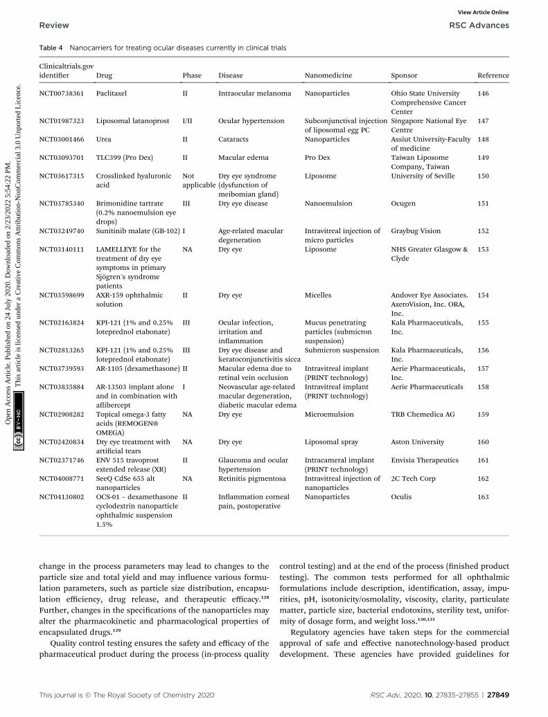
Table 4 Nanocarriers for treating ocular diseases currently in clinical trials
Clinicaltrials.govidentier Drug Phase Disease Nanomedicine Sponsor Reference
NCT00738361 Paclitaxel II Intraocular melanoma Nanoparticles Ohio State UniversityComprehensive CancerCenter
146
NCT01987323 Liposomal latanoprost I/II Ocular hypertension Subconjunctival injectionof liposomal egg PC
Singapore National EyeCentre
147
NCT03001466 Urea II Cataracts Nanoparticles Assiut University-Facultyof medicine
148
NCT03093701 TLC399 (Pro Dex) II Macular edema Pro Dex Taiwan LiposomeCompany, Taiwan
149
NCT03617315 Crosslinked hyaluronicacid
Notapplicable
Dry eye syndrome(dysfunction ofmeibomian gland)
Liposome University of Seville 150
NCT03785340 Brimonidine tartrate(0.2% nanoemulsion eyedrops)
III Dry eye disease Nanoemulsion Ocugen 151
NCT03249740 Sunitinib malate (GB-102) I Age-related maculardegeneration
Intravitreal injection ofmicro particles
Graybug Vision 152
NCT03140111 LAMELLEYE for thetreatment of dry eyesymptoms in primarySjogren's syndromepatients
NA Dry eye Liposome NHS Greater Glasgow &Clyde
153
NCT03598699 AXR-159 ophthalmicsolution
II Dry eye Micelles Andover Eye Associates.AxeroVision, Inc. ORA,Inc.
154
NCT02163824 KPI-121 (1% and 0.25%loteprednol etabonate)
III Ocular infection,irritation andinammation
Mucus penetratingparticles (submicronsuspension)
Kala Pharmaceuticals,Inc.
155
NCT02813265 KPI-121 (1% and 0.25%loteprednol etabonate)
III Dry eye disease andkeratoconjunctivitis sicca
Submicron suspension Kala Pharmaceuticals,Inc.
156
NCT03739593 AR-1105 (dexamethasone) II Macular edema due toretinal vein occlusion
Intravitreal implant(PRINT technology)
Aerie Pharmaceuticals,Inc.
157
NCT03835884 AR-13503 implant aloneand in combination withaibercept
I Neovascular age-relatedmacular degeneration,diabetic macular edema
Intravitreal implant(PRINT technology)
Aerie Pharmaceuticals 158
NCT02908282 Topical omega-3 fattyacids (REMOGEN®OMEGA)
NA Dry eye Microemulsion TRB Chemedica AG 159
NCT02420834 Dry eye treatment witharticial tears
NA Dry eye Liposomal spray Aston University 160
NCT02371746 ENV 515 travoprostextended release (XR)
II Glaucoma and ocularhypertension
Intracameral implant(PRINT technology)
Envisia Therapeutics 161
NCT04008771 SeeQ CdSe 655 altnanoparticles
NA Retinitis pigmentosa Intravitreal injection ofnanoparticles
2C Tech Corp 162
NCT04130802 OCS-01 – dexamethasonecyclodextrin nanoparticleophthalmic suspension1.5%
II Inammation cornealpain, postoperative
Nanoparticles Oculis 163
Review RSC Advances
Ope
n A
cces
s A
rtic
le. P
ublis
hed
on 2
4 Ju
ly 2
020.
Dow
nloa
ded
on 2
/23/
2022
5:5
4:22
PM
. T
his
artic
le is
lice
nsed
und
er a
Cre
ativ
e C
omm
ons
Attr
ibut
ion-
Non
Com
mer
cial
3.0
Unp
orte
d L
icen
ce.
View Article Online
change in the process parameters may lead to changes to theparticle size and total yield and may inuence various formu-lation parameters, such as particle size distribution, encapsu-lation efficiency, drug release, and therapeutic efficacy.128
Further, changes in the specications of the nanoparticles mayalter the pharmacokinetic and pharmacological properties ofencapsulated drugs.129
Quality control testing ensures the safety and efficacy of thepharmaceutical product during the process (in-process quality
This journal is © The Royal Society of Chemistry 2020
control testing) and at the end of the process (nished producttesting). The common tests performed for all ophthalmicformulations include description, identication, assay, impu-rities, pH, isotonicity/osmolality, viscosity, clarity, particulatematter, particle size, bacterial endotoxins, sterility test, unifor-mity of dosage form, and weight loss.130,131
Regulatory agencies have taken steps for the commercialapproval of safe and effective nanotechnology-based productdevelopment. These agencies have provided guidelines for
RSC Adv., 2020, 10, 27835–27855 | 27849
RSC Advances Review
Ope
n A
cces
s A
rtic
le. P
ublis
hed
on 2
4 Ju
ly 2
020.
Dow
nloa
ded
on 2
/23/
2022
5:5
4:22
PM
. T
his
artic
le is
lice
nsed
und
er a
Cre
ativ
e C
omm
ons
Attr
ibut
ion-
Non
Com
mer
cial
3.0
Unp
orte
d L
icen
ce.
View Article Online
product specication and characterization required for theproduct approval process. In the year 2017, the United StatesFood and Drug Administration (US FDA) published “DrugProducts, Including Biological Products, that Contain Nano-materials” dra guidelines for industry and suggested someattributes to be described and measured for nano-formulations.132 The guidelines comprised characteristics ofnanoformulations, including chemical composition, averageparticle size, particle size distribution (describing d10, d50, d90or polydispersity; modality), general shape and morphology(aspect ratio), physical stability (e.g., aggregation and agglom-eration or separation) and chemical stability. Further, theguidelines include specic studies, such as distribution of theactive ingredient associated with the nanomaterial, free insolution, structural attributes (lamellarity, core–shell structure),surface properties (surface area, surface charge, chemicalreactivity, ligand hydrophobicity, and roughness), coatingproperties of nanomaterials, porosity (a function related to thedrug-loading capacity), particle concentration, in vitro release,crystal form, impurities, sterility and endotoxin levels. Theabove-mentioned guidelines also provide factors to be selectedand specic characterization methods for nanoformulations.132
The US FDA also provided specic guidelines for “LiposomeDrug Products: Chemistry, Manufacturing, and Controls;Human Pharmacokinetics and Bioavailability; and LabelingDocumentation” in the year 2018 for liposomal product devel-opment.133 In 2018, the US FDA draed another guideline,“Selected FDA Publications Related to the Application, Char-acterization, Effects, and Evaluation of Nanotechnology” forcharacterization and evaluation of nanoparticulate-based drugdelivery systems.134
Recently, in October 2019, Indian regulatory bodies (CentralDrug Standard Control Organization, Indian Council of MedicalResearch, Department of Biotechnology, and Ministry of Healthand Family Welfare) issued the “Guidelines for Evaluation ofNanopharmaceuticals” with details of the evaluation parametersrequired for quality control for nanopharmaceuticals, similar tothe US FDA guidelines.135 Additionally, these guidelines sug-gested that the product should meet specications for analyticalmethod validation, stability studies of nanoformulations,comparative analysis of innovator product if applicable, preclin-ical data, clinical data, and sample testing protocols.135
The efforts of regulatory bodies will certainly ensure thesafety and efficacy of products with stringent product speci-cation for nanocarriers for ophthalmic drug delivery.
5.3. Safety and toxicity concerns for nanocarriers
Safety and toxicity are the main issues for clinical approval ofophthalmic formulations. At the preclinical level, researchershave investigated the ocular toxicity of designed nano-formulations using the Draize eye test on rabbits and in vitrohuman corneal epithelial cells for the determination of acuteocular toxicity issues.136 De et al. prepared brimonidine-loadedpolycarboxylic (polyacrylic and polyitaconic) acid nano-particles for the treatment of glaucoma. They studied thebiocompatibility of prepared nanoparticles with human corneal
27850 | RSC Adv., 2020, 10, 27835–27855
epithelial cells. The results revealed that the polyacrylic acidnanoparticles were safe, i.e., biocompatible, compared to thepolyitaconic acid nanoparticles.137 Vega et al. preparedurbiprofen-loaded poly(lactic/glycolic acid) nanoparticles.Topical application of the prepared nanoparticles to rabbit eyesshowed no sign of toxicity.138 Prow et al. compared the safetyand toxicity of chitosan, amphiphilic polyphosphoester, andmagnetic nanoparticles. They reported that intravitrealadministration of chitosan causes inammation of the eye.139
Among the different nanomaterials, lipid-based nano-carriers including nanoemulsions and liposomes were found tobe more biocompatible and safer for interaction with biologicalmembranes and showed their presence in the market.140
6. Drugs approved and undergoingclinical trials
Substantial investigations have been done on nanocarriers likenanoparticles and nanomicelles for the alleviation of anteriorand posterior ocular disorders. They have entered into clinicaltrials and shown positive results in patients. The currentophthalmic nanostructured marketed products are Restasis®(cyclosporine A nanoemulsion), Cyclokat® (cyclosporine Acationic nanoemulsion), Cequa® (cyclosporine A ophthalmicnano micellar solution), VISUDYNE® (verteporn liposomalinjection), Lacrisek® (vitamin A palmitate and vitamin E lipo-somal spray), and Artelac Rebalance® (vitamin B12 liposomaleye drops), which are used for the treatment of dry eyesyndrome. Liposome-based ocular products have gained moreattention owing to their biodegradable, biocompatible, non-toxic, and non-immunogenic nature. Moreover, Ikervis®(cyclosporine A cationic nanoemulsion) has been approved fortreating severe keratitis in dry eye disease patients.141 SunPharmaceuticals developed Cequa® (cyclosporine A ophthalmicnano micellar solution) 0.09%, which is intended to improvedrug delivery and penetration to ocular tissues. Cequa® hasbeen investigated for efficacy and safety in treating keratocon-junctivitis sicca (dry eye). The company conducted phase IIItrials with Cequa® on 744 patients and the study designinvolved two 12 week randomized and vehicle-controlled trials.The results exhibited a statistically signicant increase in theSchirmer score ($10mm) from baseline with Cequa® relative tovehicle in tear production with twice-daily dosing. Moreover,adverse events were reported by greater than 5% of patients, i.e.,22% of patients showed pain at the instillation site and 6% ofpatients showed conjunctival hyperemias, which are mostcommon for the drugs analyzed in this category. In 2018,Cequa® was the rst nanotechnology-based product to beapproved by the US FDA for treating dry eye.142
Kala Pharmaceuticals developed an innovative treatmentwith nanosized mucus penetrating particles (MPP) to improvedrug delivery to the back of the eye. MPP allow effective pene-tration through the tear lm and mucin layer, which preventsdrugs from being trapped by the tear lm and clearance byblinking. Kala Pharmaceuticals applied this MPP platformtechnology for loteprednol etabonate (KPI-121, 1% and KPI-121,
This journal is © The Royal Society of Chemistry 2020
Review RSC Advances
Ope
n A
cces
s A
rtic
le. P
ublis
hed
on 2
4 Ju
ly 2
020.
Dow
nloa
ded
on 2
/23/
2022
5:5
4:22
PM
. T
his
artic
le is
lice
nsed
und
er a
Cre
ativ
e C
omm
ons
Attr
ibut
ion-
Non
Com
mer
cial
3.0
Unp
orte
d L
icen
ce.
View Article Online
0.25%) for the treatment of several ocular diseases(NCT02163824) (NCT02813265). The phase III clinical trial resultsfor 1% KPI-121 demonstrated a signicant decrease in both painand inammation with twice a day dosing.143 In addition to this,particle replication in non-wetting template (PRINT) technologyoffers a distinctive capability to reproduce and fabricate particlesof any size.124 The clinical-stage pharmaceutical companiesEnvisia Therapeutics and Aerie Pharmaceuticals combinedPRINT particle engineering technology with biodegradable poly-mer science.144,145 With this technology, various different ocularformulations, including subconjunctival implants, intravitrealimplants, intracameral implants, and nanosuspensions, havebeen developed for controlled delivery of the drug into the oculartissue. Some clinical trials are underway, including amulti-centeropen-labeled study to evaluate the safety and efficacy study ofENV 515 (travoprost) for treating glaucoma and ocular hyper-tension (NCT02371746). AR-1105 (NCT03739593) and AR-13503(NCT03835884) designed using PRINT technology as intravitrealimplants for the treatment of age-related macular degenerationand diabetic macular edema are in clinical trials. Nanomedicinescurrently undergoing clinical trials are outlined in Table 4.146–163
7. Conclusion
Targeted drug delivery to the anterior and posterior segments ofthe eye using nanomedicine to overcome the limitations ofconventional formulations has gained remarkable attention.Advances in nanomedicine have facilitated controlled andtarget-specic drug delivery to ocular tissues to treat oculardiseases. Therapeutic delivery through nanocarriers can reducethe overall administration frequency of intravitreal injections,enhance the therapeutic efficacy, signicantly reduce treatmentcosts and improve the quality of life of ocular disease patients.Further, technological advances related to easy scale-up andreproducibility bring more hope for the cost-effective commer-cialization of nanomedicines. Various methods have beendeveloped for the production of nanomedicines with repro-ducible characteristics. Promising methods, for instancemicrouidizers, supercritical uid technology, and membraneextrusion, show scale-up capabilities. Certain ophthalmicnanomedicines, such as nanocrystals, nanoemulsions, lipo-somes, advanced PRINT, and hydrogel technologies, have scale-up potential and products of these technologies are on themarket. Regulatory agencies have draed guidelines for nano-formulation specications to ensure their safety and efficacy.
8. Future perspective
Anterior segment diseases such as cataracts, glaucoma, dry eye,and other infectious can ultimately lead to poor eyesight orblindness. For treating these conditions, eye drops are mostlyprescribed but tear uid generation, lacrimal drainage, andbarrier functions limit the efficacy of the administered drugs.Drug delivery through lms, hydrogels, and implants mayimprove the ocular residence time. However, these approachescan obstruct the vision and cause inconvenience to patients.Long-term contact with the carrier material in implants may
This journal is © The Royal Society of Chemistry 2020
compromise the safety of the ocular tissue. The reportedpreclinical and clinical outcomes for nanocarriers providefuture hope for safer and effective drug delivery for treatinganterior segment diseases. However, targeting nanocarriers tothe posterior part is challenging and there is a need to delivertherapeutics via a suitable delivery system that can overcomethe ocular barriers. Currently, injectable formulations areadministered to treat chronic retinal disorders. For example,AMD requires anti-VEGF treatment by intravitreal injection, andit requires repeated administration, and expert medical super-vision, while lacking patient compliance. Intravitreal nano-medicine injection can deliver the drug directly to the retina byovercoming the clearance of the drug molecule in the vitreousregion. This nanomedicine can stay in the vitreous region andprovide prolonged salutary drug concentrations to the targetsite. Intravitreal delivery of nanomedicines can reduce the doseand dosing frequency, and employing less invasive techniquescan reduce the medical burden for patients and healthcareprofessionals. The delivery of nanocarriers larger than 300 nmto the posterior part of the eye increases aggregation and causesdisturbance to the vision. A particle size of less than 300 nm notonly decreased the chances of aggregation but also improvedthe release and drug loading, which are of major importance.All these considerations are the bottleneck for the oculardelivery of nanoparticles.
In addition to the scale-up of nano-drug delivery systems,controlled drug release from implants, nano wafers, and 3Dprinted hydrogel technology has also been explored for thetreatment of retinal diseases. The safety, scale-up, and repro-ducibility of nanomedicine are essential for reaching commer-cial scale. Most biodegradable and natural polymers areconsidered safe for drug delivery but detailed toxicity studiesneed to be performed before commercial acceptance. Somenanocarrier preparation techniques have been investigated forscale-up and large-scale production but there is a need tofurther explore more techniques that are simple, feasible, andprovide regulatory acceptable nanomedicines.
Nanotechnology has the potential to add innovative func-tionality and provide superior therapeutic efficacy overconventional ocular drug delivery systems. The overall globalophthalmic drugs market was valued at approximately USD25.03 billion in 2017 and is expected to generate revenue ofaround USD 34.52 billion by the end of 2024, growing ata compound annual growth rate of around 4.7% between 2018and 2024.164 This reects the huge scope of ocular products forabbreviated new drug applications in future.
Conflicts of interest
Authors declare no conict of interest.
References
1 WHO, https://www.who.int/blindness/causes/priority/en/index1.html, 2018.
2 WHO, International Classication of Diseases, 11th Revision(ICD-11), https://www.who.int/classications/icd/en/, 2019.
RSC Adv., 2020, 10, 27835–27855 | 27851
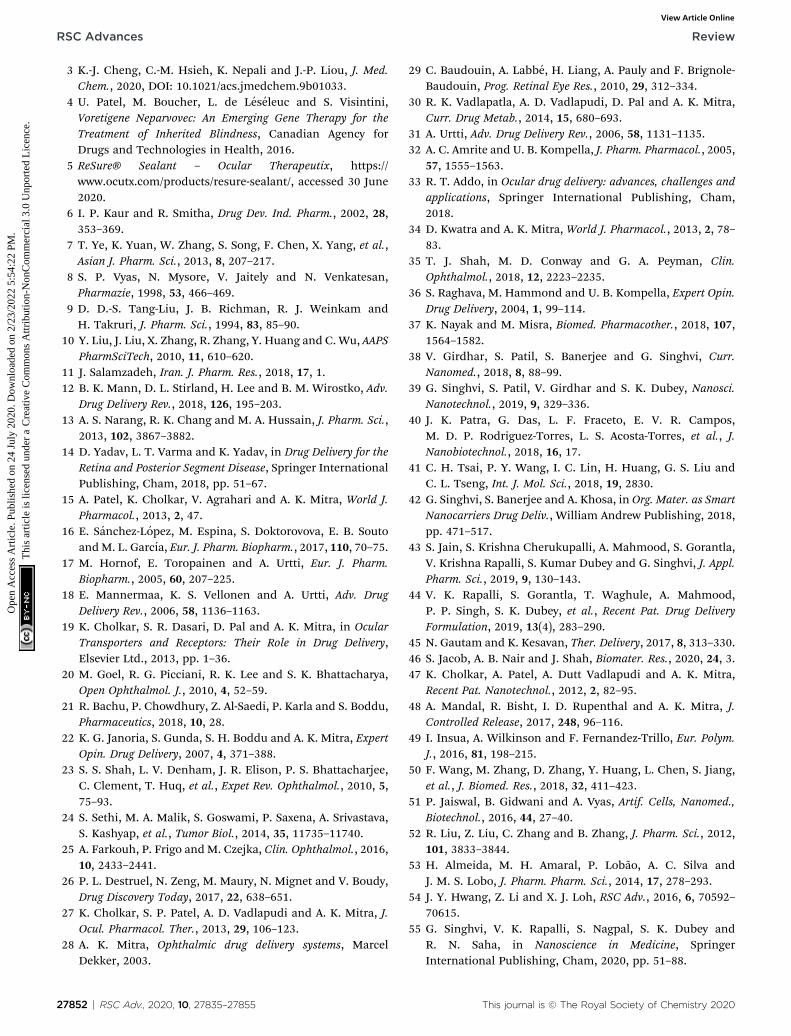
RSC Advances Review
Ope
n A
cces
s A
rtic
le. P
ublis
hed
on 2
4 Ju
ly 2
020.
Dow
nloa
ded
on 2
/23/
2022
5:5
4:22
PM
. T
his
artic
le is
lice
nsed
und
er a
Cre
ativ
e C
omm
ons
Attr
ibut
ion-
Non
Com
mer
cial
3.0
Unp
orte
d L
icen
ce.
View Article Online
3 K.-J. Cheng, C.-M. Hsieh, K. Nepali and J.-P. Liou, J. Med.Chem., 2020, DOI: 10.1021/acs.jmedchem.9b01033.
4 U. Patel, M. Boucher, L. de Leseleuc and S. Visintini,Voretigene Neparvovec: An Emerging Gene Therapy for theTreatment of Inherited Blindness, Canadian Agency forDrugs and Technologies in Health, 2016.
5 ReSure® Sealant – Ocular Therapeutix, https://www.ocutx.com/products/resure-sealant/, accessed 30 June2020.
6 I. P. Kaur and R. Smitha, Drug Dev. Ind. Pharm., 2002, 28,353–369.
7 T. Ye, K. Yuan, W. Zhang, S. Song, F. Chen, X. Yang, et al.,Asian J. Pharm. Sci., 2013, 8, 207–217.
8 S. P. Vyas, N. Mysore, V. Jaitely and N. Venkatesan,Pharmazie, 1998, 53, 466–469.
9 D. D.-S. Tang-Liu, J. B. Richman, R. J. Weinkam andH. Takruri, J. Pharm. Sci., 1994, 83, 85–90.
10 Y. Liu, J. Liu, X. Zhang, R. Zhang, Y. Huang and C. Wu, AAPSPharmSciTech, 2010, 11, 610–620.
11 J. Salamzadeh, Iran. J. Pharm. Res., 2018, 17, 1.12 B. K. Mann, D. L. Stirland, H. Lee and B. M. Wirostko, Adv.
Drug Delivery Rev., 2018, 126, 195–203.13 A. S. Narang, R. K. Chang and M. A. Hussain, J. Pharm. Sci.,
2013, 102, 3867–3882.14 D. Yadav, L. T. Varma and K. Yadav, in Drug Delivery for the
Retina and Posterior Segment Disease, Springer InternationalPublishing, Cham, 2018, pp. 51–67.
15 A. Patel, K. Cholkar, V. Agrahari and A. K. Mitra, World J.Pharmacol., 2013, 2, 47.
16 E. Sanchez-Lopez, M. Espina, S. Doktorovova, E. B. SoutoandM. L. Garcıa, Eur. J. Pharm. Biopharm., 2017, 110, 70–75.
17 M. Hornof, E. Toropainen and A. Urtti, Eur. J. Pharm.Biopharm., 2005, 60, 207–225.
18 E. Mannermaa, K. S. Vellonen and A. Urtti, Adv. DrugDelivery Rev., 2006, 58, 1136–1163.
19 K. Cholkar, S. R. Dasari, D. Pal and A. K. Mitra, in OcularTransporters and Receptors: Their Role in Drug Delivery,Elsevier Ltd., 2013, pp. 1–36.
20 M. Goel, R. G. Picciani, R. K. Lee and S. K. Bhattacharya,Open Ophthalmol. J., 2010, 4, 52–59.
21 R. Bachu, P. Chowdhury, Z. Al-Saedi, P. Karla and S. Boddu,Pharmaceutics, 2018, 10, 28.
22 K. G. Janoria, S. Gunda, S. H. Boddu and A. K. Mitra, ExpertOpin. Drug Delivery, 2007, 4, 371–388.
23 S. S. Shah, L. V. Denham, J. R. Elison, P. S. Bhattacharjee,C. Clement, T. Huq, et al., Expet Rev. Ophthalmol., 2010, 5,75–93.
24 S. Sethi, M. A. Malik, S. Goswami, P. Saxena, A. Srivastava,S. Kashyap, et al., Tumor Biol., 2014, 35, 11735–11740.
25 A. Farkouh, P. Frigo andM. Czejka, Clin. Ophthalmol., 2016,10, 2433–2441.
26 P. L. Destruel, N. Zeng, M. Maury, N. Mignet and V. Boudy,Drug Discovery Today, 2017, 22, 638–651.
27 K. Cholkar, S. P. Patel, A. D. Vadlapudi and A. K. Mitra, J.Ocul. Pharmacol. Ther., 2013, 29, 106–123.
28 A. K. Mitra, Ophthalmic drug delivery systems, MarcelDekker, 2003.
27852 | RSC Adv., 2020, 10, 27835–27855
29 C. Baudouin, A. Labbe, H. Liang, A. Pauly and F. Brignole-Baudouin, Prog. Retinal Eye Res., 2010, 29, 312–334.
30 R. K. Vadlapatla, A. D. Vadlapudi, D. Pal and A. K. Mitra,Curr. Drug Metab., 2014, 15, 680–693.
31 A. Urtti, Adv. Drug Delivery Rev., 2006, 58, 1131–1135.32 A. C. Amrite and U. B. Kompella, J. Pharm. Pharmacol., 2005,
57, 1555–1563.33 R. T. Addo, in Ocular drug delivery: advances, challenges and
applications, Springer International Publishing, Cham,2018.
34 D. Kwatra and A. K. Mitra, World J. Pharmacol., 2013, 2, 78–83.
35 T. J. Shah, M. D. Conway and G. A. Peyman, Clin.Ophthalmol., 2018, 12, 2223–2235.
36 S. Raghava, M. Hammond and U. B. Kompella, Expert Opin.Drug Delivery, 2004, 1, 99–114.
37 K. Nayak and M. Misra, Biomed. Pharmacother., 2018, 107,1564–1582.
38 V. Girdhar, S. Patil, S. Banerjee and G. Singhvi, Curr.Nanomed., 2018, 8, 88–99.
39 G. Singhvi, S. Patil, V. Girdhar and S. K. Dubey, Nanosci.Nanotechnol., 2019, 9, 329–336.
40 J. K. Patra, G. Das, L. F. Fraceto, E. V. R. Campos,M. D. P. Rodriguez-Torres, L. S. Acosta-Torres, et al., J.Nanobiotechnol., 2018, 16, 17.
41 C. H. Tsai, P. Y. Wang, I. C. Lin, H. Huang, G. S. Liu andC. L. Tseng, Int. J. Mol. Sci., 2018, 19, 2830.
42 G. Singhvi, S. Banerjee and A. Khosa, in Org. Mater. as SmartNanocarriers Drug Deliv., William Andrew Publishing, 2018,pp. 471–517.
43 S. Jain, S. Krishna Cherukupalli, A. Mahmood, S. Gorantla,V. Krishna Rapalli, S. Kumar Dubey and G. Singhvi, J. Appl.Pharm. Sci., 2019, 9, 130–143.
44 V. K. Rapalli, S. Gorantla, T. Waghule, A. Mahmood,P. P. Singh, S. K. Dubey, et al., Recent Pat. Drug DeliveryFormulation, 2019, 13(4), 283–290.
45 N. Gautam and K. Kesavan, Ther. Delivery, 2017, 8, 313–330.46 S. Jacob, A. B. Nair and J. Shah, Biomater. Res., 2020, 24, 3.47 K. Cholkar, A. Patel, A. Dutt Vadlapudi and A. K. Mitra,
Recent Pat. Nanotechnol., 2012, 2, 82–95.48 A. Mandal, R. Bisht, I. D. Rupenthal and A. K. Mitra, J.
Controlled Release, 2017, 248, 96–116.49 I. Insua, A. Wilkinson and F. Fernandez-Trillo, Eur. Polym.
J., 2016, 81, 198–215.50 F. Wang, M. Zhang, D. Zhang, Y. Huang, L. Chen, S. Jiang,
et al., J. Biomed. Res., 2018, 32, 411–423.51 P. Jaiswal, B. Gidwani and A. Vyas, Artif. Cells, Nanomed.,
Biotechnol., 2016, 44, 27–40.52 R. Liu, Z. Liu, C. Zhang and B. Zhang, J. Pharm. Sci., 2012,
101, 3833–3844.53 H. Almeida, M. H. Amaral, P. Lobao, A. C. Silva and
J. M. S. Lobo, J. Pharm. Pharm. Sci., 2014, 17, 278–293.54 J. Y. Hwang, Z. Li and X. J. Loh, RSC Adv., 2016, 6, 70592–
70615.55 G. Singhvi, V. K. Rapalli, S. Nagpal, S. K. Dubey and
R. N. Saha, in Nanoscience in Medicine, SpringerInternational Publishing, Cham, 2020, pp. 51–88.
This journal is © The Royal Society of Chemistry 2020
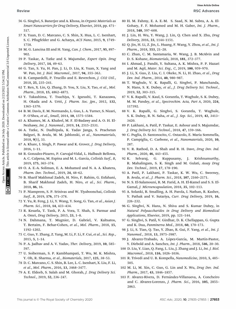
Review RSC Advances
Ope
n A
cces
s A
rtic
le. P
ublis
hed
on 2
4 Ju
ly 2
020.
Dow
nloa
ded
on 2
/23/
2022
5:5
4:22
PM
. T
his
artic
le is
lice
nsed
und
er a
Cre
ativ
e C
omm
ons
Attr
ibut
ion-
Non
Com
mer
cial
3.0
Unp
orte
d L
icen
ce.
View Article Online
56 G. Singhvi, S. Banerjee and A. Khosa, in Organic Materials asSmart Nanocarriers for Drug Delivery, Elsevier, 2018, pp. 471–517.
57 X. Yuan, D. C. Marcano, C. S. Shin, X. Hua, L. C. Isenhart,S. C. Pugfelder and G. Acharya, ACS Nano, 2015, 9, 1749–1758.
58 M. G. Lancina III and H. Yang, Can. J. Chem., 2017, 95, 897–902.
59 P. Taskar, A. Tatke and S. Majumdar, Expert Opin. DrugDelivery, 2017, 14, 49–63.
60 G. Tan, S. Yu, H. Pan, J. Li, D. Liu, K. Yuan, X. Yang andW. Pan, Int. J. Biol. Macromol., 2017, 94, 355–363.
61 R. Campardelli, P. Trucillo and E. Reverchon, J. CO2 Util.,2018, 25, 235–241.
62 T. Ren, X. Lin, Q. Zhang, D. You, X. Liu, X. Tao, et al., Mol.Pharm., 2018, 15, 4862–4871.
63 Y. Takashima, T. Tsuchiya, Y. Igarashi, T. Kanazawa,H. Okada and A. Urtti, J. Pharm. Soc. Jpn., 2012, 132,1365–1370.
64 B. M. Davis, E. M. Normando, L. Guo, L. A. Turner, S. Nizari,P. O'Shea, et al., Small, 2014, 10, 1575–1584.
65 A. Khames, M. A. Khaleel, M. F. El-Badawy and A. O. H. El-Nezhawy, Int. J. Nanomed., 2019, 14, 2515–2531.
66 A. Tatke, N. Dudhipala, K. Yadav Janga, S. PrachetanBalguri, B. Avula, M. M. Jablonski, et al., Nanomaterials,2018, 9(1), 33.
67 A. Khare, I. Singh, P. Pawar and K. Grover, J. Drug Delivery,2016, 1–11.
68 R. Gonzalez-Pizarro, P. Carvajal-Vidal, L. Halbault Bellowa,A. C. Calpena, M. Espina and M. L. Garcıa, Colloids Surf., B,2019, 175, 365–374.
69 O. A. E.-A. Soliman, E. A. Mohamed and N. A. A. Khatera,Pharm. Dev. Technol., 2019, 24, 48–62.
70 B. Sharif Makhmal Zadeh, H. Niro, F. Rahim, G. Esfahani,B. Sharif Makhmal Zadeh, H. Niro, et al., Sci. Pharm.,2018, 86, 16.
71 P. Niamprem, S. P. Srinivas and W. Tiyaboonchai, ColloidsSurf., B, 2019, 176, 371–378.
72 Y. Yu, R. Feng, J. Li, Y. Wang, Y. Song, G. Tan, et al., Asian J.Pharm. Sci., 2018, 14, 423–434.
73 R. Kesarla, T. Tank, P. A. Vora, T. Shah, S. Parmar andA. Omri, Drug Delivery, 2015, 23, 1–8.
74 N. Dahmana, T. Mugnier, D. Gabriel, V. Kaltsatos,T. Bertaim, F. Behar-Cohen, et al., Mol. Pharm., 2018, 15,1192–1202.
75 C. Guo, Y. Zhang, Z. Yang, M. Li, F. Li, F. Cui, et al., Sci. Rep.,2015, 5, 1–14.
76 P. A. Jadhav and A. V. Yadav, Ther. Delivery, 2019, 10, 585–597.
77 U. Soiberman, S. P. Kambhampati, T. Wu, M. K. Mishra,Y. Oh, R. Sharma, et al., Biomaterials, 2017, 125, 38–53.
78 D. C. Marcano, C. S. Shin, B. Lee, L. C. Isenhart, X. Liu, F. Li,et al., Mol. Pharm., 2016, 13, 3468–3477.
79 A. E. Eldeeb, S. Salah and M. Ghorab, J. Drug Delivery Sci.Technol., 2019, 52, 236–247.
This journal is © The Royal Society of Chemistry 2020
80 H. M. Fahmy, E. A. E.-M. S. Saad, N. M. Sabra, A. A. El-Gohary, F. F. Mohamed and M. H. Gaber, Int. J. Pharm.,2018, 548, 597–608.
81 J. Lin, H. Wu, Y. Wang, J. Lin, Q. Chen and X. Zhu, DrugDelivery, 2016, 23, 1144–1151.
82 Q. Jin, H. Li, Z. Jin, L. Huang, F. Wang, Y. Zhou, et al., Int. J.Pharm., 2018, 553, 21–28.
83 C. Zhan, C. M. Santamaria, W. Wang, J. B. McAlvin andD. S. Kohane, Biomaterials, 2018, 181, 372–377.
84 I. Ahmad, J. Pandit, Y. Sultana, A. K. Mishra, P. P. Hazariand M. Aqil, Mater. Sci. Eng., C, 2019, 100, 959–970.
85 J. Li, X. Guo, Z. Liu, C. I. Okeke, N. Li, H. Zhao, et al., DrugDev. Ind. Pharm., 2014, 40, 980–987.
86 T. Waghule, V. K. Rapalli, G. Singhvi, P. Manchanda,N. Hans, S. K. Dubey, et al., J. Drug Delivery Sci. Technol.,2019, 52, 303–315.
87 V. K. Rapalli, V. Kaul, S. Gorantla, T. Waghule, S. K. Dubey,M. M. Pandey, et al., Spectrochim. Acta, Part A, 2020, 224,117392.
88 V. K. Rapalli, G. Singhvi, S. Gorantla, T. Waghule,S. K. Dubey, R. N. Saha, et al., J. Sep. Sci., 2019, 42, 3413–3420.
89 P. Lakhani, A. Patil, P. Taskar, E. Ashour and S. Majumdar,J. Drug Delivery Sci. Technol., 2018, 47, 159–166.
90 C. Puglia, D. Santonocito, C. Ostacolo, E. Maria Sommella,P. Campiglia, C. Carbone, et al., Nanomaterials, 2020, 10,287.
91 V. R. Rathod, D. A. Shah and R. H. Dave, Drug Dev. Ind.Pharm., 2020, 46, 443–455.
92 K. Selvaraj, G. Kuppusamy, J. Krishnamurthy,R. Mahalingam, S. K. Singh and M. Gulati, Assay DrugDev. Technol., 2019, 17, 178–190.
93 A. Patil, P. Lakhani, P. Taskar, K. W. Wu, C. Sweeney,B. Avula, et al., J. Pharm. Sci., 2018, 107, 2160–2171.
94 N. S. El-Salamouni, R. M. Farid, A. H. El-Kamel and S. S. El-Gamal, J. Microencapsulation, 2018, 35, 102–113.
95 A. Solanki, R. Smalling, A. H. Parola, I. Nathan, R. Kasher,Y. Pathak and V. Sutariya, Curr. Drug Delivery, 2019, 16,226–232.
96 G. Singhvi, N. Hans, N. Shiva and S. Kumar Dubey, inNatural Polysaccharides in Drug Delivery and BiomedicalApplications, Elsevier, 2019, pp. 121–144.
97 G. Singhvi, S. Patil, V. Girdhar, D. K. Chellappan, G. Guptaand K. Dua, Panminerva Med., 2018, 60, 170–173.
98 J. Li, S. Tian, Q. Tao, Y. Zhao, R. Gui, F. Yang, et al., Int. J.Nanomed., 2018, 13, 3975–3987.
99 J. Alvarez-Trabado, A. Lopez-Garcıa, M. Martın-Pastor,Y. Diebold and A. Sanchez, Int. J. Pharm., 2018, 546, 20–30.
100 D. Liu, Y. Lian, Q. Fang, L. Liu, J. Zhang and J. Li, Int. J. Biol.Macromol., 2018, 116, 1026–1036.
101 R. Trivedi and U. B. Kompella, Nanomedicine, 2010, 5, 485–505.
102 M. Li, M. Xin, C. Guo, G. Lin and X. Wu, Drug Dev. Ind.Pharm., 2017, 43, 1846–1857.
103 F. Alvarez-Rivera, D. Fernandez-Villanueva, A. Concheiroand C. Alvarez-Lorenzo, J. Pharm. Sci., 2016, 105, 2855–2863.
RSC Adv., 2020, 10, 27835–27855 | 27853
RSC Advances Review
Ope
n A
cces
s A
rtic
le. P
ublis
hed
on 2
4 Ju
ly 2
020.
Dow
nloa
ded
on 2
/23/
2022
5:5
4:22
PM
. T
his
artic
le is
lice
nsed
und
er a
Cre
ativ
e C
omm
ons
Attr
ibut
ion-
Non
Com
mer
cial
3.0
Unp
orte
d L
icen
ce.
View Article Online
104 S. Gorantla, T. Waghule, V. K. Rapalli, P. P. Singh,S. K. Dubey, R. N. Saha and G. Singhvi, Recent Pat. DrugDelivery Formulation, 2019, 13(4), 291–300.
105 N. Morsi, M. Ibrahim, H. Refai and H. El Sorogy, Eur. J.Pharm. Sci., 2017, 104, 302–314.
106 N. Patel, H. Nakrani, M. Raval and N. Sheth, Drug Delivery,2016, 23, 3712–3723.
107 J. Phua, A. Hou, Y. Lui, T. Bose, G. Chandy, L. Tong, et al.,Int. J. Mol. Sci., 2018, 19, 2977.
108 V. Agrahari, Nanomedicine, 2017, 12, 819–823.109 V. K. Rapalli, A. Khosa, G. Singhvi, V. Girdhar, R. Jain and
S. K. Dubey, Pharm. Qual. by Des., 2019, pp. 255–296.110 M. L. Chen, M. John, S. L. Lee and K. M. Tyner, AAPS J.,
2017, 19, 642–651.111 A. P. Colombo, S. Briançon, J. Lieto and H. Fessi, Drug Dev.
Ind. Pharm., 2001, 27, 1063–1072.112 K. Baba and K. Nishida, Pharmaceutics, 2013, 5, 107–114.113 S. Galindo-Rodriguez, E. Allemann, H. Fessi and E. Doelker,
Pharm. Res., 2004, 21, 1428–1439.114 H. Patil, R. V. Tiwari and M. A. Repka, AAPS PharmSciTech,
2016, 17, 20–42.115 S. M. Jafari, E. Assadpoor, Y. He and B. Bhandari, Food
Hydrocolloids, 2008, 22, 1191–1202.116 V. Jenning, A. Lippacher and S. H. Gohla, J.
Microencapsulation, 2002, 19, 1–10.117 A. Fabregas, M. Minarro, E. Garcıa-Montoya, P. Perez-
Lozano, C. Carrillo, R. Sarrate, et al., Int. J. Pharm., 2013,446, 199–204.
118 D. Liu, H. Zhang, F. Fontana, J. T. Hirvonen andH. A. Santos, Adv. Drug Delivery Rev., 2018, 128, 54–83.
119 H. S. M. Ali, P. York, A. M. A. Ali and N. Blagden, J.Controlled Release, 2011, 149, 175–181.
120 A. Gdowski, K. Johnson, S. Shah, I. Gryczynski,J. Vishwanatha and A. Ranjan, J. Nanobiotechnol., 2018,16, 12.
121 C. A. Charitidis, P. Georgiou, M. A. Koklioti, A.-F. Trompetaand V. Markakis, Manuf. Rev., 2014, 1, 11.
122 J. Jung, J. Y. Clavier and M. Perrut, Mater. Sci., 2003, 3,1683–1688.
123 T. T. Pham, C. Jaafar-Maalej, C. Charcosset and H. Fessi,Colloids Surf., B, 2012, 94, 15–21.
124 J. L. Perry, K. P. Herlihy, M. E. Napier and J. M. Desimone,Acc. Chem. Res., 2011, 44, 990–998.
125 G. Acharya, C. S. Shin, M. McDermott, H. Mishra, H. Park,I. C. Kwon and K. Park, J. Controlled Release, 2010, 141, 314–319.
126 T. Meng, V. Kulkarni, R. Simmers, V. Brar and Q. Xu, DrugDiscovery Today, 2019, 24, 1524–1538.
127 S. Soares, J. Sousa, A. Pais and C. Vitorino, Front. Chem.,2018, 6, 360.
128 R. Paliwal, R. J. Babu and S. Palakurthi, AAPS PharmSciTech,2014, 15, 1527–1534.
129 S. Hua, M. B. C. de Matos, J. M. Metselaar and G. Storm,Front. Pharmacol., 2018, 9, 1–9.
130 FDA, Center for drug evaluation and research applicationnumber: 210913orig1s000 summary review, https://
27854 | RSC Adv., 2020, 10, 27835–27855
www.accessdata.fda.gov/drugsatfda_docs/nda/2018/210913Orig1s000SumR.pdf.
131 M. S. Uddin, A. Al Mamun, M. T. Kabir, J. R. Setu, S. Zaman,Y. Begum and M. S. Amran, J. Adv. Med. Pharm. Sci., 2017,14(2), 1–17.
132 FDA, CDER, Yeaton and Ayse, Drug Products, IncludingBiological Products, that Contain Nanomaterials - Guidancefor Industry, https://www.fda.gov/Drugs/GuidanceComplianceRegulatoryInformation/Guidances/default.htm.
133 FDA and CDER, Liposome Drug Products Chemistry,Manufacturing, and Controls; Human Pharmacokinetics andBioavailability; and Labeling Documentation Guidance forIndustry, 2018, http://www.fda.gov/Drugs/GuidanceComplianceRegulatoryInformation/Guidances/default.htm.
134 Selected FDA Publications Related to the Application,Characterization, Effects, and Evaluation of Nanotechnology,FDA, https://www.fda.gov/science-research/nanotechnology-programs-fda/selected-fda-publications-related-application-characterization-effects-and-evaluation-nanotechnology,accessed 31 March 2020.
135 Guidelines for Evaluation of Nanopharmaceuticals in India,2019, http://www.dbtindia.gov.in/sites/default/les/uploadles/Guidelines_For_Evaluation_of_Nanopharmaceuticals_in_India_24.10.19.pdf.
136 J. H. Draize, J. Pharmacol. Exp. Ther., 1944, 82, 377–390.137 T. K. De, E. J. Bergey, S. J. Chung, D. J. Rodman, D. J. Bharali
and P. N. Prasad, J. Microencapsulation, 2004, 21, 841–855.138 E. Vega, M. A. Egea, O. Valls, M. Espina and M. L. Garcia, J.
Pharm. Sci., 2006, 95, 2393–2405.139 T. W. Prow, Wiley Interdiscip. Rev.: Nanomed.
Nanobiotechnol., 2010, 2, 317–333.140 E. Beltran-Gracia, A. Lopez-Camacho, I. Higuera-Ciapara,
J. B. Velazquez-Fernandez and A. A. Vallejo-Cardona,Cancer Nanotechnol., 2019, 10, 1–40.
141 Y. I. Eroglu, J. Mark. Access Health Policy, 2017, 5, 1336043.142 Sun Pharma Launches Cequa for the Treatment of Dry Eye
Disease in the US – Eyewire News, https://www.eyewire.news/articles/sun-pharma-launches-cequa-for-the-treatment-of-dry-eye-disease-in-the-us/, accessed 29March 2020.
143 MPP Platform - Kala Pharmaceuticals, https://www.kalarx.com/technology/mpp-platform/, accessed 29March 2020.
144 Aerie Pharmaceuticals Announces Drug Delivery AssetAcquisition to Further Advance Its Retinal Disease Program |Business Wire, https://www.businesswire.com/news/home/20171005005490/en/Aerie-Pharmaceuticals-Announces-Drug-Delivery-Asset-Acquisition, accessed 30 March 2020.
145 Envisia Therapeutics Secures $16.5 Million In AdditionalSeries A Financing, https://www.prnewswire.com/news-releases/envisia-therapeutics-secures-165-million-in-additional-series-a-nancing-300240151.html, accessed 28March 2020.
146 Paclitaxel Albumin-Stabilized Nanoparticle Formulation inTreating Patients With Metastatic Melanoma of the Eye ThatCannot Be Removed By Surgery - Full Text View -
This journal is © The Royal Society of Chemistry 2020
Review RSC Advances
Ope
n A
cces
s A
rtic
le. P
ublis
hed
on 2
4 Ju
ly 2
020.
Dow
nloa
ded
on 2
/23/
2022
5:5
4:22
PM
. T
his
artic
le is
lice
nsed
und
er a
Cre
ativ
e C
omm
ons
Attr
ibut
ion-
Non
Com
mer
cial
3.0
Unp
orte
d L
icen
ce.
View Article Online
ClinicalTrials.gov, https://www.clinicaltrials.gov/ct2/show/NCT00738361, accessed 31 July 2019.
147 Safety and Efficacy of Liposomal Latanoprost in OcularHypertension - Full Text View - ClinicalTrials.gov, https://www.clinicaltrials.gov/ct2/show/NCT01987323, accessed29 July 2019.
148 A Randomized Controlled Trial Comparing Urea LoadedNanoparticles to Placebo: a New Concept for CataractManagement - Full Text View - ClinicalTrials.gov, https://www.clinicaltrials.gov/ct2/show/NCT03001466, accessed29 July 2019.
149 TLC399 (ProDex) in Subjects With Macular Edema Due toRetinal Vein Occlusion (RVO) - Full Text View -ClinicalTrials.gov, https://www.clinicaltrials.gov/ct2/show/NCT03093701, accessed 28 August 2019.
150 Crosslinked Hyaluronic Acid With Liposomes and Crocin inDry Eye - Full Text View - ClinicalTrials.gov, https://www.clinicaltrials.gov/ct2/show/NCT03617315, accessed 8February 2020.
151 Search of: NCT03785340 - List Results - ClinicalTrials.gov,https://www.clinicaltrials.gov/ct2/results?cond¼&term¼NCT03785340&cntry¼&state¼&city¼&dist¼,accessed 28 March 2020.
152 A Depot Formulation of Sunitinib Malate (GB-102) in SubjectsWith Neovascular (Wet) Age-related Macular Degeneration -Full Text View - ClinicalTrials.gov, https://www.clinicaltrials.gov/ct2/show/NCT03249740, accessed28 March 2020.
153 LAMELLEYE for the Treatment of Dry Eye Symptoms in pSSPatients - Full Text View - ClinicalTrials.gov, https://www.clinicaltrials.gov/ct2/show/NCT03140111, accessed28 March 2020.
154 A Study to Evaluate the Safety, Tolerability, SystemicPharmacokinetics, and Pharmacodynamics of AXR-159Ophthalmic Solution in Patients With Dry Eye Disease (DED)- Full Text View - ClinicalTrials.gov, https://www.clinicaltrials.gov/ct2/show/NCT03598699, accessed28 March 2020.
155 Safety and Efficacy of KPI-121 in Subjects With PostsurgicalInammation - Full Text View - ClinicalTrials.gov, https://
This journal is © The Royal Society of Chemistry 2020
www.clinicaltrials.gov/ct2/show/NCT02163824, accessed28 March 2020.
156 Safety and Efficacy of KPI-121 in Subjects With Dry Eye Disease- Full Text View - ClinicalTrials.gov, https://www.clinicaltrials.gov/ct2/show/NCT02813265, accessed28 March 2020.
157 Study Assessing AR-1105 in Subjects With Macular Edema Dueto Retinal Vein Occlusion (RVO) - Full Text View -ClinicalTrials.gov, https://www.clinicaltrials.gov/ct2/show/NCT03739593, accessed 28 March 2020.
158 A Study Assessing AR-13503 Implant Alone and inCombination With Aibercept in Subjects With nAMD orDME - Full Text View - ClinicalTrials.gov, https://www.clinicaltrials.gov/ct2/show/NCT03835884, accessed28 March 2020.
159 Topical Omega-3 Fatty Acids (REMOGEN® OMEGA) in theTreatment of Dry Eye - Full Text View - ClinicalTrials.gov,https://www.clinicaltrials.gov/ct2/show/NCT02908282,accessed 28 March 2020.
160 Dry Eye Treatment With Articial Tears - Full Text View -ClinicalTrials.gov, https://www.clinicaltrials.gov/ct2/show/NCT02420834, accessed 28 March 2020.
161 Safety and Efficacy of ENV515 Travoprost Extended Release(XR) in Patients With Bilateral Ocular Hypertension orPrimary Open Angle Glaucoma - Full Text View -ClinicalTrials.gov, https://www.clinicaltrials.gov/ct2/show/NCT02371746, accessed 28 March 2020.
162 Intravitreal Injection of SeeQ CdSe 655 Alt Nanoparticles forPatients With Degenerative Retinal Diseases - Full Text View- ClinicalTrials.gov, https://www.clinicaltrials.gov/ct2/show/NCT04008771, accessed 29 March 2020.
163 OCS-01 in Treating Inammation and Pain in Post-cataractPatients - Full Text View - ClinicalTrials.gov, https://www.clinicaltrials.gov/ct2/show/NCT04130802, accessed29 March 2020.
164 Global Ophthalmic Drugs Market Expected to Reach USD34.52 Billion By 2024: Zion Market Research, https://www.globenewswire.com/news-release/2018/09/21/1574283/0/en/Global-Ophthalmic-Drugs-Market-Expected-to-Reach-USD-34-52-Billion-By-2024-Zion-Market-Research.html, accessed 31 July 2019.
RSC Adv., 2020, 10, 27835–27855 | 27855