Embed Size (px)
Citation preview
• Nancy Yazinski APRN,CWCN
• Hyperbaric Medicine
LOWER EXTREMITY ARTERIAL DISEASE
•
•
• Case Review #1
• ARTERIAL ULCER
Lower Extremity Arterial Disease LEAD
• Peripheral Arterial Disease:
• narrowing or occlusion of arteries by atherosclerotic plaques.
•
Centers for Disease Control
Lower Extremity Arterial Disease
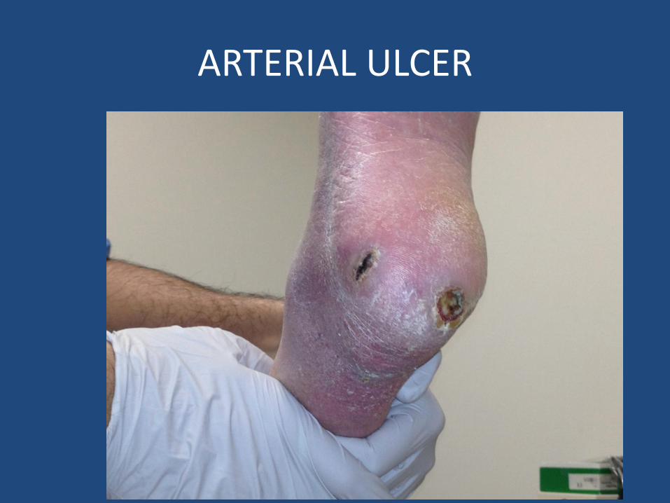
• Typical Location of Arterial Ulcers
• Feet, heels , tips of toes and between toes, nail beds of toes.
• Appearance
• Base is yellow, brown, grey or black and does not bleed, minimal drainage.
• Wound borders are punched out.
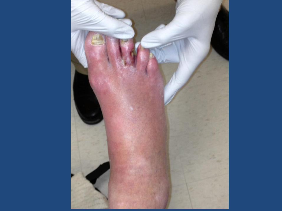
• Redness to entire foot when leg is dependent; redness turns to pale/yellow color when leg is elevated
• Pain
• Typically very painful especially at night, dangling extremity relieves pain.
CASE STUDY Lower Extremity Arterial Disease
• 80 year old male with lower extremity arterial occlusive disease.
• November, 2012, spontaneously developed ulcerations to right plantar heel, lateral second and third toes.
• Ulcers were painful with direct pressure and did not awaken him at night.
• He was seen by his PCP and referred to a vascular surgeon and wound center.
Problem List
• Peripheral Vascular Disease
• Abdominal Aortic Aneurysm
• HTN
• Hyperlipidemia
• Spinal Stenosis
• Bilateral Hip Replacement
• Type 2 Diabetes Mellitus
• Prostate cancer
• Dementia
• Social History: Smoked for 40 yrs;Quit smoking 20 yrs ago
Lower Extremity Arterial Disease
• January,2013: Seen at wound center and by a vascular surgeon.
• Physical Exam: Palpable pulses>radial, femoral and left popliteal.
• Non-palpable pulses>right popliteal, posterior tibial and dorsalis pedis .
• Full thickness ulcers right medial heel with a smaller ulcer to lateral right heel.
• Open wound to lateral right third toe with a smaller wound to lateral second toe.
Treatment ???
• Revascularization-not a good candidate for surgery
• OR
• Hyperbaric Oxygen treatments as adjunct to wound care
• Decision
• January 31,2013: Hyperbaric Medicine consult DHMC
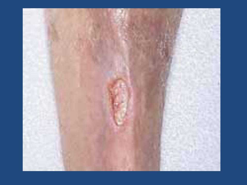
ARTERIAL ULCER
ARTERIAL ULCER
Lower Extremity Arterial Disease
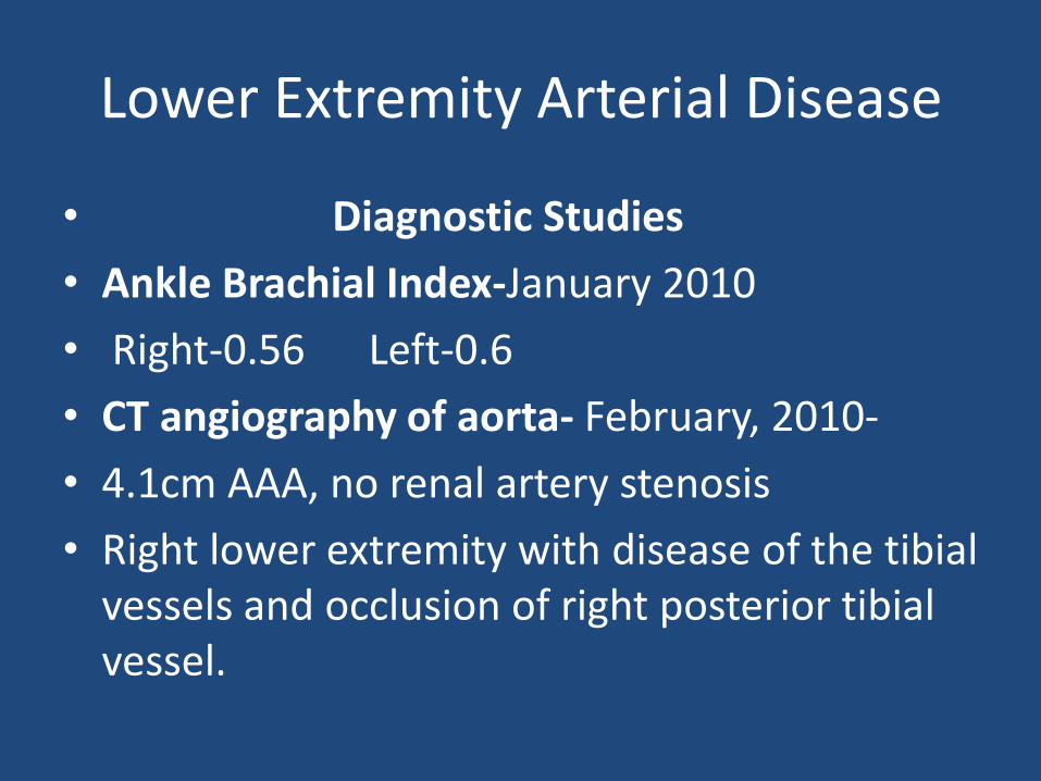
• Diagnostic Studies
• Ankle Brachial Index-January 2010
• Right-0.56 Left-0.6
• CT angiography of aorta- February, 2010-
• 4.1cm AAA, no renal artery stenosis
• Right lower extremity with disease of the tibial vessels and occlusion of right posterior tibial vessel.
Diagnostic Studies
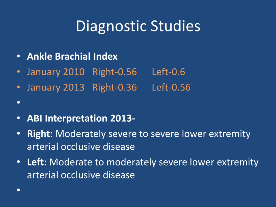
• Ankle Brachial Index
• January 2010 Right-0.56 Left-0.6
• January 2013 Right-0.36 Left-0.56
•
• ABI Interpretation 2013-
• Right: Moderately severe to severe lower extremity arterial occlusive disease
• Left: Moderate to moderately severe lower extremity arterial occlusive disease
•
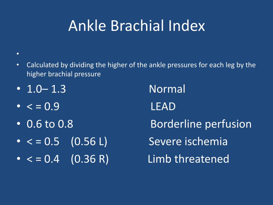
Ankle Brachial Index
•
• Calculated by dividing the higher of the ankle pressures for each leg by the higher brachial pressure
• 1.0– 1.3 Normal
• < = 0.9 LEAD
• 0.6 to 0.8 Borderline perfusion
• < = 0.5 (0.56 L) Severe ischemia
• < = 0.4 (0.36 R) Limb threatened
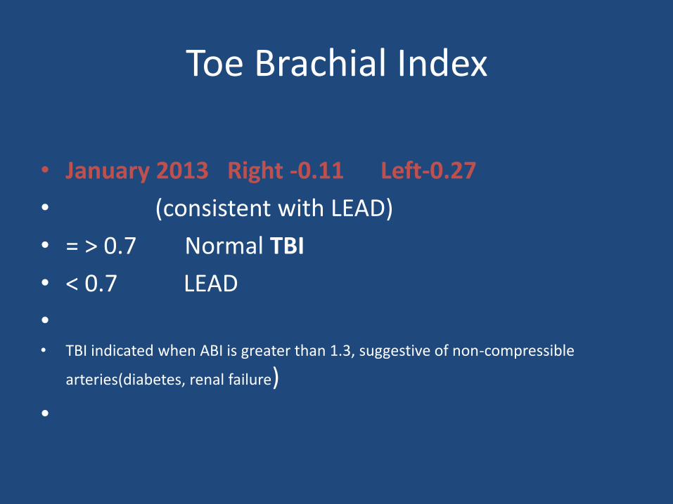
Toe Brachial Index
• January 2013 Right -0.11 Left-0.27
• (consistent with LEAD)
• = > 0.7 Normal TBI
• < 0.7 LEAD
• • TBI indicated when ABI is greater than 1.3, suggestive of non-compressible
arteries(diabetes, renal failure)
•
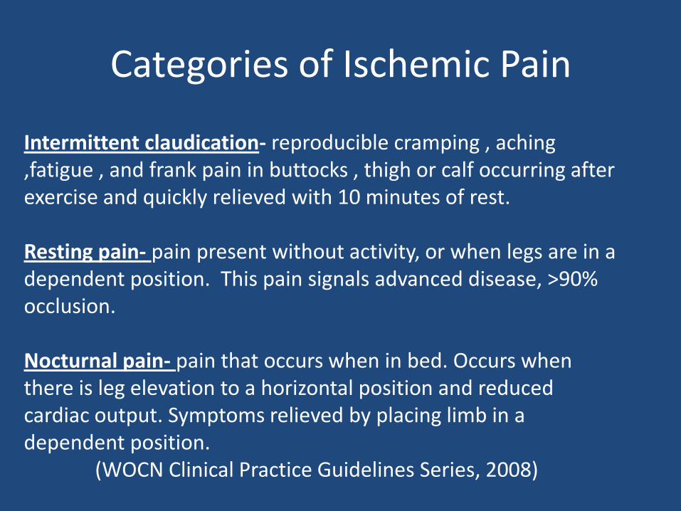
Categories of Ischemic Pain
Intermittent claudication- reproducible cramping , aching ,fatigue , and frank pain in buttocks , thigh or calf occurring after exercise and quickly relieved with 10 minutes of rest. Resting pain- pain present without activity, or when legs are in a dependent position. This pain signals advanced disease, >90% occlusion. Nocturnal pain- pain that occurs when in bed. Occurs when there is leg elevation to a horizontal position and reduced cardiac output. Symptoms relieved by placing limb in a dependent position. (WOCN Clinical Practice Guidelines Series, 2008)
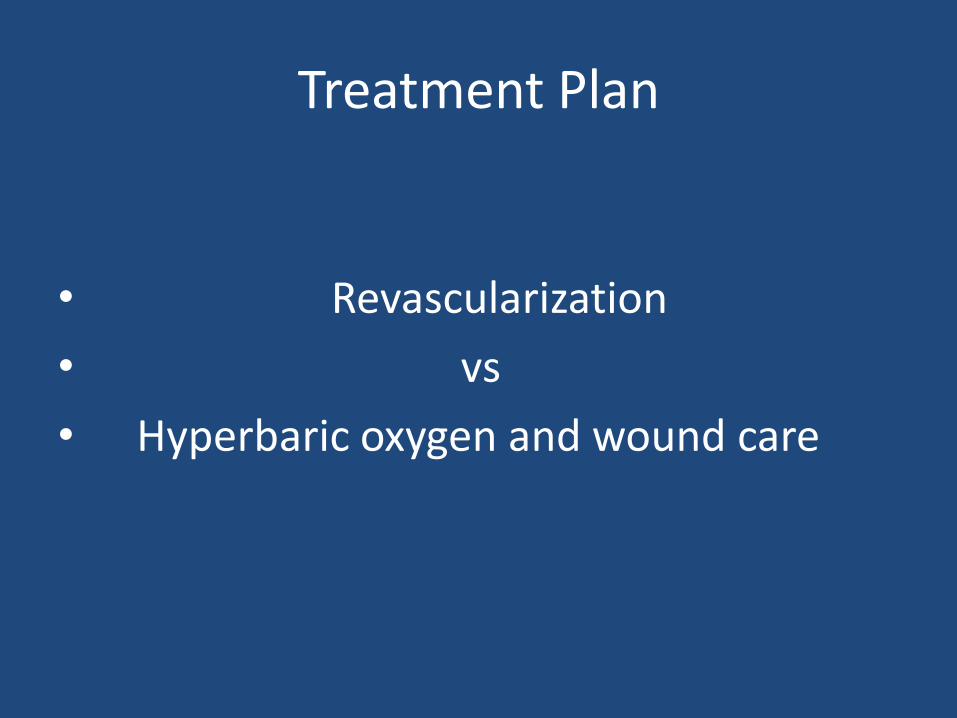
Treatment Plan
• Revascularization
• vs
• Hyperbaric oxygen and wound care
Treatment Plan
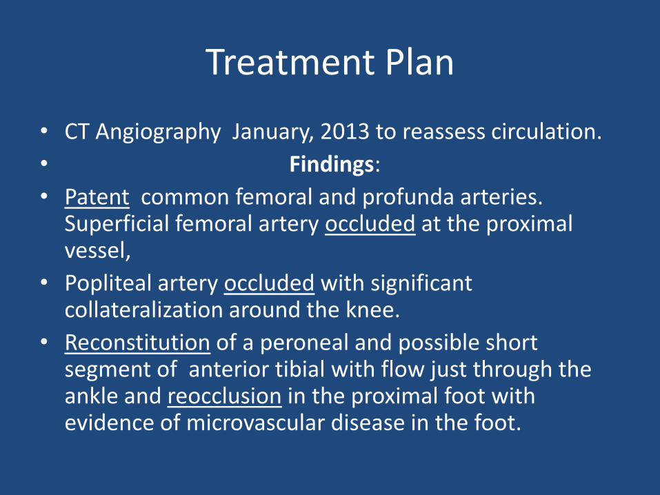
• CT Angiography January, 2013 to reassess circulation.
• Findings:
• Patent common femoral and profunda arteries. Superficial femoral artery occluded at the proximal vessel,
• Popliteal artery occluded with significant collateralization around the knee.
• Reconstitution of a peroneal and possible short segment of anterior tibial with flow just through the ankle and reocclusion in the proximal foot with evidence of microvascular disease in the foot.
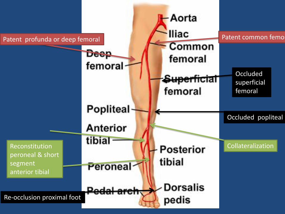
Patent profunda or deep femoral
Occluded superficial femoral
Occluded popliteal
Patent common femoral
Re-occlusion proximal foot
Reconstitution peroneal & short segment anterior tibial
Collateralization
Treatment Plan
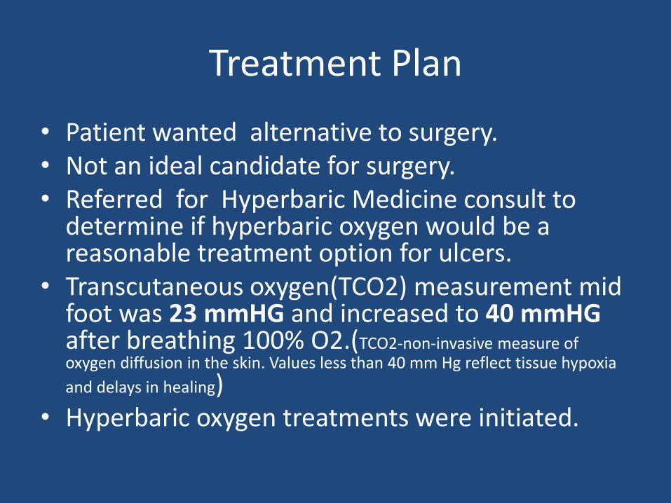
• Patient wanted alternative to surgery. • Not an ideal candidate for surgery. • Referred for Hyperbaric Medicine consult to
determine if hyperbaric oxygen would be a reasonable treatment option for ulcers.
• Transcutaneous oxygen(TCO2) measurement mid foot was 23 mmHG and increased to 40 mmHG after breathing 100% O2.(TCO2-non-invasive measure of oxygen diffusion in the skin. Values less than 40 mm Hg reflect tissue hypoxia
and delays in healing) • Hyperbaric oxygen treatments were initiated.
Hyperbaric Treatment
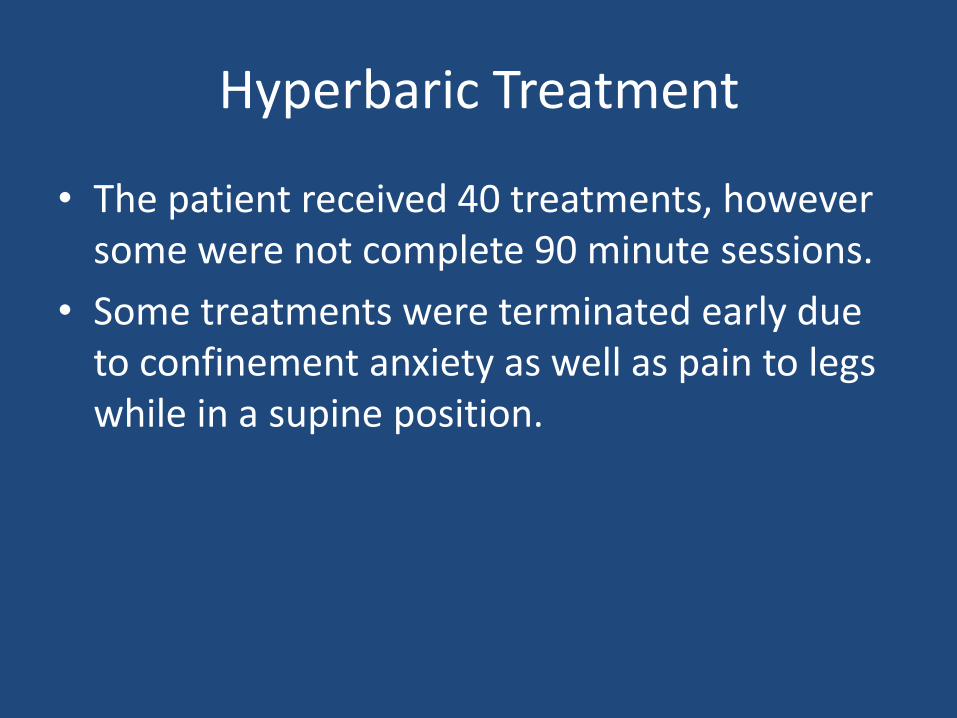
• The patient received 40 treatments, however some were not complete 90 minute sessions.
• Some treatments were terminated early due to confinement anxiety as well as pain to legs while in a supine position.
Result
• • Ulcers did not completely heal but did slightly improve. • Goal : keep wounds stable, prevent infection and sepsis.
• Wound care continued at home, managed by PCP and VNA.
• Latest update-Referred back to wound center June
2013,wounds became worse . • Referred back to vascular surgeon regarding options to
include amputation, long shot at bypass graft or continued conservative management with wound care and watchful waiting until pain or sepsis became an issue.
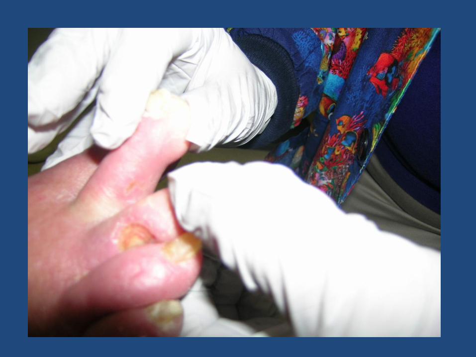
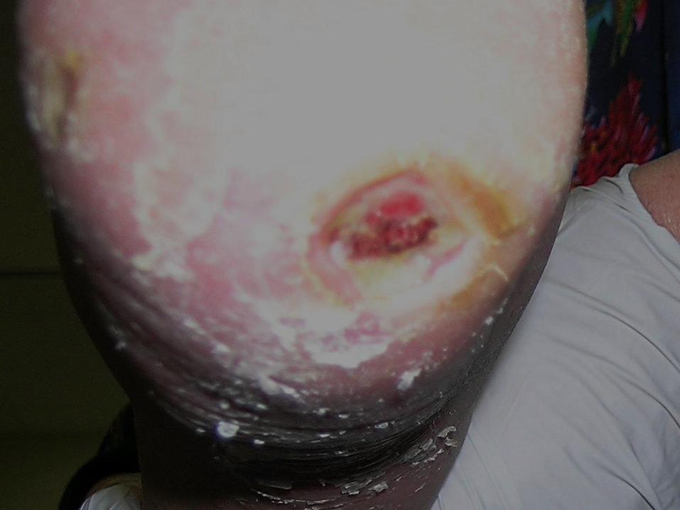
ARTERIAL ULCER
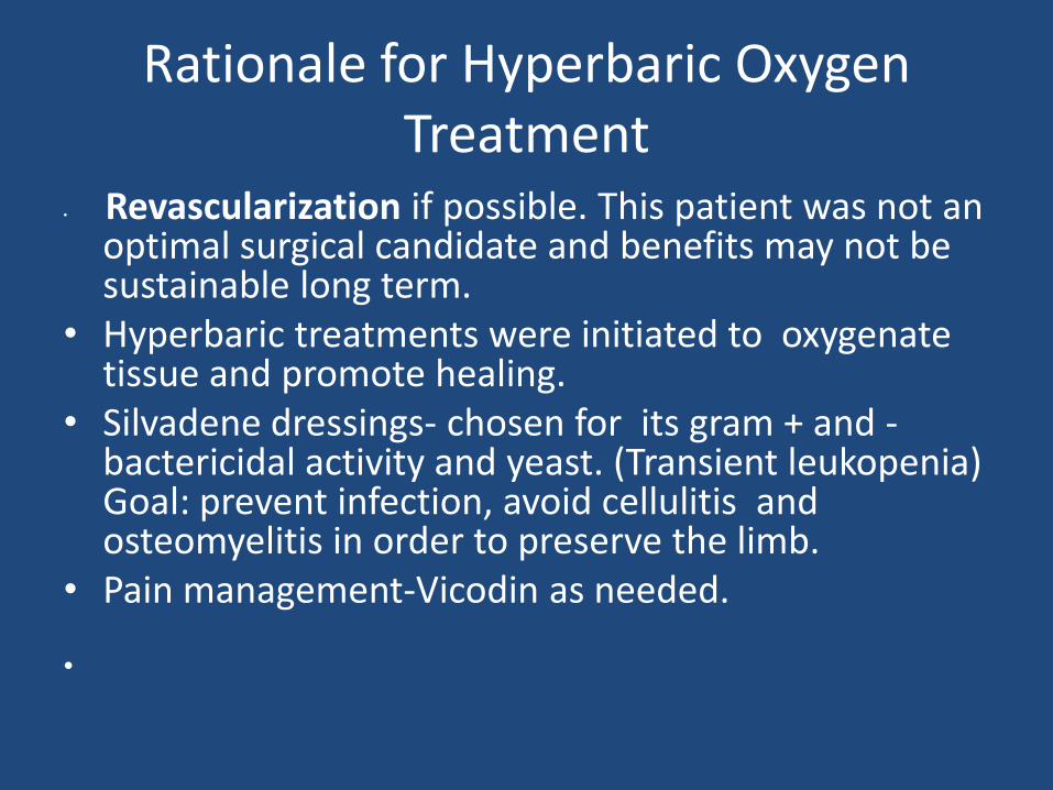
Rationale for Hyperbaric Oxygen Treatment
• Revascularization if possible. This patient was not an optimal surgical candidate and benefits may not be sustainable long term.
• Hyperbaric treatments were initiated to oxygenate tissue and promote healing.
• Silvadene dressings- chosen for its gram + and - bactericidal activity and yeast. (Transient leukopenia) Goal: prevent infection, avoid cellulitis and osteomyelitis in order to preserve the limb.
• Pain management-Vicodin as needed. •
Rationale for Treatment
• Preserve perfusion status: a)Statin-pravachol for atherosclerosis,improve ABI
• Aspirin-to prevent stroke and myocardial infarction,
• Niaspan- off label for PVD,
• Benazepril for blood pressure.
• Dementia impaired treatment-extremely forgetful, unable to correct for this.
• Exercise is strongly recommended in these patients. In this case, activity was limited related to pain and balance issues ,activity was as tolerated.
WOUND MANAGEMENT for ARTERIAL ULCERS
• Cleanse wounds with non-cytotoxic cleansers
• Avoid debridement until perfusion status is determined
• There is no consensus about use of enzymatic or autolytic debridement in arterial wounds with necrotic tissue. Each case requires careful evaluation.
• Due to concerns of infection in an ischemic limb, dressing choice should allow for frequent inspection of the wound.
• Arterial wounds with soft slough or necrotic material might benefit from a closely monitored trial of moisture retentive dressings.
• Maintain dry stable eschar- expert consensus supports maintenance of dry, stable eschar in non-infected arterial wounds to prevent wet gangrene. (Painting dry eschar with Povidone iodine and allowing it to dry)
• Adjunctive Hyperbaric oxygen therapy
• Use of topical antibiotics can result in sensitivity and resistance over time
• Identify and treat infections promptly-systemic antibiotics, surgical consult
LOWER EXTREMITY NEUROPATHIC DISEASE
CASE REVIEW #2
DIABETIC NEUROPATHIC ULCER
LOWER EXTREMITY NEUROPATHIC ULCER
• Definition: Lower extremity neuropathic disease results in autonomic dysfunction and loss of sensation further complicated by infection and ischemia.
• Diabetic neuropathy is the most common cause of neuropathic ulcers.
• (WOCN Clinical Practice Guidelines, 2004)
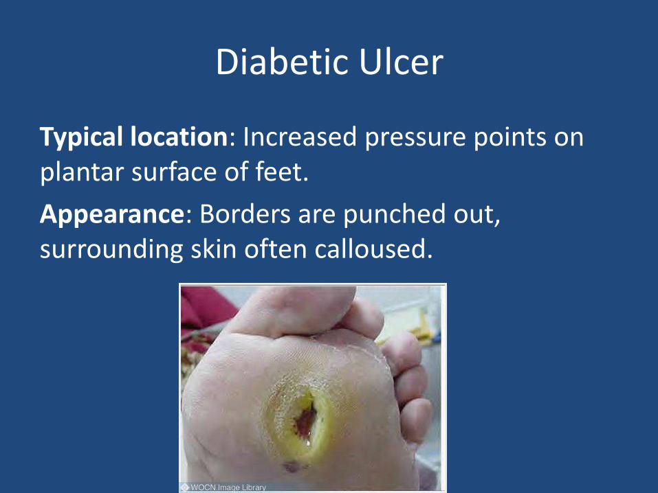
Diabetic Ulcer
Typical location: Increased pressure points on plantar surface of feet.
Appearance: Borders are punched out, surrounding skin often calloused.
Lower Extremity Neuropathic Ulcer
• Scenario: 75 year old male with 35 year history of Type 1 Diabetes Mellitus.
• May 2003-devloped ulcer to left foot that did not heal. • August-October 2003- Several surgeries for removal of
2nd digit right foot , 1st and 5th digits left foot. • Since second surgery, poor healing of ulcer to lateral
aspect left foot. • Wound therapy- daily dressing changes with
Santyl(removes necrotic debri),Granulex spray. • November, 2003-Referred for hyperbaric oxygen
treatments.
Lower Extremity Neuropathic Ulcer
• PMH: • Coronary Artery Disease, Inferior MI. • Seizures- occurred in setting of heart block. Pacemaker
placed. • Type 1 Diabetes Mellitus • Right total knee replacement • Pneumothorax after pacemaker insertion • Amputation of left great toe and fifth toe, amputation of
right third toe • History of tobacco and occasional alcohol use. • Medications • Nexium,Lipitor, Lotensin, Multi-vitamin, Insulin, ASA
Lower Extremity Neuropathic Ulcer
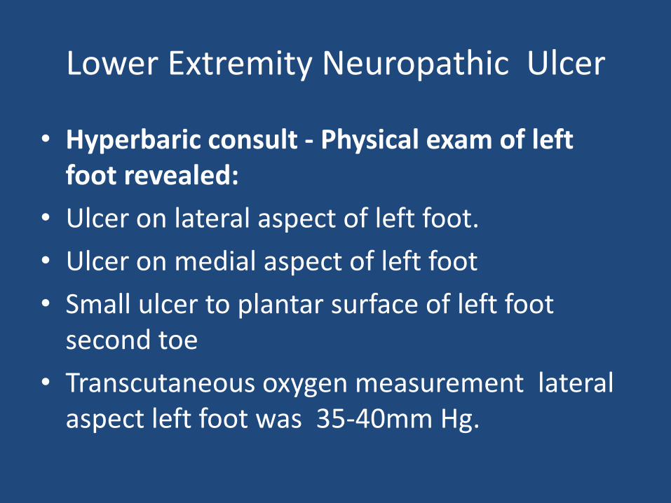
• Hyperbaric consult - Physical exam of left foot revealed:
• Ulcer on lateral aspect of left foot.
• Ulcer on medial aspect of left foot
• Small ulcer to plantar surface of left foot second toe
• Transcutaneous oxygen measurement lateral aspect left foot was 35-40mm Hg.
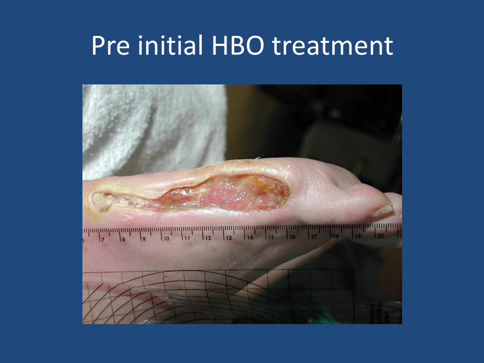
Pre initial HBO treatment
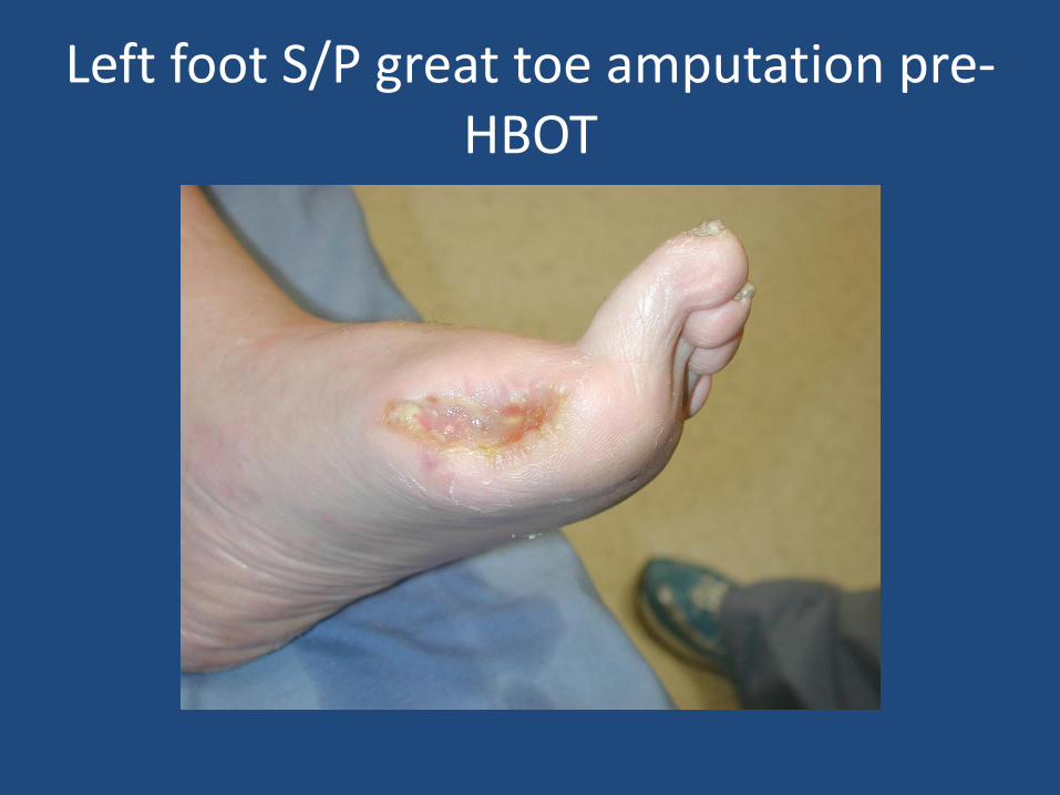
Left foot S/P great toe amputation pre-HBOT
Lower Extremity Neuropathic Ulcer
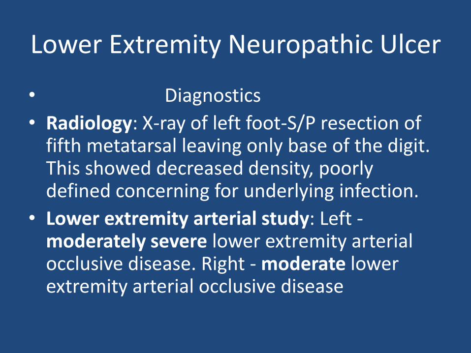
• Diagnostics
• Radiology: X-ray of left foot-S/P resection of fifth metatarsal leaving only base of the digit. This showed decreased density, poorly defined concerning for underlying infection.
• Lower extremity arterial study: Left - moderately severe lower extremity arterial occlusive disease. Right - moderate lower extremity arterial occlusive disease
Treatment Course
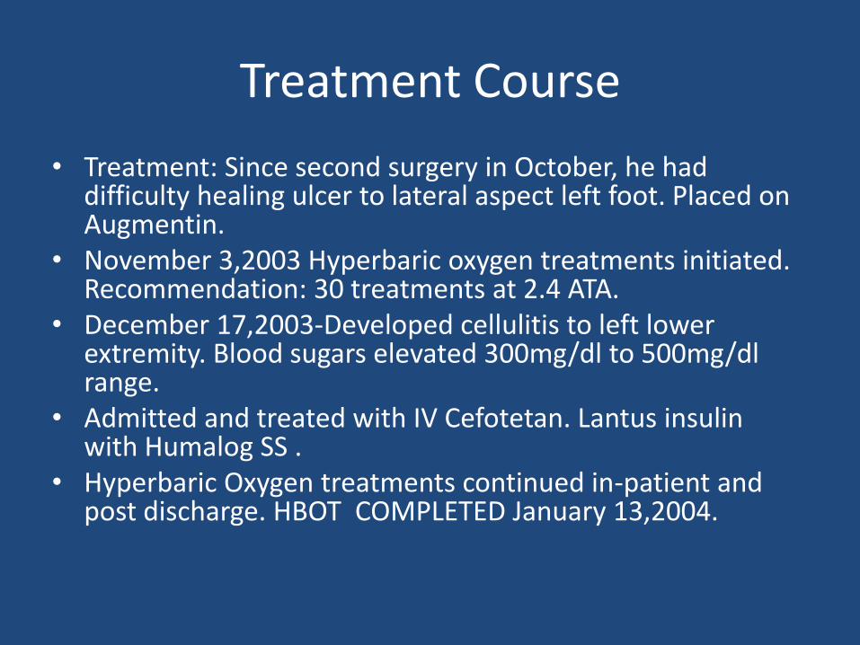
• Treatment: Since second surgery in October, he had difficulty healing ulcer to lateral aspect left foot. Placed on Augmentin.
• November 3,2003 Hyperbaric oxygen treatments initiated. Recommendation: 30 treatments at 2.4 ATA.
• December 17,2003-Developed cellulitis to left lower extremity. Blood sugars elevated 300mg/dl to 500mg/dl range.
• Admitted and treated with IV Cefotetan. Lantus insulin with Humalog SS .
• Hyperbaric Oxygen treatments continued in-patient and post discharge. HBOT COMPLETED January 13,2004.
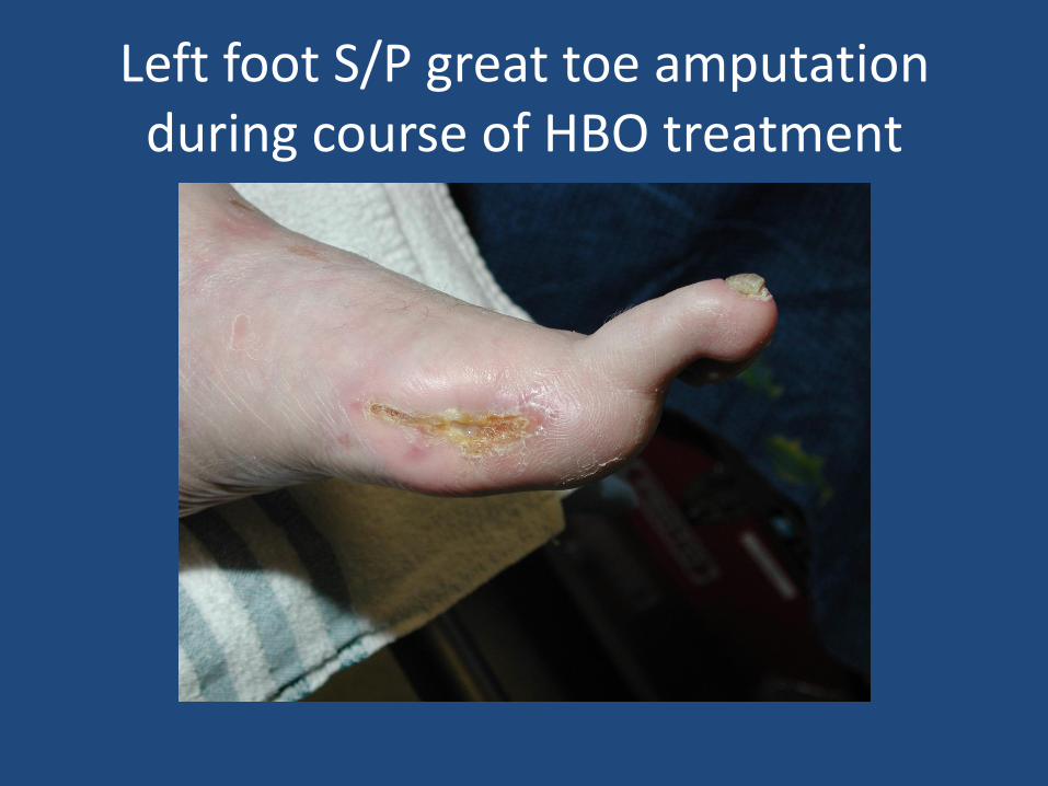
Left foot S/P great toe amputation during course of HBO treatment
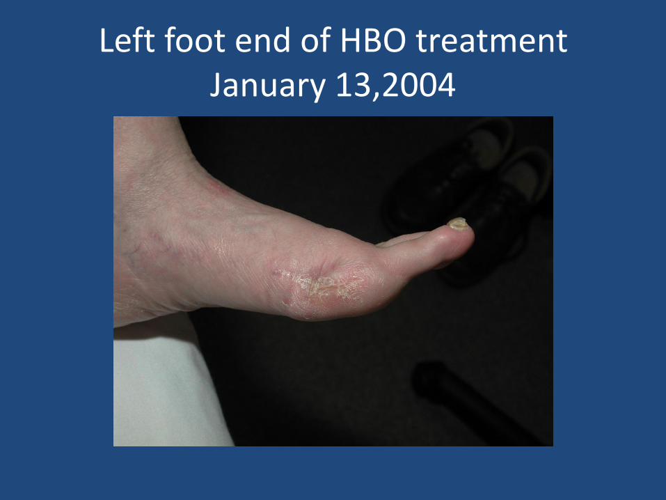
Left foot end of HBO treatment January 13,2004
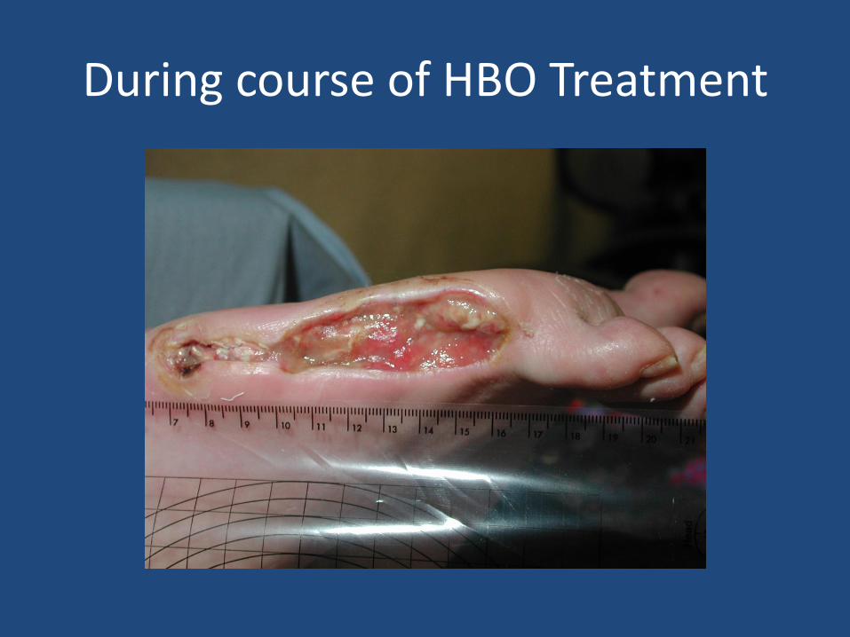
During course of HBO Treatment
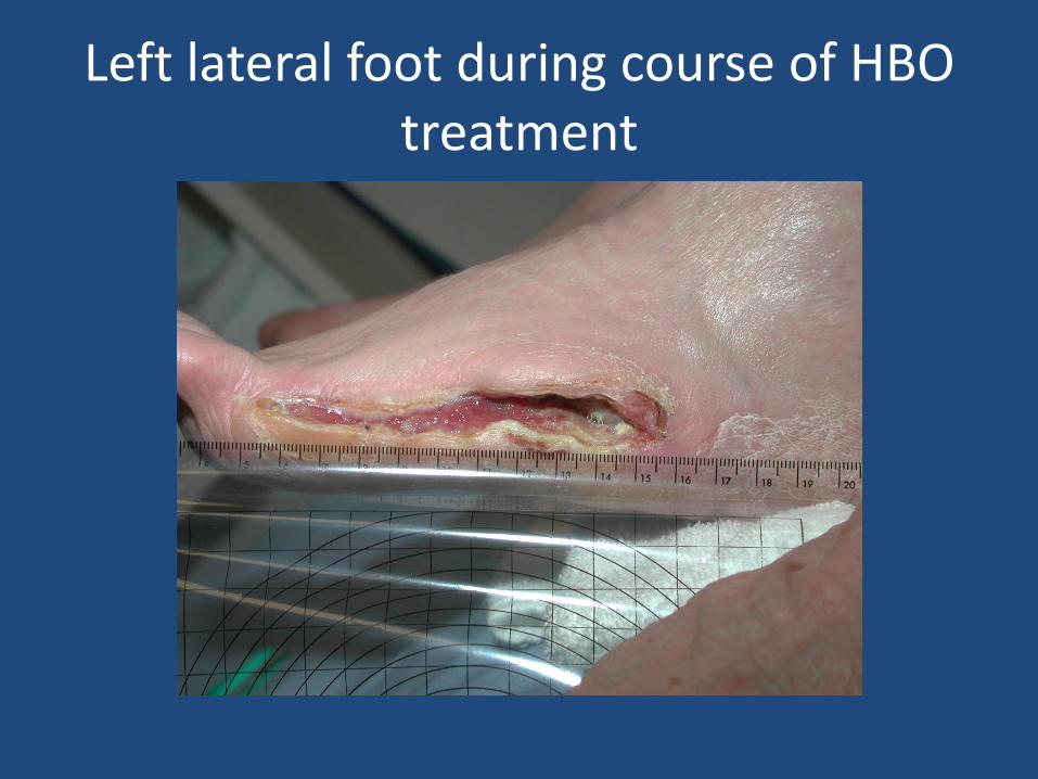
Left lateral foot during course of HBO treatment
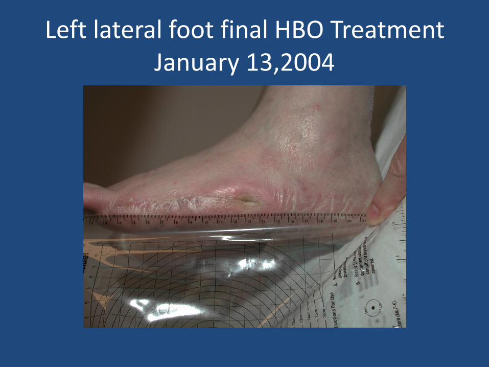
Left lateral foot final HBO Treatment January 13,2004
Rationale for Hyperbaric Oxygen Therapy
• Individuals with diabetic neuropathic ulcers and wounds typically have poor healing related to impaired arterial circulation and elevated blood sugar levels.
• Tissues lack sufficient oxygen to heal wounds.
• Hyperbaric Oxygen provides healing by supplying high oxygen levels to oxygen deprived wounds.
WOUND MANAGEMENT RECOMMENDATIONS for
NEUROPATHIC ULCERS • Assess perfusion status • Offloading • Wound management: a)maintain dry stable eschar
b)cleanse wounds with non-cytotoxic cleansers • Debridement • Dressings: a) promote moist wound environment ,b)PD-GF
platelet derived growth factor-Regranex • Treat Infection • Nutrition • Pain Management • Manage edema • Adjunctive Therapy-Hyperbaric Oxygen
References
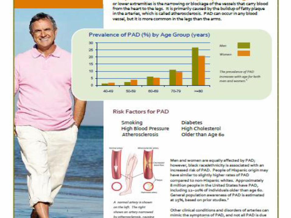
• http://www.cdc.gov. Centers for Disease Control and Prevention; Peripheral Arterial Disease Fact Sheet, July 26,2013.
• Management of Wounds in Patients with Lower Extremity Arterial Disease, pp 2-41. Wound, Ostomy and Continence Nurses Society, 2008.
• Management of Wounds in Patients with Lower-Extremity Neuropathic Disease, pp.1-40. Wound, Ostomy and Continence Nurses Society,2004.