Embed Size (px)
Citation preview
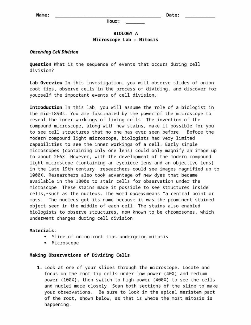
Name: _______________________________________ Date: ___________ Hour: _______
BIOLOGY AMicroscope Lab – Mitosis
Observing Cell Division
Question What is the sequence of events that occurs during cell division?
Lab Overview In this investigation, you will observe slides of onion root tips, observe cells in the process of dividing, and discover for yourself the important events of cell division.
Introduction In this lab, you will assume the role of a biologist in the mid-1890s. You are fascinated by the power of the microscope to reveal the inner workings of living cells. The invention of the compound microscope, along with new stains, make it possible for you to see cell structures that no one has ever seen before. Before the modern compound light microscope, biologists had very limited capabilities to see the inner workings of a cell. Early simple microscopes (containing only one lens) could only magnify an image up to about 266X. However, with the development of the modern compound light microscope (containing an eyepiece lens and an objective lens) in the late 19th century, researchers could see images magnified up to 1000X. Researchers also took advantage of new dyes that became available in the 1800s to stain cells for observation under the microscope. These stains made it possible to see structures inside cells, such as the nucleus. The word nucleus means “a central point or mass.” The nucleus got its name because it was the prominent stained object seen in the middle of each cell. The stains also enabled biologists to observe structures, now known to be chromosomes, which underwent changes during cell division.
Materials: Slide of onion root tips undergoing mitosis Microscope
Making Observations of Dividing Cells
1. Look at one of your slides through the microscope. Locate and focus on the root tip cells under low power (40X) and medium power (100X), then switch to high power (400X) to see the cells and nuclei more closely. Scan both sections of the slide to make your observations. Be sure to look in the apical meristem part of the root, shown below, as that is where the most mitosis is happening.
2. Find a cell that is typical of a cell in interphase, and make a detailed sketch of it on the last page of this lab packet. Include as much detail as possible. It should look much like the following image:
Apical meristem

3. Find a cell that is typical of a cell in prophase that you have observed and make a detailed sketch of it on the last page of this lab packet. Include as much detail as possible. It should look much like the following image:
4. Now scan the slide for a cell that looks typical of a cell in metaphase, and sketch it on the last page of this packet. Include as much detail as possible. Again, it should look like the image below:
5. Now scan the slide for a cell that looks typical of a cell in anaphase, and sketch it on the last page of this packet. Include as much detail as possible.
6. Now scan the slide for a cell that looks typical of a cell in telophase, and sketch it on the last page of this packet. Include as much detail as possible.
Analysis questions:

1. From your observations, in what phase of the cell cycle do cells spend most of their lives?__________________________________
2. Explain your answer to #1. State WHY this is so. ________________________________________________________________________________________________________________________________________________________________________________________________________________________
3. In mitosis, which phase seems to take the most time, based on how many cells you notice in each phase of mitosis? _______________________________
4. Explain your answer to #2 above. ________________________________________________________________________________________________________________________________________________________________________________________________________________________
5. What are three main functions of mitosis?1. ___________________________2. ___________________________3. ___________________________
6. If the onion cells have 24 chromosomes, how many chromosomes are in each new onion cell after mitosis has occurred? _____________________________
7. Explain your answer to #6 above: ________________________________________________________________________________________________________________________________________________
8. If onion cells have 24 chromosomes, how many homologous pairs do onion cells have? __________________
9. Why are individual chromosomes visible in prophase, but not in interphase? ________________________________________________________________________________________________________________________________________________
10. Why do cells do what they do to make chromosomes visible during mitosis? ________________________________________________________________________________________________________________________________________________________________________________________________________________________
11. What is the difference in cytokinensis between plant and animal cells? ________________________________________________________________________________________________________________________________________________________________________________________________________________________
Drawings:
Students should
InterphaseProphaseMetaphase Anaphase
Telophase Cytokinesis


![Lab 8 - Cell Division - Biology 105.pptx [Read-Only] 8... · 1 Cell Division Biology 105 Laboratory 8 THE MAJOR STEPS OF CELL DIVISION: When does DNA replicate? The first step of](https://img.pdfslide.us/doc/110x75/5e3ee49f130cfd05a9114a6d/lab-8-cell-division-biology-105pptx-read-only-8-1-cell-division-biology.jpg)