Embed Size (px)
Citation preview
~ 1299 ~
Journal of Entomology and Zoology Studies 2016; 4(4): 1299-1305 E-ISSN: 2320-7078 P-ISSN: 2349-6800 JEZS 2016; 4(4): 1299-1305 © 2016 JEZS Received: 19-05-2016 Accepted: 20-06-2016 Naira Mohamed Lotfi Biotechnology Research Centre, Suez Canal University, 41522 Ismailia Egypt Rowaida Saleh Ahmed Zoology Department, Faculty of Science, Suez Canal University, 41522 Ismailia, Egypt. Sahar Ahmed El-Shatoury Botany Department, Faculty of Science, Suez Canal University, 41522 Ismailia, Egypt. Amro Hanora Microbiology and Immunology Department, Faculty of Pharmacy, Suez Canal University, 41522 Ismailia, Egypt. Correspondence Naira Mohamed Lotfi Biotechnology Research Centre, Suez Canal University, 41522 Ismailia Egypt
In situ morphological abnormalities in the mouthparts of Chironomus transvaalensis (non-
biting midges) stressing their role as bioindicators
Naira Mohamed Lotfi, Rowaida Saleh Ahmed, Sahar Ahmed El-Shatoury and Amro Hanora Abstract Mouthparts deformations in the larvae of chironomus sp. are well recognized indicators of the environmental degradation. The natural incidence of morphological abnormalities in the chironomid larvae was investigated in the present study. Three sites at Lake Manzala Egypt, were chosen to cover the southern sector of the lake that is reportedly, characterized by industrial, agricultural and domestic pollution. Chironomus transvaalensis was the dominant species showing distinct deformation features with 92% of all the scanned specimens reported as deformed. Even though, elevated concentrations of heavy metals were detected in the water, sediment and larval tissue samples, the high rate of deformation could not be solely explained on the basis of one type of water pollution, this suggests that there is uninvestigated synergism of several stressors. The miscellaneous sources of pollution in the lake further support this conclusion. Applicable in situ, the environmentally inflected deformities in Chironomus transvaalensis mouthparts warrant further investigation, subsequently stressing their role as effective bioindicators to assess stressed aquatic ecosystems. Keywords: Bioindicator, non-biting midges, Chironomus, morphological deformation Introduction In situ studies are effective tools to assess the impact of a discharge or drainage into the aquatic environment. These studies are usually intended to evaluate the status of the environment during a discharge event [1]. Risk assessment studies for contaminated sites involve standardized bioassays that are performed under strict conditions. However, laboratory toxicity testing does not always generate ecologically relevant information for the area of concern [2] mainly because field situations may not be accurately simulated in the laboratory, and sample collection, storage or handling can affect sample toxicity, which is particularly relevant in sediment toxicity testing [3-5]. In situ bioassays are an efficient tool to avoid this problem [6]. Chironomids have been used as test organisms for in situ bioassays [6-10]. Organisms are good indicators of aquatic conditions because they are intimately bound to their environment and are directly subject to the sum total of all chemical, physical, and biological processes influencing the environment throughout their life cycles [1]. The non-biting midges (Chironomidae, Diptera) are widely used organisms in ecotoxicology; they are the most abundant group of insects found in freshwater ecosystems [11]. Chironomids possess several advantages making them suitable as indicator organisms; they inhabit virtually every type of aquatic habitat, the larvae are sessile so exposure to contaminants arriving at the ecosystem is direct and inescapable, short life-cycle and easily identified developmental stages [12]. The discovery of frequent morphological abnormalities in chironomid larvae collected from contaminated sites has given rise to numerous studies of possible relationships between deformities and contamination [13, 14]. The most commonly examined structures with respect to developmental abnormality are the mentum, mandibles, and antennae [16]. The frequency of deformities has been found to be generally higher at localities in which high concentrations of contaminants are present or might be assumed to be present in the environment because of the proximity of waste water discharges [15-17]. Such abnormalities are considered as early warning signals for environmental degradation by chemical contaminants [15]. In general, biological indicators provide a potential for direct observation of the overall effect of environmental contaminants by virtue of their role in aquatic ecosystems [16].
~ 1300 ~
Journal of Entomology and Zoology Studies
The study and use of chironomids as aquatic bioindicators is not applied in Egypt and only few literatures are done on chironomids in general. To summarize those: investigating an association between Chironomus transvaalensis and Vibrio cholerae [18]; identifying new species of chironomids in Egypt [19-21]; designing pictorial keys to identify Egyptian chironomid species [22]. So our study is set out not only to highlight the already well reputed method of bioindication but to introduce it to Egypt as a direct, affordable and reliable tool of environmental assessment. Materials and methods Study area and sampling sites Lake Manzala is the largest of five Egyptian Nile Delta lakes and the most productive for fisheries. It is defined as brackish, shallow water eutrophic semi-tropical lake [23]. The lake is situated at the eastern margin of the Nile Delta between latitudes 31° 00′–31° 30′ N and longitude 31° 45′–32° 22′E. It occupies an area of 1410 km2 (47 km long by 30 km wide). The study was performed at three sampling sites covering the southern sector of the lake; Bahar El-Baqar drain, Hadous drain and El-Matariya. Bahr El Baqar drain contributes to almost 25% of the total discharge into the lake, providing the largest flow rates and pollution loads contributing much to the deteriorating water quality of the lake [24]. Hadous Drain is more than a typical agricultural drain, not polluted with industrial wastes, but carrying even bigger loads of pesticides [25]. It contributes about 49% of the total inflow. El-Matariya serves 50,000 of land under agricultural reclamation. It contributes about 2% of the total inflow [26]. Determination of water quality The following parameters were measured throughout the experimental period: water temperature, turbidity (Orbeco-Hellig®), pH (BOE 5190000), conductivity (Testo®) and salinity (Testo®), chemical oxygen demand, total dissolved solids, total suspended solids were analyzed according to the standard methods of [27]. The water quality index for the three sampling sites was tested by WQI equation:
WQIMP = ∑ WYQY/∑ WY Where, Y=available parameters QY=q- values of available parameters WY = weighting factors of available parameters [28]. Heavy metals measurement The heavy metals concentrations (Cadmium, Copper, Lead, Iron and Chromium) in water, sediment and chironomids larval tissue were estimated by atomic absorption spectrophotometer (PERKEN ELMER 2380). Chironomid sampling A total of 610 chironomid Larvae were randomly collected during the study period by sampling benthos across an approximately 100 m stretch in each sampling site. A D-shaped aquatic net (20 cm in radius, 40 cm in width, 300 μm in pore size) equipped with a 1.2 m long handle was used for collecting larvae by dragging the aquatic net above 1 m of the sediment–water interface. All benthic materials collected during each sampling were transported to the laboratory on ice. In the laboratory, each sample was washed with tap water through a sieve (300 μmpore). The residue on the sieve was transferred into a white plastic pan and sufficient tap water was added to examine midge samples. The larvae were
collected with a dropper and transferred to small vials. They were preserved in 70% ethanol for subsequent taxonomic identification [14]. Chironomid identification Permanent slide mounts of 4th instar chironomid larvae were prepared. The preserved larvae were transferred to a Petri dish containing 10% KOH solution and left for 48 h to eliminate the larval muscles. Thereafter, the permanent slide mounts of the larvae were prepared following the method of Epler (2001). The slide-mounted larvae were identified to generic level using appropriate taxonomic keys [29-31]. DNA preparation Genomic DNA was extracted following a modified version of the Chelex method. Body segments of chironomid larvae were crushed to a fine powder in liquid nitrogen in a 1.5 ml Eppendorf tube. 500 µl Chelex 5% w/v was added and the mixture was incubated at 90 °C for 30 min. The solution was then shaken for 15 s and centrifuged for 2 min at 14,000 r. p. m. and stored at −20 °C. DNA preparations were thawed and centrifuged at 14,000 r. p. m. for 2 min prior to use [32]. PCR amplification and sequencing A≈510 bp fragment of the mitochondrial cytochrome oxidase subunit (COI) was amplified using primers C1-J-1718 and C1-N-2191 [33]. Reactions contained 25 μ l Master Mix; 0.5 μl of each primer; 0.5 μ L DNA template and deionized H2O to 50 μ L. Amplifications were carried using the following profile: 3 min at 94 °C, 35 cycles of 94 °C for 30 s, 45 °C for 30 s and 72 °C for 1 min and a final extension step of 5 min at 72 °C [33]. The sequences were compared to those available in the Gen Bank databases (http://www.ncbi.nlm.nih.gov) using the standard nucleotide BLAST program (BLASTN, http://www.ncbi.nlm.nih.gov), to ascertain their closest relatives. Morphological deformities screening The mouthparts of chironomid 4th instar larvae were examined for deformities at different magnifications; 10×, 20× and 40×. Mentum and mandibles were examined for deformity occurrence. A normal mentum of the genus Chironomus consists of tripartite medial teeth (one large tooth flanked on each side by a smaller tooth), with six lateral teeth to each side, generally decreasing in size with distance from the medial teeth. Mentum are considered deformed if they exhibit flattened teeth, or missing teeth including gaps, or are asymmetrical or abnormal in shape [14]. Damaged mandibles or mentum as a result of cleaning and mounting process of these mouth parts usually have abrupt breaks that are readily visible and easily distinguishable from deformed structures [34]. Deformation Frequency (DF) was calculated with the following formula which was adapted from [34]: deformity % = (number of deformed larvae/total number of examined larvae) X 100 Statistical analysis All processing of data was conducted with the software packages Microsoft Excel 2010 and SPSS version 17.0, for statistical evaluation. The results are presented as mean of values. Pearson correlation was used to compare data. Principle component analysis (PCA) was also used to deduce factors.
~ 1301 ~
Journal of Entomology and Zoology Studies
Results The mean values of the recorded physico-chemical parameters are given in Tables (1–2). The results revealed that the lake water was highly turbid at all sites exceeding the Egyptian chemical standards (10 NTU), Hadous drain showed the highest turbidity level of (79.3 NTU) while a significantly low value was recorded at El-Matariya (34.3 NTU). The pH values at the studied sites are almost neutral or slightly alkaline and fluctuated between (7.03-7.3), the values are exceeding WHO standards at the three sampling sites. Hadous and Bahar El-Baqar drains had the highest content of total suspended solids (TSS), recording (152.3 and 116.5 mg/l), respectively. While a remarkably low value
(31.5 mg/l) was recorded at El-Matariya. Similarly, total dissolved solids (TDS) recorded the highest value (2012.6 mg/l) at Bahar El-Baqar drain and lowest (840 mg/l) was recorded at El-Matariya. Both TSS and TDS readings also exceeded WHO standards at the three sites. Chemical oxygen demand values varied significantly between the sampling sites, showing the highest value (118.3 mg/l) at Bahar El-Baqar drain and the lowest value (48.2 mg/l) was recorded at El-Matariya. The water quality index (WQI) was calculated and the results indicated that the three sampling sites fell in the bad water quality range (25-50); 37.4 at Bahar El-Baqar drain, 34.6 at Hadous drain and 40.7 at El -Matariya.
Table 1: Range and mean concentration of physical and chemical parameters of the investigated sites.
Sampling Site Temperature (°C) Turbidity (NTU) pH EC (μS/cm) Range Mean Range Mean Range Mean Range Mean
Bahar El-Baqar 21-29 25.6 34-76 50.3 7.05-7.5 7.3 314.6-2867 1688.2Hadous 21-29 25.3 72-94 79.3 7.04-7.7 7.3 1853-3420 2278.6
El-Matariya 22-28 25.3 30-40 34.3 7.0-7.1 7.03 1393-2317 1801 WHO standards - - 6.5 -
Table 2: Range and mean concentration of physical and chemical parameters of the investigated sites.
Salinity (mg/l) TSS (mg/l) TDS (mg/l) COD (mg/l)
Range Mean Range Mean Range Mean Range Mean Bahar El-Baqar 997-1857 1491.3 64-197.5 116.5 1370-2530 2012.6 28.3-288.3 118.3
Hadous 657-2037 1223.6 34-300 152.3 868-2936 1705.3 35-241.6 107.2 El-Matariya 800-890 840 15-45 31.5 373-1201 897.6 18.3-98 48.2
WHO standards - <20 500 -
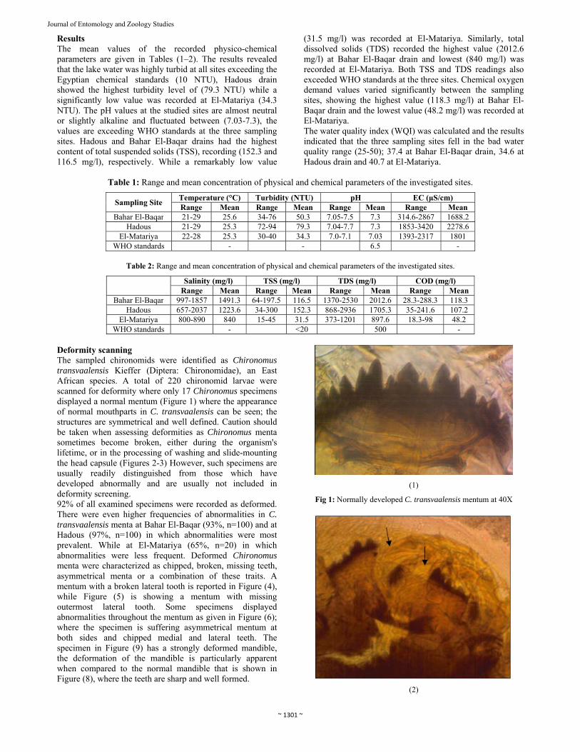
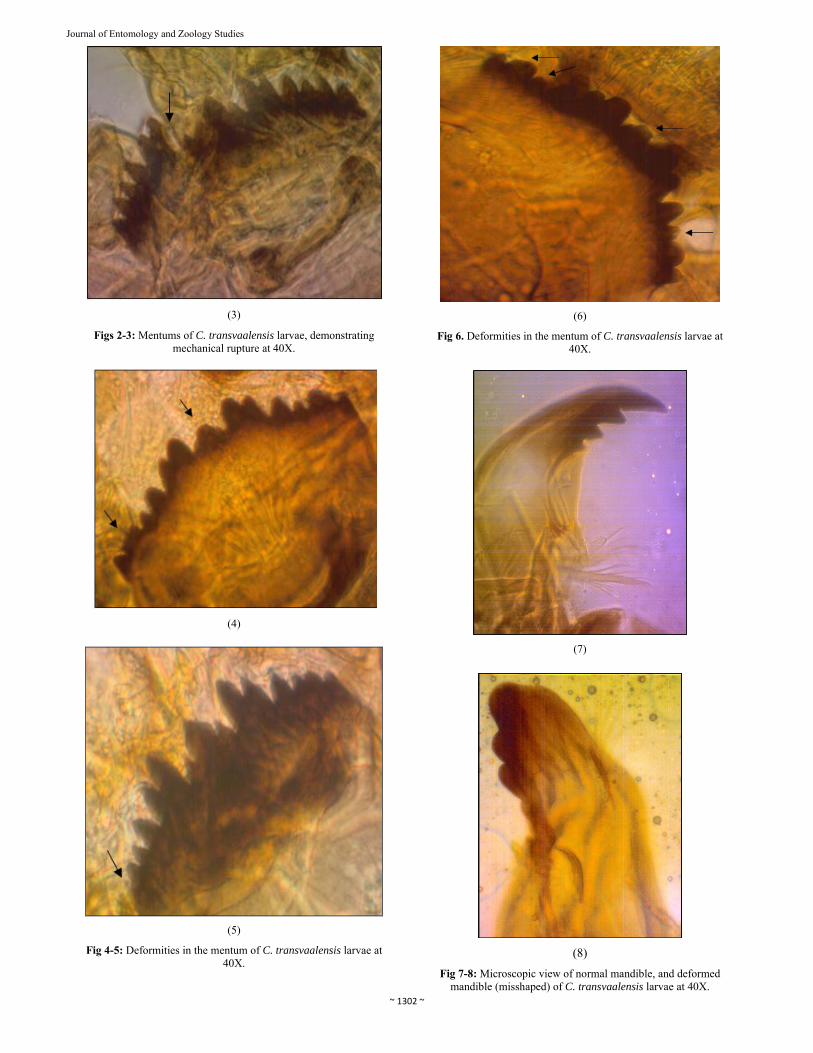
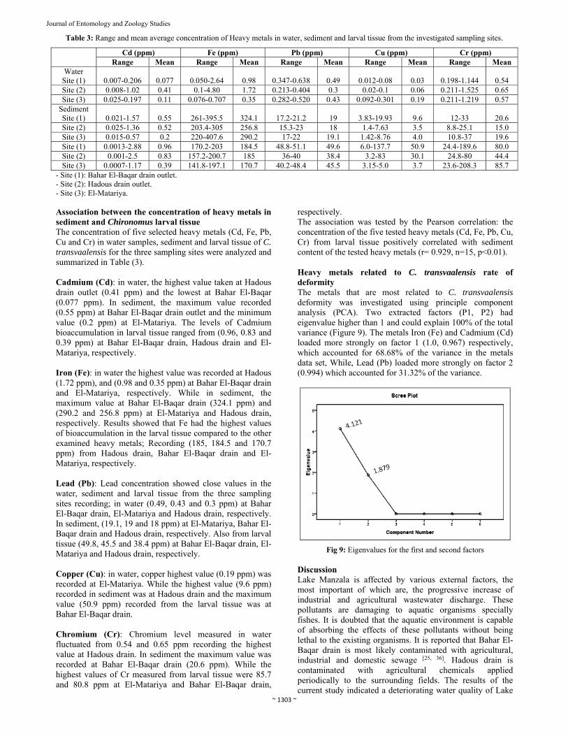
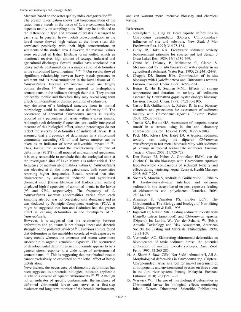
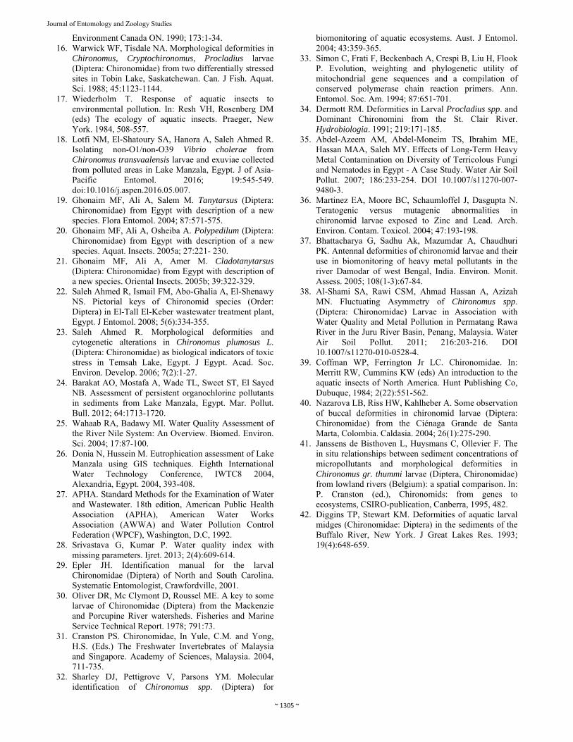
Deformity scanning The sampled chironomids were identified as Chironomus transvaalensis Kieffer (Diptera: Chironomidae), an East African species. A total of 220 chironomid larvae were scanned for deformity where only 17 Chironomus specimens displayed a normal mentum (Figure 1) where the appearance of normal mouthparts in C. transvaalensis can be seen; the structures are symmetrical and well defined. Caution should be taken when assessing deformities as Chironomus menta sometimes become broken, either during the organism's lifetime, or in the processing of washing and slide-mounting the head capsule (Figures 2-3) However, such specimens are usually readily distinguished from those which have developed abnormally and are usually not included in deformity screening. 92% of all examined specimens were recorded as deformed. There were even higher frequencies of abnormalities in C. transvaalensis menta at Bahar El-Baqar (93%, n=100) and at Hadous (97%, n=100) in which abnormalities were most prevalent. While at El-Matariya (65%, n=20) in which abnormalities were less frequent. Deformed Chironomus menta were characterized as chipped, broken, missing teeth, asymmetrical menta or a combination of these traits. A mentum with a broken lateral tooth is reported in Figure (4), while Figure (5) is showing a mentum with missing outermost lateral tooth. Some specimens displayed abnormalities throughout the mentum as given in Figure (6); where the specimen is suffering asymmetrical mentum at both sides and chipped medial and lateral teeth. The specimen in Figure (9) has a strongly deformed mandible, the deformation of the mandible is particularly apparent when compared to the normal mandible that is shown in Figure (8), where the teeth are sharp and well formed.
(1)
Fig 1: Normally developed C. transvaalensis mentum at 40X
(2)
~ 1302 ~
Journal of Entomology and Zoology Studies
(3)
Figs 2-3: Mentums of C. transvaalensis larvae, demonstrating mechanical rupture at 40X.
(4)
(5)
Fig 4-5: Deformities in the mentum of C. transvaalensis larvae at 40X.
(6)
Fig 6. Deformities in the mentum of C. transvaalensis larvae at 40X.
(7)
(8)
Fig 7-8: Microscopic view of normal mandible, and deformed mandible (misshaped) of C. transvaalensis larvae at 40X.
~ 1303 ~
Journal of Entomology and Zoology Studies
Table 3: Range and mean average concentration of Heavy metals in water, sediment and larval tissue from the investigated sampling sites.
Cd (ppm) Fe (ppm) Pb (ppm) Cu (ppm) Cr (ppm) Range Mean Range Mean Range Mean Range Mean Range Mean
Water Site (1)
0.007-0.206
0.077
0.050-2.64
0.98
0.347-0.638
0.49
0.012-0.08
0.03
0.198-1.144
0.54
Site (2) 0.008-1.02 0.41 0.1-4.80 1.72 0.213-0.404 0.3 0.02-0.1 0.06 0.211-1.525 0.65 Site (3) 0.025-0.197 0.11 0.076-0.707 0.35 0.282-0.520 0.43 0.092-0.301 0.19 0.211-1.219 0.57
Sediment Site (1)
0.021-1.57
0.55
261-395.5
324.1
17.2-21.2
19
3.83-19.93
9.6
12-33
20.6
Site (2) 0.025-1.36 0.52 203.4-305 256.8 15.3-23 18 1.4-7.63 3.5 8.8-25.1 15.0 Site (3) 0.015-0.57 0.2 220-407.6 290.2 17-22 19.1 1.42-8.76 4.0 10.8-37 19.6 Site (1) 0.0013-2.88 0.96 170.2-203 184.5 48.8-51.1 49.6 6.0-137.7 50.9 24.4-189.6 80.0 Site (2) 0.001-2.5 0.83 157.2-200.7 185 36-40 38.4 3.2-83 30.1 24.8-80 44.4 Site (3) 0.0007-1.17 0.39 141.8-197.1 170.7 40.2-48.4 45.5 3.15-5.0 3.7 23.6-208.3 85.7
- Site (1): Bahar El-Baqar drain outlet. - Site (2): Hadous drain outlet. - Site (3): El-Matariya. Association between the concentration of heavy metals in sediment and Chironomus larval tissue The concentration of five selected heavy metals (Cd, Fe, Pb, Cu and Cr) in water samples, sediment and larval tissue of C. transvaalensis for the three sampling sites were analyzed and summarized in Table (3). Cadmium (Cd): in water, the highest value taken at Hadous drain outlet (0.41 ppm) and the lowest at Bahar El-Baqar (0.077 ppm). In sediment, the maximum value recorded (0.55 ppm) at Bahar El-Baqar drain outlet and the minimum value (0.2 ppm) at El-Matariya. The levels of Cadmium bioaccumulation in larval tissue ranged from (0.96, 0.83 and 0.39 ppm) at Bahar El-Baqar drain, Hadous drain and El-Matariya, respectively. Iron (Fe): in water the highest value was recorded at Hadous (1.72 ppm), and (0.98 and 0.35 ppm) at Bahar El-Baqar drain and El-Matariya, respectively. While in sediment, the maximum value at Bahar El-Baqar drain (324.1 ppm) and (290.2 and 256.8 ppm) at El-Matariya and Hadous drain, respectively. Results showed that Fe had the highest values of bioaccumulation in the larval tissue compared to the other examined heavy metals; Recording (185, 184.5 and 170.7 ppm) from Hadous drain, Bahar El-Baqar drain and El-Matariya, respectively. Lead (Pb): Lead concentration showed close values in the water, sediment and larval tissue from the three sampling sites recording; in water (0.49, 0.43 and 0.3 ppm) at Bahar El-Baqar drain, El-Matariya and Hadous drain, respectively. In sediment, (19.1, 19 and 18 ppm) at El-Matariya, Bahar El-Baqar drain and Hadous drain, respectively. Also from larval tissue (49.8, 45.5 and 38.4 ppm) at Bahar El-Baqar drain, El-Matariya and Hadous drain, respectively. Copper (Cu): in water, copper highest value (0.19 ppm) was recorded at El-Matariya. While the highest value (9.6 ppm) recorded in sediment was at Hadous drain and the maximum value (50.9 ppm) recorded from the larval tissue was at Bahar El-Baqar drain. Chromium (Cr): Chromium level measured in water fluctuated from 0.54 and 0.65 ppm recording the highest value at Hadous drain. In sediment the maximum value was recorded at Bahar El-Baqar drain (20.6 ppm). While the highest values of Cr measured from larval tissue were 85.7 and 80.8 ppm at El-Matariya and Bahar El-Baqar drain,
respectively. The association was tested by the Pearson correlation: the concentration of the five tested heavy metals (Cd, Fe, Pb, Cu, Cr) from larval tissue positively correlated with sediment content of the tested heavy metals (r= 0.929, n=15, p<0.01). Heavy metals related to C. transvaalensis rate of deformity The metals that are most related to C. transvaalensis deformity was investigated using principle component analysis (PCA). Two extracted factors (P1, P2) had eigenvalue higher than 1 and could explain 100% of the total variance (Figure 9). The metals Iron (Fe) and Cadmium (Cd) loaded more strongly on factor 1 (1.0, 0.967) respectively, which accounted for 68.68% of the variance in the metals data set, While, Lead (Pb) loaded more strongly on factor 2 (0.994) which accounted for 31.32% of the variance.
Fig 9: Eigenvalues for the first and second factors Discussion Lake Manzala is affected by various external factors, the most important of which are, the progressive increase of industrial and agricultural wastewater discharge. These pollutants are damaging to aquatic organisms specially fishes. It is doubted that the aquatic environment is capable of absorbing the effects of these pollutants without being lethal to the existing organisms. It is reported that Bahar El-Baqar drain is most likely contaminated with agricultural, industrial and domestic sewage [25, 36]. Hadous drain is contaminated with agricultural chemicals applied periodically to the surrounding fields. The results of the current study indicated a deteriorating water quality of Lake
~ 1304 ~
Journal of Entomology and Zoology Studies
Manzala based on the water quality index categorization [28]. The present investigation shows that bioaccumulation of the tested heavy metals in the tissue of C. transvaalensis larvae depends mainly on sampling sites. This may be attributed to the difference in type and amount of wastes discharged to each site. In general, heavy metals bioaccumulation in the larval tissue showed high values at the three sites, this correlated positively with their high concentrations in sediments of the studied area. However, the maximal values were recorded at Bahar El-Baqar drain outlet, which as mentioned receives high amount of sewage; industrial and agricultural discharges. Several studies have concluded that heavy metals contamination is a major cause of deformities in the chironomid larval mentum [14, 36-38]. Our results showed significant relationship between heavy metals presence in sediment and its bioaccumulation in the larval tissue of C. transvaalensis. Because Chironomus larvae are largely bottom dwellers [39] they are exposed to hydrophobic contaminants in the sediment through their diet. They are not noticeably mobile and therefore they may reveal long term effects of intermittent or chronic pollution of sediments. Any deviation of a biological structure from its normal morphology could be considered as a deformity [16]. The occurrence of abnormal Chironomus menta is usually reported as a percentage of larvae within a given sample. Although such deformity frequencies are readily interpreted measure of this biological effect, simple percentages do not reflect the severity of deformities of individual larvae. It is assumed that a frequency of deformities in a chironomid community exceeding 8% of total larvae number can be taken as an indicator of some unfavorable impact [16, 40]. Thus, taking into account the exceptionally high rate of deformities (92.2%) in all mounted and examined specimens, it is only reasonable to conclude that the ecological state at the investigated sites of Lake Manzala is rather critical. The frequency of mentum abnormalities within C. transvaalensis was elevated at all the investigated sites, with some sites reporting higher frequencies. Results reported that sites characterized by substantial industrial and agricultural chemical input (Bahar El-Baqar and Hadous drain outlets) displayed high frequencies of abnormal menta in the larvae (93 and 97%, respectively). The frequency of C. transvaalensis mentum abnormalities varied from each sampling site, but was not correlated with abundance and as was deduced by Principle Component Analysis (PCA), it might be suggested that Iron and Cadmium had the greater effect in causing deformities in the mouthparts of C. transvaalensis. However, it is suggested that the relationship between deformities and pollutants is not always linear and depends strongly on the pollutant involved [41]. Previous studies found that deformities in the mandibles correlated with exposure to heavy metals whereas the antennae and menta were more susceptible to organic xenobiotic exposure. The occurrence of developmental deformities in chironomids appears to be a general stress response to a wide range of environmental contaminants [13]. This is suggesting that our obtained results cannot exclusively be explained on the lethal effect of heavy metals alone. Nevertheless, the occurrence of chironomid deformities has been suggested as a potential biological indicator, applicable in situ to a diverse of aquatic environments [16, 42]. Although not an indicator of specific contaminants, the incidence of deformed chironomid larvae can serve as a first-step evaluator and long term monitor of the benthic environment,
and can warrant more intensive bioassay and chemical testing. References 1. Jeysingham K, Ling N. Head capsule deformities in
Chironomus zealandicus (Diptera: Chironomidae): influence of site and substrate. New Zeal. J Mar Freshwater Res. 1997; 31:175-184.
2. Giesy JP, Hoke RA. Freshwater sediment toxicity bioassessment rationale for species and test design. J Great Lakes Res. 1989; 15(4):539-569.
3. Crane M, Delaney P, Mainstone C, Clarke S. Measurement by in situ bioassay of water quality in an agricultural catchment. Water Res. 1995; 29:2441-2448.
4. Chappie DJ, Burton JGA. Optimization of in situ bioassays with Hyalella azteca and Chironomus tentans. Environ. Toxicol. Chem. 1997; 16:559-564.
5. Beiras R, His E, Seaman MNL. Effects of storage temperature and duration on toxicity of sediments assessed by Crassostrea gigas oyster embryo bioassay. Environ. Toxicol. Chem. 1998; 17:2100-2105.
6. Castro BB, Guilhermino L, Ribeiro R. In situ bioassay chambers and procedures for assessment of sediment toxicity with Chironomus riparius. Environ. Pollut. 2003; 125:325-335.
7. Tucker KA, Burton GA. Assessment of nonpoint-source runoff in a stream using in situ and laboratory approaches. Environ. Toxicol. 1999; 18:2797-2803.
8. Peck MR, Klessa DA, Baird DJ. A tropical sediment toxicity test using the dipteran Chironomus crassiforceps to test metal bioavailability with sediment pH change in tropical acid-sulfate sediments. Environ. Toxicol. Chem. 2002; 21:720-728.
9. Den Besten PJ, Naber A, Grootelaar EMM, van de Guchte C. In situ bioassays with Chironomus riparius: laboratory-field comparisons of sediment toxicity and effects during wintering. Aqua. Ecosyst. Health Manage. 2003; 6:217-228.
10. Soares S, Moreira S, Andrade S, Guilhermino L, Ribeiro R. Freshwater–saltwater interface and estuarine sediment in situ assays based on post-exposure feeding of chironomids and polychaetes. Estuaries. 2005; 28:314-319.
11. Armitage P, Cranston PS, Pinder LCV. The Chironomidae: The Biology and Ecology of Non-Biting Midges. Chapman & Hall, 1995.
12. Ingersoll C, Nelson MK. Testing sediment toxicity with Hyalella azteca (amphipod) and Chironomus riparius (Diptera). In: Landis, W., Van der Schalie, W. (Eds.), Aquatic Toxicology and Risk Assessment, American Society for Testing and Materials, Philadelphia. 1990; 13:93-109.
13. Vermeulen AC. Elaborating chironomid deformities as bioindicators of toxic sediment stress: the potential application of mixture toxicity concepts, Ann. Zool. Fenn. 1995; 32:265-285.
14. Al-Shami S, Rawi CSM, Nor SAM, Ahmad AH, Ali A. Morphological deformities in Chironomus spp. (Diptera: Chironomidae) larvae as a tool for impact assessment of anthropogenic and environmental stresses on three rivers in the Juru river system, Penang, Malaysia. Environ. Entomol. 2010; 39(1):210-222.
15. Warwick WF. The use of morphological deformities in Chironomid larvae for biological effects monitoring Inland Waters Directorate Scientific Publications,
~ 1305 ~
Journal of Entomology and Zoology Studies
Environment Canada ON. 1990; 173:1-34. 16. Warwick WF, Tisdale NA. Morphological deformities in
Chironomus, Cryptochironomus, Procladius larvae (Diptera: Chironomidae) from two differentially stressed sites in Tobin Lake, Saskatchewan. Can. J Fish. Aquat. Sci. 1988; 45:1123-1144.
17. Wiederholm T. Response of aquatic insects to environmental pollution. In: Resh VH, Rosenberg DM (eds) The ecology of aquatic insects. Praeger, New York. 1984, 508-557.
18. Lotfi NM, El-Shatoury SA, Hanora A, Saleh Ahmed R. Isolating non-O1/non-O39 Vibrio cholerae from Chironomus transvaalensis larvae and exuviae collected from polluted areas in Lake Manzala, Egypt. J of Asia-Pacific Entomol. 2016; 19:545-549. doi:10.1016/j.aspen.2016.05.007.
19. Ghonaim MF, Ali A, Salem M. Tanytarsus (Diptera: Chironomidae) from Egypt with description of a new species. Flora Entomol. 2004; 87:571-575.
20. Ghonaim MF, Ali A, Osheiba A. Polypedilum (Diptera: Chironomidae) from Egypt with description of a new species. Aquat. Insects. 2005a; 27:221- 230.
21. Ghonaim MF, Ali A, Amer M. Cladotanytarsus (Diptera: Chironomidae) from Egypt with description of a new species. Oriental Insects. 2005b; 39:322-329.
22. Saleh Ahmed R, Ismail FM, Abo-Ghalia A, El-Shenawy NS. Pictorial keys of Chironomid species (Order: Diptera) in El-Tall El-Keber wastewater treatment plant, Egypt. J Entomol. 2008; 5(6):334-355.
23. Saleh Ahmed R. Morphological deformities and cytogenetic alterations in Chironomus plumosus L. (Diptera: Chironomidae) as biological indicators of toxic stress in Temsah Lake, Egypt. J Egypt. Acad. Soc. Environ. Develop. 2006; 7(2):1-27.
24. Barakat AO, Mostafa A, Wade TL, Sweet ST, El Sayed NB. Assessment of persistent organochlorine pollutants in sediments from Lake Manzala, Egypt. Mar. Pollut. Bull. 2012; 64:1713-1720.
25. Wahaab RA, Badawy MI. Water Quality Assessment of the River Nile System: An Overview. Biomed. Environ. Sci. 2004; 17:87-100.
26. Donia N, Hussein M. Eutrophication assessment of Lake Manzala using GIS techniques. Eighth International Water Technology Conference, IWTC8 2004, Alexandria, Egypt. 2004, 393-408.
27. APHA. Standard Methods for the Examination of Water and Wastewater. 18th edition, American Public Health Association (APHA), American Water Works Association (AWWA) and Water Pollution Control Federation (WPCF), Washington, D.C, 1992.
28. Srivastava G, Kumar P. Water quality index with missing parameters. Ijret. 2013; 2(4):609-614.
29. Epler JH. Identification manual for the larval Chironomidae (Diptera) of North and South Carolina. Systematic Entomologist, Crawfordville, 2001.
30. Oliver DR, Mc Clymont D, Roussel ME. A key to some larvae of Chironomidae (Diptera) from the Mackenzie and Porcupine River watersheds. Fisheries and Marine Service Technical Report. 1978; 791:73.
31. Cranston PS. Chironomidae, In Yule, C.M. and Yong, H.S. (Eds.) The Freshwater Invertebrates of Malaysia and Singapore. Academy of Sciences, Malaysia. 2004, 711-735.
32. Sharley DJ, Pettigrove V, Parsons YM. Molecular identification of Chironomus spp. (Diptera) for
biomonitoring of aquatic ecosystems. Aust. J Entomol. 2004; 43:359-365.
33. Simon C, Frati F, Beckenbach A, Crespi B, Liu H, Flook P. Evolution, weighting and phylogenetic utility of mitochondrial gene sequences and a compilation of conserved polymerase chain reaction primers. Ann. Entomol. Soc. Am. 1994; 87:651-701.
34. Dermott RM. Deformities in Larval Procladius spp. and Dominant Chironomini from the St. Clair River. Hydrobiologia. 1991; 219:171-185.
35. Abdel-Azeem AM, Abdel-Moneim TS, Ibrahim ME, Hassan MAA, Saleh MY. Effects of Long-Term Heavy Metal Contamination on Diversity of Terricolous Fungi and Nematodes in Egypt - A Case Study. Water Air Soil Pollut. 2007; 186:233-254. DOI 10.1007/s11270-007-9480-3.
36. Martinez EA, Moore BC, Schaumloffel J, Dasgupta N. Teratogenic versus mutagenic abnormalities in chironomid larvae exposed to Zinc and Lead. Arch. Environ. Contam. Toxicol. 2004; 47:193-198.
37. Bhattacharya G, Sadhu Ak, Mazumdar A, Chaudhuri PK. Antennal deformities of chironomid larvae and their use in biomonitoring of heavy metal pollutants in the river Damodar of west Bengal, India. Environ. Monit. Assess. 2005; 108(1-3):67-84.
38. Al-Shami SA, Rawi CSM, Ahmad Hassan A, Azizah MN. Fluctuating Asymmetry of Chironomus spp. (Diptera: Chironomidae) Larvae in Association with Water Quality and Metal Pollution in Permatang Rawa River in the Juru River Basin, Penang, Malaysia. Water Air Soil Pollut. 2011; 216:203-216. DOI 10.1007/s11270-010-0528-4.
39. Coffman WP, Ferrington Jr LC. Chironomidae. In: Merritt RW, Cummins KW (eds) An introduction to the aquatic insects of North America. Hunt Publishing Co, Dubuque, 1984; 2(22):551-562.
40. Nazarova LB, Riss HW, Kahlheber A. Some observation of buccal deformities in chironomid larvae (Diptera: Chironomidae) from the Ciénaga Grande de Santa Marta, Colombia. Caldasia. 2004; 26(1):275-290.
41. Janssens de Bisthoven L, Huysmans C, Ollevier F. The in situ relationships between sediment concentrations of micropollutants and morphological deformities in Chironomus gr. thummi larvae (Diptera, Chironomidae) from lowland rivers (Belgium): a spatial comparison. In: P. Cranston (ed.), Chironomids: from genes to ecosystems, CSIRO-publication, Canberra, 1995, 482.
42. Diggins TP, Stewart KM. Deformities of aquatic larval midges (Chironomidae: Diptera) in the sediments of the Buffalo River, New York. J Great Lakes Res. 1993; 19(4):648-659.