Embed Size (px)
Citation preview

NAACCR 2016‐2017 Webinar Series 4/13/17
Lip and Oral Cavity 1
NAACCR
2015-2016
Webinar
SeriesCollecting Cancer Data: Lip and Oral Cavity
NAACCR 2016‐2017 Webinar Series
Presented by:
Melissa Riddle [email protected]
Recinda Sherman [email protected]
Jim Hofferkamp [email protected]
Q&A
• Please submit all questions concerning webinar content through the Q&A panel.
• Reminder:
– If you have participants watching this webinar at your site, please collect their names and emails.
– We will be distributing a Q&A document in about one week. This document will fully answer questions asked during the webinar and will contain any corrections that we may discover after the webinar.

NAACCR 2016‐2017 Webinar Series 4/13/17
Lip and Oral Cavity 2
Fabulous Prizes
Agenda
• Overview
– Anatomy
– MP/H
• Staging
– Quiz
• Treatment
– Quiz
• Case Scenarios
NAACCR 2016‐2017 Webinar Series 4/13/17
Lip and Oral Cavity 3
5
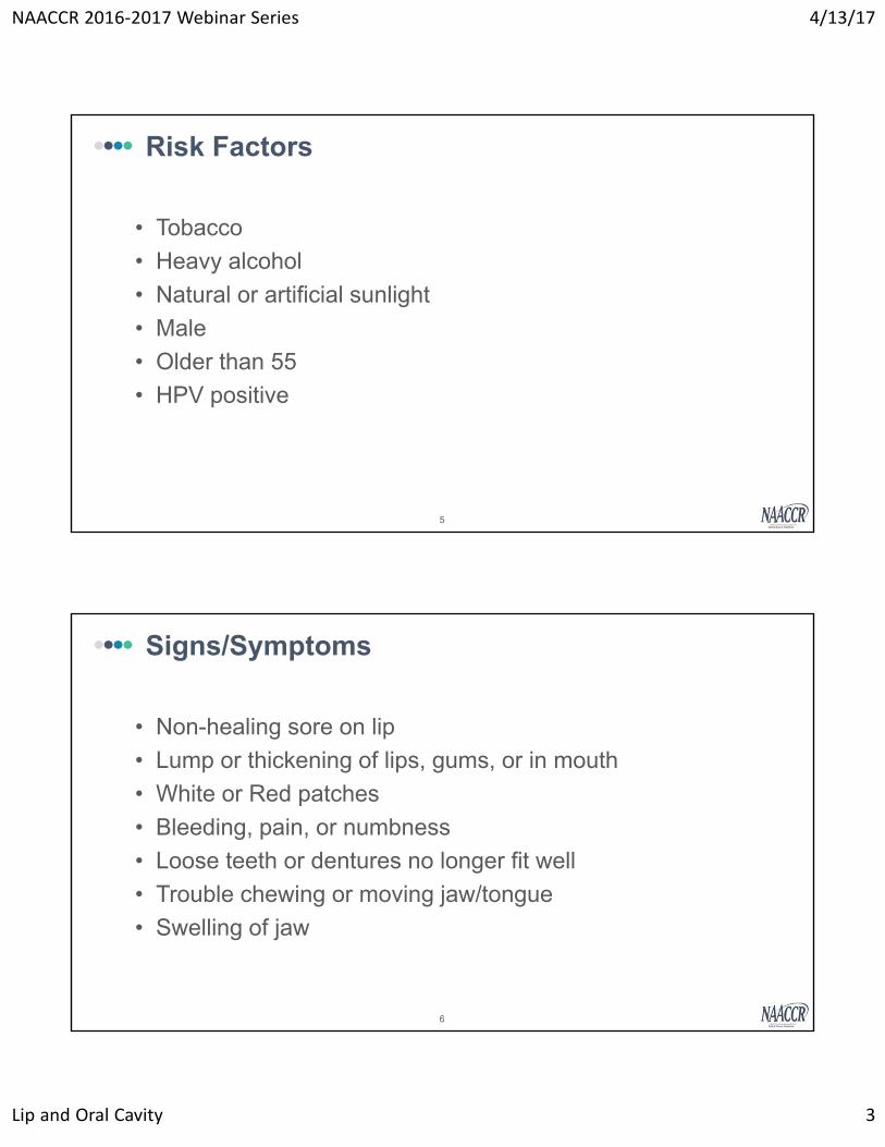
• Tobacco
• Heavy alcohol
• Natural or artificial sunlight
• Male
• Older than 55
• HPV positive
Risk Factors
6
• Non-healing sore on lip
• Lump or thickening of lips, gums, or in mouth
• White or Red patches
• Bleeding, pain, or numbness
• Loose teeth or dentures no longer fit well
• Trouble chewing or moving jaw/tongue
• Swelling of jaw
Signs/Symptoms
NAACCR 2016‐2017 Webinar Series 4/13/17
Lip and Oral Cavity 4
7
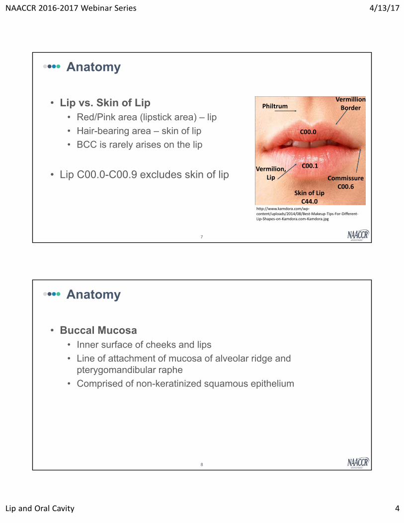
• Lip vs. Skin of Lip• Red/Pink area (lipstick area) – lip
• Hair-bearing area – skin of lip
• BCC is rarely arises on the lip
• Lip C00.0-C00.9 excludes skin of lip
Anatomy
PhiltrumVermillion Border
Vermilion, Lip Commissure
C00.6Skin of LipC44.0
C00.1
C00.0
http://www.kamdora.com/wp‐content/uploads/2014/08/Best‐Makeup‐Tips‐For‐Different‐Lip‐Shapes‐on‐Kamdora.com‐Kamdora.jpg
8
• Buccal Mucosa• Inner surface of cheeks and lips
• Line of attachment of mucosa of alveolar ridge and pterygomandibular raphe
• Comprised of non-keratinized squamous epithelium
Anatomy

NAACCR 2016‐2017 Webinar Series 4/13/17
Lip and Oral Cavity 5
9
• Lower and Upper Alveolar Ridge
• Contains tooth sockets (alveoli)
• Mucosa overlies the alveolar process
Anatomy
http://content.answcdn.com/main/content/img/elsevier/dental/f0022‐01.jpg
10
• Gingiva• Fibrous tissue covered by mucous
membrane
• Provides a seal around teeth
• Retromolar Gingiva (Trigone)• Behind the molars
• Covers retromolar pad
Anatomy

NAACCR 2016‐2017 Webinar Series 4/13/17
Lip and Oral Cavity 6
11
• Floor of the Mouth• Inferior limit of oral cavity
• Wharton’s duct
• Sublingual gland ducts
Anatomy
NCI, Alan Hoofring (Illustrator)
12
• Hard Palate• Separates oral cavity from nasal
cavities
• Oral Tongue – Anterior 2/3• 3 surfaces:
• Tip• Body• Base
• Muscles:• Extrinsic – alter position• Intrinsic – shapes tongue
Anatomy
Ventral

NAACCR 2016‐2017 Webinar Series 4/13/17
Lip and Oral Cavity 7
13
Level and Nodal Groups Cancer Sites of Lymphatic Spread
I A– SubmentalIB ‐ Submandibular
Lip; anterior tongue; floor of mouth; gingiva; buccal mucosa; anterior alveolar ridge
II – Upper Jugulo‐digastric Oral cavity; hard palate; alveolar ridge; anterior tongue
III – Middle jugular Oral cavity; hard palate; alveolar ridge
IV – Inferior jugular
V – Posterior Triangle (supraclavicular)
VI – Anterior compartment (Delphian,paratracheal)
VII – Superior mediastinal
Anatomy – Lymph Nodes
14
Anatomy – Lymph Nodes
http://anatomydiagram.info/wp‐content/uploads/2015/11/anatomy‐of‐the‐throat‐glands‐lymph+neck.jpg

NAACCR 2016‐2017 Webinar Series 4/13/17
Lip and Oral Cavity 8
15
• Approximately 30% of oral cavity primaries present with LN mets
• 50-60% anterior tongue primaries present with LN mets
• Lip, Hard palate, and Alveolar ridge rarely have LN mets
Lymph Nodes
Epi Moment
OralTheme song:
Lips are movin’

NAACCR 2016‐2017 Webinar Series 4/13/17
Lip and Oral Cavity 9
Epidemiology of oral cancer
• Analysis groups– Oral vs Head & Neck
• Laryngeal (respiratory) & esophageal (digestive) due to related etiology– Obesity‐related
• Salivary gland, nasopharynx, & hypopharnyx etiologically distinct (10% of oral cancers)
– Tobacco‐related, Alcohol‐related, HPV‐related• 3% of all cancers in US
– more common outside US; leading cause of death– India‐‐#1, Australia, France, Brazil, S. Africa
• 95% 45+, median age 63– 2:1 M:F– Highest among NH Whites (18.5 M; 6.7 F)
• Whites(17.6 M; 6.4 F); AI/AN (15.6 M; 5.8 F); Blacks (14.7 M; 5.1 F); API (10.9 M; 4.9 F); Hispanics (10.8 M; 4.1 F)
• Main histology: 90‐95% squamous
Oral cancer trends, 2009‐2013

NAACCR 2016‐2017 Webinar Series 4/13/17
Lip and Oral Cavity 10
Risk factors for oral cancers• Tobacco
– Betel Quid
• Alcohol
• Viral infections
– HPV, EBV, HIV
• Bacterial infection
– Syphilis (tx related)
• Fungal infection
– Candida
• Diet
– Salted Fish (Chinese‐style), Hot Mate
– Iron deficiency
More risk factors for oral cancers• SES, Hygiene
• Probable:
– Radiation: UV (sun exposure) & TX
– Asbestos
– Printing Processes/Inks Occupational Exposures
• Protective (probable or limited evidence)
– Non‐starchy veggies & fruits; Vit C, green tea, calcium supplements, coffee, physical activity

NAACCR 2016‐2017 Webinar Series 4/13/17
Lip and Oral Cavity 11
Oral cancer survival: 58% 5‐ year RSR overall
Oral cancer screening
• USPSTF 2013 Grade I
– Insufficient evidence to assess effectiveness
– No population‐based screening
– Systemic clinical examination
– Inspection and palpitation of oral cavity; dental check‐ups
• Dyes, laser light, rinse with acetic acid & special light
• If abnormal area found, brush biopsy/exfoliative cytology
– 2% diagnosed at in situ
• Delay in diagnosis
– Early stages asymptomatic
– Symptoms often mistaken for other health issues (toothache)

NAACCR 2016‐2017 Webinar Series 4/13/17
Lip and Oral Cavity 12
Multiple Primary & Histology
24
• Tumor Board• Staging physician’s site
assignment• Total resection of primary
tumor• Surgeon’s statement
from operative report• Final diagnosis from
pathology
Primary Site – Priority Order
• Biopsy ONLY• Endoscopy• Radiation Onc• Diagnosing physician• Primary Care physician• Other physician• Radiologist impression
from imaging• Physician state on PE

NAACCR 2016‐2017 Webinar Series 4/13/17
Lip and Oral Cavity 13
25
• Overlapping sites:• C02.8 (overlapping tongue)
• C06.8 (overlapping other and unspecified parts mouth)
• C08.8 (overlapping major salivary glands)
• C14.8 (overlapping lip, oral cavity, and pharynx)
Primary Site
26
• Paired Sites:• Parotid Glands (C07.9)
• Major Salivary glands (C08.0, C08.1)
• Tonsils (C09.0, C09.1, C09.8, C09.9
• Nasal Cavity (C30.0)
• Accessory Sinuses (C31.0, C31.2)
• Middle Ear (C30.1)
Primary Site

NAACCR 2016‐2017 Webinar Series 4/13/17
Lip and Oral Cavity 14
27
• M3 – Bilateral involvement paired sites – Multiple
• M4 – Upper Lip and Lower Lip – Multiple
• M5 – Upper gum and Lower gum – Multiple
• M7 – Topography codes different at second and/or third character – Multiple
MPH – Multiple Tumors
28
• M9 – More than 5yr apart – Multiple
• M10 – Non-specific histology w/ a more specific –Single
• Adenocarcinoma, NOS and another specific adenocarcinoma
• M11 – Histology codes different at first, second, or third number – Multiple
MPH – Multiple Tumors

NAACCR 2016‐2017 Webinar Series 4/13/17
Lip and Oral Cavity 15
29
• Squamous Cell Carcinoma, NOS• Acantholytic squamous cell carcinoma• Basaloid squamous cell carcinoma• Papillary squamous cell carcinoma• Spindle cell squamous cell carcinoma• Verrucous carcinoma
• Adenosquamous carcinoma• Mucosal Melanoma• Sarcomas
Histology
30
MPH – Chart 1

NAACCR 2016‐2017 Webinar Series 4/13/17
Lip and Oral Cavity 16
31
• H4 – Invasive and In-situ code the invasive histology
• H5 – Multiple histologies on same branch Chart 1 • Code the most specific
• Terms: pattern (in-situ), architecture (in-situ), type, subtype, predominantly, with features of, major, or with ___ differentiation
• H6 – None of the above (H1-H5)• Code highest ICD-O-3 histology code
MPH – Histology Single Tumor
32
• H10 – Code most invasive histology• Equally invasive go to next rule
• H11 – Multiple histologies all on same branch Chart 1• Code most specific using Chart 1
• H12 – None of the above (H7-H11)• Code higher ICD-O-3 code
MPH – Histology (Multiple Tumor –Single Abst)
NAACCR 2016‐2017 Webinar Series 4/13/17
Lip and Oral Cavity 17
33
• Patient has a history of SCC maxillary gingiva diagnosed on 1/13/11 and has remained disease free.
• 4/3/16 biopsy of maxillary gingiva was positive for invasive papillary carcinoma.
• 5/3/16 Radical excision w/ partial maxillectomy – 2.6cm Invasive papillary SCC, poorly diff and SCC in situ.
Pop Quiz!
How many primaries?2, M9
Primary 1:C030
Histology 1:8070/39, H3Primary 2:
C030Histology 2: 8052/33, H4
Questions?
Quiz 1

NAACCR 2016‐2017 Webinar Series 4/13/17
Lip and Oral Cavity 18
Summary Stage
Summary Stage
• Review of manual
https://seer.cancer.gov/tools/ssm/

NAACCR 2016‐2017 Webinar Series 4/13/17
Lip and Oral Cavity 19
https://seer.cancer.gov/tools/ssm/headneck.pdf
DISTINGUISHING “IN SITU” AND “LOCALIZED” TUMORS FOR LIP, ORAL CAVITY, AND PHARYNX
• Historically, carcinomas described as “confined to mucosa” have been coded as localized. In order to provide greater specificity and to rule out the possibility of classifying noninvasive tumors in this category, abstractors should determine:
– 1) if the tumor is confined to the epithelium, in which case it is in situ, OR
– 2) if the tumor has penetrated the basement membrane to invade the lamina propria, in which case it is localized and is coded to invasion of the lamina propria

NAACCR 2016‐2017 Webinar Series 4/13/17
Lip and Oral Cavity 20
Lip
1 Localized only
• Invasive tumor confined to:– Labial mucosa (inner lip)
– Lamina propria
– Multiple foci
– Musculature##
– Submucosa (superficial invasion)
– Vermilion surface
• Superficial extension to:– Skin of lip
– Subcutaneous soft tissue of lip
• Localized, NOS
Lip
2 Regional by Direct Extension
• Extension to:
– Buccal mucosa (inner cheek)
– Commissure
– Gingiva
– Opposite (both) lip(s)
• Lower lip/commissure:
– Mandible
• Upper lip/commissure:
– Maxilla
NAACCR 2016‐2017 Webinar Series 4/13/17
Lip and Oral Cavity 21
Lip
3 Regional Lymph Nodes• Cervical, NOS
• Facial, NOS:###
– Buccinators (buccal) for upper lip
– Nasolabial for upper lip
• Internal jugular, NOS***
• Deep cervical, NOS:
– Lower, NOS:
• Jugulo‐omohyoid (supraomohyoid)
– Middle
– Upper, NOS:
• Jugulodigastric (subdigastric
Lip
Distant Metastasis
• Distant lymph node(s):
– Mediastinal
– Supraclavicular (transverse cervical)
– Other distant lymph node(s)
• Extension to:
– Cortical bone
– Floor of mouth
– Inferior alveolar nerve
– Skin of face/neck
– Tongue
• Upper lip/commissure:
– Nose**
– Further contiguous extension
– Metastasis
NAACCR 2016‐2017 Webinar Series 4/13/17
Lip and Oral Cavity 22
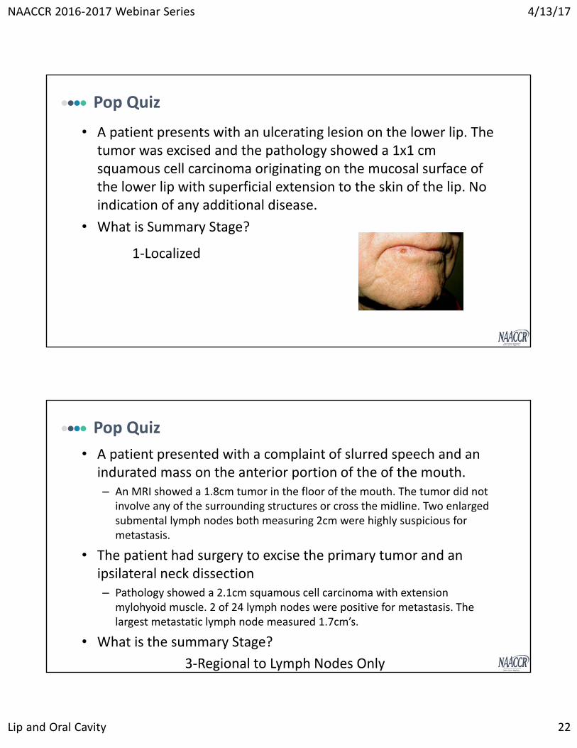
Pop Quiz
• A patient presents with an ulcerating lesion on the lower lip. The tumor was excised and the pathology showed a 1x1 cm squamous cell carcinoma originating on the mucosal surface of the lower lip with superficial extension to the skin of the lip. No indication of any additional disease.
• What is Summary Stage?
1‐Localized
Pop Quiz
• A patient presented with a complaint of slurred speech and an indurated mass on the anterior portion of the of the mouth. – An MRI showed a 1.8cm tumor in the floor of the mouth. The tumor did not
involve any of the surrounding structures or cross the midline. Two enlarged submental lymph nodes both measuring 2cm were highly suspicious for metastasis.
• The patient had surgery to excise the primary tumor and an ipsilateral neck dissection– Pathology showed a 2.1cm squamous cell carcinoma with extension
mylohyoid muscle. 2 of 24 lymph nodes were positive for metastasis. The largest metastatic lymph node measured 1.7cm’s.
• What is the summary Stage?
3‐Regional to Lymph Nodes Only

NAACCR 2016‐2017 Webinar Series 4/13/17
Lip and Oral Cavity 23
Questions?
Lip
AJCC Stage
& Oral Cavity

NAACCR 2016‐2017 Webinar Series 4/13/17
Lip and Oral Cavity 24
Rules for Classification
• Clinical Rules for Classification
– Physical exam
– Imaging
• CT
• MRI
– Primary staging classification
• Pathologic Rules for Classification
– Complete resection of the primary tumor
– Pathologic confirmation of lymph node status
Pg 29
Primary Tumor
• If the patient is found to have metastasis and no primary tumor is found, a T0 would be used if the physician believe the metastasis is from a lip or oral cavity primary.
• An in situ tumor is pTis

NAACCR 2016‐2017 Webinar Series 4/13/17
Lip and Oral Cavity 25
Primary Tumor
• Tumor size dictates the T1‐T3 values for invasive tumors that do not meet the criteria for T4.
T1 ≤ 2cm
T2 2cm ≤ 4cm
T3 > 4cm
Pop Quiz
• 1/11/16 CT Scan of maxillofacial region:
– Upper alveolar ridge soft tissue density measured 3.2 x 1 cm. The tumor is causing mucoperiosteal thickening of the right maxillary sinus, but no signs of definitive invasion.
– No enlarged lymph nodes or indications of additional metastasis.
– Biopsy confirmed squamous cell carcinoma
• Surgery was recommended, but patient refused any treatment.
Data Item Value
Clinical T
Clinical N
Clinical M
Clinical Stage
Pathologic T
Pathologic N
Pathologic M
Pathologic Stage
Summary Stage
cN0
cM0
2
99
1‐Localized
cT2

NAACCR 2016‐2017 Webinar Series 4/13/17
Lip and Oral Cavity 26
Primary Tumor
4.1cm
4.1cm tumor arising on the vermilion surface of the lip and extending to the skin of the lip is a T3.
4.1cm tumor arising on the vermilion surface of the lip and extending to the skin of the face (not skin of the lip) is a T4a.
4.1cm

NAACCR 2016‐2017 Webinar Series 4/13/17
Lip and Oral Cavity 27
Primary Tumor
Moderately advanced disease
Very advance local disease
Pop Quiz
• 1/11/16 CT Scan of maxillofacial region:
– Upper alveolar ridge soft tissue density measured 2 x 2 cm. The tumor is causing mucoperiosteal thickening of the right maxillary sinus, but no signs of definitive invasion.
– No enlarged lymph nodes or indications of additional metastasis.
– Biopsy confirmed squamous cell carcinoma
• 1/22/16 Right maxillectomy:
– 2 x 1 x 0.7 cm tumor of upper alveolar ridge, poorly differentiated squamous cell carcinoma, which infiltrates bone and mucoperiosteum of maxillary sinus.
– Metastatic squamous cell carcinoma in 2 of 3 lymph nodes.

NAACCR 2016‐2017 Webinar Series 4/13/17
Lip and Oral Cavity 28
Pop Quiz
• 1/22/16 Right maxillectomy:
– 2 x 1 x 0.7 cm tumor of upper alveolar ridge, poorly differentiated squamous cell carcinoma, which infiltrates through maxilla into the maxillary sinus.
– Metastatic squamous cell carcinoma in 2 of 3 lymph right submandibular lymph nodes. The largest metastatic lymph node measured 2.1cm. No extranodal extension.
Data Item Value
Clinical T
Clinical N
Clinical M
Clinical Stage
Pathologic T
Pathologic N
Pathologic M
Pathologic Stage
Summary Stage
cN0
cM0
1
4A
7‐Distant
cT1
pT4a
pN2b
cM0
• How many lymph nodes have metastasis?
• Are they on the same side as the primary (ipsilateral)?
• Extranodal extension?
Regional Lymph Nodes
NAACCR 2016‐2017 Webinar Series 4/13/17
Lip and Oral Cavity 29
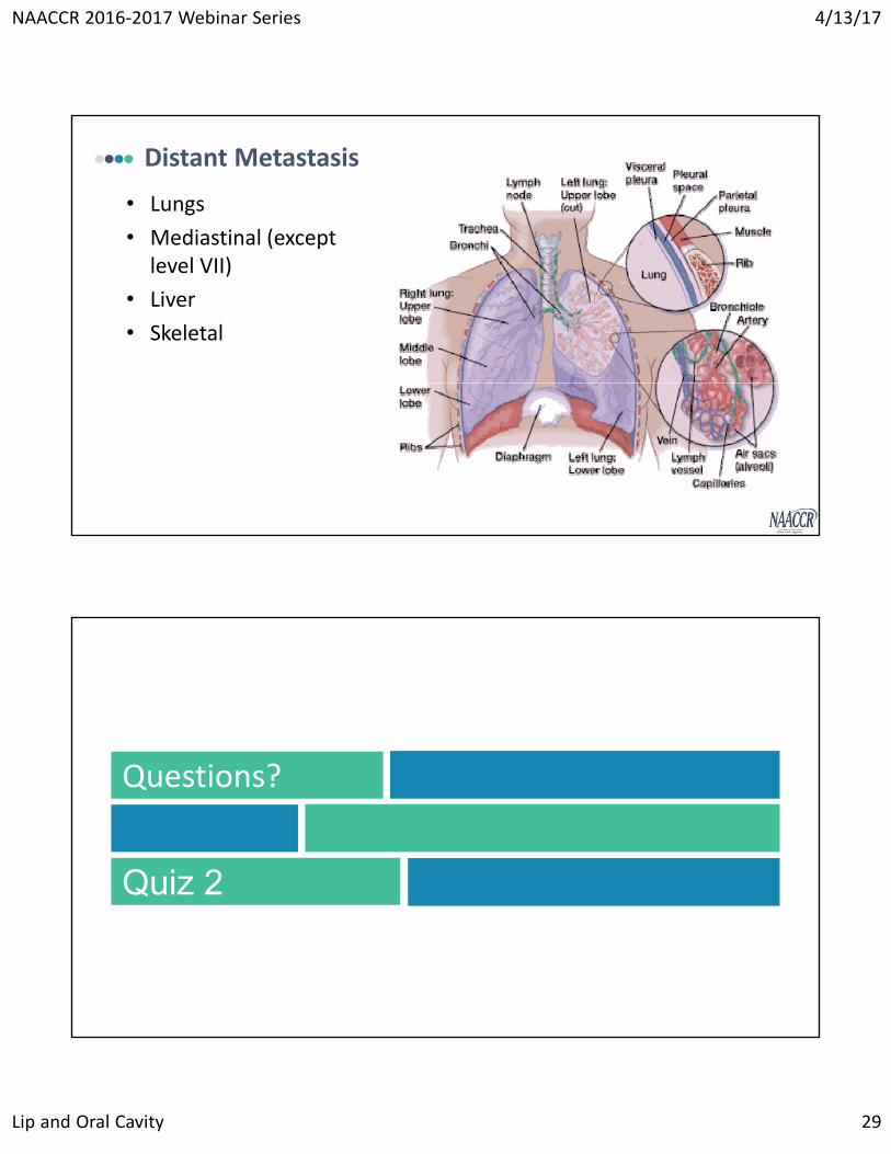
Distant Metastasis
• Lungs
• Mediastinal (except level VII)
• Liver
• Skeletal
Questions?
Quiz 2

NAACCR 2016‐2017 Webinar Series 4/13/17
Lip and Oral Cavity 30
Treatment
60
• T1-T2 and no lymph nodes involved • Surgical excision
• External Beam Radiation (IMRT)
• T3, T4 or Any T with N1-3• Excision +/- lymph node dissection
• Negative lymph nodes – no further treatment
• Positive lymph nodes – possibly chemo, radiation or re-excision
• External Beam Radiation +/- Brachytherapy or Chemotherapy
Treatment - Lip

NAACCR 2016‐2017 Webinar Series 4/13/17
Lip and Oral Cavity 31
61
• T4B or unresectable lymph nodes or newly diagnosed distant metastasis
• Clinical trials – preferred
• Standard – concurrent chemo/radiation, definitive radiation +/- systemic therapy, or supportive care
Treatment - Lip
62
• T1-2 with no lymph node involved• Surgical Excision• External Beam Radiation (IMRT)
• More than 4cm tumor without lymph node metastasis• Excision of primary with neck dissection
• T4a with any N or T1-3 with positive lymph nodes• Excise primary and neck dissection
• Advanced disease - T4b any N, unresectable LN or M1• Clinical Trials• Standard therapy: concurrent chemo and radiation, definitive
radiation +/- systemic therapy or supportive care
Treatment – Oral Cavity

NAACCR 2016‐2017 Webinar Series 4/13/17
Lip and Oral Cavity 32
63
• 30 – Wide excision, NOS• Surgeon states wide excision on operative report• Tongue – Hemiglossectomy
• 40 – Radical excision of tumor, NOS• More extensive excision of the primary tumor• 41 – Radical excision of tumor ONLY• 42 – 41 WITH mandible (marginal, segment, hemi-, or total)• 43 – 41 WITH maxilla (marginal, segment, hemi-, or total)
Surgery
64
• 4/3/16 biopsy of maxillary gingiva was positive for invasive papillary carcinoma.
• 5/3/16 Radical excision w/ partial maxillectomy –2.6cm Invasive papillary SCC, poorly diff and SCC in situ. 0/8 level 1-3 LN. Margins negative.
POP QUIZ!!!
Primary Site Surgery:43
Scope Surgical LN:5
Other/Regional Surgery:0

NAACCR 2016‐2017 Webinar Series 4/13/17
Lip and Oral Cavity 33
65
• Selective• Muscle, nerve and blood vessel in neck preserved• Depend on site, recommend for N0
• Modified Radical• Most common – All lymph nodes removed• Nerves and sometimes blood vessels or muscle spared
• Radical• All tissue from the jaw bone to the collarbone is removed
• Muscle, nerve, salivary gland, and major blood vessels removed
• NCCN term – Comprehensive, recommend for N3
Neck Dissection
66
• Primary• Low/Intermediate risk
• Primary and sites of suspected spread
• High risk• Primary and involved lymph nodes
• Select cases – Interstitial brachytherapy
• Adjuvant• Some may undergo concurrent systemic therapy as well
Radiation
NAACCR 2016‐2017 Webinar Series 4/13/17
Lip and Oral Cavity 34
Questions?
Quiz 3
Coming Up….
• Multiple Primary and Histology Rules
– 5/4/2017
• Collecting Cancer Data: Liver and Bile Ducts
– 6/1/2017

NAACCR 2016‐2017 Webinar Series 4/13/17
Lip and Oral Cavity 35
And Our Fabulous Prizes Go To…
CE Certificate Quiz Survey
• Phrase
• Link
http://www.surveygizmo.com/s3/3477379/Lip‐and‐Oral‐Cavity‐2017
NAACCR 2016‐2017 Webinar Series 4/13/17
Lip and Oral Cavity 36
Thank You!
Presented by:
Melissa Riddle [email protected] Sherman [email protected] Hofferkamp [email protected]