Embed Size (px)
Citation preview
b i o c h e m i c a l p h a r m a c o l o g y 7 1 ( 2 0 0 6 ) 1 3 7 0 – 1 3 7 6
N6-Methyl-AMP aminohydrolase activates N6-substitutedpurine acyclic nucleoside phosphonates
Marketa Schinkmanova *, Ivan Votruba, Antonın Holy
Institute of Organic Chemistry and Biochemistry, Academy of Sciences of the Czech Republic, 166 10 Prague 6, Czech Republic
a r t i c l e i n f o
Article history:
Received 24 October 2005
Accepted 24 January 2006
Keywords:
N6-Methyl-AMP aminohydrolase
N6-Methyl-AMP
N6-Cyclopropyl-2,6-diamino-9-[2-
(phosphonomethoxy)ethyl]purine
9-[2-(Phosphonomethoxy)ethyl]-
guanine
Abacavir 50-monophosphate
Carbovir 50-monophosphate
Abbreviations:
PMEDAP, 9-[2-(phosphonomethoxy)-
ethyl]-2,6-diaminopurine
cypr-PMEDAP, N6-cyclopropyl-2,6-
diamino-9-[2-(phosphonomethoxy)-
ethyl]purine
me-PMEDAP, N6-methyl-2,6-
diamino-9-[2-(phosphonomethoxy)-
ethyl]purine
me2-PMEDAP, N6-dimethyl-2,6-
diamino-9-[2-(phosphonomethoxy)-
ethyl]purine
cypr-PMEA, N6-cyclopropyl-9-[2-
(phosphonomethoxy)ethyl]adenine
me2-PMEA, N6-dimethyl-9-[2-
(phosphonomethoxy)ethyl]adenine
PMEG, 9-[2-(phosphonomethoxy)-
ethyl]guanine
a b s t r a c t
In this study we present the identification and characterization of the enzyme involved in
the N6-cyclopropyl-2,6-diamino-9-[2-(phosphonomethoxy)ethyl]purine (N6-cyclopropyl-
PMEDAP) conversion to biologically active 9-[2-(phosphonomethoxy)ethyl]guanine (PMEG)
as well as abacavir 50-phosphate to carbovir 50-phosphate. This enzyme was purified from
rat liver to homogeneity; it appears to be composed from six 42 kDa subunits and its native
form has the molecular weight 260 kDa. This so far unknown enzyme catalyzes conversion
of both N6-methyl-AMP and N6-methyl-dAMP to IMP and/or dIMP, respectively. The enzyme
acts as 6-(N-substituted amino)purine 50-nucleotide aminohydrolase with the reaction
mechanism very similar to AMP deaminase. The enzyme does not deaminate AMP and
dAMP, or the corresponding nucleosides. It is inhibited by deoxycoformycin 50-phosphate
but not by deoxycoformycin or erythro-9-(2-hydroxy-3-nonyl)adenine (EHNA).
# 2006 Elsevier Inc. All rights reserved.
avai lable at www.sc iencedi rec t .com
journal homepage: www.e lsev ier .com/ locate /b iochempharm
* Corresponding author. Tel.: +420 220 183 528; fax: +420 220 183 560.E-mail address: [email protected] (M. Schinkmanova).
0006-2952/$ – see front matter # 2006 Elsevier Inc. All rights reserved.doi:10.1016/j.bcp.2006.01.013
b i o c h e m i c a l p h a r m a c o l o g y 7 1 ( 2 0 0 6 ) 1 3 7 0 – 1 3 7 6 1371
ABC-MP, abacavir 50-
monophosphate
CBV-MP, carbovir 50-
monophosphate
me-AMP, N6-methyl-AMP
me2-AMP, N6-dimethyl-AMP
me-dAMP, N6-methyl-dAMP
EHNA, erythro-9-(2-hydroxy-3-
nonyl)adenine
dCF, deoxycoformycin
dCF-MP, deoxycoformycin
50-monophosphate
1. Introduction
N6-Substituted derivatives of PMEDAP {9-[2-(phosphono-
methoxy)ethyl]-2,6-diaminopurine} are acyclic nucleoside
phosphonates (ANPs; Fig. 1) with significant cytostatic and/
or antiviral activities [1]. The most promising compound of
this series is N6-cypr-PMEDAP {N6-cyclopropyl-2,6-diamino-9-
[2-(phosphonomethoxy)ethyl]-purine}. N6-cypr-PMEDAP is
converted to PMEG [2–4] and subsequently phosphorylated
by cellular kinases to PMEGpp [5–7], which inhibits DNA
polymerases a and e [8,9]. PMEG possesses potent antiproli-
ferative activity inhibiting the growth of various cell lines [1,6]
and increases the life-span of experimental animals suffering
from neoplasias [1,5]. Higher concentrations of PMEG induce
apoptosis while its low concentrations cause a reversible
cytostatic effect [10]. The antiproliferative effect of PMEG in
mouse hybridoma cells is accompanied by the enhancement
of the antibody production; the cell cycle analysis profile
showed a higher proportion of cells in the S and G2/M phase
[11]. Embryotoxic and genotoxic potency of this compound is
comparable with mitomycin C [12] and it is also active against
herpes simplex viruses (HSV-1, HSV-2), measles virus and
parainfluenza virus 3 [1]. Our recent experimental data show
that PMEGpp efficiently inhibits telomerase of human leu-
kaemia HL-60 cells [13].
Undoubtedly, the intracellular transformation of N6-cypr-
PMEDAP to PMEG proceeds by a mechanism, which warrants a
slower supply of the drug to its intracellular targets in order to
reach its optimum antiproliferative and/or antiviral effect. On
the basis of in vitro experiments with several cell lines and
Fig. 1 – Structures of N6-substituted acyc
their cellular extracts it has been postulated that the
formation of PMEG from N6-cypr-PMEDAP is catalyzed by
AMP deaminase or AMP deaminase-like enzyme sensitive to
20-deoxycoformycin [3,4]. Very similar results were obtained
with abacavir, the cyclopentene C-nucleoside analogue with
antiretroviral potency, which contains N6-cyclopropyl-2,6-
diaminopurine moiety and after the intracellular phospho-
rylation is converted to guanine nucleotide analogue carbovir
50-phosphate (CBV-MP). Adenylate deaminase does not cata-
lyze the formation of CBV-MP from abacavir 50-phosphate
(ABC-MP). The process is inhibited by 20-deoxycoformycin 50-
phosphate while the adenosine deaminase inhibitor EHNA is
ineffective [14]. These data indicate the existence of cytosolic
enzyme different from adenylate deaminase [14].
In this paper we describe new adenylate deaminase like
enzyme purified from rat liver that catabolizes N6-substituted
aminopurine 50-nucleotides and their analogues.
2. Materials and methods
2.1. Materials
N6-me-AMP, N6-me2-AMP and ABC-MP were synthesized from
the corresponding nucleosides [15] and purified by chromato-
graphy on POROS1 50HQ (Applied Biosystems). ANPs were
prepared by the aforementioned procedures [4]. dCF in form of
Nipent1 (pentostatin for injection) was from Parkedale
Pharmaceuticals Inc. (Rochester). dCF-MP was prepared using
ribonuclease T1 from Aspergillus oryzae, 20,30-cGMP (both from
lic nucleotide analogs and ABC-MP.
b i o c h e m i c a l p h a r m a c o l o g y 7 1 ( 2 0 0 6 ) 1 3 7 0 – 1 3 7 61372
Sigma–Aldrich) and phosphodiesterase I from Crotalus ada-
manteus venom (Amersham Biosciences) according to the
procedure described by Holy and Kowollik [16]. ABC was
kindly received from Susanne Daluge (Glaxo Wellcome Inc.).
N6-me-dAMP was synthesized by methylation of dAMP
(Calbiochem) with trimethyl phosphate according to Tanabe
et al. [17].
All other chemicals were commercial products, e.g.
Nonidet P40 (LKB Bromma), 1,1,2-trichlorotrifluoroethane
(Merck), protease inhibitor cocktail, DTT, PIPES, TOA and
TBHS (Sigma–Aldrich), acetonitrile (Fluka), EHNA (Burroughs
Wellcome Co., Research Triangle Park), 2-mercaptoethanol,
TCA and other salts (Serva).
HiPrepTM 26/10 desalting and HiPrep 16/10 DEAE FF
columns were purchased from Amersham Biosciences, Cellu-
lose Phosphate (P11) was a product from Whatman1,
POROS1HS 20 column was from PerSeptive Biosystems and
Reactive blue 2 immobilised on Sepharose CL-6B from Sigma–
Aldrich. CENTRICON1PLUS 20/PL-30 concentrators were
obtained from Millipore.
2.2. Enzyme purification
Frozen liver of Sprague–Dawley rats (�70 8C; 50 g wet weight)
were sliced, homogenized in a Dounce tissue grinder in
extraction buffer (10 mM Tris–HCl, pH 7.8, 20% glycerol),
Nonidet P40 (final concentration 0.1%) and protease inhibitor
cocktail and centrifuged 20 min at 32,000 � g and then 100 min
at 105,000 � g. Proteins were salted out from the supernatant
with ammonium sulfate (33–55%) and the precipitate desalted
(HiPrepTM 26/10 Desalting). The desalted material was applied
on P11-cellulose column (2.5 cm� 25 cm, equilibrated in buffer
A: 10 mM Tris–HCl, pH 7.4, 9 mM NaCl, 10 mM 2-mercaptoetha-
nol). The column was washed with three column volumes
(381 mL) of the same buffer and then with linear concentration
and pH gradient (318 mL) mixing 0.1 M KCl in buffer B (10 mM
Tris–HCl pH 7.6, 9 mM NaCl, 10 mM 2-mercaptoethanol) with
0.3 M KCl in buffer C (10 mM Tris–HCl pH 8.0, 9 mM NaCl, 10 mM
2-mercaptoethanol). Eluted fractions containing enzyme acti-
vity converting cypr-PMEDAP to PMEG were collected and
concentrated using CENTRICON1PLUS 20/PL-30 concentrators
to the final volume 400 mL, diluted with buffer A to the final
volume 10.5 mL and applied onto HiPrepTM 16/10 DEAE FF
column. The column was washed with 2.5 column volumes of
the same buffer and the active fractions (detected in flow-
through) were collected, concentrated, and applied on PO-
Table 1 – Purification of N6-methyl-AMP aminohydrolasea
Purification step Protein (mg)
105,000 � g supernatant 1469
33–55% (NH4)2SO4 cut 465
P-11 cellulose 39
DEAE Sepharose FF 6.3
POROS1HS 0.540
Blue Sepharose 0.010
a Purification from 50 g of Sprague–Dawley rat liver.b One enzyme unit is defined as the amount of enzyme that catalyzes
assay conditions (see Section 2).
ROS1HS 20 column (4.6 mm� 100 mm) equilibrated in buffer D
(20 mM potassium phosphate, pH 6.8). The active fractions,
which were detected in the flow-through were transferred into
buffer E (25 mM Tris–HCl, pH 7.4, 2 mM DTT, 3 mM NaN3) by gel
filtration and half of this material was mixed with an
appropriate aliquot of Blue-Sepharose equilibrated in buffer F
(10 mM Tris–HCl, pH 7.4, 9 mM NaCl, 2 mM DTT). The suspen-
sion was, after incubation on ice for 20 min, packed into a
column (0.7 cm � 1.3 cm). The column was washed with 2.5 mL
of buffer F, 2.5 mL of 0.5 M KCl in buffer F, 2.5 mL of 1 M KCl in
buffer F and the enzyme was eluted with 2.5 mL of 1 mMN6-me-
AMP in buffer F containing 1 M KCl. Collected active fractions
from specific elution that convert N6-me-AMP to IMP, represent
a pure enzyme.
2.3. Enzyme assay
During the purification procedure the reaction mixture (50 mL)
was composed of 100 mM N6-cypr-PMEDAP, 10 mM Tris–HCl
(pH 7.4), 9 mM NaCl and 10 mM 2-mercaptoethanol. The
reaction was carried out 16 h at 37 8C and stopped by the
addition of 50 mL 10% TCA. After 10 min incubation on ice the
samples were centrifuged and TCA was removed from the
supernatant with tri-n-octylamine-1,1,2-trichlorotrifluor-
oethane mixture (1:4, v/v). The aqueous phase was then
separated by centrifugation and an appropriate aliquot was
used for HPLC analysis.
For kinetic experiments the reaction mixture (50 mL) was
composed of an appropriate concentration of substrate tested,
50 mM PIPES (pH 6.8) and 2 mM DTT. The reactions were
carried out at 37 8C at different time intervals to achieve the
optimum degree of conversion and processed as described
above.
2.4. HPLC analysis
The acid-soluble extract was analysed in a Waters HPLC
system (996 PDA Detector, PDA Software Millenium32, version
3.05, 616 Pump with 600S Controller) equipped with
15 cm � 4 mm SupelcosilTM LC-18T 3 mm reverse-phase co-
lumn. The nonlinear gradient (curve 4) at a flow rate 0.75 mL/
min was used: 10–100% B, 16 min (solvent A, 50 mM potassium
dihydrogen phosphate, 3 mM tetrabutylammonium hydrogen
sulfate, pH 5.1; solvent B, 50 mM potassium dihydrogen
phosphate, 3 mM tetrabutylammonium hydrogen sulfate,
30% acetonitrile, pH 5.1). Peaks of N6-me-AMP, N6-me2-AMP,
Specific activityb (U mg�1) Total activity (U)
0.41 36,000
0.81 22,520
6.24 14,610
10.58 4,000
89.29 2,890
3362.00 2,020
conversion of 1 pmol of N6-cyprPMEDAP to PMEG per min under the
b i o c h e m i c a l p h a r m a c o l o g y 7 1 ( 2 0 0 6 ) 1 3 7 0 – 1 3 7 6 1373
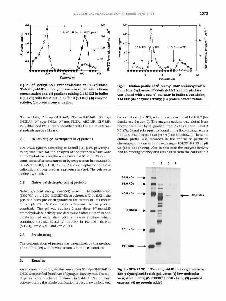
Fig. 2 – N6-Methyl-AMP aminohydrolase on P11-cellulose.
N6-Methyl-AMP aminohydrolase was eluted with a linear
concentration and pH gradient mixing 0.1 M KCl in buffer
B (pH 7.6) with 0.3 M KCl in buffer C (pH 8.0): (*) enzyme
activity; (*) protein concentration.
Fig. 3 – Elution profile of N6-methyl-AMP aminohydrolase
from Blue-Sepharose. N6-Methyl-AMP aminohydrolase
was eluted with 1 mM N6-me-AMP in buffer E containing
1 M KCl: (*) enzyme activity; (*) protein concentration.
N6-me-dAMP, N6-cypr-PMEDAP, N6-me-PMEDAP, N6-me2-
PMEDAP, N6-cypr-PMEA, N6-me2-PMEA, ABC-MP, CBV-MP,
IMP, dIMP and PMEG, were identified with the aid of external
standards spectra library.
2.5. Denaturing gel electrophoresis of proteins
SDS-PAGE system according to Laemli [18] (13% polyacryla-
mide) was used for the analysis of the purified N6-me-AMP
aminohydrolase. Samples were heated at 95 8C for 15 min (in
some cases after concentration by evaporation in vacuum) in
30 mM Tris–HCl, pH 6.8, 5% SDS, 5% 2-mercaptoethanol. LMW
calibration kit was used as a protein standard. The gels were
stained with silver.
2.6. Native gel electrophoresis of proteins
Native gradient slab gels (4–25%) were run to equilibration
(2500 Vh) on a 2050 MIDGET-Electrophoresis Unit (LKB); the
gels had been pre-electrophoresed for 30 min in Tris-borate
buffer, pH 8.3. HMW calibration kits were used as protein
standards. The gel was cut into 2-mm slices. N6-me-AMP
aminohydrolase activity was determined after extraction and
incubation of each slice with an assay mixture which
contained (150 mL): 50 mM N6-me-AMP in 100 mM Tris–HCl
(pH 7.4), 9 mM NaCl and 2 mM DTT.
2.7. Protein assay
The concentration of protein was determined by the method
of Bradford [19] with bovine serum albumin as standard.
Fig. 4 – SDS-PAGE of N6-methyl-AMP aminohydrolase in
13% polyacrylamide slab gel. Lines: (1) low-molecular-
weight standards; (2) POROS1 HS 20 eluate; (3) purified
enzyme; (4) no protein added.
3. Results
An enzyme that catalyzes the conversion N6-cypr-PMEDAP to
PMEG was purified from liver of Sprague–Dawley rats. The six-
step purification scheme is shown in Table 1. The enzyme
activity during the whole purification procedure was followed
by formation of PMEG, which was determined by HPLC (for
details see Section 2). The enzyme activity was eluted from
phosphocelullose by pH gradient from 7.7 to 7.8 at 0.15–0.20 M
KCl (Fig. 2) and subsequently found in the flow-through eluate
from DEAE Sepharose FF at pH 7.4 (data not shown). The same
elution profile was recorded in the course of perfusion
chromatography on cationic exchanger POROS1HS 20 at pH
6.8 (data not shown). Also in this case the enzyme activity
had no binding potency and was eluted from the column in a
b i o c h e m i c a l p h a r m a c o l o g y 7 1 ( 2 0 0 6 ) 1 3 7 0 – 1 3 7 61374
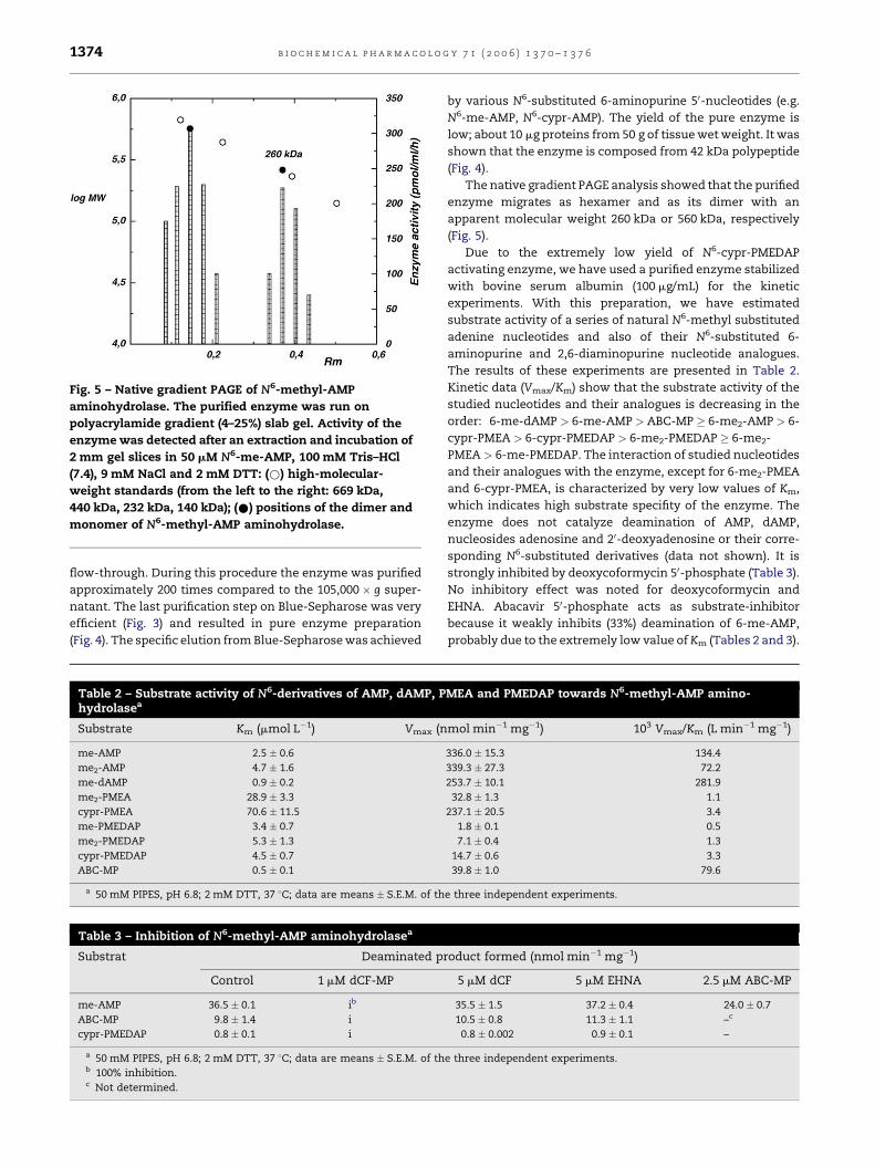
Fig. 5 – Native gradient PAGE of N6-methyl-AMP
aminohydrolase. The purified enzyme was run on
polyacrylamide gradient (4–25%) slab gel. Activity of the
enzyme was detected after an extraction and incubation of
2 mm gel slices in 50 mM N6-me-AMP, 100 mM Tris–HCl
(7.4), 9 mM NaCl and 2 mM DTT: (*) high-molecular-
weight standards (from the left to the right: 669 kDa,
440 kDa, 232 kDa, 140 kDa); (*) positions of the dimer and
monomer of N6-methyl-AMP aminohydrolase.
flow-through. During this procedure the enzyme was purified
approximately 200 times compared to the 105,000 � g super-
natant. The last purification step on Blue-Sepharose was very
efficient (Fig. 3) and resulted in pure enzyme preparation
(Fig. 4). The specific elution from Blue-Sepharose was achieved
Table 3 – Inhibition of N6-methyl-AMP aminohydrolasea
Substrat Deaminated p
Control 1 mM dCF-MP
me-AMP 36.5 � 0.1 ib
ABC-MP 9.8 � 1.4 i
cypr-PMEDAP 0.8 � 0.1 i
a 50 mM PIPES, pH 6.8; 2 mM DTT, 37 8C; data are means � S.E.M. of thb 100% inhibition.c Not determined.
Table 2 – Substrate activity of N6-derivatives of AMP, dAMP, Phydrolasea
Substrate Km (mmol L�1) Vmax (n
me-AMP 2.5 � 0.6
me2-AMP 4.7 � 1.6
me-dAMP 0.9 � 0.2
me2-PMEA 28.9 � 3.3
cypr-PMEA 70.6 � 11.5
me-PMEDAP 3.4 � 0.7
me2-PMEDAP 5.3 � 1.3
cypr-PMEDAP 4.5 � 0.7
ABC-MP 0.5 � 0.1
a 50 mM PIPES, pH 6.8; 2 mM DTT, 37 8C; data are means � S.E.M. of th
by various N6-substituted 6-aminopurine 50-nucleotides (e.g.
N6-me-AMP, N6-cypr-AMP). The yield of the pure enzyme is
low; about 10 mg proteins from 50 g of tissue wet weight. It was
shown that the enzyme is composed from 42 kDa polypeptide
(Fig. 4).
The native gradient PAGE analysis showed that the purified
enzyme migrates as hexamer and as its dimer with an
apparent molecular weight 260 kDa or 560 kDa, respectively
(Fig. 5).
Due to the extremely low yield of N6-cypr-PMEDAP
activating enzyme, we have used a purified enzyme stabilized
with bovine serum albumin (100 mg/mL) for the kinetic
experiments. With this preparation, we have estimated
substrate activity of a series of natural N6-methyl substituted
adenine nucleotides and also of their N6-substituted 6-
aminopurine and 2,6-diaminopurine nucleotide analogues.
The results of these experiments are presented in Table 2.
Kinetic data (Vmax/Km) show that the substrate activity of the
studied nucleotides and their analogues is decreasing in the
order: 6-me-dAMP > 6-me-AMP > ABC-MP � 6-me2-AMP > 6-
cypr-PMEA > 6-cypr-PMEDAP > 6-me2-PMEDAP � 6-me2-
PMEA > 6-me-PMEDAP. The interaction of studied nucleotides
and their analogues with the enzyme, except for 6-me2-PMEA
and 6-cypr-PMEA, is characterized by very low values of Km,
which indicates high substrate specifity of the enzyme. The
enzyme does not catalyze deamination of AMP, dAMP,
nucleosides adenosine and 20-deoxyadenosine or their corre-
sponding N6-substituted derivatives (data not shown). It is
strongly inhibited by deoxycoformycin 50-phosphate (Table 3).
No inhibitory effect was noted for deoxycoformycin and
EHNA. Abacavir 50-phosphate acts as substrate-inhibitor
because it weakly inhibits (33%) deamination of 6-me-AMP,
probably due to the extremely low value of Km (Tables 2 and 3).
roduct formed (nmol min�1 mg�1)
5 mM dCF 5 mM EHNA 2.5 mM ABC-MP
35.5 � 1.5 37.2 � 0.4 24.0 � 0.7
10.5 � 0.8 11.3 � 1.1 –c
0.8 � 0.002 0.9 � 0.1 –
e three independent experiments.
MEA and PMEDAP towards N6-methyl-AMP amino-
mol min�1 mg�1) 103 Vmax/Km (L min�1 mg�1)
336.0 � 15.3 134.4
339.3 � 27.3 72.2
253.7 � 10.1 281.9
32.8 � 1.3 1.1
237.1 � 20.5 3.4
1.8 � 0.1 0.5
7.1 � 0.4 1.3
14.7 � 0.6 3.3
39.8 � 1.0 79.6
e three independent experiments.
b i o c h e m i c a l p h a r m a c o l o g y 7 1 ( 2 0 0 6 ) 1 3 7 0 – 1 3 7 6 1375
4. Discussion
Our results show that enzyme activating N6-cypr-PMEDAP [2–
4], prodrug of PMEG is most probably identical with the AMP
deaminase like enzyme, which is able to catalyze the
intracellular conversion of abacavir 50-monophosphate to
carbovir 50-monophosphate [14]. We confirmed that this
enzyme accepts natural N6-methyl-, N6-dimethyl-AMP and
N6-methyl-dAMP as substrates and catalyzes their conversion
to IMP and dIMP, respectively, so that it acts as N6-methyl-
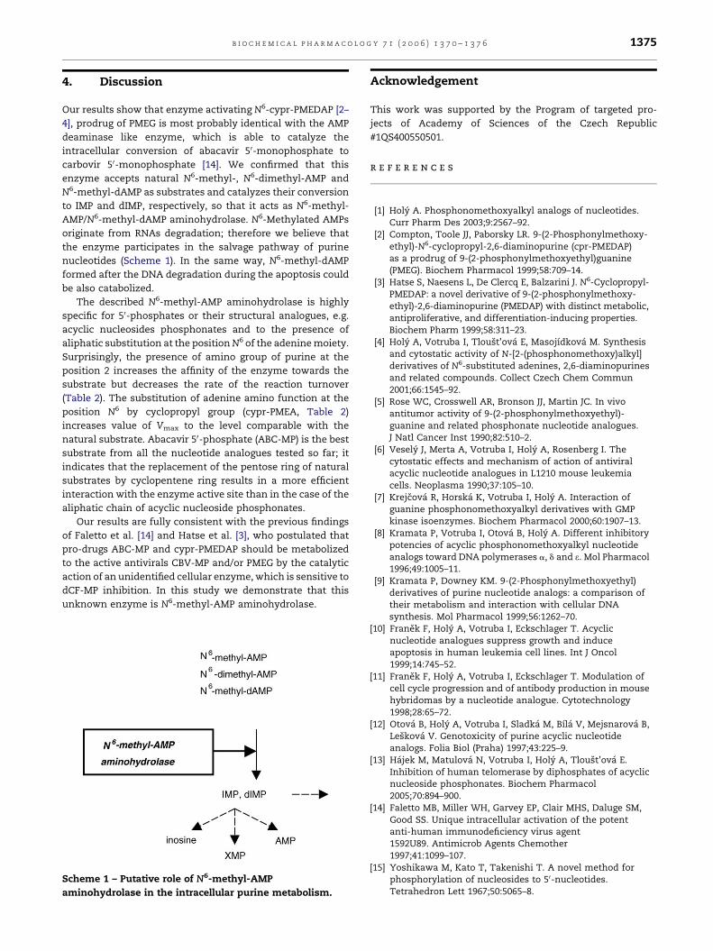
AMP/N6-methyl-dAMP aminohydrolase. N6-Methylated AMPs
originate from RNAs degradation; therefore we believe that
the enzyme participates in the salvage pathway of purine
nucleotides (Scheme 1). In the same way, N6-methyl-dAMP
formed after the DNA degradation during the apoptosis could
be also catabolized.
The described N6-methyl-AMP aminohydrolase is highly
specific for 50-phosphates or their structural analogues, e.g.
acyclic nucleosides phosphonates and to the presence of
aliphatic substitution at the position N6 of the adenine moiety.
Surprisingly, the presence of amino group of purine at the
position 2 increases the affinity of the enzyme towards the
substrate but decreases the rate of the reaction turnover
(Table 2). The substitution of adenine amino function at the
position N6 by cyclopropyl group (cypr-PMEA, Table 2)
increases value of Vmax to the level comparable with the
natural substrate. Abacavir 50-phosphate (ABC-MP) is the best
substrate from all the nucleotide analogues tested so far; it
indicates that the replacement of the pentose ring of natural
substrates by cyclopentene ring results in a more efficient
interaction with the enzyme active site than in the case of the
aliphatic chain of acyclic nucleoside phosphonates.
Our results are fully consistent with the previous findings
of Faletto et al. [14] and Hatse et al. [3], who postulated that
pro-drugs ABC-MP and cypr-PMEDAP should be metabolized
to the active antivirals CBV-MP and/or PMEG by the catalytic
action of an unidentified cellular enzyme, which is sensitive to
dCF-MP inhibition. In this study we demonstrate that this
unknown enzyme is N6-methyl-AMP aminohydrolase.
Scheme 1 – Putative role of N6-methyl-AMP
aminohydrolase in the intracellular purine metabolism.
Acknowledgement
This work was supported by the Program of targeted pro-
jects of Academy of Sciences of the Czech Republic
#1QS400550501.
r e f e r e n c e s
[1] Holy A. Phosphonomethoxyalkyl analogs of nucleotides.Curr Pharm Des 2003;9:2567–92.
[2] Compton, Toole JJ, Paborsky LR. 9-(2-Phosphonylmethoxy-ethyl)-N6-cyclopropyl-2,6-diaminopurine (cpr-PMEDAP)as a prodrug of 9-(2-phosphonylmethoxyethyl)guanine(PMEG). Biochem Pharmacol 1999;58:709–14.
[3] Hatse S, Naesens L, De Clercq E, Balzarini J. N6-Cyclopropyl-PMEDAP: a novel derivative of 9-(2-phosphonylmethoxy-ethyl)-2,6-diaminopurine (PMEDAP) with distinct metabolic,antiproliferative, and differentiation-inducing properties.Biochem Pharm 1999;58:311–23.
[4] Holy A, Votruba I, Tloust’ova E, Masojıdkova M. Synthesisand cytostatic activity of N-[2-(phosphonomethoxy)alkyl]derivatives of N6-substituted adenines, 2,6-diaminopurinesand related compounds. Collect Czech Chem Commun2001;66:1545–92.
[5] Rose WC, Crosswell AR, Bronson JJ, Martin JC. In vivoantitumor activity of 9-(2-phosphonylmethoxyethyl)-guanine and related phosphonate nucleotide analogues.J Natl Cancer Inst 1990;82:510–2.
[6] Vesely J, Merta A, Votruba I, Holy A, Rosenberg I. Thecytostatic effects and mechanism of action of antiviralacyclic nucleotide analogues in L1210 mouse leukemiacells. Neoplasma 1990;37:105–10.
[7] Krejcova R, Horska K, Votruba I, Holy A. Interaction ofguanine phosphonomethoxyalkyl derivatives with GMPkinase isoenzymes. Biochem Pharmacol 2000;60:1907–13.
[8] Kramata P, Votruba I, Otova B, Holy A. Different inhibitorypotencies of acyclic phosphonomethoxyalkyl nucleotideanalogs toward DNA polymerases a, d and e. Mol Pharmacol1996;49:1005–11.
[9] Kramata P, Downey KM. 9-(2-Phosphonylmethoxyethyl)derivatives of purine nucleotide analogs: a comparison oftheir metabolism and interaction with cellular DNAsynthesis. Mol Pharmacol 1999;56:1262–70.
[10] Franek F, Holy A, Votruba I, Eckschlager T. Acyclicnucleotide analogues suppress growth and induceapoptosis in human leukemia cell lines. Int J Oncol1999;14:745–52.
[11] Franek F, Holy A, Votruba I, Eckschlager T. Modulation ofcell cycle progression and of antibody production in mousehybridomas by a nucleotide analogue. Cytotechnology1998;28:65–72.
[12] Otova B, Holy A, Votruba I, Sladka M, Bıla V, Mejsnarova B,Leskova V. Genotoxicity of purine acyclic nucleotideanalogs. Folia Biol (Praha) 1997;43:225–9.
[13] Hajek M, Matulova N, Votruba I, Holy A, Tloust’ova E.Inhibition of human telomerase by diphosphates of acyclicnucleoside phosphonates. Biochem Pharmacol2005;70:894–900.
[14] Faletto MB, Miller WH, Garvey EP, Clair MHS, Daluge SM,Good SS. Unique intracellular activation of the potentanti-human immunodeficiency virus agent1592U89. Antimicrob Agents Chemother1997;41:1099–107.
[15] Yoshikawa M, Kato T, Takenishi T. A novel method forphosphorylation of nucleosides to 50-nucleotides.Tetrahedron Lett 1967;50:5065–8.

b i o c h e m i c a l p h a r m a c o l o g y 7 1 ( 2 0 0 6 ) 1 3 7 0 – 1 3 7 61376
[16] Holy A, Kowollik G. A simple enzymatic synthesis ofnucleoside 50-phosphates. Collect Czech Chem Commun1970;35:1013–6.
[17] Tanabe T, Yamauchi K, Kinoshita M. Methylation ofdeoxynucleotides with trimethyl phosphate inaqueous phase. Bull Chem Soc Jpn 1983;56:1826–31.
[18] Laemi UK, Favre M. Maturation of the head ofbacteriophage T4. II. Head-related, abberant tau-particles.J Biochem 1973;80:575–99.
[19] Bradford M. A rapid and sensitive method for thequantitation of microgram quantities of protein utilizingthe principle of protein-dye binding. Anal Biochem1976;72:248–54.