Embed Size (px)
Citation preview

N-Myristoyltransferase inhibition as a tool for antileishmanial drug discovery: Use in high-
throughput, de novo, and piggyback strategies for drug development
by
John Anthony Kavouris
BA, Boston University, 2011
Submitted to the Graduate Faculty of the
Dietrich School of Arts and Sciences in partial fulfillment
of the requirements for the degree of
Master of Science
University of Pittsburgh
2014

ii
UNIVERSITY OF PITTSBURGH
Dietrich School of Arts and Sciences
This thesis was presented
by
John Anthony Kavouris
It was defended on
April 25th
, 2014
and approved by
W. Seth Horne, Assistant Professor, Chemistry
Barry I. Gold, Professor and Chair, Pharmaceutical Sciences
Thesis Advisor: Scott G. Nelson, Professor, Chemistry

iii
Copyright © by John Anthony Kavouris
2014
N-Myristoyltransferase inhibition as a tool for antileishmanial drug discovery: Use in
high-throughput, de novo, and piggyback strategies for drug development
John Anthony Kavouris M.S.
University of Pittsburgh, 2014

iv
N-Myristoyltransferase inhibition as a tool for antileishmanial drug discovery: Use in high-
throughput, de novo, and piggyback strategies for drug development
John Anthony Kavouris M.S.
University of Pittsburgh, 2014
Leishmaniasis is a disease caused by the Leishmania genus of protozoan parasites, and
spread by the phlebotomine genus of sandfly. The disease is caused by at least 20 different
species of Leishmania parasites, and manifests itself in three forms: cutaneous, mucocutaneous,
and most severely, visceral leishmaniasis. Current chemotherapeutic treatments for leishmaniasis
are limited by parasite resistance to existing drugs, highly toxic side effects, and high cost of
treatment. As a neglected tropical disease, there has been relatively little research toward new
drugs, despite a large need, comprising 1.3m new cases and 20,000-50,000 deaths annually.
Fortunately, although there is a wide variety in species causing the disease, visceral
leishmaniasis is largely caused by a single species of parasite, Leishmania donovani, greatly
simplifying the search for drugs against the most dangerous form of the disease. The whole
genome of the species has been sequenced, and many crystal structures of key proteins have been
elucidated. Specifically, N-myristoyltransferase (NMT) has emerged as a promising enzyme for
target-based antileishmanial drug development, with a wide array of tools available for any level
of drug discovery: in silico modeling and docking, enzyme specific assays for protozoan and
human isoforms, high throughput in vitro assays against both the parasite itself and infected host
cells, and animal models of the disease. However, with L. donovani NMT as a newer target,
research has yet to utilize all of these individual elements synergistically with themselves, or

v
with related studies in different parasite species. Combined analysis of these methods can yield a
more efficient search for antileishmanials, regardless of screening type.

vi
TABLE OF CONTENTS
TABLE OF CONTENTS ........................................................................................................... VI
LIST OF FIGURES ................................................................................................................. VIII
LIST OF EQUATIONS .............................................................................................................. IX
ACKNOWLEDGEMENTS ........................................................................................................ X
LIST OF ABBREVIATIONS AND SYMBOLS ...................................................................... XI
1.0 LEISHMANIASIS: DISEASE OVERVIEW AND NEED FOR TREATMENT .. 1
1.1.1 Disease burden .............................................................................................. 1
1.1.2 Complications to developing treatments ..................................................... 3
1.1.3 Visceral leishmanias as focus for treatment ............................................... 5
2.0 N-MYRISTOYLTRANSFERASE ............................................................................. 7
2.1 NMT STRUCTURE, FUNCTION, AND SUBSTRATES ............................... 9
2.2 NMT UTILITY AS A LEISHMANIASIS DRUG TARGET ........................ 10
3.0 SCREENING METHODS ........................................................................................ 14
3.1 IN VITRO METHODS ...................................................................................... 14
3.1.1 Enzymatic assays ......................................................................................... 14
3.1.2 Cellular assay .............................................................................................. 16
3.1.3 Secondary screening ................................................................................... 18
3.1.4 Screen analysis and validity ....................................................................... 18

vii
3.2 IN VIVO MODELS OF VISCERAL LEISHMANIASIS .............................. 19
4.0 LIMITED STATE OF ANTILEISHMANIAL DEVELOPMENT ....................... 21
5.0 NMT STUDIES IN RELATED ORGANISMS ....................................................... 25
5.1.1 Benzofurans ................................................................................................. 26
5.1.2 Pyrazole sulfonamides ................................................................................ 28
5.1.3 Quinolines .................................................................................................... 30
5.1.4 Peptide/peptidomimetics ............................................................................ 31
6.0 FUTURE PROSPECTS IN ANTILEISHMANIAL DEVELOPMENT ............... 33
7.0 CONCLUSIONS ........................................................................................................ 39
BIBLIOGRAPHY ....................................................................................................................... 40
viii
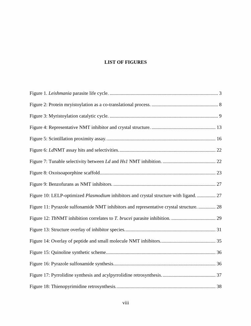
LIST OF FIGURES
Figure 1. Leishmania parasite life cycle. ........................................................................................ 3
Figure 2: Protein mryistoylation as a co-translational process. ...................................................... 8
Figure 3: Myristoylation catalytic cycle. ........................................................................................ 9
Figure 4: Representative NMT inhibitor and crystal structure. .................................................... 13
Figure 5: Scintillation proximity assay. ........................................................................................ 16
Figure 6: LdNMT assay hits and selectivities. .............................................................................. 22
Figure 7: Tunable selectivity between Ld and Hs1 NMT inhibition. ........................................... 22
Figure 8: Oxoisoaporphine scaffold. ............................................................................................. 23
Figure 9: Benzofurans as NMT inhibitors. ................................................................................... 27
Figure 10: LELP-optimized Plasmodium inhibitors and crystal structure with ligand. ............... 27
Figure 11: Pyrazole sulfonamide NMT inhibitors and representative crystal structure. .............. 28
Figure 12: TbNMT inhibition correlates to T. brucei parasite inhibition. .................................... 29
Figure 13: Structure overlay of inhibitor species. ......................................................................... 31
Figure 14: Overlay of peptide and small molecule NMT inhibitors. ............................................ 35
Figure 15: Quinoline synthetic scheme......................................................................................... 36
Figure 16: Pyrazole sulfonamide synthesis................................................................................... 36
Figure 17: Pyrrolidine synthesis and acylpyrrolidine retrosynthesis. ........................................... 37
Figure 18: Thienopyrimidine retrosynthesis. ................................................................................ 38

ix
LIST OF EQUATIONS
Equation 1: Z-Factor for HTS validity .......................................................................................... 19
Equation 2: Cheng-Prusoff equation ............................................................................................. 28

x
ACKNOWLEDGEMENTS
I’d like to begin by expressing my appreciation and gratitude for my committee members Dr.
Seth Horne, Dr. Barry Gold, and committee chair Dr. Scott Nelson. After some stressful
moments and uncertainty of what to do next in my graduate career, Scott emerged as a great help
and was encouraging of whatever I chose to pursue. Seth and Barry graciously assisted with
providing corrections to the document, and the help of all three professors has certainly
strengthened the quality and clarity of my writing.
I would also like to thank Dr. Scott Schaus and Dr. Lauren Brown at Boston University
for offering me a wonderful opportunity as the next step in my scientific career. Furthermore,
they’ve given me a chance to return to Boston, one of my favorite cities, all while being nothing
but patient while I finish work in Pittsburgh.
Finally, words cannot truly express my love, respect, and thankfulness for my aunt
Christine Oliver and my late yiayia Joan Kapsimalis. They have been an immensely positive
force throughout my life and have gone above and beyond to help me throughout my education. I
have been nothing but fortunate for their efforts.

xi
LIST OF ABBREVIATIONS AND SYMBOLS
ADMET absorption, distribution, metabolism, excretion, and toxicity
BALB/c albino, laboratory-bred strain of house mouse
C57BL/6 common, inbred mouse strain
CaNMT Candida albicans NMT
ClogP calculated LogP, P= [ocatanol]/[water]
DNDi Drugs for Negelcted Disease Initiative
GSK GlaxoSmithKline
HAT human African trypanosomaisis
HCS high-content screening
HIV human immunodeficiency virus
Hs1NMT Homo sapiens NMT1
Hs2NMT Homo sapiens NMT2
HTS high-throughput screening
IC50 half-maximal inhibitory concentration
IV intravenous
Ki inhibitory constant
Km Michaelis-Menton constant
LdNMT Leishmania donovani NMT

xii
LE ligand efficiency
LELP ligand efficiency dependency lipophilicity
LmNMT Leishmania major NMT
NMT N-Myristoyltransferase
NTD neglected tropical disease
PDB Protein Data Bank
PKDL Post kala-azar dermal leishmaniasis
PvNMT Plasmodium vivax NMT
QSAR qualitative structure-activity relationship
SAR structure-activity relationship
ScNMT Saccharomyces cerevisiae NMT
SPA scintillation proximity assay
SPPS solid-phase peptide synthesis
TbNMT Trypanosoma brucei NMT
THP-1 Human-acute monocytic leukemia cell line
VL visceral leishmaniasis

1
1.0 LEISHMANIASIS: DISEASE OVERVIEW AND NEED FOR TREATMENT
Leishmaniasis is a disease caused by the Leishmania genus of protozoan parasites and spread by
the phlebotomine genus of sandfly. The disease is endemic to tropical and sub-tropical countries,
predominantly affecting the developing world.1 Major risk factors of the disease include negative
socioeconomic conditions, malnutrition, population mobility and environmental changes, all
conditions frequently encountered in the tropics.
1.1.1 Disease burden
Leishmaniasis affects approximately 12 million people worldwide, with approximately 2
million new cases and 20-50 thousand deaths annually. After malaria, it is the second highest
parasitic killer. The disease manifests itself in a variety of forms: cutaneous, mucocutaneous, and
visceral. Cutaneous results in open sores that eventually heal on their own within a few months
to a year and a half, but scarring can occur. Mucocutaneous can affect the skin and mucous
membranes (nose, mouth, throat), yielding sores that typically heal, with scarring and potential
tissue damage. Visceral leishmaniasis (VL, also known as ‘kala-azar’), the most serious form, is
caused by parasite migration to internal organs and is fatal if left untreated.2 Even with treatment,
visceral leishmaniasis can result in organ damage. Further complications can also arise from
leishmaniasis infection, including post kala-azar dermal leishmaniasis (PKDL) in which the

2
parasite infection recurs affecting the skin and co-infection in patients with HIV. Leishmaniasis
is recognized as a neglected tropical disease (NTD).3-4
While the definition criteria vary among
different health organizations, NTDs are generally endemic to low-income areas of the tropics,
and that lack adequate available treatments.
Current treatments for leishmaniasis have improved in the last decade with the introduction
of Miltefosine as the first orally-available therapy, but existing treatments are limited by factors
of cost, oral availability, toxicity, and parasite resistance.5 Unfortunately, parasite resistance is
not only a problem of older treatments such as pentavalent antimonials, but instances of
resistance have also been shown for more recently developed treatments as well.6-9
The Drugs
for Neglected Disease Initiative (DNDi) is non-profit organization devoted to developing
treatments for neglected diseases, and includes a large variety of industry, governmental, and
academic partners, with a focus on collaborative projects. Based on DNDi efforts, the treatment
options for leishmaniasis are improving, but slowly.10
Currently, the most advanced therapies
focuses on combination of existing drugs.11
Only one novel chemotheraputic, sitamiquine, is
currently in development through GSK/Walter Reed Army Institute that is currently in phase II
clinical trials. Other treatments under development are oral and topical formulations of existing
drug amphotericin B, which has previously only available via IV therapy and is highly toxic.
Also, fexinidazole, a human African trypanosomaisis inhibitor, is beginning phase II trials as a
treatment for L. donovani infection as well.12
Palatnik-de-Sousa and Kedzierski have both proposed that the parasite’s biology may
provide a route to vaccination and there is one candidate vaccine currently in Phase I trials. 13-15
Nevertheless, until a range of treatments is approved for use, a need persists for the pre-clinical
development of antileishmanials that meet all the desired criteria for safety, efficacy, and cost.

3
1.1.2 Complications to developing treatments
Leishmania parasites are transferred to mammalian hosts via the bite of infected female
phebotomine sandflies, depicted in Figure 1.16
Within the sandfly midgut, the parasites exist as
motile metacyclic promastigotes, eventually migrating to the stomodeal valve. When the fly bites
a host, promastigote transmission occurs, however, the vector is not yet infective. Promastigotes
invade mammalian macrophages where they are phagocytized into amastigotes, the spread of
which by infection and replication within host cells affects the surrounding tissue. If transmitted
back to the sandfly, the amastigotes revert back to procyclic promastigotes in the absence of host
macrophage cells. Undergoing cell division, parasites are considered metacyclic promastigotes,
thereby restarting the cycle.
Figure 1. Leishmania parasite life cycle.
Image used from public domain.

4
In addition to the multiple parasite forms, the number of Leishmania species that
cause the disease complicates disease progression, expression, and treatment development; over
20 species of parasite have been shown to cause the differing forms of the disease. This
biological diversity accounts for the phenotypic differences in disease manifestation. Most
species are associated with a specific form of illness, but occasionally, individual species
themselves will cause different manifestations.17
Fortunately, a wide array of genomic and
proteomic data is known for leishmania parasites, including completely sequenced genomes of
several species, the characterization of many proteins necessary for parasite function, and the
solution of several crystal structures of relevant proteins.18-20
The wealth of data regarding
protein structure and function offers some mechanistic insight into known NMT inhibitors that
are effective across multiple Leishmania species.21
However, the number and variety of species
also illustrates the complexity of generating broad-spectrum antileishmanial theraputics.
Drug development for leishmaniasis has been assisted by related research on other
trypanosomatid diseases that include Trypanosoma brucei, (human African trypanomaisis,
“sleeping sickness”), and T. cruzi (South American trypanosomiasis, “Chagas diease”).22
Similar
drugs/targets have also been identified in certain fungal and bacterial infections, and in the
Plasmodium genus, responsible for malaria.23
These species share biological similarities with
Leishmania spp. and offer a large dataset related to development of antileishmanials, including
crystal structures of related target proteins, and examples of cross-species inhibition by single
compounds.24
Again, the additional information helps the understanding of existing cross-species
drug efficacy, but only further convolutes an effort for development of novel broad-spectrum
antitrypanosomal drugs. It is worth noting that, despite certain macromolecular similarities

5
between species, species pathology ultimately affects drug development as well. Considerations
for HAT drug development must account for blood-brain-barrier penetration to treat the infected
host. Under similar preliminary screening conditions, this would be an added complication for
HAT inhibitors, but is absent from Leishmania inhibitor design considerations.
The number and complexity of leishmaniasis manifestations renders the disease
an incredibly difficult candidate for rational drug design. Although a potent treatment against all
forms of the disease may be unlikely, visceral leishmaniasis infection is both a major contributor
to the global mortality and morbidity, and surprisingly well suited for rational drug design. Of
the global leishmaniasis disease burden, visceral leishmaniasis accounts for approximately
200,000-400,000 new infections, and 20,000-40,000 deaths per year.25-26
1.1.3 Visceral leishmanias as focus for treatment
Treatment of visceral leishmaniasis is aided by the high degree of similarity
between causative species. Visceral leishmaniasis is largely cited as caused by the species L.
donovani, and occasionally, by L. infantum. In fact, four individual Leishmania species have
been proposed as causes for VL: L. donovani, L. infantum, L. chagasi, and L. archibaldi.27
Despite the general classification as separate species, the distinction largely arises from
geographic origins of each sample. The perceived differences between these taxons are so minute
that markers of differentiation are often unreliable.28-29
As such, these four species are broadly
referred to as the ‘donovani complex.’ Analysis of the species has accounted for small genetic
differences and has been used to trace disease spread and evolution, however, researchers
concluded that not enough genetic variation is present within current available data to warrant
this species differentiation. Instead, a corrected differentiation of only two species is proposed:

6
donovani and infantum.30
While differences in pathology between the two species have been
studied, many considerations for rational drug design remain largely similar.31
Structural
characteristics of drug target enzymes are highly conserved across reported species, typically
with >95% sequence or structure similarities, as applicable. As such, inhibitors of both species
are likely viable through a single drug discovery campaign. However, given similar species with
same clinical outcome of VL, cross-validation of known inhibitors on both species is currently
limited.
Elements are currently in place to facilitate scalable, target-based, rational drug
design toward inhibitors of visceral leishmaniasis. Several proteins of the Leishmania donovani
complex have been investigated as potential therapeutic targets and, in particular, N-
myristoyltransferase (NMT) has emerged as a promising one. This target offers multiple design
motifs such as in silico methods using crystallographic data, enzymatic assay, in vitro assays,
and availability of in vivo disease models. The three screening methods (in silico, enzymatic, in
vitro) may be run in high-throughput or high-content applications, aided by automation to
facilitate the screening of large sized compound libraries. Furthermore, techniques relevant to
NMT-based drug development may be arranged for de novo drug design specifically targeting L.
donovani NMT, as an additional element of ‘target-free’* screening to identify an affected target
and improve hit optimization, or as a ‘piggyback’ approach based on existing NMT inhibitors in
other species. Despite the availability and scope of these methods, their combined use is
surprisingly limited. A discussion of available methods for drug development, their existing
applications, and prospective improvements or combinations is presented herein.
* ‘Target-free,’ or phenotypic screening tests potential inhibitors for protozoan growth
inhibition and limited human toxicity, irrespective of molecular targets affected.

7
2.0 N-MYRISTOYLTRANSFERASE
Protein myristoylation is an essential function in eukaryotes that occurs via the addition
of a myristoyl group to the N-glycine terminus of a set of proteins.32-33
The myristoyl group,
derived from myristic acid (n-tetradecanoic acid), is a 14-carbon saturated fatty acid serving as a
long, hydrophobic tail in a variety of functions, including protein-protein interactions, membrane
targeting, signal transduction, and apoptosis.34-35
The amide bond formation is catalyzed by the
enzyme myristoyl-CoA protein N-myristoyltransferase (NMT) that occurs as both a post- and,
more typically, co-translational modification.36

8
Figure 2: Protein mryistoylation as a co-translational process.
Image used under Creative Commons license.37
The binding events required for protein substrate myristoylation have been thoroughly
characterized and includes an understanding of substrate specificities for both the myristoyl and
protein binding sites. The mechanism proceeds via an “ordered bi bi mechanism,” in which
myristoyl-CoA first binds to the apo-enzyme.38
The myristoyl-CoA binding induces an allosteric
effect on the enzyme that positions the CoA as a functional part of the protein binding site. The
myristoyl group transfer is facilitated by the attack of the incoming N-terminal glycine on the
myristoyl-CoA thioester, shown in Figure 2. After the transfer, the free CoA is released first,
allowing the enzyme conformation to relax and release the myristoylated substrate protein; the
O
OH
myristicacid+ CoA-SH + ATP Met1-Gly2-AA3-AA4-AA-5-
Ribosome
O
S-CoA
Gly2-AA3-AA4-AA5-
Ribosome
O
NH
AA3-AA4-AA5-
O
myristoyl CoA
Ribosome
+ CoA-SH
O
NH
AA3-AA4-AA5-.....-AAn-2-AAn-1-AAn
O
myristoylated protein
membraneassociation
subcellularlocalization
protein-proteininteraction
acyl-CoA synthetase
methionyl aminopeptidase
myristoyl-CoA protein:N-myristoyltransferase (NMT)
+ AMP + PPI
9
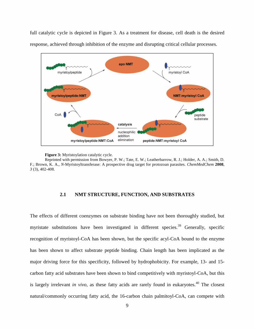
full catalytic cycle is depicted in Figure 3. As a treatment for disease, cell death is the desired
response, achieved through inhibition of the enzyme and disrupting critical cellular processes.
Figure 3: Myristoylation catalytic cycle.
Reprinted with permission from Bowyer, P. W.; Tate, E. W.; Leatherbarrow, R. J.; Holder, A. A.; Smith, D.
F.; Brown, K. A., N-Myristoyltransferase: A prospective drug target for protozoan parasites. ChemMedChem 2008,
3 (3), 402-408.
2.1 NMT STRUCTURE, FUNCTION, AND SUBSTRATES
The effects of different coenzymes on substrate binding have not been thoroughly studied, but
myristate substitutions have been investigated in different species.39
Generally, specific
recognition of myristoyl-CoA has been shown, but the specific acyl-CoA bound to the enzyme
has been shown to affect substrate peptide binding. Chain length has been implicated as the
major driving force for this specificity, followed by hydrophobicity. For example, 13- and 15-
carbon fatty acid substrates have been shown to bind competitively with myristoyl-CoA, but this
is largely irrelevant in vivo, as these fatty acids are rarely found in eukaryotes.40
The closest
natural/commonly occurring fatty acid, the 16-carbon chain palmitoyl-CoA, can compete with

10
the myristate binding.41
While a poor substrate, it is found in much greater intracellular
concentrations, yet NMT function is still determined by the myristate. Overall, the enzyme
shows little tolerance for alternate acyl-CoA substrates in vivo, and the myristoyl-CoA binding
site is highly conserved across species studied.
Unlike the myristoyl-CoA binding site, the protein binding site is not highly conserved
across species, allowing for substrate specificity for protein myristoylation and species-specific
inhibition of the NMT enzyme. Substrate specificity is accomplished by recognition of N-
terminal amino acid residues within the substrate protein with as many as 17 terminal residues
responsible for substrate recognition.42-43
These 17 residues are split into three regions; region 1
consists of the first 6 residues, which fit within the binding pocket, and must terminate with a
glycine; region 2 consists of the next 6 residues, interacting with the surface of the protein
adjacent to the binding pocket, typically consisting of smaller, polar residues; region 3 consists
of the final 5 amino acids typically recognized by the enzyme, and is usually comprised of
increasingly hydrophilic residues. The enzyme’s broader recognition of several substrate
residues allows for specificity of proteins to be myristoylated, but the existence of a specific,
well-defined binding pocket allows for enzyme inhibition by small molecules. Competitive
inhibition of protein myristoylation by small molecule binding to the substrate pocket has been
confirmed by kinetic studies, suggesting the viability of NMT inhibition as a therapeutic
target.44-45
2.2 NMT UTILITY AS A LEISHMANIASIS DRUG TARGET
Several distinctions between mammalian and protozoan NMTs have been identified and

11
may be exploited for the treatment of protozoan diseases, such as leishmaniasis. Mammals all
possess two NMT isozymes, NMT1, and NMT2, each exhibiting slightly different protein
substrate specificity, but both are highly similar to one-another and highly conserved across
species. For example, human NMT1 (Hs1NMT), and Hs2NMT share 77% sequence identity.
Homologues are, of course, expressed in mice, and the mouse NMT1 and NMT2 share 97% and
96% sequence identity with the respective human isozyme.46
This sequence homology allows for
accurate modeling of NMT inhibition in mouse models of infection.
The genus Leishmania only synthesizes one NMT isozyme which is sufficiently different
from the human forms to allow for specific inhibition. For example, the L. donovani NMT
(LdNMT) only shares 42% sequence identity with Hs1NMT and highly selective inhibition of
LdNMT over both human NMTs has been demonstrated. The NMTs of leishmania parasites are
highly conserved across different parasite species with L. major sharing 97, 96, and 96%
sequence identity with L. donovani, L. infantum, and L. mexicana, respectively. Residues
associated with the predicted peptide binding groove are fully conserved between L. donovani, L.
major, L. infantum and L. braziliensis.47
The combined similarities of parasite NMTs and
differences from host NMTs suggest that, despite a focus on treatment of visceral leishmaniasis,
expansion of a drug discovery program to successfully combat more forms of the disease may be
within reach.
N-Myristoyltransferase inhibition has been demonstrated in a variety of other organisms,
typically as a means to treat infectious disease. Closely related to antileishmanial drug
development, NMT inhibition is also currently under investigation as a treatment for other
trypanosomal diseases.24, 48-49
Initial determinations of NMT structure and function was
performed in yeasts, such as Candida albicans, commonly responsible for infection in the

12
immunocompromised. Several anti-fungal NMT inhibitors have been identified.50
NMT over
expression has also been linked to certain cancers, and NMT enzyme suppression has been
proposed as a treatment.51-53
However, cancer treatments via NMT inhibition is distinct from
other NMT inhibitor studies in that cancer cell inhibition would rely on selectivity between the
two individual human NMT isozymes, rather than selectivity between human and infection
forms. Ultimately, a continually expanding portfolio of known NMT inhibitors offers structure-
based insight toward the design of novel inhibitors and starting points for piggyback drug design.
Aside from the enzyme’s mechanism and function, a great deal of structural
information is known about NMTs beyond basic sequence information. A large number of NMT
enzyme structures have been elucidated by X-ray crystallography, most in high resolution
(~1.5Å resolution, high degree of completeness). A large majority of the crystal structures
include myristoyl-CoA bound to the protein, offering the allosteric conformation relevant for
inhibitor binding, rather than the apo-enzyme. Many species include multiple PDB entries, with
one structure crystallized with an empty binding pocket, and at least one entry with a known
small molecule NMT inhibitor bound, including Leishmania major (Figure 4), human NMT1,
Saccharomyces cerevisiae, and Candida albicans.50, 54-55
With respect to visceral leishmaniasis,
the Leishmania donovani NMT has been crystallized with a myristoyl-CoA analog (S-(2-
oxo)pentadecyl-CoA) to avoid hydrolysis, however no structure with a bound inhibitor has yet
been crystallized (PDB: 2WUU).47
Accurate structural data, allows for easily accessible in silico
studies of protein-inhibitor interaction. Furthermore, known inhibitors bound to the crystal
structures serve as means to validate computational docking models. NMT inhibition has been
biologically validated as a target in the Plasmodium genus.56
In L. donovani NMT is not fully
biologically validated, as sufficient data regarding myristoylated proteins within the parasite, or

13
specific downstream effects of NMT inhibition. Nevertheless, NMT has been shown to be an
essential enzyme for parasite survival, and inhibition of the enzyme is toxic to kinetoplastid
parasites.
Figure 4: Representative NMT inhibitor and crystal structure.
a) Inhibitor. b) L. major NMT crystal structure and computed surface. Myristoyl-CoA (blue C atoms),
inhibitor (orange C atoms). Reprinted with permission from David Robinson, University of Dundee.57
SO O
NH
N
N
Cl Cl
N N
NH

14
3.0 SCREENING METHODS
3.1 IN VITRO METHODS
A variety of in vitro cell culture and analysis methods have been shown as useful model systems
for leishmaniasis infection.58-59
These methods are scalable, enabling large compound libraries to
be assayed via high throughput screening.60
Further analysis of hit compound viability as a lead
is possible through related assays, such as toxicity and metabolic stability.
3.1.1 Enzymatic assays
For direct quantification of NMT-specific inhibition, many enzymatic assays are available.
Most assays rely on the detection of radio-labeled material incorporated into the product, through
techniques such as scintillation proximity assays (SPA).61
For this assay and others, recombinant
NMT proteins can be over-expressed and purified from E. coli. Myristoyl-CoA is introduced to
the purified protein, wherein the myristoyl group has been modified by tritiation (replacement of
hydrogen with radioactive tritium). Instead of a complete protein to be myristoylated, a model
peptide substrate is constructed by solid-phase peptide synthesis (SPPS). The sequences of these
model peptides are designed based on N-terminal (~8-residue) segments from known protein
substrates of NMT. Biotin is applied to the C-terminus of the chain as an affinity tag. Both
modified substrates (radio-labeled myristoyl-CoA and biotinylated peptide) are mixed with the
15
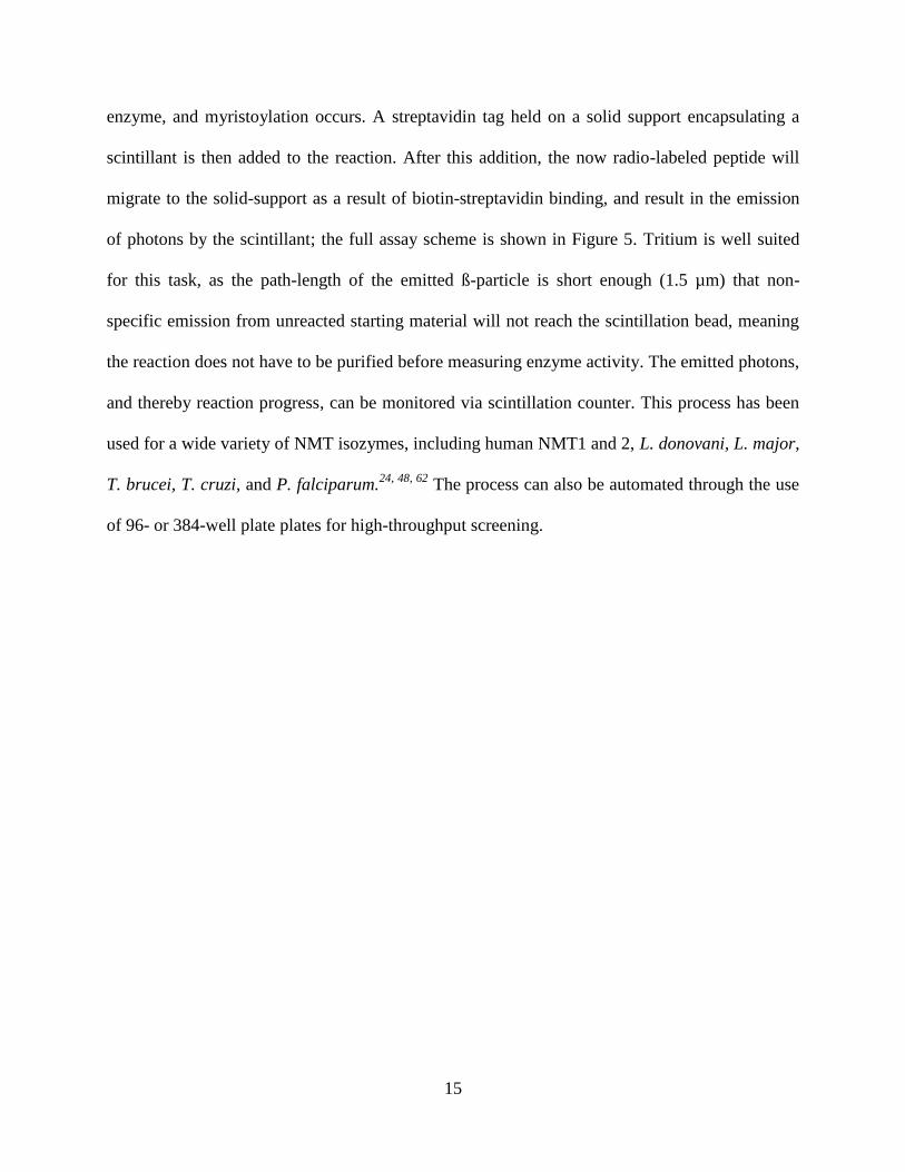
enzyme, and myristoylation occurs. A streptavidin tag held on a solid support encapsulating a
scintillant is then added to the reaction. After this addition, the now radio-labeled peptide will
migrate to the solid-support as a result of biotin-streptavidin binding, and result in the emission
of photons by the scintillant; the full assay scheme is shown in Figure 5. Tritium is well suited
for this task, as the path-length of the emitted ß-particle is short enough (1.5 µm) that non-
specific emission from unreacted starting material will not reach the scintillation bead, meaning
the reaction does not have to be purified before measuring enzyme activity. The emitted photons,
and thereby reaction progress, can be monitored via scintillation counter. This process has been
used for a wide variety of NMT isozymes, including human NMT1 and 2, L. donovani, L. major,
T. brucei, T. cruzi, and P. falciparum.24, 48, 62
The process can also be automated through the use
of 96- or 384-well plate plates for high-throughput screening.

16
Figure 5: Scintillation proximity assay.
Step 1: Radio-labeleld myristate is added to the biotin-tagged protein mimetic. Step 2: The peptide and
radio-label are attached to the scintillant bead by the biotin-streptavidin interaction. Step 3: The radio-label is
sufficiently close to the scintillant to cause a photon emission. Reprinted with permission from Bowyer, P. W.; Tate,
E. W.; Leatherbarrow, R. J.; Holder, A. A.; Smith, D. F.; Brown, K. A., N-Myristoyltransferase: A prospective drug
target for protozoan parasites. ChemMedChem 2008, 3 (3), 402-408.
3.1.2 Cellular assay
Several cellular based assays are also available to determine antileishmanial activity. These
include culture and inhibition of promastigotes, axenic amastigotes, and intracellular
amastigotes. Each of these methods may be used in a target-free screening of inhibitors, but they
may also be incorporated with NMT-based drug discovery effort, as NMT is constitutively
produced at all stages of parasite development.48

17
Promastigote-based cellular assays are the most straightforward of the three cell-based
assays. Promastigotes are grown in a media, potential inhibitors are introduced at suitable
concentrations, and growth inhibition is determined.63
High-throughput applications are available
utilizing a plate reader for analysis of cell growth via fluorescent staining, with positive and
negative controls (typically amphotericin B, and 1% DMSO, respectively). While promastigotes
are often assayed for comparative purposes, their use is waning, as techniques for the high-
throughput screening of more biologically relevant parasite forms has become commonplace.
Axenic amastigote screening provides a more biologically relevant assay than
promastigotes, since the former is the active parasite found within host mammals.64-65
Screening
is accomplished much the same way as with promastigotes in terms of controls, and hit
identification by fluorescence. However, the actual growth of the parasites differs, as they must
be chemically induced to continue multiplying as amastigotes, rather than revert to the
promastigote form. This is accomplished via manipulation of pH to mimic macrophage
conditions, and occasionally by the inclusion of 5% CO2 in the atmosphere, or incubation of cells
at biologically relevant temperatures (37 ºC).
Screening compounds against intracellular amastigotes provides the best in vitro similarity
to conditions found in vivo.66-67
In this method, promastigote cultures are allowed to infect
macrophage differentiated host cells in growth media. After a period of initial infection, the non-
internalized promastigotes are rinsed away with a buffer, and the parasites are allowed to
continue to grow under the presence of test compounds, or controls. Chemical differentiation of
the host cells into macrophages is required for two reasons - first, the macrophage best mimics
the in vivo context for a host infection, and second, without differentiation to non-dividing
macrophages, parasitized cells would be overrun by replicating cells. THP-1 (human acute

18
leukemia monocyte) cells are most often used for this purpose. Determination of inhibitor
activity is accomplished by visualization with a fluorescent DNA-binding stain. Areas rich in
DNA will fluoresce, predominantly the THP-1 cell nucleus, and parasite kinetoplasts. The
difference in size between these two structures may be exploited for high-content screening. One
assay yields not only a ratio of parasites to host cells, but also a quantification of host cells in
each sample well to serve as a measure of toxicity induced by the compounds. These
measurements can be taken by automated fluorescence microscopy, but require a software suite
for the automated/accurate image analysis.68
3.1.3 Secondary screening
After hit compounds are identified, several other in vitro methods have been used to assist
in lead identification as secondary screens. If not part of a the primary screen, assay hits may be
tested independently against uninfected THP-1 cells to determine toxicity. Metabolic stability is
also an important aspect for lead compound identification and optimization, as even the most
potent hit is ineffective if metabolized too quickly.69
Preliminary metabolic data may be gathered
by subjecting the test compound to either rat or human liver microsomal stability assay. As a
final consideration in lead identification and optimization, hit compounds may be screened
against cytochrome P450, to avoid potential drug-drug interactions.70
3.1.4 Screen analysis and validity
Large scale screening methods must provide a reliable means to identify biologically active
compounds from a large compound library, with assays performed singly (rarely in duplicate or

19
triplicate). A statistical method for determination of assay quality has been proposed, yielding a
dimensionless coefficient called the Z-factor, or Z-score (Equation 1).71
A Z-factor of 1 indicates
an ideal assay, with standard deviations of zero, and infinite dynamic range. Typically Z-factors
between 1 and 0.5 indicate a good quality assay, with a large separation band between hits and
inactive compounds.
( )
| |
Equation 1: Z-Factor for HTS validity
σ: standard deviation, μ: mean, p: positive control, n: negative control
3.2 IN VIVO MODELS OF VISCERAL LEISHMANIASIS
After identification and optimization of hit compounds through in vitro methods, animal
models are required to better mimic infection and treatment in mammalian hosts. For developing
new chemotherapies, in vivo models serve to confirm compound activity determined from
cellular and enzymatic assays, as well as provide information regarding administration routes,
distribution, metabolism, excretion, and toxicity (ADMET). Several species have been studied as
hosts for the L. donovani parasite as models for visceral leishmanias.59
Rodents, canines, and
non-human primates have all been used for this task.
Rodent models are the most accessible models of L. donovani infection of a mammalian
host. A variety of species have been studied, including C57BL/6 mouse, and Syrian golden
hamster, but the BALB/c mouse model is most common.72
Canine models of the disease have
also been used in studies of parasite biology, but with little emphasis for use on development of
chemotheraputics.59
Establishing canines as accurate models is important nonetheless, as they

20
have been shown to exhibit closer disease pathology to humans than available in rodent models.
Also, the study of leishmaniasis in canines is useful to determine their role as host reservoirs for
certain species of the parasite.73
Finally, non-human primates have been studied as the most
advanced, human-like model of leishmaniasis. Many species of primate are quite resistant to
Leishmania infection, however, the languor (Presbytis entellus) has been identified as a
promising model system for human leishmaniasis infection. The languor is susceptible to a
lasting VL infection, ultimately resulting in death, and displays the same pathogenesis and
immune response as in humans, allowing for their use in the study of antileishmanial vaccine
candidates.74

21
4.0 LIMITED STATE OF ANTILEISHMANIAL DEVELOPMENT
A wide array of experimental tools are available for the study and development of leishmania-
specific NMT inhibitors, yet there is a surprising lack of synergy and cross validation among
these models. Currently, target-free screening is gerally preferred to target-based design: many
putative targets besides NMT are characterized (albeit in less detail), and target-free screening
allows for testing compounds ‘holistically’ focusing on overall efficacy, independant of
knowledge about a particular molecular target.65-66, 69, 75-77
Choice of parasitic form
(promastigote, and axenic or intracellular amastigote) is non-trivial, but improvements in high-
throughput assays for the later stages of the life cycle have rendered promastigote assays fairly
obsolete.67, 78
Axenic and intracellular amastigote models are more biologically relevant, and are
available in for use in high-throughput applications. To date, several target-free HTS/HCS based
screens for Leishmania inhibitors has been performed, with z-scores > .5, indicating good quality
screens.79
These efforts have resulted in the identification of novel scaffolds, but no attempt has
been made to associate hits with a specific target (either by in silico methods, or enzymatic
assay).
One NMT-based HTS has been performed utilizing the scintillation proximity assay,
against the Pfizer global representative library (~150,000 compounds).62
This screen successfully
demonstrated the ability to selectively inhibit LdNMT over Hs1NMT (Figure 7), and identified
several scaffolds for NMT inhibition, representatives shown in Figure 6. However, this screen

22
was not without shortcomings, such as a lack of testing in cellular assays to confirm bioactivity
and selectivity of identified hits. Also, while each scaffold generated a series of ‘local’ hits, only
inhibition data for top hits within each family was reported, rather than observed SAR.
Figure 6: LdNMT assay hits and selectivities.
Figure 7: Tunable selectivity between Ld and Hs1 NMT inhibition.
Reprinted under Creative Commons license from Bell, A. S.; Mills, J. E.; Williams, G. P.; Brannigan, J. A.;
Wilkinson, A. J.; Parkinson, T.; Leatherbarrow, R. J.; Tate, E. W.; Holder, A. A.; Smith, D. F., Selective inhibitors
of protozoan protein N-myristoyltransferases as starting points for tropical disease medicinal chemistry programs.
PLoS Negl Trop Dis 2012, 6 (4), e1625.80
In silico analysis of antileishmanial NMT inhibitors is also limited. Despite the large
N
ONH2
OH
Cl
Cl
N
ONH2
OH
Cl
O
NH
HN
O
F
HN
HN
N N
N
S
N
N
S
N
N
N
N
Aminoacylpyrrolidines Piperidinylindoles
Biphenyl derivatives
Thienopyrimidines
LdNMT: 0.093 µM IC50
Hs1NMT: 5.2 µM IC50
LdNMT: 0.9 µM IC50
Hs1NMT:47.2 µM IC50
LdNMT: 0.10 µM IC50
Hs1NMT: 73.2 µM IC50
LdNMT: 0.16 µM IC50
Hs1NMT: 36.1 µM IC50
LdNMT: 0.48 µM IC50
Hs1NMT: 9.0 µM IC50

23
quantity of crystallographic data available for Leishmania spp. NMT enzymes, few detailed in
silico docking or modeling studies have been published. Of the few studies available, many are
not specific to NMT, but rather, focus on a cross-sectional docking study of inhibitors against a
range of potential targets. Ogungbe, et al. have published two separate reports docking plant-
based phenolic compounds (stilbenoids, phenylpropanoids, flavonoids, quinones, etc) to a large
series of crystal structures to determine potential targets.81-82
In either instance, a library of
chemicals (some of which have shown broad antiparasitic activity) is docked to the array of
structures, and energies reported. However, neither report offers a more in-depth analysis of the
data, by way of rationalizing existing cellular assay data with docking results, or collaborative
work to screen top computational hits against cellular or enzymatic assays.
Sobarzo-Sanchez et al. have studied a small series of oxoisoaporphine antileishmanials
(Figure 8) in detail, providing in vitro, in vivo, and in silico analysis.83
Their aim was similar to
Ogungbe’s work, but more successful in identifying potential targets. Utilizing 128 known
Leishmania enzymes with docked ligands as the basis for a training dataset, the group utilized
bioinformatic analysis (via ‘MARCH-INSIDE’ software) to determine 4 putative targets,
including NMT. From these results, an NMT enzymatic assay could easily confirm
oxoisoaporphines as NMT inhibitors, and provide a positive or negative training dataset for
further in silico study of oxoisoaporphines.
Figure 8: Oxoisoaporphine scaffold.
While NMT has been studied in leishmaniasis, and its inhibition has been shown as a
means to combat parasite growth, the discovery of corresponding Leishmania NMT inhibitors is
N
O

24
limited. Simple efforts to utilize existing methods synergistically can increase the efficiency in
which NMT inhibitors are discovered and optimized.

25
5.0 NMT STUDIES IN RELATED ORGANISMS
As described previously, NMT enzyme activity has been proposed as a target for a variety of
fungal and protozoan diseases, and a number of compounds have been reported to inhibit the
NMT enzyme and disease growth. However, in comparison to Leishmania NMT inhibitors,
NMT inhibition in other species has been studied in much greater detail, with inclusion of SAR,
in silico, and crystallographic supporting data. Some relevant data with regard to cross-inhibition
of Leishmania NMTs is occasionally discussed within these studies, but is rarely presented in
context of SAR studies focusing on improving antileishmanial potency. It is not uncommon for
large swaths of medicinal chemistry data to go unreported in scientific journals and only
published in patents.84
This is no different for NMT inhibitors: while there is a lack of SAR data
and discussion for antileishmanials presented within journal articles, certain data-sets can be
found in related patents, but without relevant discussion. A variety of compound classes have
been reported as NMT inhibitors in other species with some crossover studies on Leishmania
spp, including benzofurans and similar heterocycles, pyrazole sulfonamides, quinolines, and
peptides/peptidomimetics. These scaffolds and associated analyses can serve as starting points
for piggyback drug development strategies.85

26
5.1.1 Benzofurans
Benzofurans, and associated compounds (benzothiazoles, benzothiophenes, indoles) are
amongst the most studied NMT inhibitors, and this broad group of compounds has been used for
piggyback hit generation/optimization across several species.86-94
QSAR studies have been
performed on existing hit datasets to determine pharmacophore models, which have been
computationally used to generate new prospective hits.95-96
In terms of activity-based SAR, a
piggyback approach using the same or similar heterocyclic core is common amongst other types
of disease, which is visually apparent between studies on different species.97-98
Despite the core
similarities between inhibitors, each subsequent study has expanded around existing SAR
(species specific), in continually good detail.99
Furthermore, many papers include
crystallography data showing the binding pose of their top hit(s). However, no journal articles
discuss the implications of these results with respect to the development of antileishmanials.
Benzofurans and furan-like compounds have been patented for use as NMT inhibitors of
P. falciparum, and L. donovani. Despite a lack of reporting in primary literature sources, the
patent features a SAR of 140 compounds with similar heterocyclic cores, and relevant IC50
concentrations (SPA or fluorescence assay) for both L. donovani and P. falciparum.100
Of the
compounds screened, 74 have been identified by the authors as ‘hits’ (< 5 µM IC50) against
LdNMT. The most potent, shown below (Figure 9), possesses an IC50 of 0.01 µM. Unfortunately
this hit was not screened against human NMT1 to determine selectivity. The most potent
inhibitor with selectivity data displays 0.027 µM IC50 against LdNMT, and 1.00 µM IC50
inhibition against Hs1NMT, roughly 40-fold selectivity favoring parasite isozyme inhibitions.
27
Figure 9: Benzofurans as NMT inhibitors.
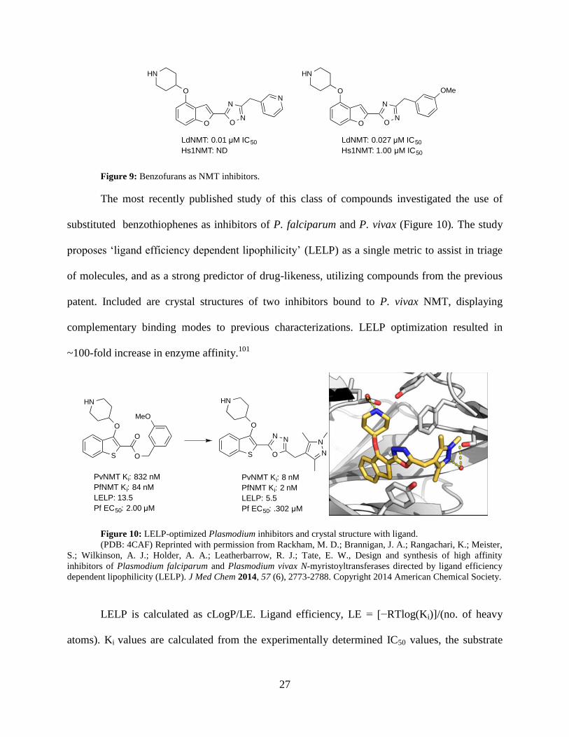
The most recently published study of this class of compounds investigated the use of
substituted benzothiophenes as inhibitors of P. falciparum and P. vivax (Figure 10). The study
proposes ‘ligand efficiency dependent lipophilicity’ (LELP) as a single metric to assist in triage
of molecules, and as a strong predictor of drug-likeness, utilizing compounds from the previous
patent. Included are crystal structures of two inhibitors bound to P. vivax NMT, displaying
complementary binding modes to previous characterizations. LELP optimization resulted in
~100-fold increase in enzyme affinity.101
Figure 10: LELP-optimized Plasmodium inhibitors and crystal structure with ligand.
(PDB: 4CAF) Reprinted with permission from Rackham, M. D.; Brannigan, J. A.; Rangachari, K.; Meister,
S.; Wilkinson, A. J.; Holder, A. A.; Leatherbarrow, R. J.; Tate, E. W., Design and synthesis of high affinity
inhibitors of Plasmodium falciparum and Plasmodium vivax N-myristoyltransferases directed by ligand efficiency
dependent lipophilicity (LELP). J Med Chem 2014, 57 (6), 2773-2788. Copyright 2014 American Chemical Society.
LELP is calculated as cLogP/LE. Ligand efficiency, LE = [−RTlog(Ki)]/(no. of heavy
atoms). Ki values are calculated from the experimentally determined IC50 values, the substrate
O
O
HN
N
ON
N
O
O
HN
N
ON
OMe
LdNMT: 0.01 µM IC50
Hs1NMT: ND
LdNMT: 0.027 µM IC50
Hs1NMT: 1.00 µM IC50
S
O
HN
N
O
N
N
N
S
O
HN
O
O
MeO
PvNMT Ki: 832 nM
PfNMT Ki: 84 nM
LELP: 13.5
Pf EC50: 2.00 µM
PvNMT Ki: 8 nM
PfNMT Ki: 2 nM
LELP: 5.5
Pf EC50: .302 µM
28
concentration ([S]), and the Michaelis–Menten constant (Km) as described by the Cheng–Prusoff
equation (Equation 2).102
Km values are used from previously experimentally determined
constants.
[ ]
Equation 2: Cheng-Prusoff equation
[S] = substrate concentration
5.1.2 Pyrazole sulfonamides
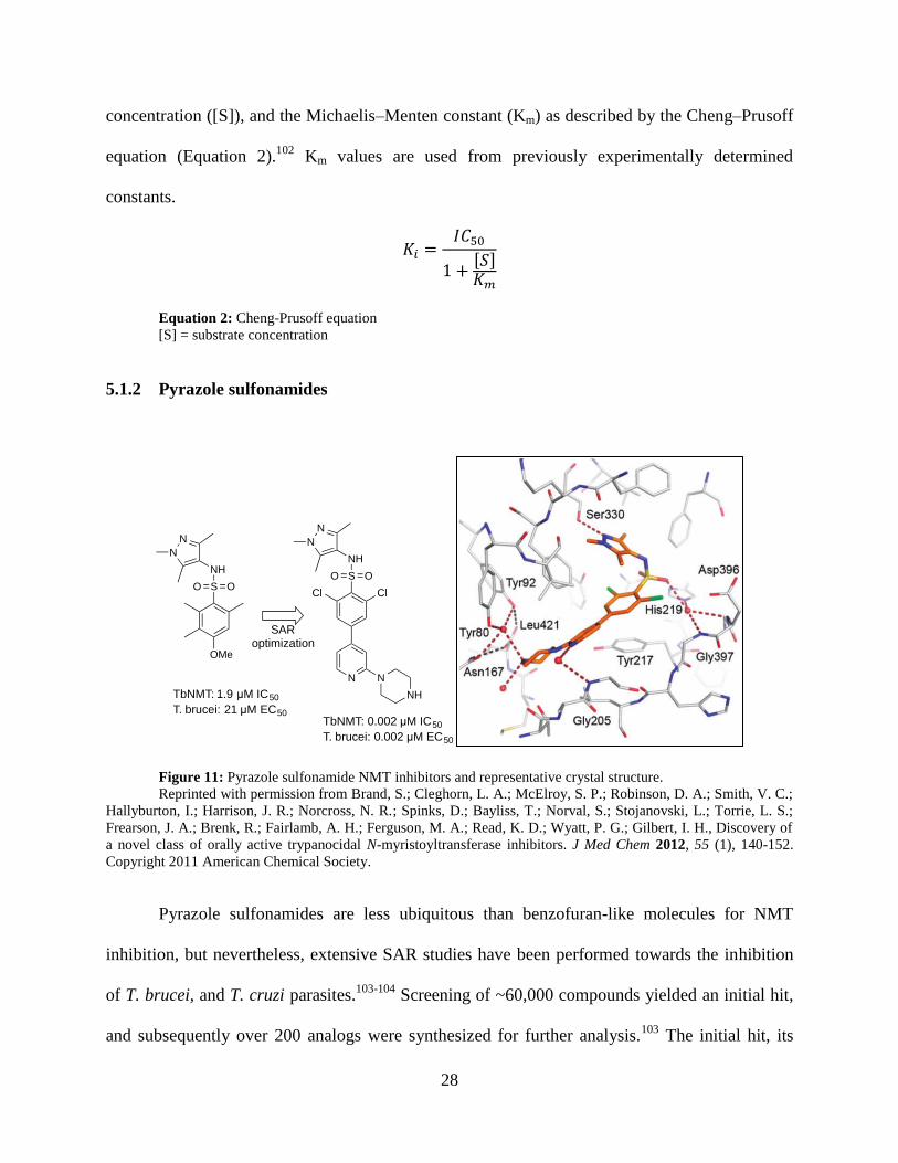
Figure 11: Pyrazole sulfonamide NMT inhibitors and representative crystal structure.
Reprinted with permission from Brand, S.; Cleghorn, L. A.; McElroy, S. P.; Robinson, D. A.; Smith, V. C.;
Hallyburton, I.; Harrison, J. R.; Norcross, N. R.; Spinks, D.; Bayliss, T.; Norval, S.; Stojanovski, L.; Torrie, L. S.;
Frearson, J. A.; Brenk, R.; Fairlamb, A. H.; Ferguson, M. A.; Read, K. D.; Wyatt, P. G.; Gilbert, I. H., Discovery of
a novel class of orally active trypanocidal N-myristoyltransferase inhibitors. J Med Chem 2012, 55 (1), 140-152.
Copyright 2011 American Chemical Society.
Pyrazole sulfonamides are less ubiquitous than benzofuran-like molecules for NMT
inhibition, but nevertheless, extensive SAR studies have been performed towards the inhibition
of T. brucei, and T. cruzi parasites.103-104
Screening of ~60,000 compounds yielded an initial hit,
and subsequently over 200 analogs were synthesized for further analysis.103
The initial hit, its
SO O
NH
N
N
OMe
SO O
NH
N
N
Cl Cl
N N
NHTbNMT: 1.9 µM IC50
T. brucei: 21 µM EC50TbNMT: 0.002 µM IC50
T. brucei: 0.002 µM EC50
SAR optimization

29
binding pose in a crystal structure, and optimized compound are shown in Figure 11. Studies on
these analogs include corresponding selectivity data between TbNMT and Hs1NMT via SPA
assay, and in vitro activity against T. brucei parasites. Beginning with an initial hit of 21 μM
(IC50) against the T. brucei parasite, the optimizations culminated in a compound with 0.002 μM
potency. The paper indicates cross-inhibition of L. major, however only inhibition by the
optimized compound (optimized specifically for T. brucei inhibition) has been reported
(LmNMT IC50, 0.002 µM), rather than data from the series of compounds.
Included in the study, 5 crystal structures with bound pyrazole sulfonamides inhibitors
are reported. However, T. brucei crystallization has not been reported, and the authors utilized
LmNMT as a surrogate model for TbNMT, but did not discuss SAR implications of these
compounds regarding antileishmanial development. Nevertheless, a wide range of analogs has
been studied, and a close correlation between NMT inhibition and T. brucei parasite inhibition
has been established (Figure 12).
Figure 12: TbNMT inhibition correlates to T. brucei parasite inhibition.
Image used with permission from Frearson, J. A.; Brand, S.; McElroy, S. P.; Cleghorn, L. A.; Smid, O.;
Stojanovski, L.; Price, H. P.; Guther, M. L.; Torrie, L. S.; Robinson, D. A.; Hallyburton, I.; Mpamhanga, C. P.;
Brannigan, J. A.; Wilkinson, A. J.; Hodgkinson, M.; Hui, R.; Qiu, W.; Raimi, O. G.; van Aalten, D. M.; Brenk, R.;
30
Gilbert, I. H.; Read, K. D.; Fairlamb, A. H.; Ferguson, M. A.; Smith, D. F.; Wyatt, P. G., N-Myristoyltransferase
inhibitors as new leads to treat sleeping sickness. Nature 2010, 464 (7289), 728-732.
Much like the benzofuran NMT inhibitors, the series of pyrazole sulfonamides inhibitors
has also been patented.105
Similar to benzofurans, the pyrazole sulfonamides have been tested
against LmNMT via SPA assay, as described in the patent, yet these results are unreported in the
original paper. Inclusion of LmNMT inhibition data is far more limited than corresponding
TbNMT data - only ~40 compounds of the total screen were also assayed for LmNMT inhibition.
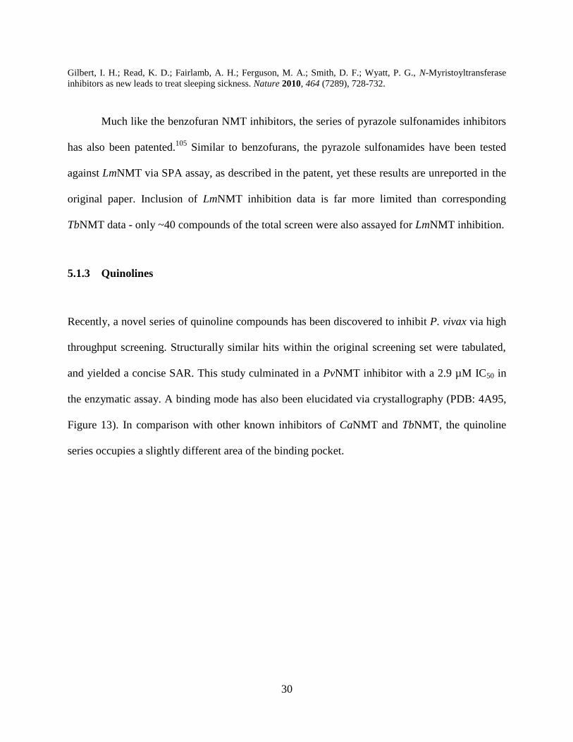
5.1.3 Quinolines
Recently, a novel series of quinoline compounds has been discovered to inhibit P. vivax via high
throughput screening. Structurally similar hits within the original screening set were tabulated,
and yielded a concise SAR. This study culminated in a PvNMT inhibitor with a 2.9 µM IC50 in
the enzymatic assay. A binding mode has also been elucidated via crystallography (PDB: 4A95,
Figure 13). In comparison with other known inhibitors of CaNMT and TbNMT, the quinoline
series occupies a slightly different area of the binding pocket.

31
Figure 13: Structure overlay of inhibitor species.
Quinoline shown in green, benzofuran in orange, pyrazole sulfonamide in blue.
Image used with permission from Goncalves, V.; Brannigan, J. A.; Whalley, D.; Ansell, K. H.; Saxty, B.; Holder, A.
A.; Wilkinson, A. J.; Tate, E. W.; Leatherbarrow, R. J., Discovery of Plasmodium vivax N-myristoyltransferase
inhibitors: Screening, synthesis, and structural characterization of their binding mode. J Med Chem 2012, 55 (7),
3578-3582. Copyright 2012 American Chemical Society.
5.1.4 Peptide/peptidomimetics
Peptide-based and peptidomimetic NMT inhibitors have been reported for C. albicans, and P.
falciparum.106-109
Detailed SAR is more limited than for small molecule inhibitors, yet activity is
similarly impressive, with as low as 0.04 µM IC50 against CaNMT. Notably, peptide and
peptidomimetic inhibitors can take advantage of the high degree of NMT substrate specificity,
with a maximum selectivity of 2200-fold demonstrated in CaNMT over Hs1NMT. Structural
SO O
NH
N
N
Cl Cl
N N
NH
N
S
O
NC
O
O
NH
N
O
N
N
Quinoline
Pyrazole sulfonamide
Benzofuran

32
characterization of a peptide bound to S. cerevisiae (PDB: 1IID) has been achieved, and analysis
of >100 synthetic peptides has been performed to determine preferred amino acid composition
for binding affinity along an 8 residue synthetic peptide.

33
6.0 FUTURE PROSPECTS IN ANTILEISHMANIAL DEVELOPMENT
Existing infrastructure for design, synthesis, and screening of L. donovani inhibitors can be better
utilized for NMT-inhibitor-based drug discovery. A simple, comprehensive improvement to
existing antileishmanial design would be the addition of NMT assay screening of target-free hits.
By testing a greatly reduced number of compounds, a minimal amount of complexity is added to
an existing screening campaign. Compounds that do not inhibit NMT may be studied and
optimized as with any other target-free screening. Targets shown to inhibit NMT activity may be
better optimized using target-based methodologies. In either case, further use of secondary
screening can be carried out to better identify lead-like compounds.
The relevance of a target-based drug development methodologies for antileishmanials
warrants discussion.110-112
Historically, the increase in computational power, and knowledge of
genetics and proteomics has made target-based screening appear as an encouraging method for
drug discovery. However, in practice, target-based drug design involves assumptions about
mechanism of action, identification and validation of a drug target, and sometimes complex
interactions between a disease and the body. Target-free screening avoids these pitfalls by
exclusively focusing on the observed drug efficacy. In the case of N-myristoyltransferase as a
target for visceral leishmaniasis treatment, the enzyme sidesteps these barriers. Crystal structures
with known inhibitors and enzymatic assay provide an immediate link to compare results of the
two methods between themselves, and between data associated with cellular assays. Transgenic

34
parasites null for the NMT-encoding gene in both L. major and L. donovani have demonstrated
the necessity for NMT enzyme expression. The parasite biology with respect to NMT inhibition
is well understood, and selective inhibition over human isozymes has been demonstrated. As an
additional metric for compound screening and triage, LELP considerations may be applied to
compound-sets. Overall, NMT presents itself as a well characterized enzyme, suitable for target-
based development.
Optimization of existing NMT inhibitors via a piggyback approach is the most immediately
accessible option for antileishmanial development. Existing SAR and crystallographic data for
other species provide a comparable framework for antileishmanial design and synthesis.
Peptide and peptidomimetics, in particular, offer a unique route to Leishmania inhibition,
with the potential for high specificity toward LdNMT over HsNMT, minimal toxicity, and easy
library synthesis of analogs via peptide coupling methods. Kinetic and inhibition studies of
known NMT-inhibiting synthetic peptides and peptidomimetics on L. donovani NMT may be a
first step, considering similarities between studied yeast NMT and Leishmania NMT. Significant
differences between the proteins occur on the opposite face of the protein and CoA binding sites,
and these differences are not purported to alter protein function, offering direct comparison of
bound ScNMT inhibiting protein with the LmNMT crystal structure (Figure 14).47
Secondary
screening of drug metabolism is also necessary to help overcome rapid metabolism commonly
associated with peptide based drugs.113

35
Figure 14: Overlay of peptide and small molecule NMT inhibitors.
Peptide (green) from ScNMT crystal structure (PDB: 1IID), inhibitor (orange) and enzyme (pink) from
LmNMT crystal structure (PDB: 4A2Z). Image used with permission form Brand, S.; Cleghorn, L. A.; McElroy, S.
P.; Robinson, D. A.; Smith, V. C.; Hallyburton, I.; Harrison, J. R.; Norcross, N. R.; Spinks, D.; Bayliss, T.; Norval,
S.; Stojanovski, L.; Torrie, L. S.; Frearson, J. A.; Brenk, R.; Fairlamb, A. H.; Ferguson, M. A.; Read, K. D.; Wyatt,
P. G.; Gilbert, I. H., Discovery of a novel class of orally active trypanocidal N-myristoyltransferase inhibitors. J Med
Chem 2012, 55 (1), 140-152. Copyright 2011 American Chemical Society.
Quinoline-based NMT inhibitors may also serve as a promising source of piggyback
development candidates. Initially, screening of the P. vivax hits against L. donovani NMT can
provide the first step to understanding the efficacy of this class of compounds against the
Leishmaniases. Synthesis of these quinolines is well established to allow for further SAR studies
and optimization.88
Notably, the nitrile-bearing thioether is attached last, allowing for relatively
easy synthesis of relevant analogs for SAR studies (Figure 15). Visually, the binding pocket is
not fully utilized by the quinoline analogs, but the nitrile group shares some overlap with other
inhibitor classes. Replacement of the nitrile group with other potent pharmacophores may yield
an increase in both activity and selectivity by utilizing a larger surface of the binding site.

36
Existing SAR and synthetic routes to benzofurans, benzothiophenes, and pyrazole
sulfonamides are extremely well established, and offer insight towards synthesis of new analogs.
Synthetic routes to these compounds are relatively simple, with numerous points to facilitate
combinatorial synthesis, or substitution of bioisosteric fragments. For example, pyrazole
sulfonamides are accessible in minimal steps, with orthogonal steps to introduce additional
moieties or substitutions (Figure 16).103
Figure 16: Pyrazole sulfonamide synthesis.
Inhibitors specific to L. donovani NMT have been identified via high throughput
enzymatic assay. Re-assay of structurally similar hits can provide a direction for future SAR
SO O
Cl
R1, X
SO O
NH
R1, X
N
HN
If X
SO O
NH
N
HN
B
SO O
NH
R1, X
N
NR2
SO O
NH
N
HN
R3
iii
iii
iv
Reagents: i)RNH2*, pyridine, DCM.
ii)R2Br, Cs2CO3, DMF. iii) bis-
pinnacolattodiboron, K3PO4,
Pd(PPh3)4. iv) RBr, K3PO4, DMF,
H2O, Pd(PPh3)4
O
O* eg: N
HN
NH2
Figure 15: Quinoline synthetic scheme.
NH
O
EtO OEt
EtO
O OR6
R1 OEt
O O
NH2
O
O
N
R6
OH
COOEt
N
OH
COOEt
R2
N
Cl
COOEt
R2
R6
N
S
COOEt
R2
R6
NCi, ii
iii
iv
iv
v
Reagents and conditions: i) 140 ºC, 1hr. ii)Ph2O, reflux, 2 hr. iii) NaH, N,N-dimethylacetamide, 120 °C, 20
min. iv) POCl3, 110 °C, 20 min. v) 3-mercaptopropanenitrile, K2CO3, THF, reflux 4hr.
37
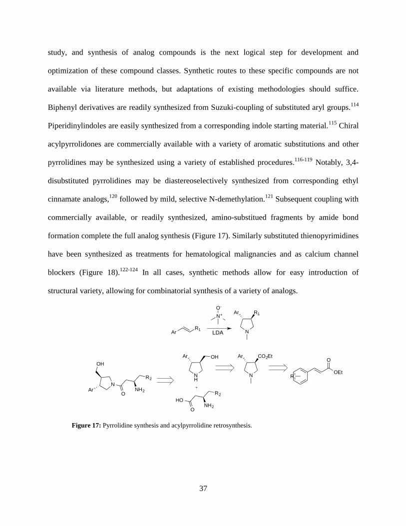
study, and synthesis of analog compounds is the next logical step for development and
optimization of these compound classes. Synthetic routes to these specific compounds are not
available via literature methods, but adaptations of existing methodologies should suffice.
Biphenyl derivatives are readily synthesized from Suzuki-coupling of substituted aryl groups.114
Piperidinylindoles are easily synthesized from a corresponding indole starting material.115
Chiral
acylpyrrolidones are commercially available with a variety of aromatic substitutions and other
pyrrolidines may be synthesized using a variety of established procedures.116-119
Notably, 3,4-
disubstituted pyrrolidines may be diastereoselectively synthesized from corresponding ethyl
cinnamate analogs,120
followed by mild, selective N-demethylation.121
Subsequent coupling with
commercially available, or readily synthesized, amino-substitued fragments by amide bond
formation complete the full analog synthesis (Figure 17). Similarly substituted thienopyrimidines
have been synthesized as treatments for hematological malignancies and as calcium channel
blockers (Figure 18).122-124
In all cases, synthetic methods allow for easy introduction of
structural variety, allowing for combinatorial synthesis of a variety of analogs.
Figure 17: Pyrrolidine synthesis and acylpyrrolidine retrosynthesis.
O
OEtRN
Ar CO2Et
NH
Ar
ONH2
R2
HO
N
Ar R1
ArR1
N+
O-
LDA
ArN
ONH2
R2
OH
OH

38
Figure 18: Thienopyrimidine retrosynthesis.
Finally, purely in silico methods may provide the easiest route for screening large
compound libraries. While there is currently a lack of computer based drug design toward
antileishmanials, selecting NMT as a target offers a simple, robust approach for development.
Preliminary screening in silico can be accomplished through tandem screening of compounds
against Leishmania and human NMT isozymes. This can be done with relatively little computing
power (dedicated desktop as opposed to multicore servers) and at low cost (several free docking
programs offer comparable docking performance to commercial software suites).125-126
Any
molecular library may be screened, with the substantial ZINC database being an excellent
starting point.127
Docking may be validated based on comparison with ligand positions in crystal
structures, and by NMT enzyme assay of top hits. Likewise, existing SAR studies may be
applied to provide extensive training datasets for validation of in silico methods. Computational
methods may be inserted at any point of NMT-based antileishmanial discovery to achieve an
optimal relationship between screening, validation, and generation of new compounds to test.
N
N SN
N
N
HN N
N SCl
NR1R2
HNR3R4
N
N SCl
Cl
HH2NR1R2
HN
N SO
O
H2N
RO2C
S
R6
R5 R5
R6
R5
R6
R5
R6

39
7.0 CONCLUSIONS
The global disease burden and lack of sufficient clinical treatments warrant further investigation
of antileishmanial compounds. N-myristoyltransferase has been demonstrated as a promising
target for development of drugs treating leishmaniasis, particularly visceral leishmaniasis.
Unfortunately, the current body of research on NMT-specific leishmania inhibitors is limited. In
this case, existing target-free screening techniques may be easily combined to include screening
for NMT inhibitors. The body of research on NMT inhibition in similar parasite and fungal
species is thorough, with detailed SAR and crystallographic supporting evidence of binding
modes on highly similar molecular targets. This offers an excellent starting point for piggyback
development of Leishmania-specific NMT inhibitors. Of the few specific NMT inhibitors of L.
donovani shown, no optimization has been performed on the parent chemical scaffolds, offering
an opportunity to determine efficient syntheses, and relevant SAR for these compounds. Finally,
in silico methods may be applied through out, in tandem with enzymatic assay results. In this
manner, more efficient drug discovery methods may be accomplished, regardless of initial
development method. The currently limited state of NMT-inhibitor development has been
discussed, within the context of more expansive methods, applications, and relevant studies.
Although there is a noticeable gap between these similar areas of research, several solutions have
been proposed to further study of NMT inhibition of L. donovani.

40
BIBLIOGRAPHY
1. World Health Organization. Leishmaniasis fact sheet no 375.
http://www.who.int/mediacentre/factsheets/fs375/en/ (accessed Apr 06, 2014).
2. McCall, L. I.; Zhang, W. W.; Matlashewski, G., Determinants for the development of
visceral leishmaniasis disease. PLoS Pathog. 2013, 9 e1003053.
3. Drugs for neglected diseases initiative. Leishmaniasis. http://www.dndi.org/diseases-
projects/diseases/vl.html (accessed Apr 16, 2014).
4. World Health Organization. Leishmaniasis. http://www.who.int/leishmaniasis/en/
(accessed Apr 16, 2014).
5. den Boer, M. L.; Alvar, J.; Davidson, R. N.; Ritmeijer, K.; Balasegaram, M.,
Developments in the treatment of visceral leishmaniasis. Expert Opin. Emerg. Drugs 2009, 14
395-410.
6. Croft, S. L.; Sundar, S.; Fairlamb, A. H., Drug resistance in leishmaniasis. Clin.
Microbiol. Rev. 2006, 19 111-126.
7. Decuypere, S.; Vanaerschot, M.; Brunker, K.; Imamura, H.; Muller, S.; Khanal, B.; Rijal,
S.; Dujardin, J. C.; Coombs, G. H., Molecular mechanisms of drug resistance in natural
Leishmania populations vary with genetic background. PLoS Negl. Trop. Dis. 2012, 6 e1514.
8. Jhingran, A.; Chawla, B.; Saxena, S.; Barrett, M. P.; Madhubala, R., Paromomycin:
Uptake and resistance in Leishmania donovani. Mol. Biochem. Parasitol. 2009, 164 111-117.
9. Ephros, M.; Bitnun, A.; Shaked, P.; Waldman, E.; Zilberstein, D., Stage-specific activity
of pentavalent antimony against Leishmania donovani axenic amastigotes. Antimicrob. Agents
Chemother. 1999, 43 278-282.
10. Renslo, A. R.; McKerrow, J. H., Drug discovery and development for neglected parasitic
diseases. Nat. Chem. Biol. 2006, 2 701-710.
11. Nwaka, S.; Hudson, A., Innovative lead discovery strategies for tropical diseases. Nat.
Rev. Drug Discov. 2006, 5 941-955.

41
12. Croft, S. L.; Coombs, G. H., Leishmaniasis– current chemotherapy and recent advances
in the search for novel drugs. Trends Parasitol. 2003, 19 502-508.
13. Palatnik-de-Sousa, C. B., Vaccines for leishmaniasis in the fore coming 25 years.
Vaccine 2008, 26 1709-1724.
14. Kedzierski, L., Leishmaniasis vaccine: Where are we today? J. Glob. Infect. Dis. 2010, 2
177-185.
15. Chakravarty, J.; Kumar, S.; Trivedi, S.; Rai, V. K.; Singh, A.; Ashman, J. A.; Laughlin,
E. M.; Coler, R. N.; Kahn, S. J.; Beckmann, A. M.; Cowgill, K. D.; Reed, S. G.; Sundar, S.;
Piazza, F. M., A clinical trial to evaluate the safety and immunogenicity of the LEISH-F1+MPL-
SE vaccine for use in the prevention of visceral leishmaniasis. Vaccine 2011, 29 3531-3537.
16. Bates, P. A., Transmission of Leishmania metacyclic promastigotes by phlebotomine
sand flies. Int. J. Parasitol. 2007, 37 1097-1106.
17. Wilson, M. E.; Jeronimo, S. M.; Pearson, R. D., Immunopathogenesis of infection with
the visceralizing Leishmania species. Microb. Pathog. 2005, 38 147-160.
18. Peacock, C. S.; Seeger, K.; Harris, D.; Murphy, L.; Ruiz, J. C.; Quail, M. A.; Peters, N.;
Adlem, E.; Tivey, A.; Aslett, M.; Kerhornou, A.; Ivens, A.; Fraser, A.; Rajandream, M. A.;
Carver, T.; Norbertczak, H.; Chillingworth, T.; Hance, Z.; Jagels, K.; Moule, S.; Ormond, D.;
Rutter, S.; Squares, R.; Whitehead, S.; Rabbinowitsch, E.; Arrowsmith, C.; White, B.; Thurston,
S.; Bringaud, F.; Baldauf, S. L.; Faulconbridge, A.; Jeffares, D.; Depledge, D. P.; Oyola, S. O.;
Hilley, J. D.; Brito, L. O.; Tosi, L. R.; Barrell, B.; Cruz, A. K.; Mottram, J. C.; Smith, D. F.;
Berriman, M., Comparative genomic analysis of three Leishmania species that cause diverse
human disease. Nat. Genet. 2007, 39 839-847.
19. Leifso, K.; Cohen-Freue, G.; Dogra, N.; Murray, A.; McMaster, W. R., Genomic and
proteomic expression analysis of Leishmania promastigote and amastigote life stages: The
Leishmania genome is constitutively expressed. Mol. Biochem. Parasitol. 2007, 152 35-46.
20. Rogers, M. B.; Hilley, J. D.; Dickens, N. J.; Wilkes, J.; Bates, P. A.; Depledge, D. P.;
Harris, D.; Her, Y.; Herzyk, P.; Imamura, H.; Otto, T. D.; Sanders, M.; Seeger, K.; Dujardin, J.
C.; Berriman, M.; Smith, D. F.; Hertz-Fowler, C.; Mottram, J. C., Chromosome and gene copy
number variation allow major structural change between species and strains of Leishmania.
Genome Res. 2011, 21 2129-2142.
21. Chawla, B.; Madhubala, R., Drug targets in Leishmania. J. Parasit. Dis. 2010, 34 1-13.
22. Gilbert, I. H., Target-based drug discovery for human African trypanosomiasis: Selection
of molecular target and chemical matter. Parasitology 2014, 141 28-36.
23. Gilbert, I. H., Drug discovery for neglected diseases: Molecular target-based and
phenotypic approaches. J. Med. Chem. 2013, 56 7719-7726.

42
24. Tate, E. W.; Bell, A. S.; Rackham, M. D.; Wright, M. H., N-Myristoyltransferase as a
potential drug target in malaria and leishmaniasis. Parasitology 2014, 141 37-49.
25. Guerin, P. J.; Olliaro, P.; Sundar, S.; Boelaert, M.; Croft, S. L.; Desjeux, P.; Wasunna, M.
K.; Bryceson, A. D. M., Visceral leishmaniasis: Current status of control, diagnosis, and
treatment, and a proposed research and development agenda. Lancet Infect. Dis. 2002, 2 494-
501.
26. Chappuis, F.; Sundar, S.; Hailu, A.; Ghalib, H.; Rijal, S.; Peeling, R. W.; Alvar, J.;
Boelaert, M., Visceral leishmaniasis: What are the needs for diagnosis, treatment and control?
Nat. Rev. Microbiol. 2007, 5 873-882.
27. Kuhls, K.; Keilonat, L.; Ochsenreither, S.; Schaar, M.; Schweynoch, C.; Presber, W.;
Schonian, G., Multilocus microsatellite typing (mlmt) reveals genetically isolated populations
between and within the main endemic regions of visceral leishmaniasis. Microbes Infect. 2007, 9
334-343.
28. Mauricio, I. L.; Howard, M. K.; Stothard, J. R.; Miles, M. A., Genomic diversity in the
Leishmania donovani complex. Parasitology 1999, 119 ( Pt 3) 237-246.
29. Jamjoom, M. B.; Ashford, R. W.; Bates, P. A.; Chance, M. L.; Kemp, S. J.; Watts, P. C.;
Noyes, H. A., Leishmania donovani is the only cause of visceral leishmaniasis in east africa;
previous descriptions of L. Infantum and “L. Archibaldi” from this region are a consequence of
convergent evolution in the isoenzyme data. Parasitology 2004, 129 399-409.
30. Lukes, J.; Mauricio, I. L.; Schonian, G.; Dujardin, J. C.; Soteriadou, K.; Dedet, J. P.;
Kuhls, K.; Tintaya, K. W.; Jirku, M.; Chocholova, E.; Haralambous, C.; Pratlong, F.; Obornik,
M.; Horak, A.; Ayala, F. J.; Miles, M. A., Evolutionary and geographical history of the
Leishmania donovani complex with a revision of current taxonomy. Proc. Natl. Acad. Sci. U. S.
A. 2007, 104 9375-9380.
31. Depledge, D. P.; MacLean, L. M.; Hodgkinson, M. R.; Smith, B. A.; Jackson, A. P.; Ma,
S.; Uliana, S. R.; Smith, D. F., Leishmania-specific surface antigens show sub-genus sequence
variation and immune recognition. PLoS Negl. Trop. Dis. 2010, 4 e829.
32. Raju, R. V. S.; Datla, R. S. S.; Moyana, T. N.; Kakkar, R.; Carlsen, S. A.; Sharma, R. K.,
N-myristoyltransferase. Mol. Cell. Biochem. 2000, 204 135-155.
33. Rioux, V.; Legrand, P., Saturated fatty acids: Simple molecular structures with complex
cellular functions. Curr. Opin. Clin. Nutr. Metab. Care 2007, 10 752-758.
34. Martin, D. D.; Beauchamp, E.; Berthiaume, L. G., Post-translational myristoylation: Fat
matters in cellular life and death. Biochimie 2011, 93 18-31.
35. Perinpanayagam, M. A.; Beauchamp, E.; Martin, D. D.; Sim, J. Y.; Yap, M. C.;
Berthiaume, L. G., Regulation of co- and post-translational myristoylation of proteins during
apoptosis: Interplay of N-myristoyltransferases and caspases. FASEB J. 2013, 27 811-821.

43
36. Farazi, T. A.; Waksman, G.; Gordon, J. I., The biology and enzymology of protein N-
myristoylation. J. Biol. Chem. 2001, 276 39501-39504.
37. Creative commons. Attribution-sharealike 3.0 license.
http://creativecommons.org/licenses/by-sa/3.0/legalcode (accessed Apr 16, 2014).
38. Rudnick, D. A.; McWherter, C. A.; Rocque, W. J.; Lennon, P. J.; Getman, D. P.; Gordon,
J. I., Kinetic and structural evidence for a sequential ordered bi bi mechanism of catalysis by
Saccharomyces cerevisiae myrisyoyl-CoA:Protein N-myristoyltransferase. J. Biol. Chem. 1991,
266 9732-9739.
39. Wright, M. H.; Heal, W. P.; Mann, D. J.; Tate, E. W., Protein myristoylation in health
and disease. J. Chem. Biol. 2010, 3 19-35.
40. Doering, T. L.; Lu, T.; Werbovetz, K. A.; Gokel, G. W.; Hart, G. W.; Gordon, J. I.;
Englund, P. T., Toxicity of myristic acid analogs toward African trypanosomes. Proc. Natl.
Acad. Sci. U. S. A. 1994, 91 9735-9739.
41. Bhatnagar, R. S.; Jackson-Machelski, E.; McWherter, C. A.; Gordon, J. I., Isothermal
titration calorimetric studies of Saccharomyces cerevisiae myristoyl-coaprotein N-
myristoyltransferase J. Biol. Chem. 1994, 269 11045-11053
.
42. Maurer-Stroh, S.; Eisenhaber, B.; Eisenhaber, F., N-terminal N-myristoylation of
proteins: Refinement of the sequence motif and its taxon-specific differences. J. Mol. Biol. 2002,
317 523-540.
43. Maurer-Stroh, S.; Eisenhaber, B.; Eisenhaber, F., N-terminal N-myristoylation of
proteins: Prediction of substrate proteins from amino acid sequence. J. Mol. Biol. 2002, 317 541-
557.
44. Roberts, A. J.; Torrie, L. S.; Wyllie, S.; Fairlamb, A. H., Biochemical and genetic
characterization of Trypanosoma cruzi N-myristoyltransferase. Biochem. J. 2014, 459 323-332.
45. Panethymitaki, C.; Bowyer, P. W.; Price, H. P.; Leatherbarrow, R. J.; Brown, K. A.;
Smith, D. F., Characterization and selective inhibition of myristoyl-CoA:Protein N-
myristoyltransferase from Trypanosoma brucei and Leishmania major. Biochem. J. 2006, 396
277-285.
46. Giang, D. K., A second mammalian N-myristoyltransferase. J. Biol. Chem. 1998, 273
6595-6598.
47. Brannigan, J. A.; Smith, B. A.; Yu, Z.; Brzozowski, A. M.; Hodgkinson, M. R.; Maroof,
A.; Price, H. P.; Meier, F.; Leatherbarrow, R. J.; Tate, E. W.; Smith, D. F.; Wilkinson, A. J., N-
Myristoyltransferase from Leishmania donovani: Structural and functional characterisation of a
potential drug target for visceral leishmaniasis. J. Mol. Biol. 2010, 396 985-999.

44
48. Price, H. P.; Menon, M. R.; Panethymitaki, C.; Goulding, D.; McKean, P. G.; Smith, D.
F., Myristoyl-CoA:Protein N-myristoyltransferase, an essential enzyme and potential drug target
in kinetoplastid parasites. J. Biol. Chem. 2003, 278 7206-7214.
49. Bowyer, P. W.; Tate, E. W.; Leatherbarrow, R. J.; Holder, A. A.; Smith, D. F.; Brown, K.
A., N-Myristoyltransferase: A prospective drug target for protozoan parasites. ChemMedChem
2008, 3 402-408.
50. Sogabe, S.; Masubuchi, M.; Sakata, K.; Fukami, T. A.; Morikami, K.; Shiratori, Y.;
Ebiike, H.; Kawasaki, K.; Aoki, Y.; Shimma, N.; D'Arcy, A.; Winkler, F. K.; Banner, D. W.;
Ohtsuka, T., Crystal structures of Candida albicans N-myristoyltransferase with two distinct
inhibitors. Chem. Biol. 2002, 9 1119-1128.
51. Shrivastav, A.; Suri, S. S.; Mohr, R.; Janardhan, K. S.; Sharma, R. K.; Singh, B.,
Expression and activity of N-myristoyltransferase in lung inflammation of cattle and its role in
neutrophil apoptosis. Vet. Res. 2010, 41 9.
52. Shrivastav, A.; Pasha, M. K.; Selvakumar, P.; Gowda, S.; Olson, D. J. H.; Ross, A. R. S.;
Dimmock, J. R.; Sharma, R. K., Potent inhibitor of N-myristoylation: A novel molecular target
for cancer. Cancer Res. 2003, 63 7975-7978.
53. Ducker, C. E.; Upson, J. J.; French, K. J.; Smith, C. D., Two N-myristoyltransferase
isozymes play unique roles in protein myristoylation, proliferation, and apoptosis. Mol. Cancer.
Res. 2005, 3 463-476.
54. Bhatnagar, R. S.; Futterer, K.; Farazi, T. A.; Korolev, S.; Murray, C. L.; Jackson-
Machelski, E.; Gokel, G. W.; Gordon, J. I.; Waksman, G., Structure of N-myristoyltransferase
with bound myristoyl-CoA and peptide substrate analogs. Nat. Struct. Biol. 1998, 5 1091-1097.
55. Farazi, T. A.; Waksman, G.; Gordon, J. I., Structures of Saccharomyces cerevisiae N-
myristoyltransferase with bound myristoyl-CoA and peptide provide insights about substrate
recognition and catalysis. Biochemistry 2001, 40 6335-6343.
56. Wright, M. H.; Clough, B.; Rackham, M. D.; Rangachari, K.; Brannigan, J. A.; Grainger,
M.; Moss, D. K.; Bottrill, A. R.; Heal, W. P.; Broncel, M.; Serwa, R. A.; Brady, D.; Mann, D. J.;
Leatherbarrow, R. J.; Tewari, R.; Wilkinson, A. J.; Holder, A. A.; Tate, E. W., Validation of N-
myristoyltransferase as an antimalarial drug target using an integrated chemical biology
approach. Nat. Chem. 2014, 6 112-121.
57. European synchrotron research facility. N-myristoyltransferase inhibitors as new leads to
treat neglected diseases.
http://www.esrf.eu/UsersAndScience/Publications/Highlights/2010/sb/sb06 (accessed Apr 16,
2014).
58. Fumarola, L.; Spinelli, R.; Brandonisio, O., In vitro assays for evaluation of drug activity
against Leishmania spp. Res. Microbiol. 2004, 155 224-230.

45
59. Gupta, S.; Nishi, Visceral leishmaniasis: Experimental models for drug discovery. Indian
J. Med. Res. 2011, 133 27-39.
60. Don, R.; Ioset, J. R., Screening strategies to identify new chemical diversity for drug
development to treat kinetoplastid infections. Parasitology 2014, 141 140-146.
61. French, K. J.; Zhuang, Y.; Schrecengost, R. S.; Copper, J. E.; Xia, Z.; Smith, C. D.,
Cyclohexyl-octahydro-pyrrolo[1,2-a]pyrazine-based inhibitors of human N-myristoyltransferase-
1. J. Pharmacol. Exp. Ther. 2004, 309 340-347.
62. Bell, A. S.; Mills, J. E.; Williams, G. P.; Brannigan, J. A.; Wilkinson, A. J.; Parkinson,
T.; Leatherbarrow, R. J.; Tate, E. W.; Holder, A. A.; Smith, D. F., Selective inhibitors of
protozoan protein N-myristoyltransferases as starting points for tropical disease medicinal
chemistry programs. PLoS Negl. Trop. Dis. 2012, 6 e1625.
63. Mikus, J.; Steverding, D., A simple colorimetric method to screen drug cytotoxicity
against Leishmania using the dye alamar blue®. Parasitology Int. 2000, 48 265-269.
64. Gupta, N.; Goyal, N.; Rastogi, A. K., In vitro cultivation and characterization of axenic
amastigotes of Leishmania. Trends Parasitol. 2001, 17 150-153.
65. Callahan, H. L.; Portal, A. C.; Devereaux, R.; Grogl, M., An axenic amastigote system
for drug screening. Antimicrob. Agents Chemother. 1997, 41 818-822.
66. Siqueira-Neto, J. L.; Moon, S.; Jang, J.; Yang, G.; Lee, C.; Moon, H. K.; Chatelain, E.;
Genovesio, A.; Cechetto, J.; Freitas-Junior, L. H., An image-based high-content screening assay
for compounds targeting intracellular Leishmania donovani amastigotes in human macrophages.
PLoS Negl. Trop. Dis. 2012, 6 e1671.
67. De Rycker, M.; Hallyburton, I.; Thomas, J.; Campbell, L.; Wyllie, S.; Joshi, D.;
Cameron, S.; Gilbert, I. H.; Wyatt, P. G.; Frearson, J. A.; Fairlamb, A. H.; Gray, D. W.,
Comparison of a high-throughput high-content intracellular Leishmania donovani assay with an
axenic amastigote assay. Antimicrob. Agents Chemother. 2013, 57 2913-2922.
68. Fero, M.; Pogliano, K., Automated quantitative live cell fluorescence microscopy. Cold
Spring Harb. Perspect. Biol. 2010, 2 a000455.
69. Siqueira-Neto, J. L.; Song, O. R.; Oh, H.; Sohn, J. H.; Yang, G.; Nam, J.; Jang, J.;
Cechetto, J.; Lee, C. B.; Moon, S.; Genovesio, A.; Chatelain, E.; Christophe, T.; Freitas-Junior,
L. H., Antileishmanial high-throughput drug screening reveals drug candidates with new
scaffolds. PLoS Negl. Trop. Dis. 2010, 4 e675.
70. Crespi, C. L.; Miller, V. P.; Penman, B. W., Microtiter plate assays for inhibition of
human, drug-metabolizing cytochromes p450. Anal. Biochem. 1997, 248 188-190.
71. Zhang, J. H., A simple statistical parameter for use in evaluation and validation of high
throughput screening assays. J. Biomol. Screen. 1999, 4 67-73.

46
72. Lang, T.; Ave, P.; Huerre, M.; Milon, G.; Antoine, J. C., Macrophage subsets harbouring
Leishmania donovani in spleens of infected balb/c mice: Localization and characterization. Cell
Microbiol. 2000, 2 415-430.
73. Hassan, M. M.; Osman, O. F.; El-Raba'a, F. M.; Schallig, H. D.; Elnaiem, D. E., Role of
the domestic dog as a reservoir host of Leishmania donovani in eastern sudan. Parasit. Vectors
2009, 2 26.
74. Carneiro, L. A.; Laurenti, M. D.; Campos, M. B.; Gomes, C. M. d. C.; Corbett, C. E. P.;
Silveira, F. T., Susceptibility of peritoneal macrophage from different species of neotropical
primates to ex vivo Leishmania (L.) infantum chagasi-infection. Rev. I. Med. Trop. 2012, 54 95-
102.
75. Goncalves, V.; Brannigan, J. A.; Thinon, E.; Olaleye, T. O.; Serwa, R.; Lanzarone, S.;
Wilkinson, A. J.; Tate, E. W.; Leatherbarrow, R. J., A fluorescence-based assay for N-
myristoyltransferase activity. Anal. Biochem. 2012, 421 342-344.
76. De Muylder, G.; Ang, K. K.; Chen, S.; Arkin, M. R.; Engel, J. C.; McKerrow, J. H., A
screen against Leishmania intracellular amastigotes: Comparison to a promastigote screen and
identification of a host cell-specific hit. PLoS Negl. Trop. Dis. 2011, 5 e1253.
77. St George, S.; Bishop, J. V.; Titus, R. G.; Selitrennikoff, C. P., Novel compounds active
against Leishmania major. Antimicrob. Agents Chemother. 2006, 50 474-479.
78. Vermeersch, M.; da Luz, R. I.; Tote, K.; Timmermans, J. P.; Cos, P.; Maes, L., In vitro
susceptibilities of Leishmania donovani promastigote and amastigote stages to antileishmanial
reference drugs: Practical relevance of stage-specific differences. Antimicrob. Agents Chemother.
2009, 53 3855-3859.
79. Sharlow, E. R.; Close, D.; Shun, T.; Leimgruber, S.; Reed, R.; Mustata, G.; Wipf, P.;
Johnson, J.; O'Neil, M.; Grogl, M.; Magill, A. J.; Lazo, J. S., Identification of potent chemotypes
targeting Leishmania major using a high-throughput, low-stringency, computationally enhanced,
small molecule screen. PLoS Negl. Trop. Dis. 2009, 3 e540.
80. Creative commons. Attribution 2.5 generic license.
http://creativecommons.org/licenses/by/2.5/legalcode (accessed Apr 16, 2014).
81. Ogungbe, I. V.; Setzer, W. N., In silico Leishmania target selectivity of antiparasitic
terpenoids. Molecules 2013, 18 7761-7847.
82. Ogungbe, I. V.; Erwin, W. R.; Setzer, W. N., Antileishmanial phytochemical phenolics:
Molecular docking to potential protein targets. J. Mol. Graph. Model. 2014, 48 105-117.
83. Sobarzo-Sanchez, E.; Bilbao-Ramos, P.; Dea-Ayuela, M.; Gonzalez-Diaz, H.; Yanez, M.;
Uriarte, E.; Santana, L.; Martinez-Sernandez, V.; Bolas-Fernandez, F.; Ubeira, F. M., Synthetic
oxoisoaporphine alkaloids: In vitro, in vivo and in silico assessment of antileishmanial activities.
PLoS One 2013, 8 e77560.

47
84. Suriyawongkul, I.; Southan, C.; Muresan, S., The cinderella of biological data
integration: Addressing the challenges of entity and relationship mining from patent sources. In
Lecture notes in bioinformatics, Lambrix, P.; Kemp, G., Eds. Springer Verlag: 2010; pp 106-
121.
85. Gelb, M. H.; Van Voorhis, W. C.; Buckner, F. S.; Yokoyama, K.; Eastman, R.;
Carpenter, E. P.; Panethymitaki, C.; Brown, K. A.; Smith, D. F., Protein farnesyl and N-
myristoyl transferases: Piggy-back medicinal chemistry targets for the development of
antitrypanosomatid and antimalarial therapeutics. Mol. Biochem. Parasit. 2003, 126 155-163.
86. Sheng, C.; Xu, H.; Wang, W.; Cao, Y.; Dong, G.; Wang, S.; Che, X.; Ji, H.; Miao, Z.;
Yao, J.; Zhang, W., Design, synthesis and antifungal activity of isosteric analogues of
benzoheterocyclic N-myristoyltransferase inhibitors. Eur. J. Med. Chem. 2010, 45 3531-3540.
87. Rackham, M. D.; Brannigan, J. A.; Moss, D. K.; Yu, Z.; Wilkinson, A. J.; Holder, A. A.;
Tate, E. W.; Leatherbarrow, R. J., Discovery of novel and ligand-efficient inhibitors of
Plasmodium falciparum and Plasmodium vivax N-myristoyltransferase. J. Med. Chem. 2013, 56
371-375.
88. Goncalves, V.; Brannigan, J. A.; Whalley, D.; Ansell, K. H.; Saxty, B.; Holder, A. A.;
Wilkinson, A. J.; Tate, E. W.; Leatherbarrow, R. J., Discovery of Plasmodium vivax N-
myristoyltransferase inhibitors: Screening, synthesis, and structural characterization of their
binding mode. J. Med. Chem. 2012, 55 3578-3582.
89. Delmas, F.; Avellaneda, A.; Di Giorgio, C.; Robin, M.; De Clercq, E.; Timon-David, P.;
Galy, J. P., Synthesis and antileishmanial activity of (1,3-benzothiazol-2-yl) amino-9-(10h)-
acridinone derivatives. Eur. J. Med. Chem. 2004, 39 685-690.
90. Masubuchi, M.; Ebiike, H.; Kawasaki, K.-i.; Sogabe, S.; Morikami, K.; Shiratori, Y.;
Tsujii, S.; Fujii, T.; Sakata, K.; Hayase, M.; Shindoh, H.; Aoki, Y.; Ohtsuka, T.; Shimma, N.,
Synthesis and biological activities of benzofuran antifungal agents targeting fungal N-
myristoyltransferase. Bioorg. Med. Chem. 2003, 11 4463-4478.
91. Yamazaki, K.; Kaneko, Y.; Suwa, K.; Ebara, S.; Nakazawa, K.; Yasuno, K., Synthesis of
potent and selective inhibitors of Candida albicans N-myristoyltransferase based on the
benzothiazole structure. Bioorg. Med. Chem. 2005, 13 2509-2522.
92. Masubuchi, M.; Kawasaki, K.; Ebiike, H.; Ikeda, Y.; Tsujii, S.; Sogabe, S.; Fujii, T.;
Sakata, K.; Shiratori, Y.; Aoki, Y.; Ohtsuka, T.; Shimma, N., Design and synthesis of novel
benzofurans as a new class of antifungal agents targeting fungal N-myristoyltransferase. Part 1.
Bioorg. Med. Chem. Lett. 2001, 11 1833-1837.
93. Ebiike, H.; Masubuchi, M.; Liu, P. L.; Kawasaki, K.; Morikami, K.; Sogabe, S.; Hayase,
M.; Fujii, T.; Sakata, K.; Shindoh, H.; Shiratori, Y.; Aoki, Y.; Ohtsuka, T.; Shimma, N., Design
and synthesis of novel benzofurans as a new class of antifungal agents targeting fungal N-
myristoyltransferase. Part 2. Bioorg. Med. Chem. Lett. 2002, 12 607-610.

48
94. Kawasaki, K.; Masubuchi, M.; Morikami, K.; Sogabe, S.; Aoyama, T.; Ebiike, H.;
Niizuma, S.; Hayase, M.; Fujii, T.; Sakata, K.; Shindoh, H.; Shiratori, Y.; Aoki, Y.; Ohtsuka, T.;
Shimma, N., Design and synthesis of novel benzofurans as a new class of antifungal agents
targeting fungal N-myristoyltransferase. Part 3. Bioorg. Med. Chem. Lett. 2003, 13 87-91.
95. Hasegawa, K.; Morikami, K.; Shiratori, Y.; Ohtsuka, T.; Aoki, Y.; Shimma, N., 3D-
QSAR study of antifungal N-myristoyltransferase inhibitors by comparative molecular surface
analysis. Chemometr. Intell. Lab. 2003, 69 51-59.
96. Taha, M. O.; Qandil, A. M.; Al-Haraznah, T.; Khalaf, R. A.; Zalloum, H.; Al-Bakri, A.
G., Discovery of new antifungal leads via pharmacophore modeling and QSAR analysis of
fungal N-myristoyltransferase inhibitors followed by in silico screening. Chem. Biol. Drug. Des.
2011, 78 391-407.
97. Hasegawa, K.; Shindoh, H.; Shiratori, Y.; Ohtsuka, T.; Aoki, Y.; Ichihara, S.; Horii, I.;
Shimma, N., Cassette dosing approach and quantitative structure-pharmacokinetic relationship
study of antifungal N-myristoyltransferase inhibitors. J. Chem. Inf. Comput. Sci. 2002, 42 968-
975.
98. Bowyer, P. W.; Gunaratne, R. S.; Grainger, M.; Withers-Martinez, C.; Wickramsinghe,
S. R.; Tate, E. W.; Leatherbarrow, R. J.; Brown, K. A.; Holder, A. A.; Smith, D. F., Molecules
incorporating a benzothiazole core scaffold inhibit the N-myristoyltransferase of Plasmodium
falciparum. Biochem. J. 2007, 408 173-180.
99. Liu, Y.; Wang, Y.; Dong, G.; Zhang, Y.; Wu, S.; Miao, Z.; Yao, J.; Zhang, W.; Sheng,
C., Novel benzothiazole derivatives with a broad antifungal spectrum: Design, synthesis and
structure–activity relationships. MedChemComm 2013, 4 1551.
100. Leatherbarrow, R.; Tate, E.; Yu, Z.; Rackham, M. Novel compounds and their use in
therapy. WO 2013083991 A1, 2013.
101. Rackham, M. D.; Brannigan, J. A.; Rangachari, K.; Meister, S.; Wilkinson, A. J.; Holder,
A. A.; Leatherbarrow, R. J.; Tate, E. W., Design and synthesis of high affinity inhibitors of
Plasmodium falciparum and Plasmodium vivax N-myristoyltransferases directed by ligand
efficiency dependent lipophilicity (LELP). J. Med. Chem. 2014, 57 2773-2788.
102. Yung-Chi, C.; Prusoff, W. H., Relationship between the inhibition constant (ki) and the
concentration of inhibitor which causes 50 per cent inhibition (i50) of an enzymatic reaction.
Biochem. Pharmacol. 1973, 22 3099-3108.
103. Brand, S.; Cleghorn, L. A.; McElroy, S. P.; Robinson, D. A.; Smith, V. C.; Hallyburton,
I.; Harrison, J. R.; Norcross, N. R.; Spinks, D.; Bayliss, T.; Norval, S.; Stojanovski, L.; Torrie, L.
S.; Frearson, J. A.; Brenk, R.; Fairlamb, A. H.; Ferguson, M. A.; Read, K. D.; Wyatt, P. G.;
Gilbert, I. H., Discovery of a novel class of orally active trypanocidal N-myristoyltransferase
inhibitors. J. Med. Chem. 2012, 55 140-152.
104. Frearson, J. A.; Brand, S.; McElroy, S. P.; Cleghorn, L. A.; Smid, O.; Stojanovski, L.;
Price, H. P.; Guther, M. L.; Torrie, L. S.; Robinson, D. A.; Hallyburton, I.; Mpamhanga, C. P.;

49
Brannigan, J. A.; Wilkinson, A. J.; Hodgkinson, M.; Hui, R.; Qiu, W.; Raimi, O. G.; van Aalten,
D. M.; Brenk, R.; Gilbert, I. H.; Read, K. D.; Fairlamb, A. H.; Ferguson, M. A.; Smith, D. F.;
Wyatt, P. G., N-Myristoyltransferase inhibitors as new leads to treat sleeping sickness. Nature
2010, 464 728-732.
105. Brand, S.; Wyatt, P. N-Myristoyltransferase inhibitors. WO2010026365 A1, 2010.
106. Devadas, B.; Zupec, M. E.; Freeman, S. K.; Brown, D. L.; Nagarajan, S.; Sikorski, J. A.;
McWherter, C. A.; Getman, D. P.; Gordon, J. I., Design and syntheses of potent and selective
dipeptide inhibitors of Candida albicans myristoyl-CoA:Protein N-myristoyltransferase. J. Med.
Chem. 1995, 38 1837-1840.
107. Devadas, B.; Freeman, S. K.; Zupec, M. E.; Lu, H. F.; Nagarajan, S. R.; Kishore, N. S.;
Lodge, J. K.; Kuneman, D. W.; McWherter, C. A.; Vinjamoori, D. V.; Getman, D. P.; Gordon, J.
I.; Sikorski, J. A., Design and synthesis of novel imidazole-substituted dipeptide amides as
potent and selective inhibitors of Candida albicans myristoylcoa:Protein N-myristoyltransferase
and identification of related tripeptide inhibitors with mechanism-based antifungal activity. J.
Med. Chem. 1997, 40 2609-2625.
108. Tate, E. W.; Bowyer, P. W.; Brown, K. A.; Smith, D. F.; Holder, A. A.; Leatherbarrow,
R. J., Peptide-based inhibitors of N-myristoyltransferase generated from a lipid/combinatorial
peptide chimera library. Signal Transduct. 2006, 6 160-166.
109. Sikorski, J. A.; Devadas, B.; Zupec, M. E.; Freeman, S. K.; Brown, D. L.; Lu, H.-F.;
Nagarajan, S.; Mehta, P. P.; Wade, A. C.; Kishore, N. S.; Bryant, M. L.; Getman, D. P.;
McWherter, C. A.; Gordon, J. I., Selective peptidic and peptidomimetic inhibitors of Candida
albicans myristoylcoa: Protein N-myristoyltransferase: A new approach to antifungal therapy.
Biopolymers 1997, 43 43-71.
110. Swinney, D. C., Phenotypic vs. Target-based drug discovery for first-in-class medicines.
Clin. Pharmacol. Ther. 2013, 93 299-301.
111. Samsdodd, F., Target-based drug discovery: Is something wrong? Drug Discov. Today
2005, 10 139-147.
112. Schreiber, S. L., Target-oriented and diversity-oriented organic synthesis in drug
discovery. Science 2000, 287 1964-1969.
113. Katsila, T.; Siskos, A. P.; Tamvakopoulos, C., Peptide and protein drugs: The study of
their metabolism and catabolism by mass spectrometry. Mass Spectrom. Rev. 2012, 31 110-133.
114. Baltus, C. B. Suzuki-miyaura mediated biphenyl synthesis: A spotlight on the boronate
coupling partner. University of Greenwich, 2011.
115. Bignan, G. C.; Battista, K.; Connolly, P. J.; Orsini, M. J.; Liu, J.; Middleton, S. A.; Reitz,
A. B., 3-(4-piperidinyl)indoles and 3-(4-piperidinyl)pyrrolo-[2,3-b]pyridines as ligands for the
ORL-1 receptor. Bioorg. Med. Chem. Lett. 2006, 16 3524-3528.

50
116. Bubenik, M.; Chan, C. K. L.; Courchesne, M.; Moinet, C.; Louis Vaillancourt Novel
spirotropane compounds and methods for the modulation of chemokine receptor activity. WO
2006060918 A1, 2006.
117. Fujita, K.; Fujii, T.; Yamaguchi, R., CpIr complex-catalyzed N-heterocyclization of
primary amines with diols: A new catalytic system for environmentally benign synthesis of
cyclic amines. Org. Lett. 2004, 6 3525-3528.
118. Hamid, M. H.; Allen, C. L.; Lamb, G. W.; Maxwell, A. C.; Maytum, H. C.; Watson, A.
J.; Williams, J. M., Ruthenium-catalyzed N-alkylation of amines and sulfonamides using
borrowing hydrogen methodology. J. Am. Chem. Soc. 2009, 131 1766-1774.
119. Xu, F.; Simmons, B.; Reamer, R. A.; Corley, E.; Murry, J.; Tschaen, D.,
Chlorination/cyclodehydration of amino alcohols with socl2: An old reaction revisited. J. Org.
Chem. 2008, 73 312-315.
120. Gray, D.; Davoren, J.; Harris, A.; Nason, D.; Xu, W., Remarkable [3+2] annulations of
electron-rich olefins with unstabilized azomethine ylides. Synlett 2010, 2010 2490-2492.
121. Bhat, R. G.; Ghosh, Y.; Chandrasekaran, S., A mild and selective method for N-
dealkylation of tertiary amines. Tetrahedron Lett. 2004, 45 7983-7985.
122. Madge, D.; CHhan, F.; John, D. E.; Edwards, S. D.; Blunt, R.; Hartzoulakis, B.; Lindsay
Brown Thieno- and furo - pyrimidines and pyridines, useful as potassium channel inhibitors. WO
2013072694 A1, 2013.
123. Liu, R.; Arrington, M. P.; Hopper, A.; Tehim, A. Thienopyrimidine derivatives as
phosphodiesterase 10 inhibitors. WO2006071988 A1, 2006.
124. Allen J. Ebens, J.; Friedman, L. Combinations of phosphoinositide 3-kinase inhibitor
compounds and chemotherapeutic agents for the treatment of hematopoietic malignancies.
WO2010105008 A3, 2010.
125. Selzer, P. M., In silico models for drug discovery. Edited by sandhya kortagere.
ChemMedChem 2014, 9 856-857.
126. Hartenfeller, M.; Schneider, G., De novo drug design. Methods Mol. Biol. 2011, 672 299-
323.
127. ZINC database. http://zinc.docking.org/ (accessed Apr 6, 2014).