Embed Size (px)
Citation preview

1
N-formyl kynurenine as a marker of high light stress in photosynthesis
Tina M. Dreaden, 1Jun Chen, 2Sascha Rexroth, and Bridgette A. Barry From the School of Chemistry and Biochemistry and the Petit Institute of Bioengineering and Bioscience,
Georgia Institute of Technology, Atlanta, GA 30332 Current addresses: 1Dalian National Laboratory for Clean Energy,
Dalian Institute of Chemical Physics, Chinese Academy of Science, Dalian 116023, China 2Department of Biology, Ruhr-Universität, Bochum, Germany
Running Title: N-formylkynurenine in PSII
Address correspondence to: Prof. Bridgette A. Barry, School of Chemistry and Biochemistry, 901 Atlantic Drive NW, Atlanta, GA 30332; Tel. (404) 385-6085; Fax (404) 894-2295; E-mail: [email protected] Key words: UV resonance Raman, mass spectrometry, water oxidation, tryptophan, photoinhibition, metalloproteins, membrane proteins, photosynthesis, post translational modification, protein conformation
Abbreviations: 2D, two dimensional; B5A, 5-(biotinamido)-pentylamine; chl, chlorophyll; CN-PAGE, clear native polyacrylamide gel electrophoresis; DCBQ, 2,6 dichlorobenzoquinone; DCMU, (3-(3,4-dichlorophenyl)-1,1-dimethylurea; HEPES, 4-(2-hydroxyethyl)-1-piperazineethanesulfonic acid; HPLC, high pressure liquid chromatography; MS/MS, tandem mass spectrometry; MSP, manganese stabilizing protein; NFK, N-formylkynurenine; PSII, photosystem II; PTM, post-translational modification; ROS, reactive oxygen species; SDS-PAGE, sodium dodecyl sulfate polyacrylamide gel electrophoresis; TFA, trifluoroacetic acid; Tris, tris (hydroxymethyl)aminomethane; TW PSII, Tris-washed PSII; UVRR, ultraviolet resonance Raman
Photosystem II (PSII) is the membrane protein complex that catalyzes the photo-induced oxidation of water at a manganese-calcium active site. Light-dependent damage and repair occur in PSII under conditions of high light stress. The core reaction center complex is composed of the D1, D2, CP43, and CP47 intrinsic polypeptides. In this study, a new chromophore formed from the oxidative post-translational modification of tryptophan is identified in the CP43 subunit. Tandem mass spectrometry peptide sequencing is consistent with the oxidation of the CP43 tryptophan side chain, Trp-365, to produce N-formylkynurenine (NFK). Characterization with ultraviolet-visible absorption and ultraviolet resonance Raman spectroscopies supports this assignment. An optical assay suggests that the yield of NFK increases two fold (2.2 + 0.5) under high light illumination. A concomitant 2.4 + 0.5 fold decrease is observed in the steady state rate of oxygen evolution under the same high light conditions. NFK is the product formed from reaction of
tryptophan with highly reactive singlet oxygen, which can be produced under high-light stress in PSII. Reactive oxygen species reactions lead to oxidative damage to the reaction center, D1 protein turnover, and inhibition of electron transfer. Our results are consistent with a role for the CP43 NFK modification in photoinhibition.
Oxygenic photosynthesis is the enzyme-
catalyzed conversion of light energy to biochemical energy, and this process occurs in the membranes of plants, algae, and cyanobacteria. In oxygenic photosynthesis, Photosystem II (PSII) catalyzes the light-driven oxidation of water and reduction of plastoquinone. On the acceptor side of PSII, electrons are transferred sequentially to two quinone molecules, QA and QB (1). On the donor side, a Mn4Ca active site is the binding site for water and the site of oxygen production. Each monomer is composed of 20 protein subunits, chlorophylls, carotenoids, and redox-active plastoquinones (2,3). Calcium and chloride are required for activity under physiological
http://www.jbc.org/cgi/doi/10.1074/jbc.M110.212928The latest version is at JBC Papers in Press. Published on April 28, 2011 as Manuscript M110.212928
Copyright 2011 by The American Society for Biochemistry and Molecular Biology, Inc.
by guest on July 16, 2018http://w
ww
.jbc.org/D
ownloaded from
2
conditions (4). The chloride binding site has been assigned near the active site (2,3).
The D1, D2, CP43, and CP47 polypeptides form the intrinsic core complex of PSII. The D1 and D2 membrane spanning proteins bind the electron transfer cofactors active in water oxidation (2,3). This central heterodimeric core is symmetrically flanked by the CP43 and CP47 proteins, which bind light-harvesting antennae chlorophyll (chl) molecules (5). Each of these core polypeptides is composed of intrinsic membrane-spanning helices, as well as several hydrophilic loops that protrude into the interior lumen of the thylakoid membrane (2,3). The lumenal loop regions of CP43 have been implicated as important in assembly and protection from photoinhibition (see ref 5 and references therein).
The active site of water oxidation, the Mn4Ca cluster, is located on the lumenal surface and is protected by three extrinsic polypeptides (6). In plants, these extrinsic proteins, the 18-kDa, 24-kDa, and the psbO (or the 33-kDa, manganese stabilizing protein, MSP), are essential for maximal oxygen evolution under physiological conditions (6). Both cyanobacterial and plant PSII contain an intrinsic cytochrome b559 (7), while cyanobacterial PSII also contains an extrinsic cytochrome c550 (2,8-11). The structure of cyanobacterial PSII has been solved to 1.9 Å resolution (3, and also see 2,8-11). In contrast, the resolution of a plant PSII structure remains at 8 Å resolution (12). Given its structural and functional complexity, many aspects of PSII function remain elusive. In particular, the roles of post-translational modifications (PTMs) of amino acid side chains are not thoroughly understood. The biological relevance of PTMs is evident in their wide range of functions, including roles in cellular regulation (13,14) and catalysis (15). Modifications of the intrinsic subunits of PSII have been described previously (16,17). For example, in the D1 subunit, the N-terminal methionine is removed, the N-terminal amino acid is acylated, and the carboxyl terminus is processed by a specific lumenal protease, CtpA (18,19). In CP43, fourteen amino acids are cleaved from the N-terminus, which is then N-acetylated. In D2 and CP47, amino terminal residues are removed, and the subunits are also N-acetylated. In addition, in a
PSII reaction center preparation, in which CP47 and CP43 have been removed with chaotropes, a susceptibility to oxidation of D1/D2 has been reported (20). A proteomics-based study of Arabidopsis has shown a increased prevalence of oxidative modifications under high light stress (21). Despite improvements in PSII structure resolution, detection of PTMs based on available X-ray structures is not yet possible.
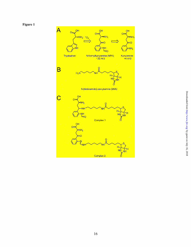
Other PTMs of PSII proteins have been identified, including oxidation of tryptophan to kynurenine (Fig. 1A) (22), reduction of aspartic acid to aspartyl aldehyde (23), acyl activation of glutamic acid to a species that binds primary amines (24), as well as numerous phosphorylations (25). Core PSII subunits contain multiple unidentified PTM residues that covalently bind amines (23,26,27). These reactions were attributed to reactive, carbonyl-containing amino acid side chains close to the active site, and covalent binding was proposed to occur via a Schiff base complex. The addition of chloride was observed to inhibit amine binding, suggesting that the binding sites were near the water oxidizing complex (28,29). Importantly, amines are well-known inhibitors of photosynthetic water oxidation (28,29), this reactivity was found in plants and cyanobacteria, and experiments showed that amines were oxidized to produce aldehydes (26,27). These experiments imply that the amine-binding residues may play a role in the structure, function, or assembly of PSII.
In addition to kynurenine, another known PTM of tryptophan in proteins is N-formylkynurenine (NFK). NFK may bind amines and is created as a stable double oxidation intermediate in the formation of kynurenine (Fig. 1A) (21,30-32). In this work, we use tandem mass spectrometry (MS/MS) and UV resonance Raman (UVRR) to show that PSII contains NFK. The unique ~325 nm absorption band of NFK is employed in the purification of NFK-containing CP43 peptides. By MS/MS peptide sequencing, NFK is identified as a +32 m/z modification of Trp-365 in CP43. A vibrational band at 1044 cm-1 is observed, which is characteristic of the oxidized indole ring in NFK. Quantitative analysis of the HPLC chromatogram was compared to the amount of inhibition under high light conditions. This comparison suggests that the CP43 NFK
by guest on July 16, 2018http://w
ww
.jbc.org/D
ownloaded from
3
modification can be induced by high light stress in PSII membrane preparations.
EXPERIMENTAL PROCEDURES
PSII Preparations, Oxygen Evolution Measurements, and Purification of PSII peptides: PSII was isolated from spinach (33) with the modifications previously described (27). Unless otherwise noted, all procedures were performed at 4°C and under dim green light illumination. Chlorophyll (34) and oxygen assays (35) were performed, and steady-state rates of oxygen evolution were ≥ 600 µmol O2/ (mg chl•h).
The 18- and 24-kDa extrinsic subunits were removed by treatment with 2 M NaCl for 30 minutes in the dark (36). Removal of psbO and the Mn4Ca cluster (Fig. S1, step 1) was performed by incubation with 800 mM tris(hydroxymethyl)aminomethane (Tris)-NaOH, pH 8.0 for 45 minutes at room temperature in the light (37). These Tris-washed (TW) PSII membranes were washed three times with a buffer of 400 mM sucrose, 50 mM 4-(2-hydroxyethyl)-1-piperazineethanesulfonic acid (HEPES)-NaOH, pH 7.5 and finally resuspended in the same buffer to yield a chlorophyll concentration of 2-4 mg/mL. Samples were stored at -70 °C.
Supporting information describes the purification of PSII peptides, including derivatization with a primary amine-biotin conjugate, 5-(biotinamido)-pentylamine (B5A) (Fig. 1B), in-situ trypsin digestion, high-pressure liquid chromatography (HPLC), 2-Dimensional (2D) electrophoresis, clear native polyacrylamide gel electrophoresis (CN-PAGE), in-gel digestion, and avidin affinity chromatography.
Synthesis of the Model Compound, NFK: NFK (Fig. 1A) was synthesized by formylation of commercially available kynurenine (95% purity, Sigma Aldrich, St. Louis, MO). The method has been previously described (38) and is known to produce a mixture of the single formylated NFK, and a double formylated compound, N’, Nα-formylkynurenine (39). ESI MS analysis was used to characterize the product. A Micromass Quattro LC, a triple quadrupole tandem mass spectrometer, was employed. The MH+ peaks observed were 236.8 and 265.0 m/z, consistent with the predicted MH+ masses for the singly and doubly formulated NFK at 237.2 and 265.2 m/z. The relative intensities of the two peaks were approximately
1:2 (236.8:265:0), consistent with the expectation that a mixture of the singly and doubly formulated species was produced. The electronic spectra of the singly and doubly formylated compounds have been reported to be indistinguishable (39).
UV-Visible Spectrophotometry: Optical spectra in Fig. 3C and Fig. 4 were recorded at room temperature from 200-750 nm on a Hitachi (U3000) spectrophotometer. The quartz cuvettes contained 200 µL, the slit width was 2 nm, and the scan speed was 120 nm min-1. The optical spectra in Fig. 3A and B were derived from the chromatogram through the use of a Beckman System Gold® HPLC (Brea, CA), equipped with a 125 solvent module, a 168 photodiode array detector (1 cm path length, 2 nm scan interval), and 32 Karat Software, version 7.0. Peptide samples were suspended in 200 µL of 50% acetonitrile/ 0.1% trifluoroacetic acid (TFA). The model compounds, 40 µM L-tryptophan (Sigma-Aldrich, St. Louis, MO), L-kynurenine (Sigma-Aldrich, St. Louis, MO), and NFK (synthesis described above) were suspended either in H2O or 50% acetonitrile/ 0.1% TFA. Reduction of NFK with NaBH4 was performed by incubation of 40 µM NFK with 400 µM NaBH4 for 30 minutes at room temperature (40). Reduction of a NFK-B5A mixture with 400 µM NaBH4 was performed using 40 µM NFK and 160 µM B5A.
UVRR Spectroscopy: A Renishaw (Hoffman Estates, IL) microprobe resonance Raman spectrometer was employed, as described (41,42). A 15x objective was used to focus the laser beam on the sample and to collect backscattered radiation. Experiments were conducted at room temperature, and the slit width was 50 µm.
To reduce the fluorescence background, PSII peptides were both HPLC and affinity purified (Fig. S1, steps 5B and 6A). Lyophilized peptide samples were suspended in water/ 0.1% TFA to increase solubility. A 3 µL peptide sample and a 360 µW, 325 nm probe beam from a He-Cd laser (KIMMON, Tokyo, Japan) were used. The 325 nm probe was chosen to give resonance enhancement of the PSII chromophore. The total exposure time for each spectrum was two minutes (41), and data from three individual experiments were averaged. The spectral resolution was 6 cm-1.
by guest on July 16, 2018http://w
ww
.jbc.org/D
ownloaded from

4
A 220 µW, 229 nm probe beam from a frequency-doubled Ar-ion laser (Cambridge LEXEL 95, Fremont, CA) was used to record Raman spectra of the model compounds, kynurenine and NFK, which were dissolved in water. These samples were recirculated at a flow rate ~4.5 m/s through a 120 µm diameter nozzle, which formed a jet (41). The total exposure time for each spectrum was two minutes (41). The spectral resolution was 10 cm-1.
Peptide Sequencing with MS/MS: Lyophylized peptide samples were reconstituted in 50 µL buffer A (99.9% water, 0.1% TFA) and analyzed on a Waters nanoHPLC C18 column (75 µm x 150 mm, 130 Å, 1.7 µm). For reverse phase chromatography, a gradient of buffer A (99.9 % H2O, 0.1 % TFA) and buffer B (99.9 % acetonitrile, 0.1 % TFA) was used. For MS analysis, a Thermo LTQ Orbitrap mass spectrometer was operated in a duty cycle consisting of one 400-2000 m/z FT-MS and four MS/MS LTQ scans.
MS/MS Data Analysis: For analysis of the LC-MS/MS data, the Sequest algorithm (43), implemented in the Bioworks software (Thermo Scientific, Waltham, MA), was applied for peptide identification versus a database. The database consisted of all spinach protein sequences present in National Center for Biotechnology Information (NCBI). For detection of modified peptides, a tryptophan modification of 31.98928 m/z was used as a parameter during the search. Photoinhibition experiments: Photoinhibition experiments were conducted with intact PSII (22,44,45). Samples were illuminated with white light from a Dolan-Jenner (Boxborough, MA) Fiber-Lite illuminator. The applied light intensity was ~9,000 µmol photons/ (m2•s) when measured with a Li-Cor (Lincoln, NE) Light Meter (model LI-189, with a ~8 cm diameter sensor) before the sample. The light intensity was ~7,000 µmol photons/ (m2•s) when measured after an empty sample tube. During illumination, PSII samples were maintained at 25 ºC by immersion in a water bath. The same two hour illumination experiment was also conducted without the water bath. During this time, the temperature was observed to increase to 37 ºC. As dark controls, PSII samples were incubated for two hours either at room temperature (~ 25 ºC) or at 37 ºC.
These conditions are similar to those described in the literature. For example, in spinach PSII membranes, at 25 ºC, and a light intensity of 4,000 µmol photons/ (m2•s), the half-time for oxygen evolution was reported as ~30 minutes (46). In spinach thylakoid membranes, at 20 ºC, and a light intensity of 7,000 µmol photons/ (m2•s), the half-time was ~25 minutes (47). A light intensity of 5,000 µmol photons/ (m2•s) at 25 ºC was used for studies of photoinhibition and degradation of the spinach CP43 subunit in spinach PSII membranes (48). For quantitation of the amount of NFK induced by photoinhibition, the intact PSII samples were digested with trypsin, and an HPLC assay was performed (see Supporting Information). Briefly, tryptic peptides were injected onto a C18 column, and the elution was monitored with a diode array detector, as described above. To quantitate the yield of the NFK-containing peptide, the area of the 350 nm peak was calculated using instrument software. This value was normalized to the total integrated area in the 220 nm chromatogram (0-50 min). This normalization provides an internal standard and corrects for any changes in the yield of tryptic peptides. Experiments were performed 3-7 times, and the values were averaged. Oxygen evolution experiments (35) were performed under the same conditions, i.e., after a two hour dark incubation or a two hour illumination (with water bath at 25oC). Measurements were performed six times, and the values were averaged.
RESULTS Isolation of the Amine-Binding
Chromophore: Following purification from spinach (33), PSII membranes were depleted of the 18-kDa, 24-kDa, and psbO extrinsic polypeptides, as well as the Mn4Ca cluster (36,37). These modifications allow access to the sterically-hindered core complex where covalent amine-binding occurs (Fig. S1, step 1) (26,27). TW PSII membranes were reacted with a primary amine-biotin conjugate, B5A (Fig. 1B) by incubation in the light (Fig. S1, step 2). Previous work demonstrated that PSII core subunits form stable covalent adducts with amines under these conditions, and binding was attributed to reactive carbonyl groups in PTMs (23,26,27). In our experiments, the biotin-linked amine allowed the
by guest on July 16, 2018http://w
ww
.jbc.org/D
ownloaded from

5
selective purification of peptides by avidin affinity chromatography (Fig. S1 steps 6A & B). Binding of B5A was confirmed by Western blot of a sodium dodecyl sulfate-polyacrylamide gel (SDS-PAGE) of B5A-derivatized PSII and by detection with an avidin-alkaline phosphatase conjugate (data not shown) (49).
Following derivatization, in-situ digestion was employed to release modified, surface exposed peptides. This method was used previously to identify surface-exposed phosphorylation sites in Arapidopsis thylakoid membranes (50). B5A-derivatized TW PSII was trypsin digested overnight (Fig. S1 step 4B), cleaved peptides were separated from undigested PSII by centrifugation, and the peptides were subjected to HPLC (Fig. S1 step 5B). When a 10-60% acetonitrile/ 0.1% TFA gradient was used and the peptide elution was monitored at 350 nm, three fractions with unique red-shifted absorption peaks were observed (Fig. S2). The fractions had retention times of ~28 min (fraction 1), ~35 min (fraction 2), and ~36 min (fraction 3). Unlabeled peptides gave a similar 350 nm chromatogram (Fig. S2). These peaks were also present in intact PSII membranes (Fig. 2).
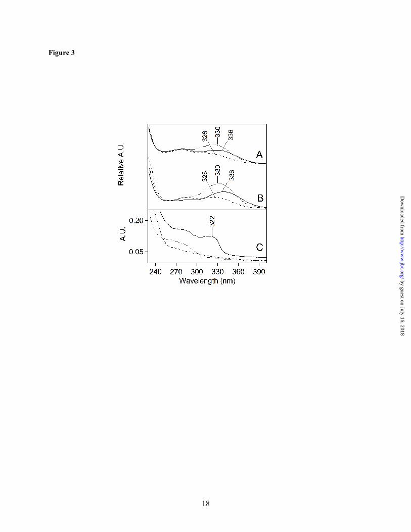
The absorption spectra of these unlabeled and B5A-labeled fractions are shown in Fig. 3A and B, respectively. As derived from the HPLC detector, the spectra all exhibited maxima between 326 and 336 nm. Small shifts may be due to overlap with a 280 nm shoulder, which is indicative of tyrosine absorption (51). The spectra of the fractions also showed strong 220 nm absorption from the peptide bond (52), but no visible absorption, which is characteristic of photosynthetic pigments. The observation of these 220 and 280 nm absorption bands supports the conclusion that the fractions contain peptides, which have been post-translationally modified to produce a chromophore with a ~325 nm absorption maximum.
To identify the 325 nm chromophore in the peptide samples, comparison was made to model compounds. Some oxidative tryptophan products, such as hydroxytryptophan, dioxyindolylalanine, and oxyindolylalanine, absorb near 295 nm, only slightly red-shifted from the tryptophan absorption band (53,54). However, other PTMs of tryptophan, including NFK and kynurenine (Fig. 1A), show more red-shifted
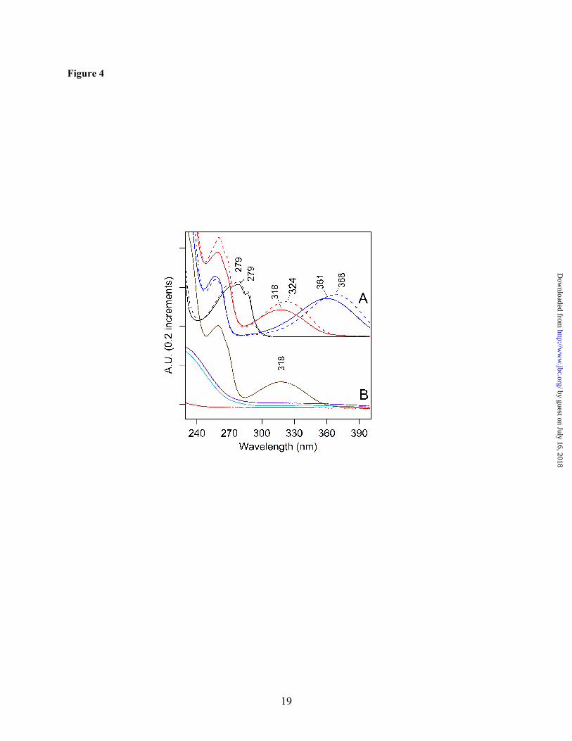
absorption (40,55). To compare with the peptide spectra, NFK was synthesized by formylation of commercially available kynurenine. As shown in Fig. 4A, NFK had an absorption peak at ~320 nm (Fig. 4A, red), while kynurenine had a longer wavelength absorption maximum at ~365 nm (Fig. 4A, blue). Both spectra were red-shifted compared to the tryptophan absorption maximum at ~280 nm (Fig. 4A, black). The absorption maximum of NFK was slightly solvent dependent, showing a shift from 318 to 324 nm when water (Fig. 4A, solid red) was compared to 50% acetonitrile/0.1% TFA (Fig. 4A, dashed red). The peptide spectra (Fig. 3A and B) exhibited a clear similarity with the NFK spectrum (Fig. 4A, red), making NFK a candidate for the PTM.
MS/MS Identifies a NFK Modification in the CP43 Subunit: HPLC fractions 1-3 were purified by a second round of chromatography, avidin affinity (Fig. S1, step 6A). Applying LC-MS/MS analysis to the purified peptide samples resulted in unambiguous identification of a CP43 peptide, 363AP[W*]LEPLRGPNGLDLSR379, in fraction 1 with a p-value of 10-7 and displaying a mass shift of +32 m/z on Trp-365 (Fig. 5). These results are indicative of an NFK modification. In fraction 2, a peptide of CP24, 169PDSQSVE[W*]ATPWSR184, with a NFK modification was observed (data not shown). There were no spectra detected consistent with the B5A-labeled peptides, however, suggesting that the adduct of NFK and B5A was not stable under the conditions employed for mass spectrometry.
2D Gel Electrophoresis: To confirm that the chromophore arises from a PTM in a PSII peptide, PSII core peptides were purified by 2D gel electrophoresis. For these experiments, B5A-labeled PSII was solubilized and electrophoresized in the first dimension by non-denaturing CN-PAGE (56), which separates the PSII membranes into dimer complexes with varying amounts of light-harvesting proteins (24). All gels were run without Coomassie or bromophenol blue to eliminate possible spectral artifacts from the dyes. The PSII dimer complex, which is deficient in light-harvesting proteins (24), was excised and resolved into individual polypeptides in the second dimension (Fig. S1, step 3) (57). Due their similarity in electrophoretic mobility, the CP43 and CP47 protein bands were not fully resolved. MS/MS analysis, following in-gel digestion and
by guest on July 16, 2018http://w
ww
.jbc.org/D
ownloaded from

6
peptide extraction (Fig. S1, step 4C), validated the band identities (data not shown). The identities were also substantiated by previous work (24,58). Although in-situ these polypeptides bind many pigment molecules, the non-covalently bound pigments were separated from the proteins under the denaturing gel conditions in the second dimension. Therefore, this experiment eliminated chlorophyll or carotenoid (or their degradation products) as possible sources of the optical absorption. Before affinity chromatography (Fig. S1, step 6B), the absorption spectrum of the gel extracted sample exhibited only absorption characteristic of tyrosine containing peptides, with a 280 nm absorption band (51) (Fig. 3C, dotted line). However, upon affinity purification of the gel-extracted peptide mixture, a ~322 nm peak was observed (Fig. 3C, solid line). This band resembled the chromophore absorption observed in the HPLC purified peptides (Fig. 3A and B). The λmax was similar to the spectra recorded from the HPLC fractions, given the increased scattering background in the gel-extracted samples. Fig. 3C also shows that the B5A compound itself does not contribute to the optical absorption (Fig. 3C, dashed line), although affinity chromatography was essential for the selection of the modified peptides. Proposed Structure of the B5A Adduct: NFK is expected to react with hydrazines, hydrazides, and amines (59-61). Fig. 1C presents two possible structures for the B5A-NFK adduct formed in our experiments. The small 2 m/z mass difference between the two structures may not be distinguishable by low resolution peptide mass spectrometry. Fig. 1C, complex 1, is a covalent adduct, formed by nucleophilic addition of the B5A label at the C4 position of NFK and a subsequent reduction reaction. Fig. 1C, complex 2, is another possibility for the formation of a B5A-derived amidine, in which the nucleophilic addition occurs at the N-formyl carbon. While complex 1 should be quite stable during MS and should be observed in MS/MS spectra, complex 2 is expected to break preferentially at the NFK-B5A interface and escape MS/MS detection. To distinguish between these two possible structures, we considered the effect of reduction on the optical spectrum. As shown in Fig. 4B, addition of sodium borohydride (NaBH4) and
reduction of the C4 carbonyl group eliminated the 318 nm absorption of NFK (compare Fig. 4A, red and Fig. 4B, violet). Addition of sodium borohydride to a mixture of NFK and B5A had a similar effect (Fig. 4B, brown and cyan). This result suggests that reduction of the carbonyl group of NFK will eliminate the 322 nm absorption. Because the 322 nm band was observed in the labeled peptides, consideration of these optical properties supports an assignment of complex 2 as the covalent adduct.
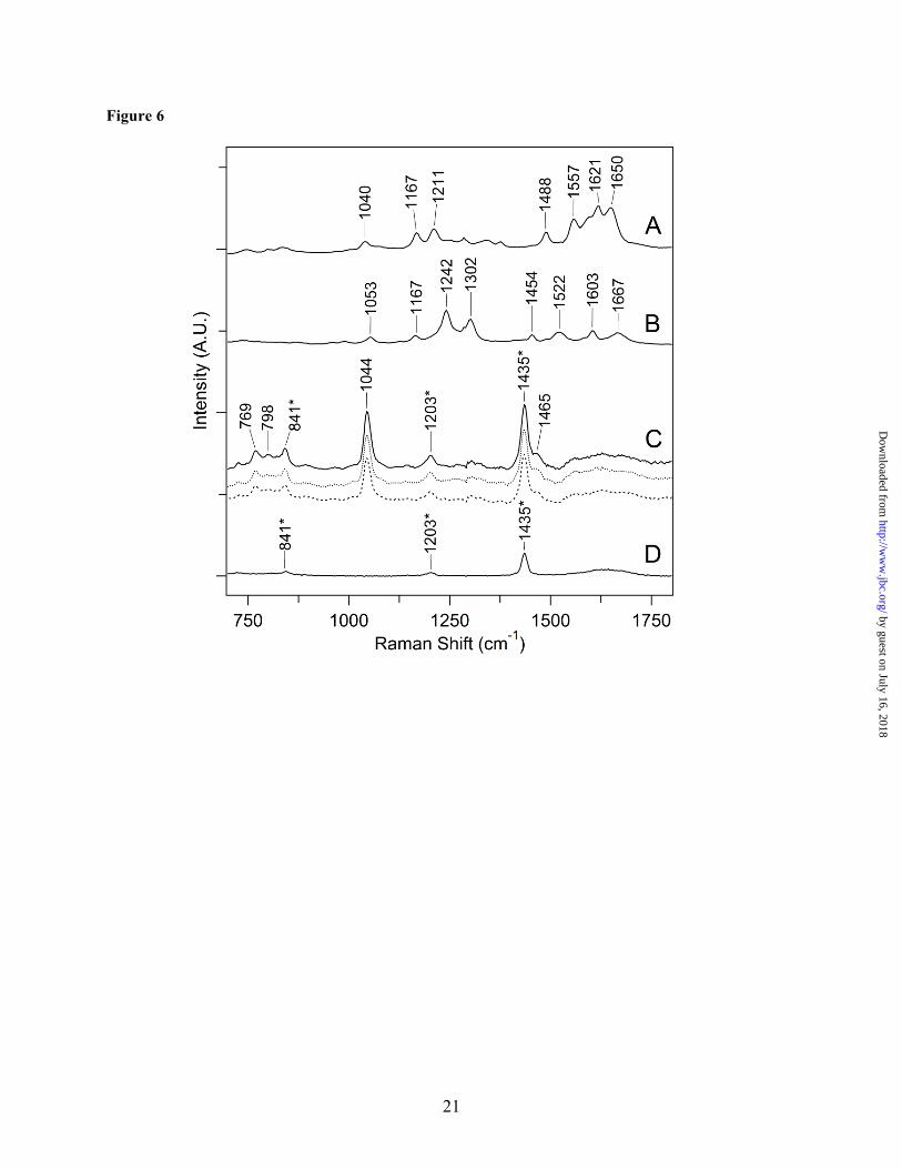
UVRR Spectroscopy of the Chromophore: To obtain more information concerning the structure of the B5A peptide complex, UV resonance Raman (UVRR) was employed (62). Fig. 6A and B are the Raman spectra of kynurenine and NFK model compounds, respectively. These data were obtained with 229 nm excitation. The Raman spectrum of kynurenine could not be acquired at 325 nm, due to a large fluorescence background. The Raman spectrum of NFK, obtained either with 229 or 325 nm probe beams, displayed a band at 1242 cm-1. There was no observable signal from the B5A label alone, either at 229 or 325 nm, due to lack of resonance enhancement (data not shown).
In Fig. 6B, unique bands assignable to the NFK N-formyl group were observed at 1667 and 1242 cm-1. A band between 1040 and 1053 cm-1 was observed both in the kynurenine and the NFK Raman spectra (Fig. 6A and B). These bands are characteristic of the oxidized indole group (31,63). These spectral features were not observed in the UV Raman spectra of the aromatic amino acids, histidine, tryptophan, tyrosine, and phenylalanine (Fig. S3). For example, unmodified tryptophan exhibited a benzene breathing mode at 1009 cm-1 (Fig. S3). In addition, these bands were not observed in spectra derived from chl a or a carotenoid, after correction for solvent scattering (Fig. S4)
Previous FT-Raman measurements on NFK assigned a band at 1050 cm-1 to the ring system, a band at 1604 cm-1 to a ring stretching mode, and bands at 1239 and 1685 cm-1 to the N-formyl group (63). In addition, bands at 1052 and 1050 cm-1 were observed for NFK modifications in lysozyme (63) and egg white ovalbumin (31), respectively. Formamide gave rise to Raman bands at 1670, 1599, 1391, 1313, 1098, 1048, and 983 cm-1 (64). In anilides, the carbonyl band was
by guest on July 16, 2018http://w
ww
.jbc.org/D
ownloaded from

7
observed at higher frequency (1704 cm-1 in formanilide), due to delocalization of the unpaired electrons on the formamide nitrogen into the phenyl ring (65).
Because the UV Raman spectrum is specific for the contribution of NFK, the Raman spectrum of derivatized peptide samples was obtained with a 325 nm probe (Fig. 6C), which resonantly enhances the chromophore. In peptides purified by HPLC only (Fig. S1, step 5B), a strong fluorescence background obscured the Raman signal. Therefore, samples were subjected to purification by affinity chromatography (Fig. S1, step 6A), which reduced the background fluorescent signal. The resulting Raman spectra of all three HPLC, 350 nm absorbing fractions were similar (Fig. 6C). These results imply that all three fractions contain the same PTM. Comparison with the TFA buffer spectrum (Fig. 6D) showed that bands at 841, 1203, and 1435 cm-1 arise from TFA. The Raman spectra of all three peptide fractions displayed a band at 1044 cm-1, which is consistent with the presence of NFK in all three fractions. The shift from the 1053 cm-1 frequency observed in NFK alone (Fig. 6B) may be due to reaction with the B5A label. No vibrational bands from the N-formyl group (1242 and 1667 cm-1) were observed, supporting the interpretation that complex 2 (Fig. 1C) is the stable structure.
Yield of NFK in TW PSII: Using an extinction coefficient of 3750 M-1 cm-1 at 321 nm (55), the yield of NFK in TW PSII can be estimated. For the HPLC experiment, fraction 1 was collected, the sample was concentrated to 200 µl, and the absorption spectrum was measured on a Hitachi spectrophotometer (see Materials and Methods). Starting with 6 mg chl or 24 nmole PSII reaction center (66), the NFK yield (on a reaction center basis) was estimated as ~7% in the HPLC method. For the 2-D gel experiment, peptides were extruded from the gel, concentrated to 200 µl, and the absorption spectrum was measured as described above (Fig. 3C). Starting with 13 mg chl or 52 nmole PSII reaction center (66), the NFK yield was estimated as ~6% in the 2D gel method.
Photoinhibition Increases the Yield of NFK in Intact PSII: Our MS/MS data support the interpretation that NFK is formed by PTM of Trp-365 in the CP43 subunit. NFK can be generated
from tryptophan by ROS (67,68). These species, including singlet oxygen (1O2,), hydrogen peroxide (H2O2), superoxide anion (O2
-), and hydroxyl radical (•OH) (69), have been proposed to be involved with photoinhibition in PSII. However, the mechanism of their involvement remains controversial (reviewed in 70,71). High light conditions have been linked to oxidative modification of Arabidopsis PSII in proteomic studies (21). We have previously reported that substitutions at Trp-365 increase the rate of photoinhibition (22). The magnitude of the change depends on light intensity (data not shown) and will be described in a future publication.
To probe for a possible connection between the yield of NFK and photoinhibition, we compared 350 nm chromatograms of tryptic peptides obtained from intact PSII (Fig. 2). The integrated area of the 350 nm peak, derived from fraction 1, was corrected for the total integrated absorption at 220 nm. This normalization is an internal standard, which corrects for any change in the total yield of tryptic peptides. In this experiment, intact PSII samples were maintained in the dark at room temperature (~25 ºC) (Fig. 2A) or in the dark at 37 ºC (Fig. 2B). These dark-maintained, intact PSII samples gave a 350 nm fraction, with a similar retention time to TW PSII fraction 1 (Fig. 2, shaded peaks and Fig. S2). The fraction 1 yield in dark maintained PSII was estimated as 0.3% on a reaction center basis. The NFK yield was not significantly altered by an increase in temperature in the dark. Comparison of the integrated areas (Fig. 2, shaded peaks, fraction 1), derived from dark-maintained PSII at 25 ºC and at 37 ºC, gave a ratio of 1.1 + 0.1. Intact PSII samples were also illuminated with white light for two hours under temperature-controlled conditions at 25 ºC (Fig. 2C) or under conditions (Fig. 2D), in which the temperature of the sample increased to 37 ºC. An increase in peak height was observed after two hours of illumination. This increase was observed when the temperature was controlled at 25oC (ratio 2.4 + 0.8, Fig. 2C, shaded peak) or when the temperature was allowed to increase to 37 ºC (ratio 2.2 + 0.5, Fig. 2D, shaded peak). The steady state rate of oxygen evolution was also measured under the same conditions (35). Before illumination, the average rate was 740 + 50 µmol/mg chl-hr. After two hours in the dark, the
by guest on July 16, 2018http://w
ww
.jbc.org/D
ownloaded from
8
rate was 630 + 30 µmol/mg-hr. However, with a two hour illumination, the rate declined to 270 + 60 µmol/mg chl-hr. The 2.4 + 0.5 fold decrease in activity is similar to the increase observed in NFK yield. Therefore, these results suggest that the NFK modification at Trp-365 is induced by illumination and high light stress in intact PSII.
DISCUSSION
Summary: In this paper, we provide evidence that PSII contains a modified form of tryptophan, NFK. Mass spectrometry on purified peptides shows a mass shift of +32 m/z for the 363AP[W*]LEPLRGPNGLDLSR379 peptide from CP43. This mass shift and peptide sequencing by MS/MS are consistent with a double oxidation of Trp-365. Optical absorption and UV resonance Raman data support the conclusion that CP43 peptides contain NFK. In these experiments, NFK was observed following in-situ and in-gel tryptic digestion. The yield of NFK in TW PSII was significant, and the yield increased when intact PSII was subjected to photoinhibitory conditions.
Generation of NFK and NFK in Other Proteins: NFK has been identified in other proteins by mass spectrometry, including bovine heart mitochondrial proteins (72), rat skeletal muscle proteins (73), bovine α -crystalline (74), and spinach LHCII (32). NFK is formed by the reaction of ROS with tryptophan side chains in proteins (67). One potential reactive species is singlet oxygen, 1O2 (68). Initial reaction of tryptophan with 1O2 has been proposed to form one of two unstable intermediates. A dioxetane derivative intermediate can form across the C2-C3 indole ring bond; subsequent ring cleavage gives NFK. On the other hand, an intermediate hydroperoxide, formed at the C3 position on the indole ring, can also decompose to form NFK (75). Therefore, we propose that NFK is formed by a reaction between Trp-365 and 1O2.
Other Modifications at Trp-365: Other modifications of Trp-365 in the lumenal loop of CP43 (Trp-352 in Synechocystis sp. PCC 6803) have been reported previously (22). The data were obtained by tandem mass spectrometry (22) and were consistent with modification of the side chain to kynurenine (+4 m/z), oxindolalanine (+16 m/z), and a hydroxy-indole derivative (+18 m/z). The oxindolalanine and hydroxy-indole derivatives were proposed to be intermediates produced
during oxidative cleavage to give kynurenine (22). None of these species are expected to show an absorption maximum at 325 nm (53,54), as observed here for the NFK-containing peptides. NFK can be formed as a stable intermediate during the production of kynurenine (Fig. 1A) (38). In previous work, kynurenine was observed to be present in PSII which had not been TW and had not been subjected to gel electrophoresis (22). Kynurenine was also observed in PSII, which had been maintained in the dark. Taken together with the results described here, these data support the conclusion that oxidative modification of Trp-365 is relevant in vivo.
Analysis of Three Different HPLC Fractions: In this work, three 350 nm absorbing fractions, with reproducible retention times, were observed with HPLC purification of TW PSII peptides. UV resonance Raman studies suggest that all three fractions contained the same, B5A-derivatized NFK chromophore. The NFK modification in CP43 was confirmed by MS/MS of fraction 1. An NFK modification in a light harvesting protein (CP24) was observed in fraction 2. However, the modified peptide detected in fraction 1 was the result of incomplete tryptic digestion. Therefore, it is possible that other fractions contain a different cleavage product of the same CP43 peptide. In addition to 325 nm absorption, all three fractions exhibited a 280 nm peak, which is indicative of tyrosine absorption. It should be noted that the NFK containing CP43 sequence does not contain tyrosine (363AP[W*]LEPLRGPNGLDLSR379). Thus, other tyrosine-containing peptides must be present in all three eluting fractions. Our MS analysis of the fractions provided evidence for ~40 peptides, even after HPLC and affinity purification (data not shown). Such complexity can be attributed to the challenges of MS/MS as applied to membrane associated peptides.
Proposed Structure of the B5A-Labeled, NFK Complex: In some of our experiments, a biotinylated amine was used to label the NFK-containing peptide. Amines and hydrazines are expected to label activated carbonyl groups (26 and references therein). In other proteins, it has been suggested that kynurenine and NFK react with hydrazine. These proteins include low-density lipoprotein (LDL) (76), cucumber microsomal membrane proteins (60) and ribulose-
by guest on July 16, 2018http://w
ww
.jbc.org/D
ownloaded from
9
1,5-bisphosphate carboxylase oxygenase (Rubisco) (60). In bovine serum albumin (59) and α-crystallin (59), NFK was proposed to crosslink with lysine residues. On the other hand, in PSII, a complex between kynurenine and a hydrazide labeling reagent was not observed by MS/MS (22).
NFK may react with amines to form an adduct at the C4 position (Fig. 1C, complex 1). Reaction of the amine and electrophilic carbon produces a Schiff base, which can be reduced to give the stable product shown in complex 1. Previous work has indicated that reducing equivalents are produced during PSII light reactions. These reducing equivalents were observed to stabilize amine-PTM complexes (27). On the other hand, reaction of the amine with the NFK formamide group would give the structure shown in Fig. 1C, complex 2. Although amide groups do not usually react with amines, a similar product complex was observed between N-acetyl-formylkynurenine and dimethyl-p-benzoquinonediimine in solution (77). Delocalization of the unpaired electrons on the nitrogen into the aromatic ring may help to activate the formamide carbon (65). It should be noted that we have not observed the labeled NFK adduct by MS/MS. However, our optical and resonance Raman data support binding of the amine label, B5A, at the formamide group, as shown in complex 2. An addition at the N-formyl carbon would provide a breaking point during collision induced dissociation, which could lead to the neutral loss of the B5A moiety during MS analysis.
Photoinhibition and High Light Stress: Examination of CP43 protein sequences in other organisms, both prokaryotes and eukaryotes, indicates strict sequence conservation of Trp-365. This conservation suggests a functional role for this residue. We hypothesize that Trp-365 may play a role in protection from photoinhibition (22). In PSII, photoinhibition is the light-induced inactivation of photosynthetic activity induced by excess light energy (reviewed in 78). The results are a decreased efficiency in electron transfer, damage and degradation of the D1 and other PSII subunits, and finally repair by de novo protein synthesis. When the rate of repair is slower than the rate of degradation, a loss of PSII activity is observed (78). This cycle of damage and repair
must be coordinated, and the mechanisms of these reactions have not yet been elucidated.
Photoinhibition may occur by two different mechanisms (reviewed in 71,78,79,80). In the acceptor side photoinhibition model, damage is initiated by charge separation in reaction centers that contain a reduced quinone, QA
-. Recombination leads to the product of triplet chlorophyll, 3chl, species (81). Changes in the midpoint potential of QA
- may alter the susceptibility of PSII to photoinhibition (reviewed in 79). In donor side photoinhibition, damage is initiated by inactivation of the OEC and water oxidation. Under these conditions, P680
+, which has a high potential, has a long lifetime and can act as an oxidant for prosthetic groups and amino acid residues (reviewed in 79).
ROS and High Light Stress in PSII: We attribute the formation of NFK to the reaction of Trp-365 with 1O2. In PSII, 1O2 can be formed by photo-excitation of chlorophyll molecules, which results in formation of 3chl (82). ROS and 3chl are generated by charge recombination in acceptor side inhibition (81,83). 1O2 has been detected by spin trapping in photoinhibited PSII (70,84) and by a fluorescence sensor in Arabidopsis leaves (85). Chemical trapping in PSII reaction center preparations, which lack the quinone acceptors, has also detected 1O2 (86). Other ROS species may be formed in donor side photoinhibition (70,84).
While a correlation between ROS production and photodamage is largely accepted, there is no consensus on the specific role of 1O2 (see 84 and references therein). One view suggests direct involvement of ROS in damage and increased turnover of the reaction center D1 protein (87). This can occur directly, because ROS damage can lead to peptide bond cleavage (67). Alternatively, ROS induced modifications could cause a protein conformational change, which allows access of specific proteases to the D1 subunit (22,88,89). Finally, 1O2 may inhibit the D1 repair cycle (for example, see 90). These roles of ROS are not mutually exclusive. Proposed Role for NFK-365 in Photoinhibition: In this paper, we provide evidence that NFK is present in dark maintained PSII, and that the yield of NFK increases by a factor of two under high light intensity in intact PSII. This change was accompanied by a 2 fold
by guest on July 16, 2018http://w
ww
.jbc.org/D
ownloaded from
10
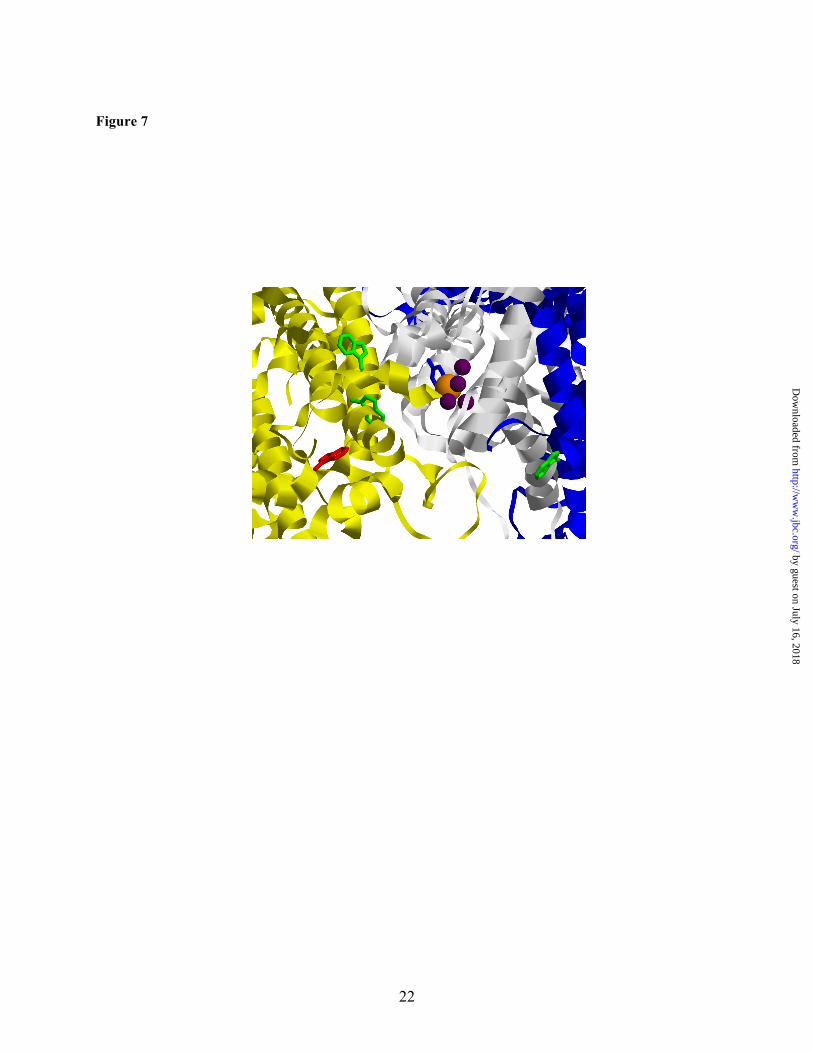
decrease in steady state oxygen evolution rate. Previous preliminary characterization of site directed mutations at Trp-365 reported that the Trp provides photoprotection (22). The 1.9 Å resolution crystal structure (3) of cyanobacterial PSII shows that Trp-365 is ~17 Å from the water oxidizing complex (Fig. 7). Trp-365 is found in a CP43 loop region, which is in close proximity to the D1 subunit.
Taken together, these observations suggest a role for modification of Trp-365 in the high light induced, damage/repair cycle. This could be accomplished by two different mechanisms. In the first, Trp-365 may act as a ROS scavenger, and in the second, Trp-365 may serve as signal, which facilitates reaction center repair. It should be noted that Trp-359 and 291 in CP43, as well as
Trp-328 in D2, are also within 17 Å of the OEC (Fig. 7). In our MS/MS experiments, we have seen no evidence for NFK modifications of these sidechains, although Trp-359 was observed in the +16 m/z form (data not shown).
Conclusions: We have identified an oxidative modification of tryptophan in the CP43 subunit of PSII. This NFK is a UV absorbing chromophore, which is formed by the oxidation of a Trp side chain by ROS. We propose that NFK plays a role in protection and repair during photoinhibition. The evolutionarily conserved residue may act as a 1O2 scavenger. Alternatively, oxidation of the tryptophan may promote repair by signaling for degradation or enabling efficient removal of the damaged D1.
REFERENCES
1. Nelson, N., and Yocum, C. F. (2006) Annu. Rev. Plant Biol. 57, 521-565 2. Guskov, A., Kern, J., Gabdulkhakov, A., Broser, M., Zouni, A., and Saenger, W. (2009) Nat.
Struct. Mol. Biol. 16, 334- 342 3. Umena, Y., Kawakami, K., Shen, J.-R., and Kamiya, N. (2011) Nature In press 4. Yocum, C. F. (2008) Coord. Chem. Rev. 252, 296-305 5. Bricker, T. M., and Frankel, L. K. (2002) Photosynth. Res. 72, 131-146 6. Miyao, M., and Murata, N. (1989) Biochim. Biophys. Acta 977, 315- 321 7. MacDonald, G. M., Boerner, R. J., Everly, R. M., Cramer, W. A., Debus, R. J., and Barry, B. A.
(1994) Biochemistry 33, 4393-4400 8. Zouni, A., Witt, H.-T., Kern, J., Fromme, P., Krauss, N., Saenger, W., and Orth, P. (2001) Nature
409, 739-743 9. Ferreira, K. N., Iverson, T. M., Maghlaoui, K., Barber, J., and Iwata, S. (2004) Science 303,
1831-1838 10. Kamiya, N., and Shen, J. R. (2003) Proc. Natl. Acad. Sci. U. S. A. 100, 98-103 11. Loll, B., Kern, J., Saenger, W., Zouni, A., and Biesiadka, J. (2005) Nature 438, 1040-1044 12. Rhee, K.-H., Morris, E. P., Barber, J., and Kühlbrandt, W. (1998) Nature 396, 283-286 13. Allen, J. F. (1992) Biochim. Biophys. Acta 1098, 275-335 14. Stadtman, E. R. (1990) Biochemistry 29, 6323-6331 15. Janes, S. M., Mu, D., Wemmer, D., Smith, A. J., Kaur, S., Maltby, D., Burlingame, A. L., and
Klinman, J. P. (1990) Science 248, 981-987 16. Whitelegge, J. P., Faull, K. F., Gundersen, C. B., and Gómez, S. M. (1999) in Photosynthesis:
Mechanisms and Effects (Garab, G. ed.), Kluwer Academic Publishers, Dordrecht, Netherlands. pp 4381 -4384
17. Whitelegge, J. P., Gundersen, C. B., and Faull, K. F. (1998) Protein Sci. 7, 1423-1430 18. Bowyer, J. R., Packer, J. C. L., McCormack, B. A., Whitelegge, J. P., Robinson, C., and Taylor,
M. A. (1992) J. Biol. Chem. 267, 5424-5433 19. Liao, D. I., Qian, J., Chisholm, D. A., Jordan, D. B., and Diner, B. A. (2000) Nat. Struct. Biol. 7,
749-753 20. Sharma, J., Panico, M., Shipton, C. A., Nilsson, F., Morris, H. R., and Barber, J. (1997) J. Biol.
Chem. 272, 33158-33166
by guest on July 16, 2018http://w
ww
.jbc.org/D
ownloaded from

11
21. Galetskiy, D., Lohscheider, J. N., Kononikhin, A. S., Popov, I. A., Nikolaev, E. N., and Adamska, I. (2010) Rapid Commun. Mass Spectrom. 25, 184-190
22. Anderson, L. B., Maderia, M., Ouellette, A. J. A., Putnam-Evans, C., Higgins, L., Krick, T., MacCoss, M. J., Lim, H., Yates III, J. R., and Barry, B. A. (2002) Proc. Natl. Acad. Sci. U. S. A. 99, 14676-14681
23. Anderson, L. B., Ouellette, A. J. A., Eaton-Rye, J., Maderia, M., MacCoss, M. J., Yates III, J. R., and Barry, B. A. (2004) J. Am. Chem. Soc. 126, 8399-8405
24. Rexroth, S., Wong, C. C. L., Park, J. H., Yates III, J. R., and Barry, B. A. (2007) J. Biol. Chem. 282, 27802-27809
25. Mamedov, F., Rintamäki, E., Aro, E. M., Andersson, B., and Styring, S. (2002) Photosynth. Res. 74, 61-72
26. Ouellette, A. J. A., Anderson, L. B., and Barry, B. A. (1998) Proc. Natl. Acad. Sci. U. S. A. 95, 2204-2209
27. Anderson, L. B., Ouellette, A. J. A., and Barry, B. A. (2000) J. Biol. Chem. 275, 4920-4927 28. Sandusky, P. O., and Yocum, C. F. (1984) Biochim. Biophys. Acta 766, 603-611 29. Sandusky, P. O., and Yocum, C. F. (1986) Biochim. Biophys. Acta 849, 85-93 30. Previero, A., Coletti-Previero, M. A., and Jollès, P. (1967) J. Mol. Biol. 24, 261-268 31. Rokos, H., Wood, J. M., Hasse, S., and Schallreuter, K. U. (2008) J. Raman Spectrosc. 39, 1214-
1218 32. Rinalducci, S., Campostrini, N., Antonioli, P., Righetti, P. G., Roepstorff, P., and Zolla, L. (2005)
J. Proteome Res. 4, 2327-2337 33. Berthold, D. A., Babcock, G. T., and Yocum, C. F. (1981) FEBS Lett. 134, 231-234 34. Lichtenthaler, H. K. (1987) Methods Enzymol. 148, 350-382 35. Barry, B. A. (1995) Methods Enzymol. 258, 303-319 36. Ghanotakis, D. F., Topper, J. N., Babcock, G. T., and Yocum, C. F. (1984) FEBS Lett. 170, 169-
173 37. Yamamoto, Y., Doi, M., Tamura, N., and Nishimura, M. (1981) FEBS Lett. 133, 265-268 38. Simat, T., Meyer, K., and Steinhart, H. (1994) J. Chromatogr. A 661, 93-99 39. Jacobson, K. B. (1978) Arch. Biochem. Biophys. 186, 84-88 40. Pirie, A. (1971) Biochem. J 125, 203- 208 41. Chen, J., and Barry, B. A. (2008) Photochem. Photobiol. 84, 815-818 42. Chen, J., Bender, S. L., Keough, J. M., and Barry, B. A. (2009) J. Phys. Chem. B 113, 11367-
11370 43. Eng, J. K., McCormack, A. L., and Yates III, J. R., (1994) J. Am. Soc. Mass. Spectrom. 5, 976-
989 44. Rosenberg, C., Christian, J., Bricker, T. M., and Putnam-Evans, C. (1999) Biochemistry 38,
15994-16000 45. Knoepfle, N., Bricker, T. M., and Putnam-Evans, C. (1999) Biochemistry 38, 1582-1588 46. Henmi, T., Miyao, M., and Yamamoto, Y. (2004) Plant Cell Physiol. 45, 243-250 47. Virgin, I., Styring, S., and Andersson, B. (1988) FEBS Lett. 233, 408-412 48. Yamamoto, Y., and Akasada, T. (1995) Biochemistry 34, 9038-9045 49. Towbin, H., Staehelin, T., and Gordon, J. (1979) Proc. Natl. Acad. Sci. U. S. A. 76, 4350-4354 50. Vener, A. V., Harms, A., Sussman, M. R., and Vierstra, R. D. (2001) J. Biol. Chem. 276, 6959-
6966 51. Edelhoch, H. (1967) Biochemistry 6, 1948-1954 52. Ham, J. S., and Platt, J. R. (1952) J. Chem. Phys. 20, 335-336 53. Zhao, H., Sagert, J., Hwang, D. S., and Waite, J. H. (2009) J. Biol. Chem. 284, 23344-23352 54. Huang, H. V., Bond, M. W., Hunkapiller, M. W., and Hood, L. E. (1983) Methods Enzymol. 91,
318-324 55. Mehler, A. H., and Knox, W. E. (1950) J. Biol. Chem. 187, 431-438 56. Schägger, H., and von Jagow, G. (1991) Anal. Biochem. 199, 223-231
by guest on July 16, 2018http://w
ww
.jbc.org/D
ownloaded from
12
57. Schägger, H., Cramer, W. A., and von Jagow, G. (1994) Anal. Biochem. 217, 220-230 58. Kügler, M., Jänsch, L., Kruft, V., Schmitz, U. K., and Braun, H.-P. (1997) Photosynth. Res. 53,
35-44 59. Fujimori, E. (1981) FEBS Lett. 135, 257-260 60. Caldwell, C. R. (1993) Plant Physiol. 101, 947-953 61. Yang, C.-Y., Gu, Z.-W., Yang, H.-X., Yang, M., Gotto, A. M., Jr., and Smith, C. V. (1997) Free
Radical Biol. Med. 23, 82-89 62. Asher, S. A. (1993) Anal. Chem. 65, A59-A66 63. Bieker, L., and Schmidt, H. (1979) FEBS Lett. 106, 268--270 64. Puranik, P. G., and Ramiah, K. V. (1959) J. Mol. Spectrosc. 3, 486-495 65. Chalapathi, V. V., and Ramiah, K. V. (1968) J. Mol. Spectrosc. 26, 444-453 66. Patzlaff, J. S., and Barry, B. A. (1996) Biochemistry 35, 7802-7811 67. Berlett, B. S., and Stadtman, E. R. (1997) J. Biol. Chem. 272, 20313-20316 68. Gracanin, M., Hawkins, C. L., Pattison, D. I., and Davies, M. J. (2009) Free Radical Biol. Med.
47, 92-102 69. Asada, K. (1999) Annu. Rev. Plant Physiol. Plant Mol. Biol. 50, 601-639 70. Krieger, A., Rutherford, A. W., Vass, I., and Hideg, E. (1998) Biochemistry 37, 16262-16269 71. Nishiyama, Y., Allakhverdiev, S. I., and Murata, N. (2006) Biochim. Biophys. Acta 1757, 742-
749 72. Hunzinger, C., Wozny, W., Schwall, G. P., Poznanović, S., Stegmann, W., Zengerling, H.,
Schoepf, R., Groebe, K., Cahill, M. A., Osiewacz, H. D., Jägemann, N., Bloch, M., Dencher, N. A., Krause, F., and Schrattenholz, A. (2006) J. Proteome Res. 5, 625-633
73. Fedorova, M., Todorovsky, T., Kuleva, N., and Hoffmann, R. (2010) Proteomics 10, 2692-2700 74. Finley, E. L., Dillon, J., Crouch, R. K., and Schey, K. L. (1998) Protein Sci. 7, 2391-2397 75. Ronsein, G. E., Oliveira, M. C. B., Miyamoto, S., Medeiros, M. H. G., and Di Mascio, P. (2008)
Chem. Res. Toxicol. 21, 1271-1283 76. Yang, C.-Y., Gu, Z.-W., Yang, M., Lin, S.-N., Siuzdak, G., and Smith, C. V. (1999) Biochemistry
38, 15903-15908 77. Eilstein, J., Giménez-Arnau, E., Duché, D., Rousset, F., and Lepoittevin, J.-P. (2006) Chem. Res.
Toxicol. 19, 1248-1256 78. Adir, N., Zer, H., Shochat, S., and Ohad, I. (2003) Photosynth. Res. 76, 343-370 79. Krieger-Liszkay, A., Fufezan, C., and Trebst, A. (2008) Photosynth. Res. 98, 551-564 80. Nixon, P. J., Michoux, F., Yu, J. F., Boehm, M., and Komenda, J. (2010) Ann. Bot. 106, 1-16 81. Vass, I., Styring, S., Hundal, T., Koivuniemi, A., Aro, E. M., and Andersson, B. (1992) Proc.
Natl. Acad. Sci. U. S. A. 89, 1408-1412 82. Knox, J. P., and Dodge, A. D. (1985) Phytochemistry 24, 889-896 83. Keren, N., Berg, A., van Kan, P. J. M., Levanon, H., and Ohad, I. (1997) Proc. Natl. Acad. Sci. U.
S. A. 94, 1579-1584 84. Hideg, E., Spetea, C., and Vass, I. (1994) Biochim. Biophys. Acta 1186, 143-152 85. Flors, C., Fryer, M. J., Waring, J., Reeder, B., Bechtold, U., Mullineaux, P. M., Nonell, S.,
Wilson, M. T., and Baker, N. R. (2006) J. Exp. Bot. 57, 1725-1734 86. Telfer, A., Bishop, S. M., Phillips, D., and Barber, J. (1994) J. Biol. Chem. 269, 13244-13253 87. Mishra, N. P., Francke, C., Vangorkom, H. J., and Ghanotakis, D. F. (1994) Biochim. Biophys.
Acta 1186, 81-90 88. Aro, E. M., Virgin, I., and Andersson, B. (1993) Biochim. Biophys. Acta 1143, 113-134 89. Prasil, O., Adir, N., and Ohad, I. (1992) in The Photosystems: Structure, Function and Molecular
Biology (Barber, J. ed.), Elsevier, Amsterdam. pp 295-348 90. Nishiyama, Y., Allakhverdiev, S. I., Yamamoto, H., Hayashi, H., and Murata, N. (2004)
Biochemistry 43, 11321-11330
by guest on July 16, 2018http://w
ww
.jbc.org/D
ownloaded from
13
ACKNOWLEDGMENTS We thank Prof. Cindy Putnam-Evans for helpful discussions concerning the photoinhibition
experiments. We are also grateful to Dr. Adam Offenbacher for the UV resonance Raman spectra of the aromatic amino acid residues. Supported by NSF 08-42246 to B.A.B.
SUPPORTING INFORMATION
Supporting information, including experimental procedures for PSII peptide purification, a purification overview (Fig. S1), 350 nm chromatograms from TW PSII (Fig. S2), and UV Raman spectra of model compounds (Figs. S3 and S4), is available.
by guest on July 16, 2018http://w
ww
.jbc.org/D
ownloaded from
14
FIGURE LEGENDS Figure 1. Structures of NFK and the PSII labeling reagent. A. Oxidation of tryptophan to form NFK and kynurenine. B. B5A reagent used for derivatization. C. Possible covalent complexes of B5A with NFK. Figure 2. HPLC chromatograms of tryptic peptides from intact PSII membranes. The shaded peak at ~27 minutes corresponds to fraction 1 and contains a CP43 peptide with Trp-365 modified to NFK. Elution was monitored at 350 nm. PSII was maintained in the dark at room temperature (~25 ºC) (A) or 37 ºC (B) for two hours. PSII was illuminated with white light (C and D) at a light intensity of ~7,000 µmol photons/ (m2•s) for two hours. In (C), the temperature under illumination was maintained at 25 ºC. In (D), the temperature under illumination was allowed to increase to 37oC (see Materials and Methods). Chromatograms were displaced in the y-direction for comparison, and the y-axis tick marks correspond to 20 milli-absorbance units. As an internal standard, the chromatograms were normalized to the total 220 nm absorption, which was integrated from 0-50 min. The 350 nm peak at 26 min is not observed in tryptic digests of TW PSII (Supporting information). Figure 3. Absorption spectra of peptides, derived from tryptic digestion of TW PSII. In A and B, the peptides were separated by HPLC. The 350 nm chromatogram exhibited three fractions with approximate retention times of 28 (fraction 1), 35 (fraction 2), and 36 (fraction 3) min (Supporting information, Fig. S2). A. Absorption spectra of fraction 1 (solid line), fraction 2 (dotted line), and fraction 3 (dashed line) from samples in which PSII was not treated with B5A. B. Absorption spectra of fraction 1 (solid line), fraction 2 (dotted line), and fraction 3 (dashed line) from samples in which PSII was treated with B5A. Fraction 1 corresponds to a CP43 peptide. C. Absorption spectra of 2D gel-purified, B5A labeled-CP43 peptides before (dotted line) and after (solid line) affinity chromatography. The data in the dashed black line in C is the spectrum of B5A alone. Spectra shown in A and B were derived from the HPLC chromatogram and are an arbitrary y scale. The spectra shown in C were measured on a Hitachi spectrophotometer. See Materials and Methods for more information. Figure 4. Absorption spectra of model compounds. A. Spectra of 40 µM tryptophan in water (black, solid line) and in 50% acetonitrile/ 0.1% TFA (black, dashed line). Spectra of 40 µM kynurenine in water (blue, solid line) and in 50% acetonitrile/ 0.1% TFA (blue, dashed line). Spectra of 40 µM NFK in water (red, solid line) and in 50% acetonitrile/ 0.1% TFA (red, dashed line). B. Spectra of a mixture of 40 µM NFK and 160 µM B5A (brown) and when treated with 400 µM NaBH4 in water (cyan). Spectrum of 160 µM B5A when treated with 400 µM NaBH4 in water (red). Spectrum of 40 µM NFK when treated with 400 µM NaBH4 (violet) in water. Spectra shown in A and B were displaced by an arbitrary amount on the y-axis for comparison. Data were recorded on a Hitachi spectrophotometer. See Materials and Methods for more information. Figure 5. MS/MS spectrum assigned to the triply charged CP43 peptide. 363AP[W*]LEPLRGPNGLDLSR379. The labels in the figure indicate the N-terminal amino acids for the b-fragments and the C-terminal amino acids for the y-fragments. W is bold face because this residue carries a +32 m/z modification. The mass shift of +32 m/z can be unambiguously assigned to Trp-365 due to the y2+-ion series and b-ion series. All relevant signals in the MS/MS spectra, which are complex due to the presence of singly, doubly and triply charged fragment ions, can be explained by the peptide sequence. Figure 6. UVRR spectra of chromophore-containing PSII peptides and model compounds. Spectra of kynurenine (A) and NFK (B) in water, recorded with 220 µW, 229 nm laser excitation. C. Spectra of the chromophore-containing peptides from HPLC fractions 1 (solid line), 2 (dotted line), and 3 (bold dotted line) (see Fig. S2B), recorded with 360 µW, 325 nm laser excitation. The peptides were B5A-derivatized, purified both by HPLC and by affinity chromatography, and suspended in H2O/ 0.1% TFA. D. shows the
by guest on July 16, 2018http://w
ww
.jbc.org/D
ownloaded from

15
UVRR spectrum of H2O/ 2% TFA only, and the bands assigned to TFA are indicated with a “*”. The spectra were displaced by an arbitrary amount on the y-axis for comparison. Each y-axis tick mark corresponds to 6,500 arbitrary intensity units. Figure 7. Location of CP43 Trp-365, a site of NFK modification, in the PSII structure from the cyanobacterium, Thermosynechococcus vulcanus (3) (PDB 3ARC). The CP43 protein backbone is shown in yellow, and the D1 and D2 proteins are shown in white and blue, respectively. The Mn4Ca cluster is displayed in purple and orange; the Tyrz side chain is shown in blue. Trp residues located within 20 Å of the Mn4Ca cluster are shown. The side chain of CP43 Trp-365, which is modified to NFK, is shown in red. Trp-359 (CP43), Trp-291 (CP43), and Trp-328 (D2) are shown in green. The measured distances to the Mn4Ca are 17 Å for Trp-365 (CP43), 16 Å for Trp-359 (CP43), 11 Å for Trp-291 (CP43), and 15 Å for Trp-328 (D2).
by guest on July 16, 2018http://w
ww
.jbc.org/D
ownloaded from
Tina M. Dreaden, Jun Chen, Sascha Rexroth and Bridgette A. BarryN-formyl kynurenine as a marker of high light stress in photosynthesis
published online April 28, 2011J. Biol. Chem.
10.1074/jbc.M110.212928Access the most updated version of this article at doi:
Alerts:
When a correction for this article is posted•
When this article is cited•
to choose from all of JBC's e-mail alertsClick here
Supplemental material:
http://www.jbc.org/content/suppl/2011/04/28/M110.212928.DC1
by guest on July 16, 2018http://w
ww
.jbc.org/D
ownloaded from