Embed Size (px)
Citation preview
CHARACTERIZATION OF ENTERIC BACTERIA
FROM HOUSE FLIES IN HEALTH CAMPUS, USM
SITTI NANDA BINTI ZAINAL
UNIVERSITI SAINS MALAYSIA
2018
SIT
TI N
AN
DA
BIN
TI Z
AIN
AL
C
HA
RA
CT
ER
IZA
TIO
N O
F E
NT
ER
IC B
AC
TE
RIA
FR
OM
HO
US
E F
LIE
S IN
HE
AL
TH
CA
MP
US
, US
M
2018
M.S
c.
(MIX
ED
MO
DE
)
M.S
c.
(MIX
ED
MO
DE
)

CHARACTERIZATION OF ENTERIC BACTERIA
FROM HOUSE FLIES IN HEALTH CAMPUS, USM
by
SITTI NANDA BINTI ZAINAL
Thesis submitted in partial fulfillment of the
requirements for the degree of
Master Science (Biomedicine) Mixed Mode
DECEMBER 2018

ii
CERTIFICATE
This is to certify that the thesis entitle “Characterization of Enteric Bacteria from House
Flies in Health Campus, USM” is fide record of research work done by Ms Sitti Nanda
binti Zainal during the period from February 2018 to December 2018 under my
supervision.
Supervisor,
......................................................
Dr Noor Izani Noor Jamil
Lecturer,
School of Health Sciences,
Universiti Sains Malaysia,
Health Campus,
16150 Kubang Kerian,
Kelantan, Malaysia.
Date: ...................................
Co-Supervisor,
................................................................
Dr Wan Nor Amilah Wan Abdul Wahab
Lecturer,
School of Health Sciences,
Universiti Sains Malaysia,
Health Campus,
16150 Kubang Kerian,
Kelantan, Malaysia.
Date: ...................................

iii
DECLARATION
I hereby declare that this thesis is the result of my own investigations, except where
otherwise stated and duly acknowledged. I also declare that it has not been previously or
concurrently submitted as a whole for any other masters at Universiti Sains Malaysia or
other institutions. I grant Universiti Sains Malaysia the right to use the thesis for
teaching, research and promotional purposes.
........................................................
(SITTI NANDA BINTI ZAINAL)
Date:........................................
iv
ACKNOWLEDGEMENTS
Alhamdulillah, all praises to Allah for the strengths and His blessings, I get to complete
this research project and thesis in a good health. First and foremost, I would like to show
my deepest gratitude to my main supervisor, Dr Noor Izani Noor Jamil for giving me the
opportunity to be supervised by him throughout this research project. Thank you for
being patient and for all the guidance, valuable knowledge, opinions and advices given.
Special thanks to my co-supervisor, Dr Wan Nor Amilah Wan Abdul Wahab for the
guidance and assistance given for this research.
Next, I would like to give a handful appreciation to all USM staffs, especially
from the School of Health Sciences for providing facilities, professional training and the
knowledge, information and skills shared. Not forget to mention, the research assistant
and postgraduate students under Dr Noor Izani Noor Jamil’s supervision for their
assistance and contribution in completing this research project. They had provided a
great help and support related to my experimental works.
I would also like to express thousands of appreciation towards my parents and
family members for their constant prayers, understandings and supports, especially in
the terms of financial needs. To all of my friends and classmates, I would like to express
my sincere thanks for their help, kindness and moral support during my research. Thanks
for the friendship and memories. Last but not least, I would like to thank everyone who
had been involved in this research project, directly or indirectly. For all, I beg for
forgiveness and thank you.
v
TABLE OF CONTENTS
CERTIFICATE
DECLARATION
ACKNOWLEDGEMENTS
ii
iii
iv
TABLE OF CONTENTS v
LIST OF FIGURES AND TABLES vii
LIST OF ABBREVIATIONS AND SYMBOLS viii
ABSTRAK ix
ABSTRACT x
CHAPTER 1 INTRODUCTION 1
1.1 BACKGROUND OF STUDY 1
1.2 RATIONALE OF STUDY 2
1.3 OBJECTIVES OF STUDY 3
CHAPTER 2 LITERATURE REVIEW 4
2.1 HOUSE FLY 4
2.1.1 Morphology and biology 4
2.1.2 Behavioral characteristics and habitat 7
2.1.3 Public health importance 9
2.2 FOOD-BORNE DISEASES 10
2.2.1 Food-borne diseases in Malaysia 12
2.3 ENTERIC BACTERIA 13
CHAPTER 3 METHODOLOGY 16
3.1 MATERIALS 16
3.2 STUDY DESIGN 16
3.3 STUDY LOCATION 17
3.4 COLLECTION AND IDENTIFICATION OF HOUSE
FLIES
17
3.5 ISOLATION OF BACTERIA FROM M. domestica 18

vi
3.6 DETERMINATION OF HETEROTROPHIC
BACTERIAL COUNTS
18
3.7 IDENTIFICATION OF BACTERIA 19
3.7.1 Selective and/or differential media 20
3.7.2 Biochemical tests 21
3.7.2.1 Oxidase test 21
3.7.2.2 Triple Sugar Iron 22
3.7.2.3 Sulphide-Indole-Motility 22
3.7.2.4 Methyl red 23
3.7.2.5 Citrate utilization 23
3.7.2.6 Urease test 23
CHAPTER 4 RESULTS 25
4.1 HETEROTROPHIC BACTERIAL COUNTS 25
4.2 DETECTION OF MICROORGANISMS 27
CHAPTER 5 DISCUSSION 29
CHAPTER 6 CONCLUSION 34
REFERENCES 35
APPENDICES 38
Appendix A List of chemicals and reagents 38
Appendix B List of laboratory apparatus and consumables 39
Appendix C List of laboratory instruments 39
Appendix D Preparation of media for bacterial isolation 40
Appendix E Preparation of media for biochemical tests 42

vii
LIST OF FIGURES AND TABLES
Number Pages
Figure 2.1
Figure 3.1
Life cycle of a house fly
Flowchart of study
6
17
Table 4.1 Total CFU/ml of heterotrophic bacteria from different house
flies collection sites grown on nutrient agar after 24 hours
incubation
26
Table 4.2 Presumptive identification of isolates obtained based on
their biochemical tests results
28

viii
LIST OF ABBREVIATIONS AND SYMBOLS
% Percentage
°C degree Celcius
BPW Buffered Peptone Water
CFU/ml Colony-forming units per milliliter
FBD Food Borne Diseases
FDA Food and Drug Administration
g gram
MAC MacConkey agar
MOH Ministry of Health
MRVP Methyl red-Vogues Proskauer
NA Nutrient agar
pH Potential of hydrogen
PPSK Pusat Pengajian Sains Kesihatan (School
of Health Sciences)
SSA Salmonella-Shigella agar
SIM Sulphide-Indole-Motility
TCBS Thiosulphate-citrate-bile salt-sucrose agar
TNTC too numerous to count
TSI Triple Sugar Iron
USM Universiti Sains Malaysia
WHO World Health Organization

ix
PENCIRIAN BAKTERIA ENTERIK DARIPADA LALAT RUMAH DI KAMPUS
KESIHATAN, USM
ABSTRAK
Musca domestica, atau dikenali sebagai lalat rumah merupakan serangga sinantrofik
yang memainkan peranan penting sebagai vektor mekanikal dalam penyebaran penyakit.
Lalat rumah boleh menyebabkan pencemaran makanan berlaku apabila mereka mendarat
pada makanan, air dan perkakasan. Lalat rumah telah dikaitkan dengan pelbagai
patogen, termasuklah yang boleh menyebabkan penyakit bawaan makanan seperti
kolera, salmonelosis dan shigelosis. Maka, kajian ini dijalankan untuk memencil dan
mengenalpasti bakteria enterik daripada lalat rumah yang dikumpul di Kampus
Kesihatan USM. Pemencilan dan pengenalpastian bakteria enterik dilakukan melalui
kaedah konvensional. Terdapat 16 pencilan yang berjaya dikenalpasti daripada enam
lokasi terpilih iaitu tiga kafe; Harmoni, Murni dan PPSK, kolam kumbahan, tempat
pembuangan sampah dan rumah haiwan. Bakteria daripada genus Escherichia,
Enterobacter, Klebsiella, Proteus dan Serratia telah dikenalpasti. Bakteria E. coli dan
Proteus spp. menunjukkan bilangan pencilan paling banyak iaitu masing-masing dengan
n=5, diikuti oleh Enterobacter spp., n=3, Klebsiella spp., n=2 dan Serratia sp., n=1.
Kajian ini menunjukkan bahawa lalat rumah boleh mengangkut bakteria enterik bersifat
patogen dan menyebarkannya ke habitat manusia serta terdapat kemungkinan untuk
menyebabkan berlakunya penyakit bawaan makanan.
x
CHARACTERIZATION OF ENTERIC BACTERIA FROM HOUSE FLIES IN
HEALTH CAMPUS, USM
ABSTRACT
Musca domestica, or known as house flies are synantrophic insect that play a role in
being a mechanical vector in transmitting diseases. They are able to cause food
contamination easily by landing on food, water or utensils. House flies have been
associated with various pathogens, especially those that can cause food borne diseases
such as cholerae, salmonellosis and shigellosis. Hence, this study was conducted to
isolate and identify enteric bacteria from house flies collected from various locations in
Health Campus, USM. Isolation and identification of bacteria was performed using
conventional culture and identification methods. Total of 16 isolates as identified from
six locations in the vicinity of Health Campus, USM which were three of the cafes;
Harmoni, Murni and PPSK, sewage pond, garbage dumping site and animal house.
Bacteria from genera Escherichia, Enterobacter, Klebsiella, Proteus and Serratia were
identified. Both E. coli and Proteus spp. exhibited the highest number of isolates, n=5,
respectively, followed by Enterobacter spp., n=3, Klebsiella spp., n=2 and Serratia sp.,
n=1. This study has confirmed the ability of house flies in carrying pathogenic enteric
bacteria and may spread them into human habitations which have the possibility to cause
food borne illnesses.
CHAPTER 1
INTRODUCTION
1.1 BACKGROUND OF STUDY
Food-borne disease (FBD) is one of the public health concerns around the world.
Most cases of FBD with high prevalence occur in under-developed countries due
to health and economical loss (Parvez et al., 2016). The contributing factors to
FBD are contamination of food with microorganisms, especially pathogenic
microbes and intoxication with biological or chemical components ingested with
the food (Parvez et al., 2016). Meanwhile, food contamination can occur at any
stages, which includes food processing, preparation and delivery. The sources of
contamination may come from environmental factor such as air and water which
can deliver microbes. However, most of the time food contamination is caused
by poor hygiene behavior of food handlers and lack of sanitation of the
surroundings.
Since pests such as house flies are usually considered as an indicator of
unhygienic and poor sanitation environment, its presence also contributed to food
contamination. This is because Musca domestica, or also known as house fly is a
synantrophic insect and can be easily found in human surroundings, thus, it can
easily contaminate food, water or utensils. In addition, the behavioral
characteristics of the insect ensure its contact with microorganisms from food,
2
garbage, excreta and other sources of filth on their legs, mouthparts and other
body parts, and then transferred to food when they landed on it (Nazni et al.
2005).
House flies are an important mechanical vector for numbers of
pathogenic microorganisms. This insect has been recognized by the US Food and
Drug Administration (FDA) as a major agent in spreading diseases such as
cholera, shigellosis, and salmonellosis (Nazari et al., 2017). Most bacteria
associated with house flies include food-borne pathogenic bacteria such as
Escherichia coli, Vibrio cholerae, Salmonella spp., Shigella spp., and others
(Barreiro et al. 2013). The house fly also acts as a carrier for Campylobacter
jejuni, Staphylococcus aureus, Pseudomonas aeruginosa and Enterococcus
fecalis (Bahrndoff et al., 2017).
1.2 RATIONALE OF STUDY
House flies as the mechanical vector in bacterial transmission and causing FBD
is often overlooked by people even though previous studies has shown there are
few possibilities of how the microbes can be transmitted from the insect, such as
Kelly et al. (1994) showed that fecal-oral route of transmission is feasible since
viable bacteria was isolated from feces. Another study by Nazni et al. (2005) in
Malaysia has shown that the feces and vomitus carried by the house flies contain
bacteria such as Bacillus sp. and Staphylococcus sp. It also reported that the
external body parts of house flies can come into contact with viable
microorganisms and carries them around.
3
Additionally, the development of antibiotic resistance among clinical
bacterial isolates and commensal bacteria of people and animals, as well as
bacteria in other habitats, raises a concern that house flies can potentially be a
competent vector not only for specific pathogens such as enteric bacteria, but
also for nonpathogenic bacteria carrying antibiotic resistance genes (Barreiro et
al., 2013). Plus, there are only few studies on the identification of microorganism
from the common flies has been conducted in Malaysia (Nazni et al., 2005; Tan
et al., 1997). This study was conducted in Kelantan since it is endemic to
cholera, shigellosis and salmonellosis, besides having one of the highest number
of cases of FBD in the country (Ang et al., 2010; Baddam et al., 2012). The level
of hygiene in various sectors of the society and in institutional of higher studies
are not satisfactory at all. Hence, the bacteriological characterization of the house
flies in Health Campus, USM is performed.
1.3 OBJECTIVES OF STUDY
1.3.1 General objective
To isolate and identify bacteria from house flies sampled from various locations
in Health Campus, Universiti Sains Malaysia, USM.
1.3.2 Specific objectives
1. To determine the bacterial load carried by house flies.
2. To isolate enteric bacteria from house flies using conventional culture
methods.
3. To identify the isolated enteric bacteria using conventional identification
methods.
CHAPTER 2
LITERATURE REVIEW
2.1 HOUSE FLY
Musca domestica, or also known as the common house fly, belongs to the order Diptera
and family Muscidae (Nazari et al., 2017). There are about 70 species of flies in the
genus Musca but M. domestica is the most common species worldwide (Service, 2012).
It is categorized as synantrophic flies which are flies adapted to live in close association
with human habitations and are capable of transmitting microorganisms either
mechanically or biologically through this close interaction (Chaiwong et al., 2012). The
fly species in the family Muscidae such as house fly has evolved to live and adapt in
close association with human habitations changes and development. The ability of house
flies to act as the mechanical vector in transmitting microorganisms into human habitat
is contributed by of their morphology and biology, behavioral characteristics and habitat.
2.1.1 Morphology and biology
According to the Medical Entomology for Students (2012), house flies are non-metallic
flies with the length of about 6–9 mm long and color variation from light to dark grey
with some darker markings. They have four black longitudinal stripes, or specifically
four longitudinal black vittae on the dorsal surface of the thorax, also known as the
mesonotum. Each fly has antenna with prominent hair known as arista. This insect has a
proboscis, or the mouthpart which are adapted for feeding. There are three pairs of legs
5
on a M. domestica, which the end of them are equipped with paired claws and a pair of
pulvilli, a fleshy pad-like structures supplied with grandular hairs. These external
morphologies of house flies become the factor that allow them to become the
mechanical vector in transmitting microorganism.
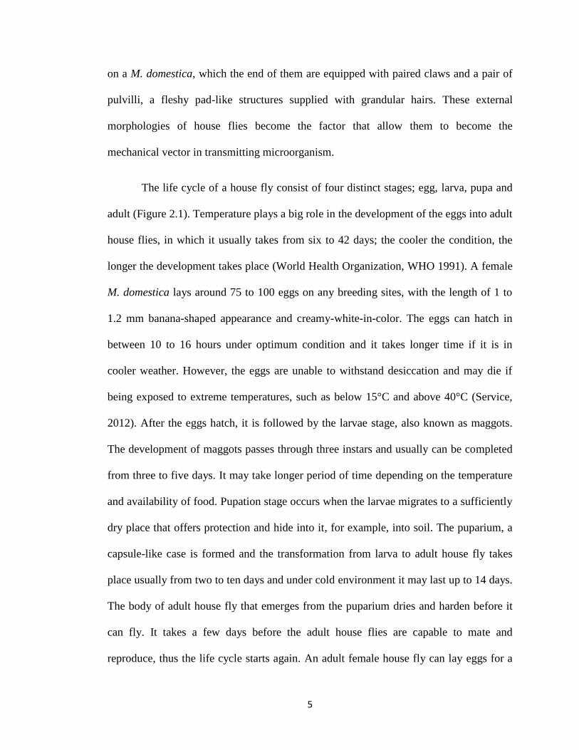
The life cycle of a house fly consist of four distinct stages; egg, larva, pupa and
adult (Figure 2.1). Temperature plays a big role in the development of the eggs into adult
house flies, in which it usually takes from six to 42 days; the cooler the condition, the
longer the development takes place (World Health Organization, WHO 1991). A female
M. domestica lays around 75 to 100 eggs on any breeding sites, with the length of 1 to
1.2 mm banana-shaped appearance and creamy-white-in-color. The eggs can hatch in
between 10 to 16 hours under optimum condition and it takes longer time if it is in
cooler weather. However, the eggs are unable to withstand desiccation and may die if
being exposed to extreme temperatures, such as below 15°C and above 40°C (Service,
2012). After the eggs hatch, it is followed by the larvae stage, also known as maggots.
The development of maggots passes through three instars and usually can be completed
from three to five days. It may take longer period of time depending on the temperature
and availability of food. Pupation stage occurs when the larvae migrates to a sufficiently
dry place that offers protection and hide into it, for example, into soil. The puparium, a
capsule-like case is formed and the transformation from larva to adult house fly takes
place usually from two to ten days and under cold environment it may last up to 14 days.
The body of adult house fly that emerges from the puparium dries and harden before it
can fly. It takes a few days before the adult house flies are capable to mate and
reproduce, thus the life cycle starts again. An adult female house fly can lay eggs for a

6
few times in a lifetime but as mentioned by WHO (1991), under natural conditions they
can rarely lay eggs more than five times.
Figure 2.1 Life cycle of house fly, Musca domestica (Source: Service, 2007)
7
2.1.2 Behavioral characteristics and habitat
House flies feed on all kinds of substances including food, garbage and excreta from
humans and animals – almost any organic material. It has been reported that their
feeding sites includes horse manure, cow manure, human excreta, fermenting vegetables
and fruits, garbage and kitchen wastes and commonly exposed human foods (Ahmadu et
al., 2016). The proboscis is the most important part for feeding as it is specially adapted
for sucking up fluid and semifluid foods (Service, 2007). The structure of proboscis
consists of a pair of oval-shaped labella, with very fine channels named pseudotracheae.
The fluids are sucked up through those channels. When a house fly feeds, the proboscis
will be extended downwards towards the food source and when it is not in use, the
proboscis is partially withdrawn into the head capsule.
The flies have different approaches of feeding which are affected by the physical
state of the food (Service, 2007). When the flies feed on fluids such as milk, the labella
are placed directly in contact with the food and it will be sucked up through small
openings in the pseudotracheae. If the feedings are sourced from semisolid state such as
vomit or animal dung, the labella are completely everted and the food is sucked up
directly into the food channel. As for foods in solid state, the flies have to wet it with
their saliva or perform regurgitation in the crop before ingestion occurs. The acts of
regurgitation and moistening the food are very conducive in the spreading of variety of
pathogens. Since house flies have the same feeding sites and breeding sites, another
factor that helps with the widespread of pathogens is when the flies landed on these
sites, the microorganisms are able to attach to the pulvilli on the flies’ legs. The sticky
hairs enable the house flies to adhere onto any surfaces, including smooth surface such
8
as glass and plastics. Not to mention, the exterior surfaces of the house flies are exposed
to the surroundings, making them having the potential to carry the pathogens. A study in
2007 by Yap et al. has shown that the wings of the house flies have the capability to
carry V. cholerae in droplets. However, they have concluded that the wings did not play
a significant role in mechanical transmission of non-adhering liquid medium because of
the low transfer rate of the bacteria to the wings and poor retention of bacteria on the
wings during normal house fly activities. Still, the exposure of the exterior body parts of
the flies towards sources of microorganisms in the surroundings helps in making them a
very important mechanical vector to introduce pathogens into human habitat.
In addition, the house flies distribution is greatly influenced by several factors
(WHO, 1991). This comprises by their reactions to light, temperature, humidity, surface
color and texture. During the day, they are mainly gathered near their feeding and
breeding sites, where mating and resting also take place. While during the night, they are
normally inactive so most of the time they are at their resting sites like ceilings. House
flies favor the temperature from 20°C to 25ºC, which is also the temperature of when
they reach the highest density of distribution in the environment. As mentioned by WHO
(1991), the preferred temperature for resting is between 35°C to 40°C. If the temperature
gets lower than 10°C or higher than 40°C, the flies distribution can be undetectable. All
of these details about the house flies may give an insight towards their role as carriers of
diseases and why they become a public health importance.

9
2.1.3 Public health importance
House flies can be an important nuisance towards people, especially when a large
number of them are involved. Plus, their presence is considered as a sign of unhygienic
conditions (WHO, 1991). According to Nayduch and Burrus (2016), house flies serve as
bridges between clean and unclean environments, moving freely between contaminated
materials such as waste to domestic and peridomestic environments, food and water
sources. Their synantrophic nature strengthened their potential as mechanical vector in
transmitting diseases to humans. Other than the two ways they are able to transmit the
microorganisms; through their exterior body surface and feeding method, there is
another one possible route of transmission, which is by defecation. According to Sasaki
et al. (2000), house flies defecate while feeding or resting, leaving specks and organisms
passing through their digestive system. This is a simple mechanical transfer of microbes
by a house fly that act as the vector, whose behavior places the contaminants from filth
sources onto new food or host source they visit (Holt et al., 2007)
Few studies in the past had shown the microorganisms associated with house
flies and the diseases they cause. According to Sukontason et al. (2000), the house flies
are able to carry microbes that can cause eye infection, such as trachoma, caused by
Chlamydia trachomatis. It was also considered as a potential carrier of bird flu virus,
which can cause harm towards humans’ health and livestocks industry (Nazari et al.,
2017). House flies can also transmit polio virus, which causes poliomyelitis and leads to
paralysis (WHO, 2017). In 1997, Grubel et al. had stated that house flies probably can
act as vectors in the transmission of Helicobacter pylori if they carry the bacterium and
contaminate human food. The house flies were also reported to act as carrier for
10
Campylobacter jejuni, Staphylococcus aureus, Pseudomonas aeruginosa and
Enterococcus fecalis (Bahrndoff et al., 2017). Most importantly, this insect has long
been associated with foodborne pathogens such as E. coli, V. cholerae, Salmonella spp.,
Shigella spp. and others (Barreiro et al., 2013) that may cause foodborne diseases such
as diarrhea, cholera, typhoid, shigellosis and other food poisoning conditions,
respectively. According to the study conducted by Chavasse in 1999, the use of effective
measures for controlling the population of house flies would reduce the prevalence of
gastrointestinal symptoms like diarrhea.
2.2 FOOD-BORNE DISEASES
Foodborne diseases (FBD) are responsible for a large burden of illnesses (morbidity)
and death (mortality) in both resource-rich and resource-poor countries (Kirk et al.,
2015). Globally, an estimated 2 million people died from diarrheal diseases in 2005;
approximately 70% of diarrheal diseases are foodborne. Diarrheal diseases alone, a
considerable proportion of which is foodborne, kills 1.8 million children every year
worldwide (WHO, 2007). There are more than 200 diseases that can be transmitted to
people from ingesting contaminated food biologically (with microorganisms) or the ones
contaminated with chemicals. Food contamination can occur at any stages of food
production and the source of contamination can arise from water, soil and air pollution,
or through lack of good food-handling practices such as unwashed hands before
handling food or usage of dirty utensils, plus low level of sanitation in the surroundings.
According to WHO (2011), the population in developing countries is more prone
to suffer from foodborne illnesses because of multiple reasons, including lack of access

11
to clean water for food preparation; inappropriate transportation and storage of foods;
and lack of awareness regarding safe and hygienic food practices. According to Kirk et
al. (2015) again, FBD usually presented with gastrointestinal symptoms such as stomach
cramps, diarrhea and vomiting. These are the most common symptoms for any FBD
cases. However, some FBD can have symptoms that affect other parts of the body or
causes serious sequelae. For example, certain infection with other E. coli strains can lead
to kidney failure (Nordstrom et al., 2013). Generally, symptoms of FBD are self-limiting
or mild. However, severe cases can occur towards the high risk groups. These include
infants, young children, the elderly and immunocompromised people (Fleury et al.,
2008). Even though FBD are prevalent (Hoffman et al., 2005), but the magnitude of
illness and associated deaths are not accurately reflected by the data available in both
developed and developing countries (Jahan, 2012). It can be considered as an unknown
burden, especially towards the specific causative agents of FBD cases that occurred.
This is mainly because the person involved does not seek medical attention whenever
they came up with FBD, leading to lack of laboratory-tested results that may be used to
fill the gaps of FBD prevalence data (WHO 2011).
Recognizing that contaminated food is an important cause of human disease,
estimates of disease burden of the various FBD has been sought to enable advocacy for
improved food safety and to assist governments to prioritize efforts for enhancing food
safety (Kirk et al., 2015). Many different diseases, including those due to bacteria,
viruses, parasites, chemicals, and prions, may be transmitted to humans by contaminated
food (Scallan et al., 2011). Thus, some of these factors that cause food contamination
12
can be related to house flies as the mechanical vector in transmitting pathogens, causing
cross-contamination between the filth to the food sources.
2.2.1 Food-borne diseases in Malaysia
In Malaysia, the reported food and waterborne diseases in 2009, such as cholera,
dysentery, typhoid and Hepatitis A were low, ranging from 0.14 to 1.07 cases per
100,000 people in a population (MOH, 2009). In contrast, food poisoning cases is on the
rise as evident by the incidence rate of 62.47 cases per 100,000 population in 2008 and
36.17 in 2009 according to the Ministry of Health (MOH 2009, 2010). In 2007, MOH
has stated that there are five food and waterborne diseases (FWBD) that must be
reported and notified under the Prevention and Control of Infectious Diseases Act 1988
(Act 342). These are cholera, typhoid/paratyphoid fevers, viral hepatitis A, food
poisoning and dysentery. FBD outbreaks are believed to be lead by locations of foods
consumed in institutions and other food services which were demonstrated by Olsen et
al. (2000). Most of the implicated food settings occurred in schools’ and academic
institutions’ food preparation premises and inappropriate food handling practices, meals
prepared too early and kept at ambient temperature until served and unhygienic practices
were the causes of food poisoning cases (Soon et al., 2011).
In Malaysia, the main contributing factor was identified as insanitary food
handling procedures and lack of cleanliness in food preparation establishments which
accounted for more than 50% of the poisoning episodes (MOH 2007). It is hereby
believed that there are association between the presence of house flies as the indicator of
unsanitary environment, them being the mechanical vector of food borne pathogens and
13
transmission of the microbes into human habitations. Hence, this study is conducted to
have preliminary observation of the variation of microorganisms carried by the house
flies in the vicinity of USM’s Health Campus, an academic institution in the state of
Kelantan. In addition, Kelantan always has higher incidence of typhoid compared to
other states in Malaysia (Baddam et al., 2012). There was also a cholera outbreak in this
state from November to December 2009 which V. cholerae O1 was isolated (Ang et al.,
2010). Thus, this study was performed to see whether these bacteria can be isolated from
the house flies collected, which may in fact strengthened the hypothesis of having this
insect associated with being the mechanical vector in transmitting food borne pathogen.
2.3 ENTERIC BACTERIA
Enterobacteriaceae is a family of bacteria that consist of around 53 genera with hundreds
of species. They are a group of facultatively anaerobic Gram negative rod bacteria and
they are distributed worldwide, which can also be found in soil and plants. Also, their
natural habitat is in the intestinal tract of human and animals (Kayser, 2005). However,
some of the bacteria in this family are facultative pathogenic. The nomenclature of the
Enterobacteriaceae is complicated and has been based on biochemical and antigenic
characteristics. The application of new technologies such as DNA hybridization has
resulted in numerous changes in classification of the Enterobacteriaceae (Hong et al.,
2007). Many new genera and species have been discovered, some unusual and rare, and
many species have also been reclassified to other genera. Characteristics of
Enterobacteriaceae also included having peritrichous flagella except bacteria from genus
Tatumella, Shigella and Klebsiella which are non-motile. Usually bacteria from this
family are divided by their ability of fermenting lactose.

14
There are a few of genera in Enterobacteriaceae that are considered medically
important according to the UK Standards for Microbiology Investigations (2015). First,
is from genus Citrobacter. Ten out of eleven species bacteria from this genus has been
isolated from clinical material (Euzeby, 2013). They may be found in the faeces of
humans and animals as part of the normal flora. Second is bacteria from genus
Enterobacter. They have the general characteristics of Klebsiella species but can be
differentiated because they are motile and ornithine positive. Enterobacter species are
widely distributed in nature. They are found in the soil, water, dairy products, and in the
intestines of animals as well as humans. Only 10 out of 26 species in this genus has been
isolated from clinical materials (Euzeby, 2013). Third, bacteria from genus Klebsiella
which there are four species related to humans and they include K. pneumoniae
subspecies pneumoniae, ozaenae, and rhinoscleromatis; K. oxytoca; K. granulomatis
and K. variicola. Bacteria from this genus are known to cause bacteremia and hepatic
infections. They also have been isolated from a number of unusual infections, including
endocarditis, peritonitis, acute cholecystitis, crepitant myonecrosis, pyomyositis,
necrotising fasciitis, psoas muscle abscess, fascial space infections of the head and neck,
and septic arthritis (Janda & Abbott, 2006).
Then, there is a genus well-known in this family, which is Proteus spp. Proteus
includes pathogens responsible for many human urinary tract infections (Guentzel,
1996). P. mirabilis is often found as a free-living organism in soil and water. It can
cause wound and urinary tract infections. Once attached to the urinary tract, P. mirabilis
can affect and infect kidney more commonly than E. coli. The other species well-known
under this genus is P. vulgaris. It occurs naturally in the intestines of humans and a wide
15
variety of animals, and in manure, soil, and polluted waters. However, this organism is
isolated less often in the laboratory and usually only targets immunosuppressed
individuals. Next are the most commonly known bacteria from this family, which are
Escherichia, Salmonella and Shigella. The genus Shigella consists of four species;
Shigella dysenteriae, Shigella flexneri, Shigella boydii, and Shigella sonnei (Euzeby,
2013). They can cause shigellosis, and dysentery which can be classified into different
level of symptoms severity; fever, stomach cramp and diarrhea with blood (Kaper et al.,
2004). Mostly, bacteria from Enterobacteriaceae can cause diarrheal symptoms in
human, only with different level of severity according to the risk groups affected.
Together with the family of Vibrionaceae, the genus of Vibrio is focused in this
study. This bacterium is a gram negative with comma shape and motile with a single
polar flagellum, giving the ‘darting’ motion. 10 species of bacteria from this genus have
cause gastrointestinal and extra-intestinal diseases in man; most importantly cholera,
caused by V. cholerae.

CHAPTER 3
METHODOLOGY
3.1 MATERIALS
Lists of chemicals and reagents, consumables as well as laboratory apparatus used in this
study are shown in Appendix A, B and C respectively. Preparation methods for media
are shown in Appendix D and E while chemical and reagents used in this study has been
prepared by the laboratory staff and also purchased ready-to-use packages or kits.
3.2 STUDY DESIGN
This is a cross-sectional study in which the house flies, Musca domestica from several
locations within the vicinity of Health Campus, USM were collected. The study was
done between the months of May to October 2018. Bacteria were isolated from the
house flies using conventional culture method for heterotrophic plate counts as well as
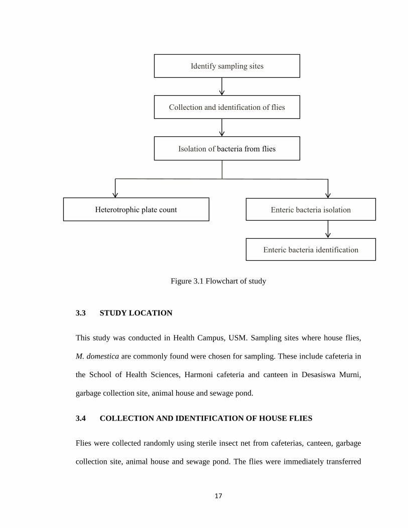
enteric bacteria isolation and identification. Figure 3.1 shows the flowchart of the study.
17
Figure 3.1 Flowchart of study
3.3 STUDY LOCATION
This study was conducted in Health Campus, USM. Sampling sites where house flies,
M. domestica are commonly found were chosen for sampling. These include cafeteria in
the School of Health Sciences, Harmoni cafeteria and canteen in Desasiswa Murni,
garbage collection site, animal house and sewage pond.
3.4 COLLECTION AND IDENTIFICATION OF HOUSE FLIES
Flies were collected randomly using sterile insect net from cafeterias, canteen, garbage
collection site, animal house and sewage pond. The flies were immediately transferred
Identify sampling sites
Collection and identification of flies
Isolation of bacteria from flies
Heterotrophic plate count Enteric bacteria isolation
Enteric bacteria identification
18
from the net into the individual sterile containers from each collection site. The flies
were then stored at 4°C overnight to immobilize them. Following immobilization, the
flies were aseptically handled and identified according to Nazni et al. (2011). Flies other
than M. domestica were not included for bacterial isolation in this study.
3.5 ISOLATION OF BACTERIA FROM M. domestica
About 1 g of M. domestica were added to 9 ml sterile buffered peptone water (BPW) and
allowed to stand for 15 minutes before vigorously agitated thrice at interval of one
minute to dislodge all bacteria from the flies. The flies’ body parts were removed from
the BPW and the solution was used in the determination of heterotrophic plate counts
and isolation and identification of enteric bacteria.
3.6 DETERMINATION OF HETEROTROPHIC BACTERIAL COUNTS
Serial dilutions were performed in which 1 ml of BPW solution from Section 3.5 was
diluted into a series of test tubes containing 9 ml of sterile BPW up to 1 x 10⁻⁶
. Aliquots
of 0.1 ml from each tube were inoculated onto the surface of nutrient agar (NA) using
spread plate technique for bacterial enumeration. The plates were incubated at 37°C for
24 hours. Discrete colonies that grew were counted to obtain the colony forming units
(CFU/ml), and the total number of bacterial load from the flies can be estimated (Davari
et al., 2010). After 24 hours incubation period, the plates are viewed with the bottom
side up and the number of colony on the surface and embedded within the agar were
counted. Ideally, only plates with the range of 30 to 300 colonies are used. Hence, any
plates with number of colony of more than 300 is considered as “too numerous too

19
count” (TNTC). To calculate the concentration of viable cells in the sample, the
following equation by Lammert (2007) was used:
number of colonies on plate
dilution of sample
For example, suppose that 30 colonies were counted on the 10-5
plate, and when these
numbers were used in the equation above it will be,
Thus, the viable cells load from the sample is estimated to be 3.0 107 CFU/ml. Since
the plate count method has at least 10% margin of error, only two significant numbers
appear in the final value.
3.7 IDENTIFICATION OF BACTERIA
MacConkey agar (MAC) and Salmonella-Shigella agar (SSA) were primarily used for
the detection and isolation of enteric bacteria. Thiosulphate citrate-bile salt sucrose agar
(TCBS) was used to detect and isolate Vibrio spp. Dilutions from previous BPW
solutions were plated onto each agar medium and the plates were incubated at 37°C for
24 hours. The bacterial colonies that emerged from the agar plates were differentiated
based on the colony morphology. The presumed different colonies were sub-cultured
onto new NA plates to obtain pure colonies. Then, the pure colonies were stored on
nutrient agar slant.
The pure isolates were identified at least up to genus level by conventional
identification method; microscopy and biochemical tests. First, Gram staining was
20
performed to determine bacterial morphology microscopically. Then, biochemical tests
were performed which involve oxidase, catalase, triple-sugar iron (TSI), sulphide-
indole-motility (SIM), indole, methyl-red and Voges-Proskauer (MRVP), citrate
utilization and urease test. For bacteria that were suspected as Vibrio spp., hanging-drop
method was performed to observe ‘darting’ movement, which is a distinct characteristic
of V. cholerae. Identification of bacteria was performed by referring to Bergey’s Manual
of Determinative Bacteriology (Holt et al., 1994).
3.7.1 Selective and/or differential media
As mention before, MAC, SSA and TCBS were used to primarily isolate bacteria as
these three types of agar are considered as selective and/or differential media. For MAC
(Oxoid UK), it was used to detect and differentiate among gram-negative enteric bacilli
based on their ability to grow on the medium and to ferment lactose. Lactose was added
in this media, making it differential to which bacteria that are able to ferment lactose and
produce acids will have pink to red colonies because of color change in neutral red, a pH
indicator in the medium. For non-lactose fermenting bacteria, they will produce a
colorless colony. Gram positive bacteria are unable to grow on MAC since their growth
are inhibited by the presence of crystal violet and bile salts, making MAC as a selective
media. Few examples of lactose-fermenter (LF) Enterobacteriaceae are from genus
Escherichia, Klebsiella, and Citrobacter, while for non-lactose fermenter (NLF) they
came from genus Salmonella, Shigella and Proteus. For SSA (Oxoid UK), it was used to
primarily detect the presence of Salmonella and Shigella from the house fly external
wash, based on the formation of colorless colony with black center from sulphide
production that may be the indicator of Salmonella typhimurium presence and colorless
21
colony without black center that is indicative of Shigella sonnei presence. SSA contains
some selective inhibitory components such as bile salts, thiosulphate and citrate that are
able to inhibit the growth of gram positive and coliform organisms. As for TCBS, it was
used to primarily detect the presence of Vibrio spp., specifically V. cholerae and V.
parahaemolyticus, based on their morphology on the agar plate, which are flat, circular,
yellow colonies and blue to green centered colonies, respectively (Hardy Diagnostics
USA). TCBS also able to inhibit the growth of gram positive bacteria and coliforms.
3.7.2 Biochemical tests
Before biochemical tests were performed, all the isolates were subcultured onto NA to
obtain pure colonies and identified microscopically using Gram stain method in order to
confirm the morphology of the bacteria which are gram negative rod for enteric bacteria
and gram negative with “comma” shape or slightly curved rod for Vibrio spp. Then, the
biochemical tests were performed based on the techniques in microbiology (Lammert,
2007) which include the followings:
3.7.2.1 Oxidase test
This test was performed to determine whether the bacteria have cytochrome oxidase, a
participant in electron transport chain during respiration. When electrons are added to
the oxidase reagent by cytochrome oxidase, a positive outcome will turns the reagent
color from dark blue to purple within few seconds. All Enterobacteriaceae are oxidase-
negative microorganism except for Plesiomonas shigelloides, which caused the reagent
to turn colorless. While for Vibrio spp., it will give positive result.

22
3.7.2.2 Triple Sugar Iron
This test was performed to differentiate among the Enterobacteriaceae as to their ability
to ferment glucose, lactose and sucrose, also their ability to produce H2S. This media
contain 1% of lactose and sucrose and 0.1% glucose. If any of these sugars are
fermented by the bacteria, it will produce organic acids as waste products, causing drop
in pH level. The acidic condition will turn the phenol red color that act as pH indicator
into yellow, while alkaline end product will produce red color of medium. TSI are
prepared in slant. The interpretations of this test are; entire tube (slant and butt) are
yellow, glucose and lactose and/or sucrose are fermented, if red slant and yellow butt,
only glucose are fermented, if entire tube is red then no sugar are fermented, indicating
bacteria presence are not from Enterobacteriaceae. Bubbles produced in agar indicates
byproduction of gas from fermentation and finally black precipitate formation indicates
that H2S was produced.
3.7.2.3 Sulphide-Indole-Motility
Through this test, three conditions of bacteria biochemical and morphology can be
observed which are their ability to produce H2S, their ability to split the amino acid
tryptophan into indole and pyruvic acid since some bacteria able to use tryptophan as the
energy source and producing indole as their byproduct, and their motility. E.coli is
positive in indole and form a ‘red-ring’ when Kovac’s reagent are added into the tube.
While black precipitate is produced if the bacteria is positive in H2S production, a
byproduct from the ability of bacteria to reduce thiosulfate, such as Proteus spp. The
motility of the bacteria can be observed through their growth along the stabbing line, in
23
which growth along the line means the bacteria is non-motile and if they grow spreaded
from the stabbing line they are motile.
3.7.2.4 Methyl Red
This test was performed in order to determine the ability of bacteria to ferment glucose
via mixed-acid fermentation. Methyl red is added to the medium as it act as the pH
indicator. Whenever the pH of the medium is 4.5 and below, methyl red reagent will
remain red in color, giving a positive result of acid formation from glucose mixed-acid
fermentation.
3.7.2.5 Citrate utilization
This test was performed to determine whether a bacterium is able to utilize citrate as the
sole source of carbon and energy. Bacteria that carry citrate permease have the ability to
convert citrate into pyruvate and carbon dioxide. Bromothymol blue is used as pH
indicator in this medium, which it changes color to blue, denoting a positive result from
alkaline pH changes. For example, E. coli does not have the ability to utilize citrate.
Thus, the green color of the medium remains the same after incubation over 48 hours.
3.7.2.6 Urease test
In this test, it was performed to determine the ability of bacteria to hydrolyze urea with
the presence of enzyme urease that rapidly degrade urea into carbon dioxide and
ammonia. Among Enterobacteriaceae, bacteria from genus Proteus (especially P.
mirabilis) are a rapid hydrolyzer of urea. This is shown when a urease-producing
bacterium hydrolyze urea in a medium, causing accumulation of ammonia that made the

24
medium more alkaline. This causes pH changes of phenol red that turns the color of
medium from yellow to bright pink.