Embed Size (px)
Citation preview
Kodak’s dental radiograph series
Radiation safety in dental radiography
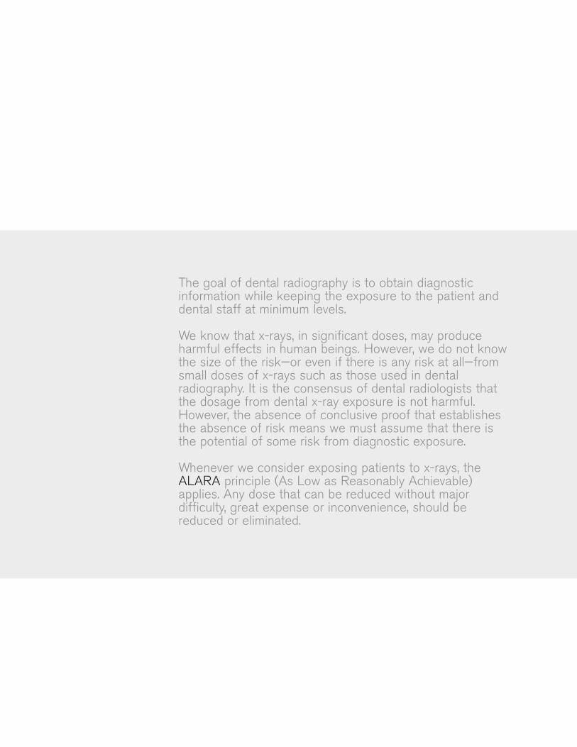
The goal of dental radiography is to obtain diagnosticinformation while keeping the exposure to the patient anddental staff at minimum levels.
We know that x-rays, in significant doses, may produceharmful effects in human beings. However, we do not knowthe size of the risk—or even if there is any risk at all—fromsmall doses of x-rays such as those used in dentalradiography. It is the consensus of dental radiologists thatthe dosage from dental x-ray exposure is not harmful.However, the absence of conclusive proof that establishesthe absence of risk means we must assume that there isthe potential of some risk from diagnostic exposure.
Whenever we consider exposing patients to x-rays, theALARA principle (As Low as Reasonably Achievable)applies. Any dose that can be reduced without majordifficulty, great expense or inconvenience, should bereduced or eliminated.
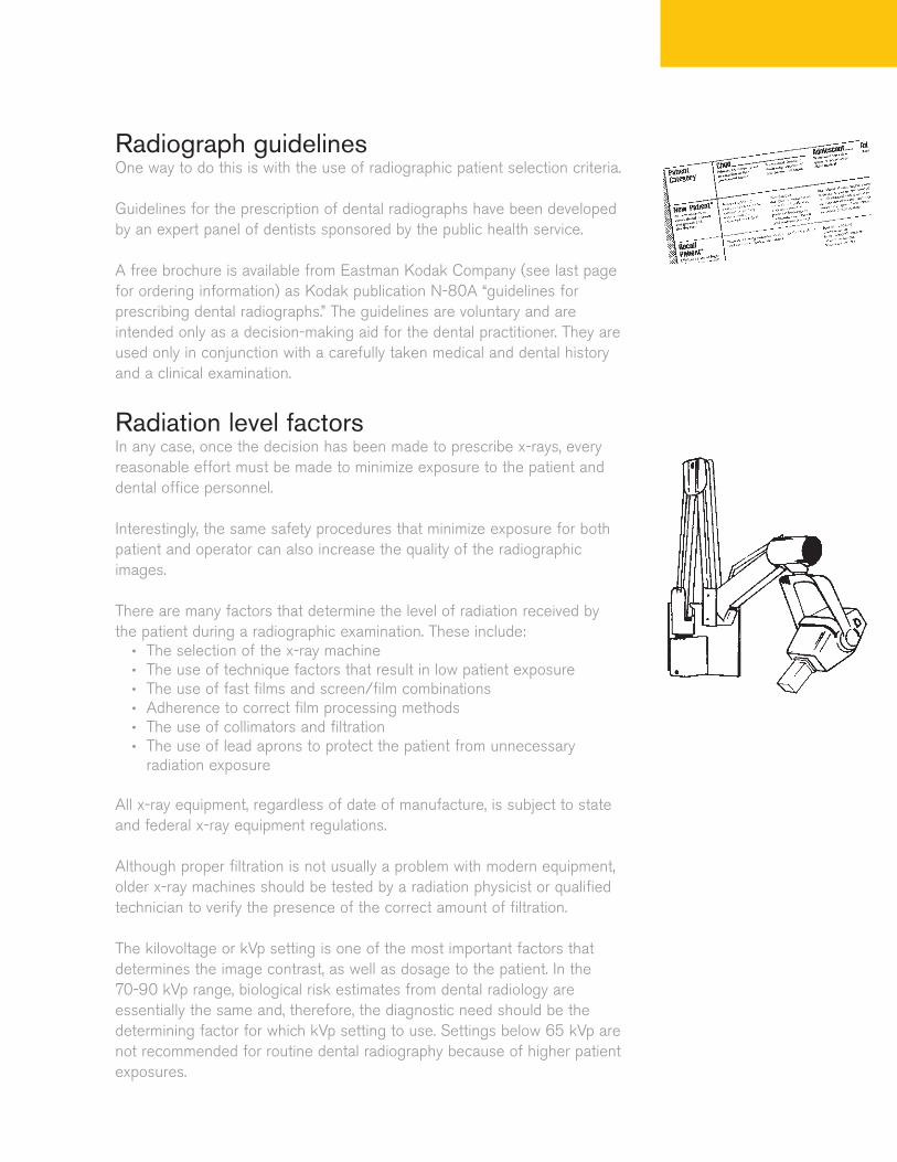
Radiograph guidelinesOne way to do this is with the use of radiographic patient selection criteria.
Guidelines for the prescription of dental radiographs have been developedby an expert panel of dentists sponsored by the public health service.
A free brochure is available from Eastman Kodak Company (see last pagefor ordering information) as Kodak publication N-80A “guidelines forprescribing dental radiographs.” The guidelines are voluntary and areintended only as a decision-making aid for the dental practitioner. They areused only in conjunction with a carefully taken medical and dental historyand a clinical examination.
Radiation level factorsIn any case, once the decision has been made to prescribe x-rays, everyreasonable effort must be made to minimize exposure to the patient anddental office personnel.
Interestingly, the same safety procedures that minimize exposure for bothpatient and operator can also increase the quality of the radiographicimages.
There are many factors that determine the level of radiation received bythe patient during a radiographic examination. These include:
• The selection of the x-ray machine• The use of technique factors that result in low patient exposure• The use of fast films and screen/film combinations• Adherence to correct film processing methods• The use of collimators and filtration• The use of lead aprons to protect the patient from unnecessary
radiation exposure
All x-ray equipment, regardless of date of manufacture, is subject to stateand federal x-ray equipment regulations.
Although proper filtration is not usually a problem with modern equipment,older x-ray machines should be tested by a radiation physicist or qualifiedtechnician to verify the presence of the correct amount of filtration.
The kilovoltage or kVp setting is one of the most important factors thatdetermines the image contrast, as well as dosage to the patient. In the 70-90 kVp range, biological risk estimates from dental radiology areessentially the same and, therefore, the diagnostic need should be thedetermining factor for which kVp setting to use. Settings below 65 kVp arenot recommended for routine dental radiography because of higher patientexposures.
ExposureEach of us is exposed to radiation from a variety of naturally occurringsources. Most exposure comes from breathing radon in the atmosphere.We’re exposed to cosmic radiation from space and terrestrial radiationfrom radioactive isotopes in stone and building materials. We’re evenexposed from internal sources. A radioisotope of potassium is found in allliving things.
In addition to these natural sources of radiation, we get small doses frommiscellaneous sources including tobacco, watches with luminous dials,color television, and others. A significant source of man-made radiation isdiagnostic exposure in the healing arts.
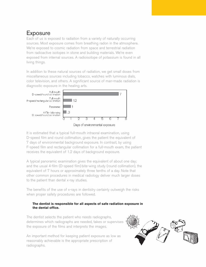
It is estimated that a typical full-mouth intraoral examination, using D-speed film and round collimation, gives the patient the equivalent of 7 days of environmental background exposure. In contrast, by using F-speed film and rectangular collimation for a full-mouth exam, the patientreceives the equivalent of 1.2 days of background exposure.
A typical panoramic examination gives the equivalent of about one day;and the usual 4-film (D-speed film) bite-wing study (round collimation), theequivalent of 7 hours or approximately three tenths of a day. Note thatother common procedures in medical radiology deliver much larger dosesto the patient than dental x-ray studies.
The benefits of the use of x-rays in dentistry certainly outweigh the riskswhen proper safety procedures are followed.
The dentist is responsible for all aspects of safe radiation exposure inthe dental office.
The dentist selects the patient who needs radiographs,determines which radiographs are needed, takes or supervisesthe exposure of the films and interprets the images.
An important method for keeping patient exposure as low asreasonably achievable is the appropriate prescription ofradiographs.
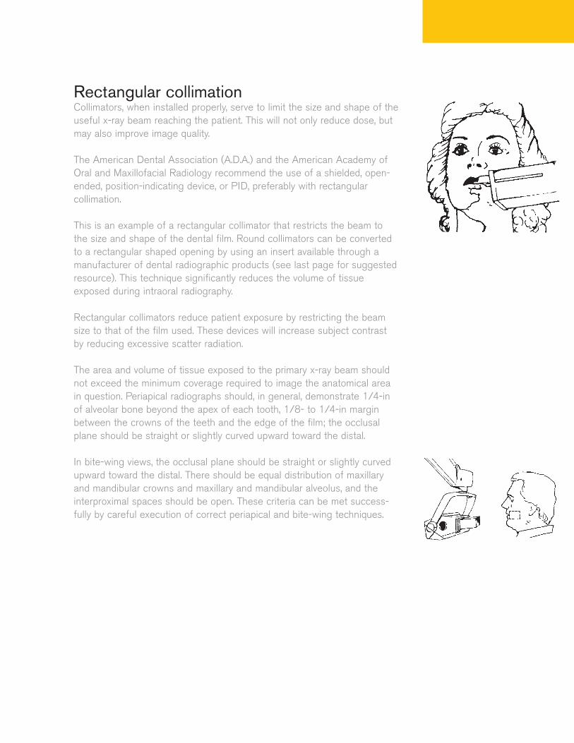
Rectangular collimationCollimators, when installed properly, serve to limit the size and shape of theuseful x-ray beam reaching the patient. This will not only reduce dose, butmay also improve image quality.
The American Dental Association (A.D.A.) and the American Academy ofOral and Maxillofacial Radiology recommend the use of a shielded, open-ended, position-indicating device, or PID, preferably with rectangularcollimation.
This is an example of a rectangular collimator that restricts the beam tothe size and shape of the dental film. Round collimators can be convertedto a rectangular shaped opening by using an insert available through amanufacturer of dental radiographic products (see last page for suggestedresource). This technique significantly reduces the volume of tissueexposed during intraoral radiography.
Rectangular collimators reduce patient exposure by restricting the beamsize to that of the film used. These devices will increase subject contrastby reducing excessive scatter radiation.
The area and volume of tissue exposed to the primary x-ray beam shouldnot exceed the minimum coverage required to image the anatomical areain question. Periapical radiographs should, in general, demonstrate 1/4-inof alveolar bone beyond the apex of each tooth, 1/8- to 1/4-in marginbetween the crowns of the teeth and the edge of the film; the occlusalplane should be straight or slightly curved upward toward the distal.
In bite-wing views, the occlusal plane should be straight or slightly curvedupward toward the distal. There should be equal distribution of maxillaryand mandibular crowns and maxillary and mandibular alveolus, and theinterproximal spaces should be open. These criteria can be met success-fully by careful execution of correct periapical and bite-wing techniques.
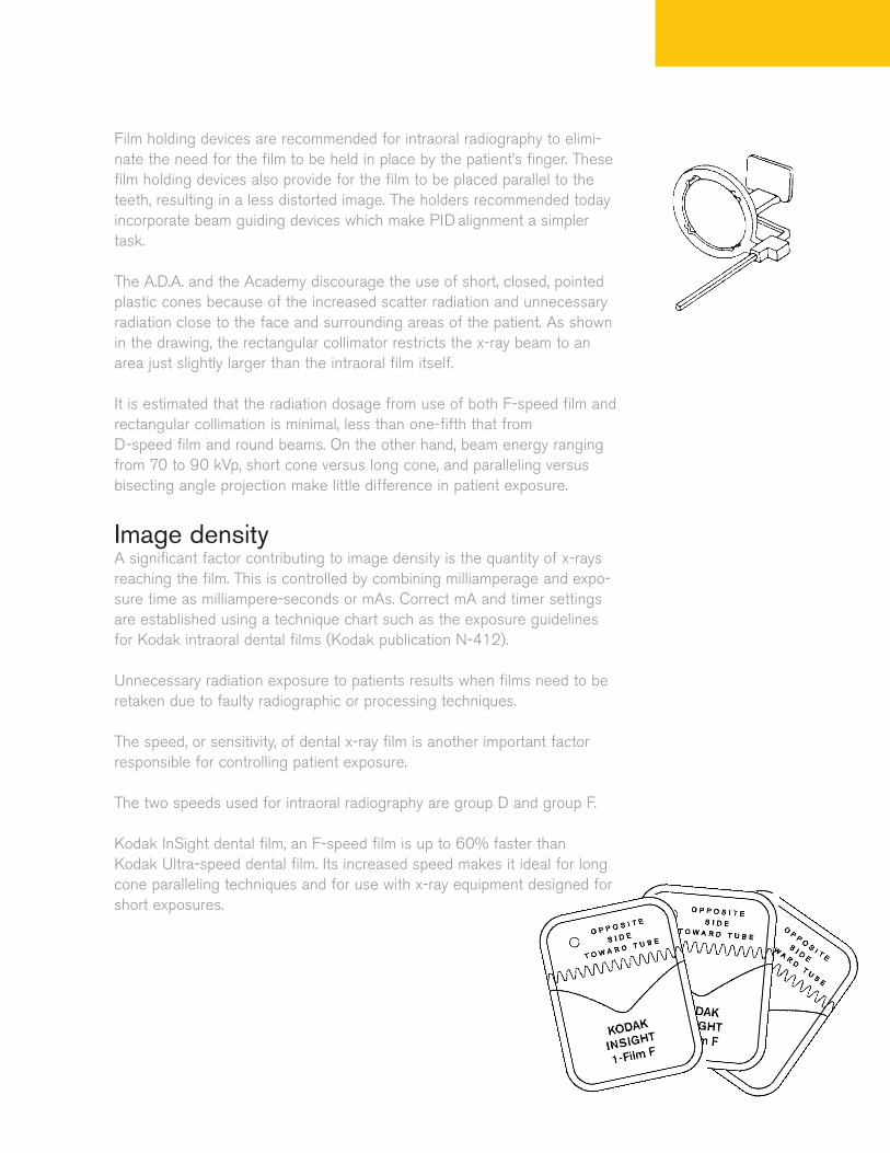
Film holding devices are recommended for intraoral radiography to elimi-nate the need for the film to be held in place by the patient’s finger. Thesefilm holding devices also provide for the film to be placed parallel to theteeth, resulting in a less distorted image. The holders recommended todayincorporate beam guiding devices which make PID alignment a simplertask.
The A.D.A. and the Academy discourage the use of short, closed, pointedplastic cones because of the increased scatter radiation and unnecessaryradiation close to the face and surrounding areas of the patient. As shownin the drawing, the rectangular collimator restricts the x-ray beam to anarea just slightly larger than the intraoral film itself.
It is estimated that the radiation dosage from use of both F-speed film andrectangular collimation is minimal, less than one-fifth that from D-speed film and round beams. On the other hand, beam energy rangingfrom 70 to 90 kVp, short cone versus long cone, and paralleling versusbisecting angle projection make little difference in patient exposure.
Image densityA significant factor contributing to image density is the quantity of x-raysreaching the film. This is controlled by combining milliamperage and expo-sure time as milliampere-seconds or mAs. Correct mA and timer settingsare established using a technique chart such as the exposure guidelinesfor Kodak intraoral dental films (Kodak publication N-412).
Unnecessary radiation exposure to patients results when films need to beretaken due to faulty radiographic or processing techniques.
The speed, or sensitivity, of dental x-ray film is another important factorresponsible for controlling patient exposure.
The two speeds used for intraoral radiography are group D and group F.
Kodak InSight dental film, an F-speed film is up to 60% faster than Kodak Ultra-speed dental film. Its increased speed makes it ideal for longcone paralleling techniques and for use with x-ray equipment designed forshort exposures.
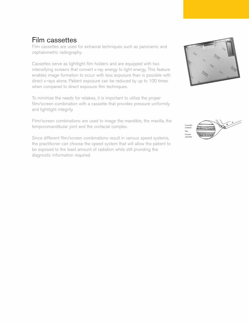
Film cassettesFilm cassettes are used for extraoral techniques such as panoramic andcephalometric radiography.
Cassettes serve as lighttight film holders and are equipped with twointensifying screens that convert x-ray energy to light energy. This featureenables image formation to occur with less exposure than is possible withdirect x-rays alone. Patient exposure can be reduced by up to 100 timeswhen compared to direct exposure film techniques.
To minimize the needs for retakes, it is important to utilize the properfilm/screen combination with a cassette that provides pressure uniformityand lighttight integrity.
Film/screen combinations are used to image the mandible, the maxilla, thetemporomandibular joint and the orofacial complex.
Since different film/screen combinations result in various speed systems,the practitioner can choose the speed system that will allow the patient tobe exposed to the least amount of radiation while still providing thediagnostic information required.
Minimal exposureKodak Lanex regular intensifying screens are used with a green sensitivefilm, such as Kodak T-Mat G/RA film. Exposures are usually one quarter toone half those needed with the earlier generation blue-light emittingphosphors. Beside dose reduction, the newer phosphors maintain excellentimage detail, help eliminate motion blur by the use of shorter exposuretimes, and produce less wear on the x-ray tube.
Kodak Ektavision extraoral imaging system also provides a similarexposure reduction, while providing a further increase in sharpnessresulting from new film and screen technology.
Proper exposure and processing of film is another factor in keepingexposure as low as reasonably achievable. Errors can result in the need foradditional radiographs and increased exposure.
Quality assurance is any systematic action to ensure that a dental officewill produce consistently high-quality images with minimal exposure topatients and personnel. When operators are presented with clearguidelines for quality assurance, patient exposure is dramatically reduced.
Besides diagnosis, films are used for insurance claims, teaching, patientreferrals and legal purposes. The use of duplicate radiographs reducespatient x-ray exposure because the need to re-expose patients iseliminated. When duplicate radiographs are needed, there are severalmethods available to produce them. Kodak offers duplicating film, in avariety of film sizes that range from size 2 to an 8 x 10-in size.
Kodak also offers two-film dental packets that contain two separateintraoral dental films together for producing two identical radiographs withone exposure. The two-film packet requires no adjusting or resetting ofequipment or additional exposure to the patient. The same double loadingtechnique can be done with extraoral film by using a combination of T-Mat H film and Lanex regular screens.
Exposure protectionEven though the thyroid gland may be out of the primary beam path whenrectangular collimation is used, exposure of that gland may be significantwhen the round positioning device is used. Lap aprons are available withthyroid collars attached. Separate thyroid collars are also available.
The most commonly used leaded aprons cover the entire chest and lap,effectively reducing scatter radiation reaching underlying tissues. Lapaprons should be used for all dental radiographic procedures.
The best way of limiting the possibility of occupational exposure is theestablishment of radiation safety procedures that are understood andfollowed by all personnel.
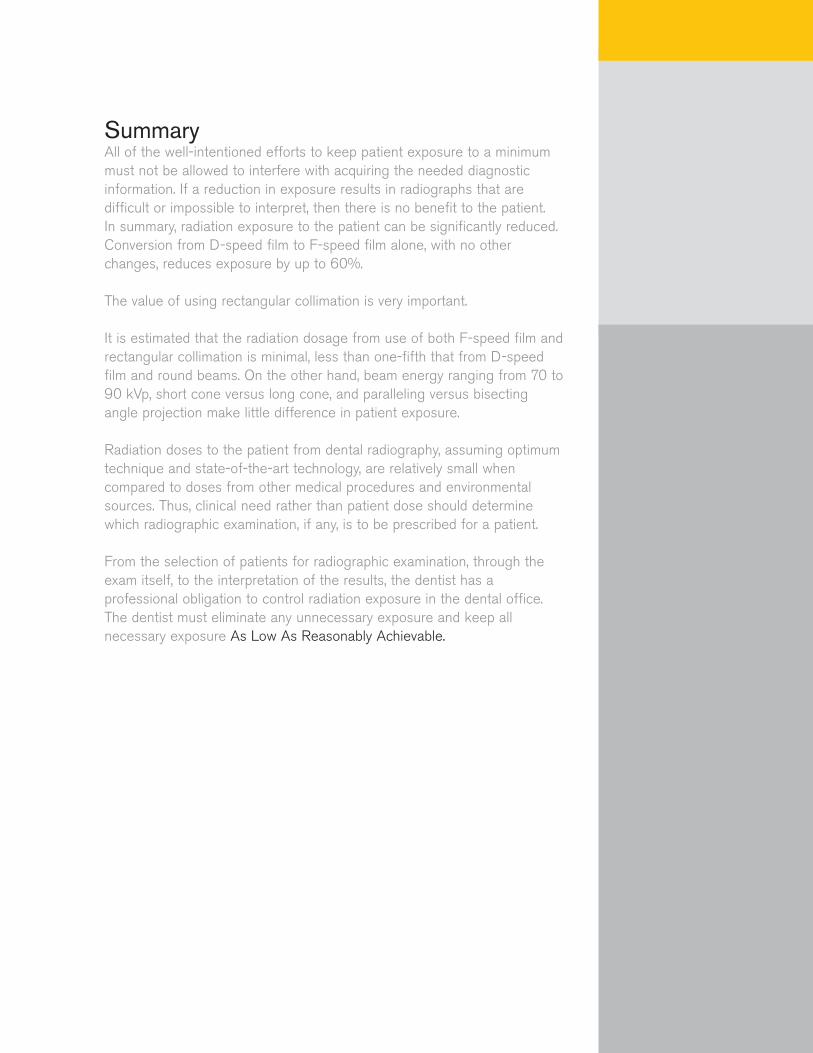
The most important factor in reducing personnel exposure to radiation isfor the operator to stand behind a radiation barrier during the exposure.This is usually accomplished by installing the exposure button in a locationoutside the dental operatory.
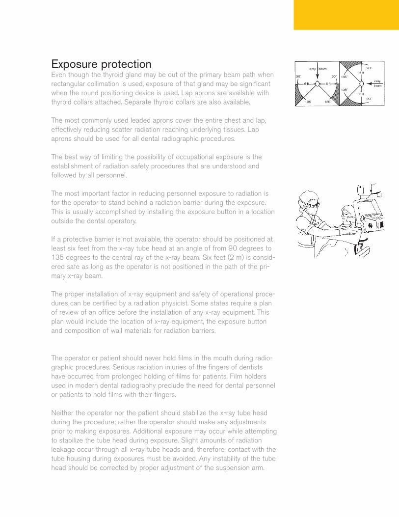
If a protective barrier is not available, the operator should be positioned atleast six feet from the x-ray tube head at an angle of from 90 degrees to135 degrees to the central ray of the x-ray beam. Six feet (2 m) is consid-ered safe as long as the operator is not positioned in the path of the pri-mary x-ray beam.
The proper installation of x-ray equipment and safety of operational proce-dures can be certified by a radiation physicist. Some states require a planof review of an office before the installation of any x-ray equipment. Thisplan would include the location of x-ray equipment, the exposure buttonand composition of wall materials for radiation barriers.
The operator or patient should never hold films in the mouth during radio-graphic procedures. Serious radiation injuries of the fingers of dentistshave occurred from prolonged holding of films for patients. Film holdersused in modern dental radiography preclude the need for dental personnelor patients to hold films with their fingers.
Neither the operator nor the patient should stabilize the x-ray tube headduring the procedure; rather the operator should make any adjustmentsprior to making exposures. Additional exposure may occur while attemptingto stabilize the tube head during exposure. Slight amounts of radiationleakage occur through all x-ray tube heads and, therefore, contact with thetube housing during exposures must be avoided. Any instability of the tubehead should be corrected by proper adjustment of the suspension arm.
SummaryAll of the well-intentioned efforts to keep patient exposure to a minimummust not be allowed to interfere with acquiring the needed diagnosticinformation. If a reduction in exposure results in radiographs that aredifficult or impossible to interpret, then there is no benefit to the patient.In summary, radiation exposure to the patient can be significantly reduced.Conversion from D-speed film to F-speed film alone, with no otherchanges, reduces exposure by up to 60%.
The value of using rectangular collimation is very important.
It is estimated that the radiation dosage from use of both F-speed film andrectangular collimation is minimal, less than one-fifth that from D-speedfilm and round beams. On the other hand, beam energy ranging from 70 to90 kVp, short cone versus long cone, and paralleling versus bisectingangle projection make little difference in patient exposure.
Radiation doses to the patient from dental radiography, assuming optimumtechnique and state-of-the-art technology, are relatively small whencompared to doses from other medical procedures and environmentalsources. Thus, clinical need rather than patient dose should determinewhich radiographic examination, if any, is to be prescribed for a patient.
From the selection of patients for radiographic examination, through theexam itself, to the interpretation of the results, the dentist has aprofessional obligation to control radiation exposure in the dental office.The dentist must eliminate any unnecessary exposure and keep allnecessary exposure As Low As Reasonably Achievable.
FOR MORE INFORMATION, VISIT:
www.kodak.com/go/dental
ReferencesTechnical Report of the Commission on Dental Products:Recommendations for radiographic procedures. International DentalJournal, June 1989.
White, S.C. 1992 Assessment of radiation risk from dental radiography.Dentomaxillofacial Radiology, 1992, V.21, Aug. 118-26.
The Benefits of Dental X-Ray Examinations,American Dental Association W138, 2000.
To obtain a free copy of any of the Kodak publications referred to in thispamphlet:
In the U.S.A., please call Kodak at 1-800-933-8031
Order the publication by name and code number.
Outside the U.S.A., all publication requests should be directed to thenearest Kodak company, or write:
Eastman Kodak CompanyDental BusinessHealth Imaging Division343 State StreetRochester, New York 14650-1122 U.S.A.
To obtain assistance in converting to the F-speed films mentioned in thispamphlet, or to obtain further information on other Kodak products or rec-ommendations:
• In the U.S.A., please call 1-800-933-8031• In Canada, please call 1-800-465-6325
© Eastman Kodak Company, 2004.Kodak, Ultra-speed, Ektavision, InSight, Lanex andT-Mat are trademarks of Eastman Kodak Company.
N-414 CAT No. 129 7019
Eastman Kodak CompanyDental BusinessHealth Imaging Division343 State StreetRochester, New York 14650-1122
FOR MORE INFORMATION, CALL: 1-800-933-8031 (U.S.A.)1-800-465-6325, EXT. 35147 (Canada) or visit www.kodak.com/go/dentalIf you are outside of the U.S.A. or Canada, please call your local Kodakcompany for more information.