Embed Size (px)
Citation preview
Myotrauma &
Muscle-Protective Ventilation:New Language for an Old Problem
Ewan C. Goligher MD PhDToronto General Hospital & Mount Sinai Hospital
Interdepartmental Division of Critical Care Medicine
University of Toronto

Disclosures
• Conflicts of Interest
– Equipment from GE
– Equipment and speakers’ honoraria from Getinge
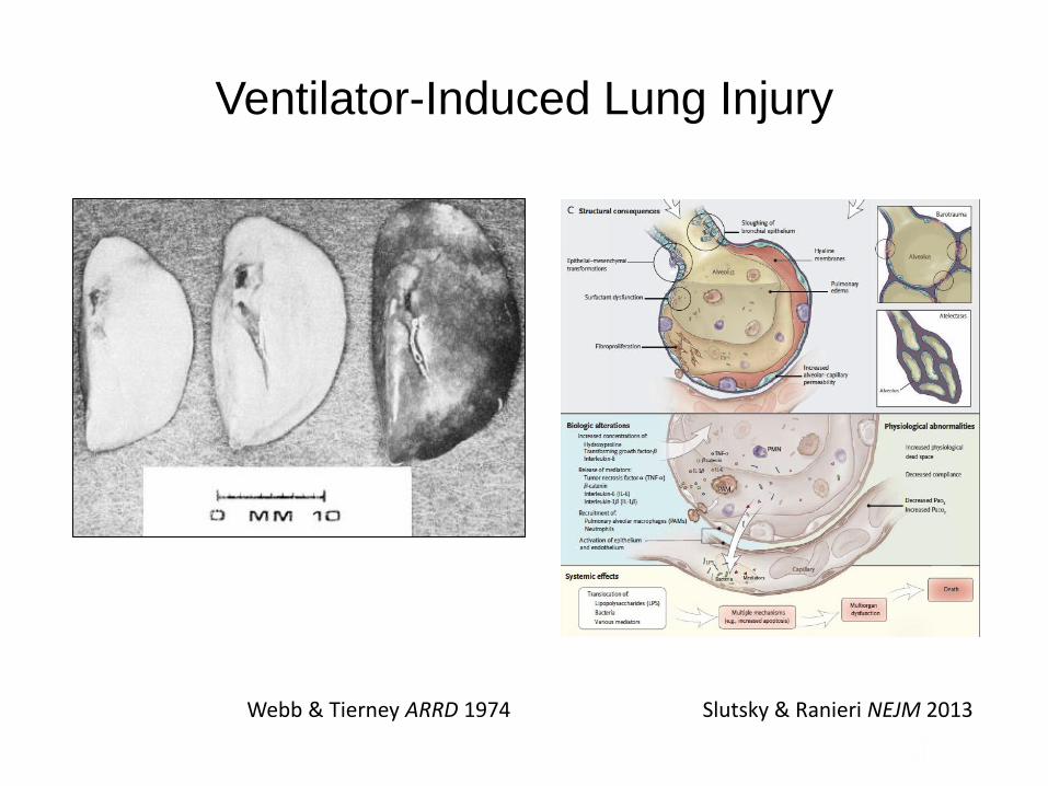
Ventilator-Induced Lung Injury
Webb & Tierney ARRD 1974 Slutsky & Ranieri NEJM 2013

Gauthier et al J Appl Phys 1994
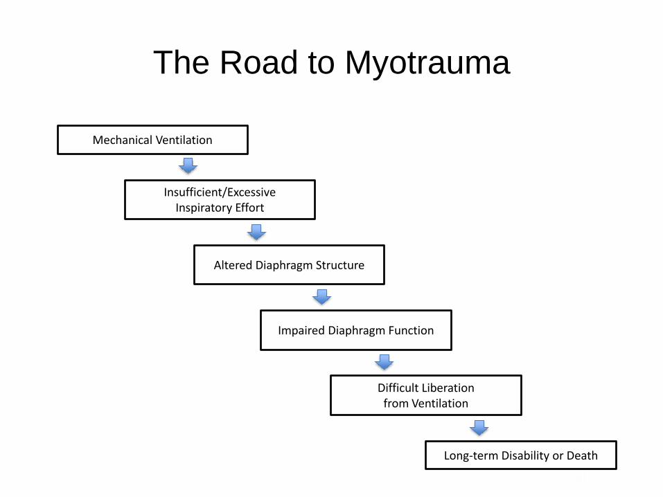
The Road to Myotrauma
Mechanical Ventilation
Insufficient/Excessive Inspiratory Effort
Altered Diaphragm Structure
Impaired Diaphragm Function
Difficult Liberation from Ventilation
Long-term Disability or Death

Myotrauma: 1980s

Myotrauma: 1990s
Baboon #1
Baboon #2
Baboon #3
Day 1 of MV
Day 11 of MV
Anzueto et al CCM 1997
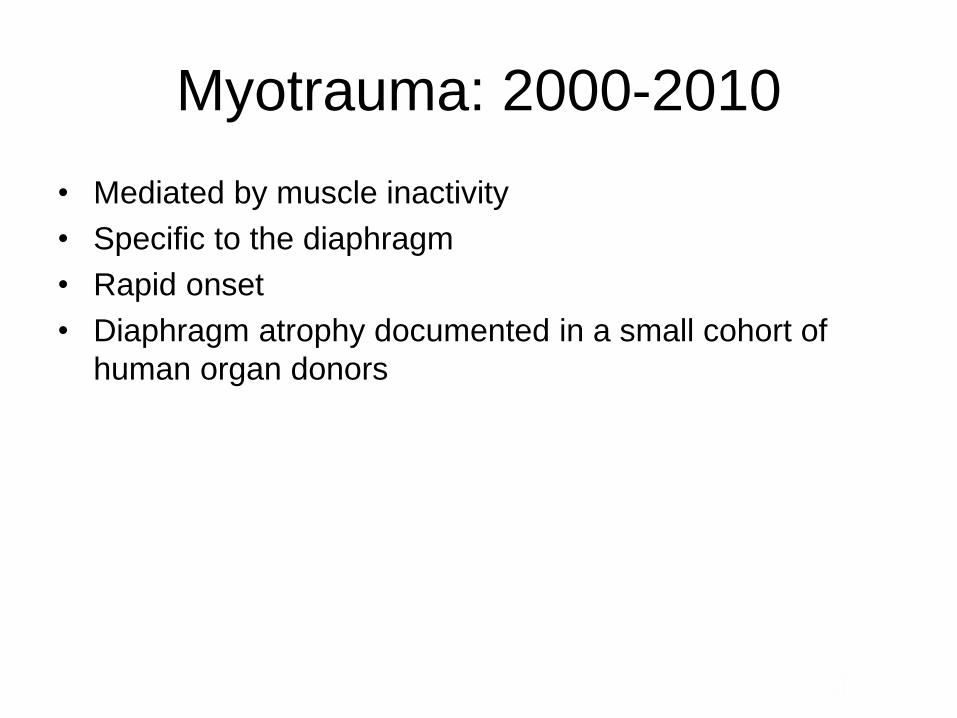
Myotrauma: 2000-2010
• Mediated by muscle inactivity
• Specific to the diaphragm
• Rapid onset
• Diaphragm atrophy documented in a small cohort of
human organ donors
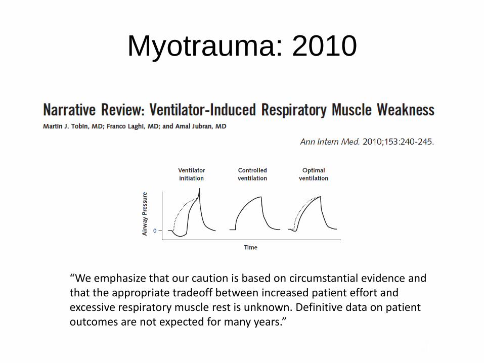
Myotrauma: 2010
“We emphasize that our caution is based on circumstantial evidence and that the appropriate tradeoff between increased patient effort and excessive respiratory muscle rest is unknown. Definitive data on patient outcomes are not expected for many years.”
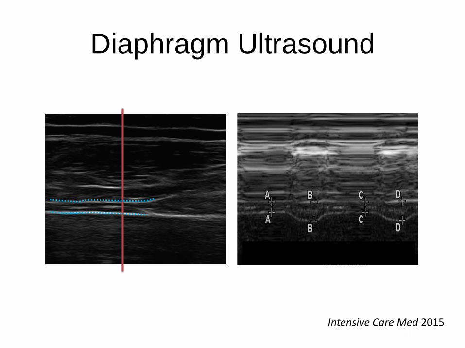
Diaphragm Ultrasound
A Window into Diaphragmatic Kinetics: Feasibility,
Precision, and Physiological Meaning of Ultrasound
Measurements of Diaphragm Thickness!Ewan C. Goligher1,2, Franco Laghi3, Brian P. Kavanagh1, !
Gordon D. Rubenfeld1,4, Martin J. Tobin3, Niall D. Ferguson1,2 !1University of Toronto - Toronto ON, 2University Health Network - Toronto ON, !
3Loyola University Stritch School of Medicine - Chicago IL 4Sunnybrook Health Sciences Center - Toronto ON !
Background++
Methods
Results
Conclusions
Abstract!
deemedinvalid if thetwoclear bright parallel linesof thepleural and peritoneal membranes were not plainlyidentified at each moment of the respiratory cycle.Ultrasonographic recordings were stored on compactdisks, and a subsequent computer-assisted quantitativeanalysis was performed by a trained investigator whowas unaware of the ventilatory condition. The measure-ments included diaphragm thickness at end-expiration(TEE) and at end-inspiration (TEI). When airway pressurecouldnot bedisplayedonthescreenof theEcho-Dopplermachine to match theultrasound tracings to the respira-tory cycle(duringSB), TEE wasmeasured just beforethethickening start and TEI was measured at maximalthickening. Measurementswereaveraged out of threeormore consecutive breaths on the last valid image recor-ded at the end of each period. The thickening fraction(TF) wascalculated as(TEI - TEE)/TEE andexpressed asapercentage (Fig. 1c).
Flow and pressuremeasurements
Flow was measured using a Fleisch N°2 pneumotacho-graph (Fleisch, Lausanne, Switzerland) connected to adifferential (±2 cmH2O) pressure transducer (MP45,Validyne, Northridge, CA) and placed between the facemask and the ventilator Y connector. Airway openingpressurewasmeasuredbetween theventilator circuit andthepneumotachographusingapressuretransducer (MP45,±100 cmH2O). Oesophageal (Pes) and gastric pressures(Pga) were measured using a double-balloon catheter(Marquat, Boissy Saint Leger, France) as previouslydescribed with appropriate placement checked and arte-factseliminated (seeSupplementary Material for details)[20, 21]. Transdiaphragmatic pressure(Pdi) wasobtainedby electronic subtraction of the Pes signal from thePgasignal over at least tenconsecutivebreathsselectedatthe end of the pressure and flow recordings. The
A
B
C
TEI, thickness at end inspiration; TEE, thickness at end expiration.
Fig. 1 Probeplacement toexplore thediaphragmin thezoneof apposition (a), with theultrasonographic view of thenormal diaphragmin thezoneof apposition (b) andillustration of themeasurementof diaphragmthicknessat end-inspiration and end-expirationin TM mode(c). TEI thicknessat end-inspiration, TEE thicknessat end-expiration
798
Cur r en t Concept s
n engl j med 366;10 nejm.org march 8, 2012 939
Tr eat men t
The treatment of patients with diaphragmatic dys-
function depends on the cause and on the pres-
ence or absence of symptoms and nocturnal hy-
poventilation. Examples of treatable causes of
diaphragmatic dysfunction include myopathies re-
lated to metabolic disturbances such as hypoka-
lemia, hypomagnesemia, hypocalcemia, and hy-
pophosphatemia. Correction of electrolyte and
hormonal imbalances and avoidance of neuro-
pathic or neuromuscular blocking agents can re-
store strength in the diaphragm. Myopathies due to
parasitic infection (e.g., trichinosis) may respond
to appropriate antimicrobial agents.61 Idiopathic
diaphragmatic paralysis or paralysis due to neu-
ralgic amyotrophy may improve spontaneously.13
When diaphragmatic dysfunction persists or pro-
gresses, ventilatory support, ranging from noctur-
nal to continuous, may be needed. The need for
ventilatory support may be temporary, as in cases
of diaphragmatic paralysis after cardiac surgery, or
it may be permanent, as in cases of progressive
neuromuscular diseases. The generally accepted in-
dications for initiating nocturnal noninvasive venti-
lation in patients with symptoms include a partial
pressure of carbon dioxide of 45 mm Hg or high-
er in the arterial blood in the daytime, oxygen
saturation of 88% or less for 5 consecutive min-
utes at night, or progressive neuromuscular dis-
ease with a maximal static inspiratory pressure of
less than 60 cm of water or a forced vital capacity
of less than 50% of the predicted value.62 Most
patients with neuromuscular disease will eventu-
ally require mechanical ventilation, whether it is
provided by invasive means (tracheostomy or endo-
tracheal tube) or noninvasive means (nasal can-
nula or face mask).
Plication of the diaphragm is a procedure in
which the flaccid hemidiaphragm is made taut by
oversewing the membranous central tendon and
the muscular components of the diaphragm. The
indications and timing for this procedure are not
fully defined, given that most studies are retro-
spective and uncontrolled, but it may be offered to
patients with unilateral diaphragmatic paralysis
C D
A B
Normal
Diaphragm
Paralyzed
Diaphragm
DiaphragmDiaphragm
Chest Wall
Chest Wall
Lung
Lung Lung
Liver Liver
Liver
Pleura
PleuraPleura
Peritoneum
PeritoneumPeritoneum
Diaphragm
Diaphragm
Figure 5. Ultrasonographic Images of Normal and Paralyzed Diaphragms.
Panels A and B show the end-expiration and end-inspiration stages, respectively, in a normal diaphragm. Panels C
and D show the end-expiration and end-inspiration stages, respectively, in a paralyzed diaphragm. During inspiration,
the normal diaphragm thickens, whereas the paralyzed diaphragm does not thicken.
The New England Journal of Medicine
Downloaded from nejm.org at UNIVERSITY OF TORONTO on March 8, 2012. For personal use only. No other uses without permission.
Copyright © 2012 Massachusetts Medical Society. All rights reserved.
B-mode!
Zone of Apposition Ultrasound!
M-mode!
Experimental Setup and Inspiratory Maneuvers!
Tidal breathing!
Threshold-loaded breathing (20 cm H2O)!
Maximal inspiratory transdiaphragmatic
pressure effort !
Rest @ FRC!
Inhale to target!
Relax & hold @ target!
25% IC! 50% IC! 75% IC! IC!
deemedinvalid if thetwoclear bright parallel linesof thepleural and peritoneal membranes were not plainlyidentified at each moment of the respiratory cycle.Ultrasonographic recordings were stored on compactdisks, and a subsequent computer-assisted quantitativeanalysis was performed by a trained investigator whowas unaware of the ventilatory condition. The measure-ments included diaphragm thickness at end-expiration(TEE) and at end-inspiration (TEI). When airway pressurecouldnot bedisplayedonthescreenof theEcho-Dopplermachine to match the ultrasound tracings to the respira-tory cycle(during SB), TEE wasmeasured just beforethethickening start and TEI was measured at maximalthickening. Measurements wereaveraged out of threeormore consecutive breaths on the last valid image recor-ded at the end of each period. The thickening fraction(TF) wascalculated as(TEI - TEE)/TEE andexpressed asapercentage (Fig. 1c).
Flow and pressuremeasurements
Flow was measured using a Fleisch N°2 pneumotacho-graph (Fleisch, Lausanne, Switzerland) connected to adifferential (±2 cmH2O) pressure transducer (MP45,Validyne, Northridge, CA) and placed between the facemask and the ventilator Y connector. Airway openingpressurewasmeasured between theventilator circuit andthepneumotachographusingapressuretransducer (MP45,±100 cmH2O). Oesophageal (Pes) and gastric pressures(Pga) were measured using a double-balloon catheter(Marquat, Boissy Saint Leger, France) as previouslydescribed with appropriate placement checked and arte-factseliminated (seeSupplementary Material for details)[20, 21]. Transdiaphragmatic pressure(Pdi) wasobtainedby electronic subtraction of the Pes signal from thePgasignal over at least tenconsecutivebreathsselectedatthe end of the pressure and flow recordings. The
A
B
C
TEI, thickness at end inspiration; TEE, thickness at end expiration.
Fig. 1 Probeplacement toexplore thediaphragm in thezoneof apposition (a), with theultrasonographic view of thenormal diaphragm in thezoneof apposition (b) andillustration of themeasurementof diaphragm thicknessat end-inspiration and end-expirationin TM mode(c). TEI thicknessat end-inspiration, TEE thicknessat end-expiration
798
Pga!
EAdi!
Pes!
Spirometer!
Threshold load valve!
5 healthy subjects!
Preliminary Findings from Sonographic Data!
Inspiratory+Thickening+Frac3on+
Diaphragm thickening fraction at increasing levels of inspiratory effort. Means and standard deviations of sonographic thickening fraction are shown (p<0.01 for
difference in means). Note that thickening appears greater at inspiratory capacity than during Pdi,max maneuver (p=0.24 for difference)!
Thickening+Frac3on+
Inspiratory+Volume+(%+Inspiratory+Capacity)+
Relationship between diaphragm thickness and inspiratory effort vs. lung volume. Peak diaphragm thickening (blue dots) and post-inspiratory
resting diaphragm thickening (red dots) are displayed as a function of lung volume (%inspiratory capacity). Linear regressions with slopes and
r2 values are shown. At low lung volumes, diaphragm thickening predominantly represents muscular effort, but at higher lung volumes,
diaphragm thickening indicates both increasing effort and increasing lung
volume (p<0.001 for difference in relative proportions with increasing lung volume). !
Intensive Care Med 2015
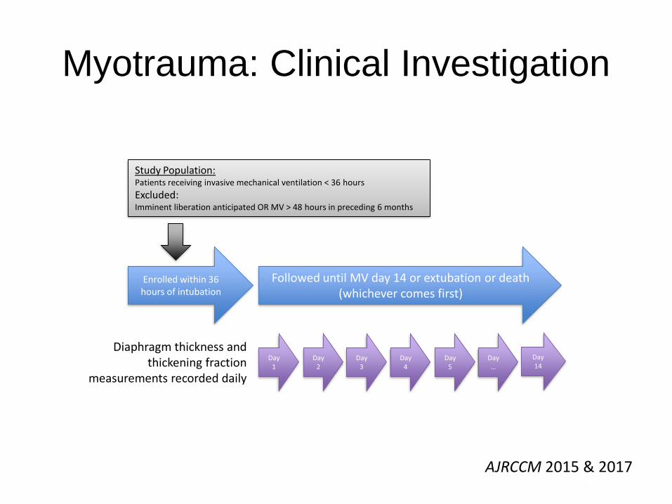
Myotrauma: Clinical Investigation
Enrolled within 36 hours of intubation
Day 1
Day 2
Day 3
Day 4
Day 5
Day …
Day 14
Diaphragm thickness and thickening fraction
measurements recorded daily
Followed until MV day 14 or extubation or death (whichever comes first)
Study Population:Patients receiving invasive mechanical ventilation < 36 hours
Excluded:Imminent liberation anticipated OR MV > 48 hours in preceding 6 months
AJRCCM 2015 & 2017

Myotrauma: Clinical Investigation
47
47
41
3934
30
27
21
4744
3228
22
18 15 1313
13
11
11
11
11
11
10
-30%
-20%
-10%
0%
+10%
+20%
+30%
1 2 3 4 5 6 7 8
Day of Study
Cha
nge
in
dia
ph
ragm
th
ickne
ss o
ve
r tim
e (
% o
f b
ase
line
)
Group: Diaphragm Thickness Change
>10% loss on or before day 8
<10% change on or before day 8
>10% gain on or before day 8
AJRCCM 2017

Myotrauma: Clinical Investigation
0
2
4
6
80%
20%
40%
60%
80%
+10%
0%
-10%
-20%
Duration of Ventilation (Days) Diaphra
gm Contra
ctile
Act
ivity
(Tid
al Thick
enin
g Fra
ctio
n)
Ch
an
ge
in
Dia
ph
rag
m T
hic
kn
ess O
ve
r T
ime
(%
of
Ba
se
line
)
-4
-2
0
2
4
6
0 20 40 60 80
Daily diaphragm thickening fraction (%)
Ra
te o
f ch
an
ge
in
dia
ph
rag
m t
hic
kn
ess (
%/d
ay)
-4
-2
0
2
4
6
0 5 10 15 20
Mean daily diaphragm electrical activity (mV)
Ra
te o
f cha
ng
e in
dia
ph
rag
m th
ickne
ss (
%/d
ay)
-6
-4
-2
0
2
Controlled Partially assisted
Mode of ventilation
Ra
te o
f cha
ng
e in
dia
ph
rag
m th
ickne
ss (
%/d
ay)
-6
-4
-2
0
2
£ 10 cm H2O >10 cm H2O
Applied driving pressure in partially assisted modes
Ra
te o
f cha
ng
e in
dia
ph
rag
m t
hic
kne
ss (
%/d
ay)
AJRCCM 2015 & 2017
Ultrasound: Thickening Fraction Diaphragm electrical activity

Myotrauma: also a Load-Induced Injury?
Control
LPS + MV
LPS
Peake et al JAP 2015
▲ = Swelling/thickness
Jiang, Reid, Road AJRCCM 1997

Myotrauma: Clinical Outcomes
VariableDecreased
thickness (>10%)Unchanged thickness
Increased thickness(>10%)
N 78 (41%) 66 (35%) 47 (24%)
Severity of illness
Cause of illness
Baseline ventilator settings
Timing of classification
Fluid balance at 72 hours
Baseline diaphragm thickness
2.6 mm 2.3 mm 2.0 mm
AJRCCM 2017
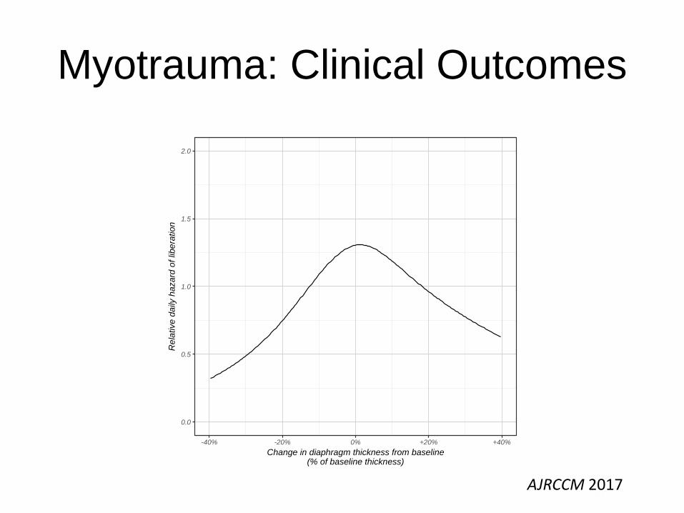
Myotrauma: Clinical Outcomes
0.0
0.5
1.0
1.5
2.0
-40% -20% 0% +20% +40%
Change in diaphragm thickness from baseline(% of baseline thickness)
Re
lative d
aily
haza
rd o
f lib
era
tion
AJRCCM 2017

Myotrauma: Clinical Outcomes
*
*
0%
20%
40%
60%
80%
100%
0 7 14 21
Duration of follow-up (days)
Cum
ula
tive incid
en
ce
of
libe
ration
or
de
ath
Initial change in diaphragm thickness on or before day 7 of ventilation
No change from baseline (n=66)
>10% decrease (n=78)
>10% increase (n=47)
Status at disconnection from ventilator
Alive
Dead
AJRCCM 2017
Myotrauma: Clinical Outcomes
33 51 72 32 2133 51 72 32 210
25
50
75
100
>20% decrease 10-20% decrease <10% change 10-20% increase >20% increase
Change in diaphragm thickness
Fre
que
ncy o
f a
t le
ast 1
co
mp
lication
(%
)
Complication type
Death in hospital
Reintubation or tracheostomy or MV > 14 days
ICU Length-of-Stay Complications (Reintubation, Tracheostomy, Prolonged MV, Death)
0
10
20
30
>20% decrease 10-20% decrease <10% change 10-20% increase >20% increase
Early change in diaphragm thickness
Dura
tio
n o
f IC
U s
tay (
days)
AJRCCM 2017

The Road to Myotrauma
Mechanical Ventilation
Insufficient/Excessive Inspiratory Effort
Altered Diaphragm Structure
Impaired Diaphragm Function
Difficult Liberation from Ventilation
Long-term Disability or Death
Muscle-Protective Ventilation
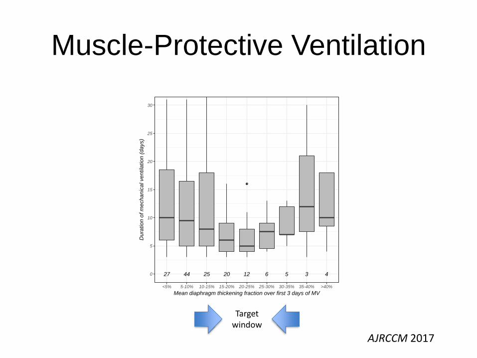
Muscle-Protective Ventilation
27 44 25 20 12 6 5 3 40
5
10
15
20
25
30
<5% 5-10% 10-15% 15-20% 20-25% 25-30% 30-35% 35-40% >40%
Mean diaphragm thickening fraction over first 3 days of MV
Du
ratio
n o
f m
ech
an
ica
l ve
ntila
tio
n (
da
ys)
AJRCCM 2017
Target window
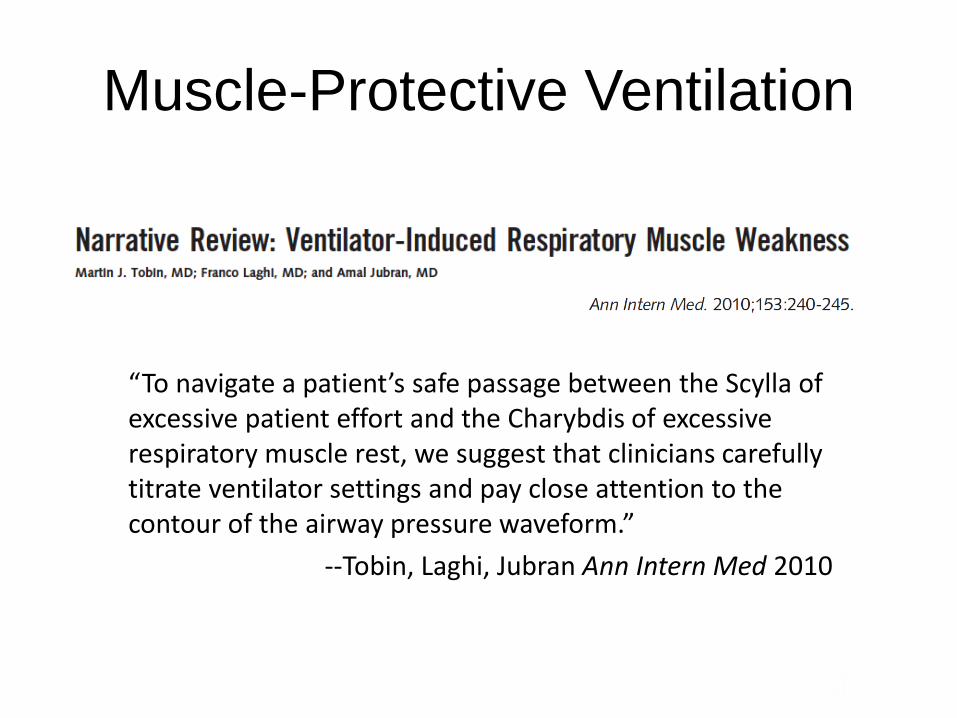
Muscle-Protective Ventilation
“To navigate a patient’s safe passage between the Scylla of excessive patient effort and the Charybdis of excessive respiratory muscle rest, we suggest that clinicians carefully titrate ventilator settings and pay close attention to the contour of the airway pressure waveform.”
--Tobin, Laghi, Jubran Ann Intern Med 2010
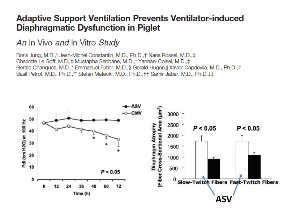
ASV

Myotrauma in 2017
• What can clinicians do now?
1. Use ‘myotrauma’ in your clinical lexicon
2. Monitor and consider inspiratory effort in ventilated patients
3. Develop skill in diaphragm ultrasound and esophageal
manometry
• Future directions
1. Further characterization of underlying biology
2. Impact on long-term functional outcomes
3. Muscle-protective ventilation strategies
4. Targeted rehabilitation
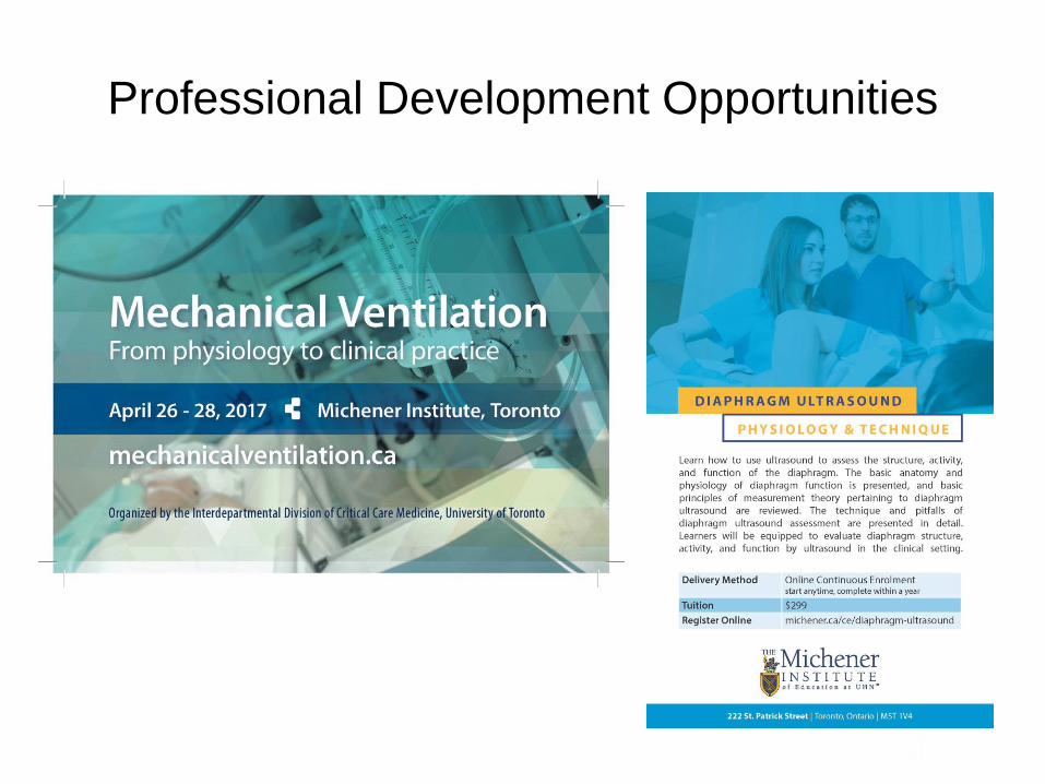
Professional Development Opportunities

Acknowledgments
• Mentorship
– Dr. Niall Ferguson
– Dr. Laurent Brochard
– Dr. Brian Kavanagh
– Dr. Gordon Rubenfeld
– Dr. Eddy Fan
– Dr. Margaret Herridge
– Dr. Darlene Reid
– Dr. Art Slutsky
• Research Team
– Stefannie Vorona
– Dr. Martin Dres
– Dr. Michael Sklar
– Dr. Cristian Urrea
– Alistair Murray
– Debbie Brace
– Ashley Lanys
– Dr. Nuttapol Rittayamai