Embed Size (px)
Citation preview
BioMed CentralJournal of Biomedical Science
ss
Open AcceResearchMyofibril-Inducing RNA (MIR) is essential for tropomyosin expression and myofibrillogenesis in axolotl heartsChi Zhang1, Pingping Jia1, Xupei Huang1, Gian Franco Sferrazza1, Gagani Athauda1, Mohan P Achary2, Jikui Wang3, Sharon L Lemanski3, Dipak K Dube4 and Larry F Lemanski*3,5Address: 1Department of Biomedical Science, Florida Atlantic University, Boca Raton, FL 33431, USA, 2Department of Radiation Oncology, Temple University School of Medicine, Philadelphia, PA 19140, USA, 3Department of Anatomy and Cell Biology and The Cardiovascular Research Center, Temple University, Philadelphia, PA 19140, USA, 4Department of Medicine, Upstate Medical University, Syracuse, NY 13210, USA and 5Department of Biological and Environmental Sciences, Texas A&M University-Commerce, Commerce, TX 75429-3011, USA
Email: Chi Zhang - [email protected]; Pingping Jia - [email protected]; Xupei Huang - [email protected]; Gian Franco Sferrazza - [email protected]; Gagani Athauda - [email protected]; Mohan P Achary - [email protected]; Jikui Wang - [email protected]; Sharon L Lemanski - [email protected]; Dipak K Dube - [email protected]; Larry F Lemanski* - [email protected]
* Corresponding author
AbstractThe Mexican axolotl, Ambystoma mexicanum, carries the naturally-occurring recessive mutant gene'c' that results in a failure of homozygous (c/c) embryos to form hearts that beat because of anabsence of organized myofibrils. Our previous studies have shown that a noncoding RNA,Myofibril-Inducing RNA (MIR), is capable of promoting myofibrillogenesis and heart beating in themutant (c/c) axolotls. The present study demonstrates that the MIR gene is essential fortropomyosin (TM) expression in axolotl hearts during development. Gene expression studies showthat mRNA expression of various tropomyosin isoforms in untreated mutant hearts and in normalhearts knocked down with double-stranded MIR (dsMIR) are similar to untreated normal.However, at the protein level, selected tropomyosin isoforms are significantly reduced in mutantand dsMIR treated normal hearts. These results suggest that MIR is involved in controlling thetranslation or post-translation of various TM isoforms and subsequently of regulating cardiaccontractility.
IntroductionThe Mexican axolotl, Ambystoma mexicanum, has proven tobe a unique animal model in the study of cardiac develop-ment. The axolotl (a neotenous salamander) carries a nat-urally occurring recessive mutation, gene c, firstdiscovered and characterized by Humphrey [1], whichresults in abnormal cardiac development in homozygousrecessive "c/c" embryos. The mutant embryonic hearts
develop, but fail to beat, making them distinguishablefrom normal embryonic hearts which start to beat at stage35. The myocytes of the mutant hearts fail to form organ-ized myofibrils and the embryos survive only to stage 42,the hatching stage, due to a lack of circulation.
Among the various myofibril structural proteins, tropo-myosin has been shown by SDS-PAGE [2], radio-immu-
noassay [3], 2D gel electrophoresis [4] and
Published: 3 September 2009
Journal of Biomedical Science 2009, 16:81 doi:10.1186/1423-0127-16-81
Received: 8 May 2009Accepted: 3 September 2009
This article is available from: http://www.jbiomedsci.com/content/16/1/81
© 2009 Zhang et al; licensee BioMed Central Ltd. This is an Open Access article distributed under the terms of the Creative Commons Attribution License (http://creativecommons.org/licenses/by/2.0), which permits unrestricted use, distribution, and reproduction in any medium, provided the original work is properly cited.
Page 1 of 20(page number not for citation purposes)
The cost of publication in Journal of Biomedical Scienceis bourne by the National Science Council, Taiwan.
Journal of Biomedical Science 2009, 16:81 http://www.jbiomedsci.com/content/16/1/81
confocal microscopy of whole hearts to be drasticallyreduced in the mutants [5-7]. Interestingly, other myofi-bril structural proteins such as actin, myosin and myosinbinding protein C, however, were found to be at or nearnormal levels in the mutant hearts [8-10].
Using this animal model, Myofibril-Inducing RNA (MIR),a small bioactive RNA, was shown in previous studies tobe able to restore tropomyosin protein synthesis, promotemyofibrillogenesis, and initiate heartbeat in the mutantembryonic hearts in organ culture [11]. The MIR appearsto function through its unique secondary structure since itis a non-coding RNA [7,11].
In mammals, birds and amphibians, altogether four dif-ferent types of tropomyosin genes have been identified:alpha gene (TPM1), beta gene (TPM2), gamma gene(TPM3) and TM4 type gene (TPM4) [12]. More recently,TM4, a cytoskeletal tropomyosin, also has been associatedwith growth and regeneration in response to injury, dis-ease state and stress in skeletal muscle of mouse andhumans [13]. Moreover, in zebrafish embryos, a heartspecific isoform of TM4 is essential for normal myofibrilformation and developing a heartbeat [14]. In addition, ithas been found recently that tropomyosin is likely neces-sary for actin filament formation in motile cells thatemploy lamellipodia and filopodia for locomotion [15].Thus, tropomyosin appears to play a major role in actinfilament modulation and contractility in both muscle andnonmuscle contractile systems. Striated muscle-specificalpha-tropomyosin is the predominant isoform in cardiacmuscle, with low levels of beta-tropomyosin expressedduring fetal development in the mouse heart [16]. Inamphibian models, such as Xenopus, the cardiac muscletropomyosins are synthesized from the alpha-TM and TM-4 genes [17]. In all of these animal models, the alpha-tro-pomyosin gene is the major contributor to the tropomy-osin proteins in the cardiac myofibril structures.
There are at least three striated muscle isoforms of tropo-myosin present in the axolotl. Two isoforms of tropomy-osin cDNA have been identified which apparently arederived from the single alpha-tropomyosin gene (TPM1)through alternative splicing [18]. Spinner et al. [19]cloned another tropomyosin cDNA which is the productof a TM4 type tropomyosin gene from axolotl heart. Ourresults from the axolotl model of heart development areconsistent with the findings in Xenopus in that both alpha(ATmC-1 and ATmC-2) and TM4 type (ATmC-3) tropo-myosin transcripts are expressed in axolotl hearts [18,19].
In the present studies, we conducted a series of experi-ments to further understand the role of MIR in the expres-sion of tropomyosin and myofibrillogenesis using ourmutant axolotl heart model. First, we have cloned the full-
length cDNA sequence of MIR. Sequence analyses suggestthat MIR is a noncoding RNA molecule. However, func-tional studies indicate that the MIR is essential for tropo-myosin expression as well as myofibrillogenesis in axolotlhearts during development at the level of translation orpost-translation.
Materials and methodsProcurement of animal tissuesThe embryos used in the study were derived from adultanimals maintained in our colony. We followed NIHGuidelines for the Care and Use of Laboratory Animalsand all animal protocols were approved by the Institu-tional Animal Care and Use Committee. Embryos atstages 35-38 were collected and dissected in Steinberg'ssolution (SS) as described previously [20]. The inner tho-racic cavities were exposed and the hearts removed andused in the various bioassays.
Bioassays with cationic liposome transfection and confocal microscopyLiposome reagents (0.1-0.16 μg/μl) and MIR sense (500nt) (0.022 μg/μl), antisense RNA (500 nt) (0.022 μg/μl)or dsRNA (500 bp, 0.002 μg/μl) were diluted in SS with-out antibiotics for 30-45 minutes at room temperature(RT). The two solutions were mixed and drops of transfec-tion solution were prepared. The hearts are transferredwith SS into drops containing the transfection solution fora total volume of 20 μl and appropriate concentrations ofRNAs (7 ng/μl) or double-stranded RNA (4 ng/μl), andlipofectin. 10× of SS was added to the transfectionmedium to dilute the liposome reagents after 24 hoursand the hearts were cultured for an additional 2 days (formutant hearts + MIR RNA) or an additional 9 days (fornormal hearts + dsRNA) in a 17°C incubator. The heartswere stained for immunofluorescent observation and ana-lyzed by confocal microscopy following our previouslypublished procedures [7,21,22].
Synthesis of double-stranded MIRSense and antisense MIR are synthesized in vitro using T7RNA polymerase (Ambion, TX) on PCR generated MIRcDNA templates with T7 promoter added to either 5' or 3'end sequences. 20 μg of both sense and antisense RNA aremixed in annealing buffer (pH 7.4) and denatured at68°C for 5 minutes. Sense and antisense RNAs areannealed to each other to form dsRNA by gradually reduc-ing the temperature to 25°C. The products were run on1.5% agarose gels and the dsRNA was recovered from thegels. FITC-labeled dsRNA was synthesized as describedabove but using 1:3 FITC-conjugated UTP and unlabeledUTP for single-stranded RNA transcription.
Page 2 of 20(page number not for citation purposes)
Journal of Biomedical Science 2009, 16:81 http://www.jbiomedsci.com/content/16/1/81
Real-time RT-PCRStage 36/37 normal and mutant embryonic hearts areexplanted into 15 μl droplet cultures of Steinberg's buff-ered salt solution containing antibiotics [20]. Mutanthearts are treated with 40 ng/heart MIR sense (166 nt) orMIR antisense (166 nt) and incubated at 14°C for 36hours or 72 hours. Each treatment group consisted of 40
hearts. As controls, 40 normal and 40 mutant hearts areleft untreated. The total RNA is extracted from the axolotlhearts using TRI Reagent (Sigma). Reverse transcription isperformed using the Thermoscript RT system from Invit-rogen, CA. Quantitative PCR is performed in a Lightcyclersystem using a Roche's Fast Start SYBR Green I Kit, follow-ing our published methods [23]. The primers were
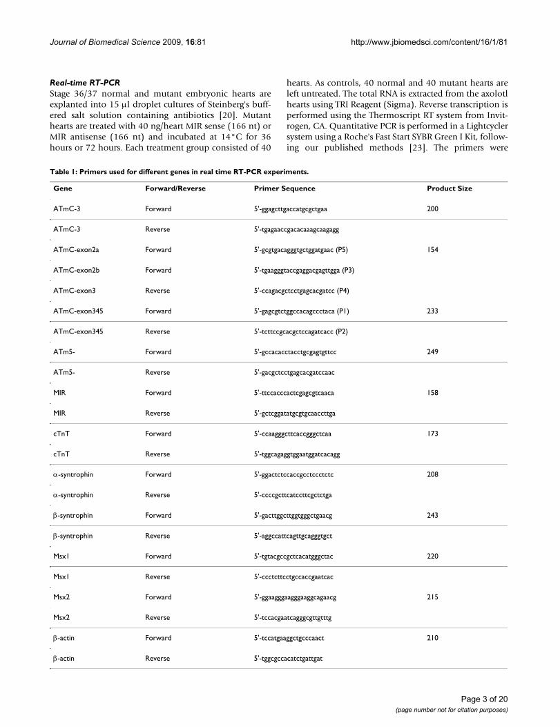
Table 1: Primers used for different genes in real time RT-PCR experiments.
Gene Forward/Reverse Primer Sequence Product Size
ATmC-3 Forward 5'-ggagcttgaccatgcgctgaa 200
ATmC-3 Reverse 5'-tgagaaccgacacaaagcaagagg
ATmC-exon2a Forward 5'-gcgtgacagggtgctggatgaac (P5) 154
ATmC-exon2b Forward 5'-tgaagggtaccgaggacgagttgga (P3)
ATmC-exon3 Reverse 5'-ccagacgctcctgagcacgatcc (P4)
ATmC-exon345 Forward 5'-gagcgtctggccacagccctaca (P1) 233
ATmC-exon345 Reverse 5'-tcttccgcacgctccagatcacc (P2)
ATm5- Forward 5'-gccacacctacctgcgagtgttcc 249
ATm5- Reverse 5'-gacgctcctgagcacgatccaac
MIR Forward 5'-ttccacccactcgagcgtcaaca 158
MIR Reverse 5'-gctcggatatgcgtgcaaccttga
cTnT Forward 5'-ccaagggcttcaccgggctcaa 173
cTnT Reverse 5'-tggcagaggtggaatggatcacagg
α-syntrophin Forward 5'-ggactctccaccgcctccctctc 208
α-syntrophin Reverse 5'-ccccgcttcatccttcgctctga
β-syntrophin Forward 5'-gacttggcttggtgggctgaacg 243
β-syntrophin Reverse 5'-aggccattcagttgcagggtgct
Msx1 Forward 5'-tgtacgccgctcacatgggctac 220
Msx1 Reverse 5'-ccctcttcctgccaccgaatcac
Msx2 Forward 5'-ggaagggaagggaaggcagaacg 215
Msx2 Reverse 5'-tccacgaatcagggcgttgtttg
β-actin Forward 5'-tccatgaaggctgcccaact 210
β-actin Reverse 5'-tggcgccacatctgattgat
Page 3 of 20(page number not for citation purposes)
Journal of Biomedical Science 2009, 16:81 http://www.jbiomedsci.com/content/16/1/81
designed by the Primer 3 program from the MassachusettsInstitute of Technology as listed in the Table 1.
Cloning of exon 1b sequence for axolotl alpha-TMWe have cloned exon 1b of the axolotl alpha-TM. 5'RACEexperiments using primers designed from the exon 3sequence of the axolotl alpha-TM (ATmC1-exon3-rev-1:5'-CCAGACGCTCCTGAGCACGATCC; ATmC1-exon3-rev-2:5'-CCAGCTGGATACGCCTGT TC) and a 5' adaptorsequence revealed both PCR bands corresponding to theATmC-1 sequence and a shorter PCR band after agarosegel electrophoresis. The shorter band was cloned into apGEM-T easy vector (Promega, WI) and sequenced. Thesequence shows high homology to the 5' ends of alpha-TM5a or 5b from other vertebrates. The sequence is: 5'-GTACTGTTGAGGCATCCACGTCTTCACTATTACT-GGGGCATTTGTAGTCCCTTGGAATTTGAG CTGACCT-TATCGCTACTCGCCTCATATGATAGAGGCGCCACACCTACCTGCGAGTGTTCCGTT CCCCGGCCCTCAGCAT-GTCTGGGGGCACCTCCCTGGAAGCGGTGCGGCG-GAAGATCCGCGCCCTGCAGGAGCAGGCGGACTCCGCTGAAGCCCG-GGCGTGTAGCCTGCAGCGGGAACGAGAC GCT-GAGCGGCAGCTGCGAGAGGCGGCTGAGAGTGATGTAGCCTCCCTGAACAGGCGTATCCA GCTGGTTGAGGAA-GAGTTGGAT CGTGCTCAGGAGCGTCTGG. The boldedATG represents a putative translation start site. The clonedsequence corresponding to exon 3 shows 100% identity toalpha-TM but with variations to axolotl ATmC-3, demon-strating the origin of this fragment from axolotl alpha-TM.We have named this isoform ATm5.
Two-dimensional gel electrophoresis and Western blottingIsoelectrical focusingThe immobilized pH gradient (IPG) gel strips (pH range3-6, 11 cm, Bio-Rad, CA) were rehydrated in 200 μl Read-yPrep Rehydration/Sample Buffer that contained heartsamples for 12 hours. Isoelectric focusing (IEF) was per-formed on the rehydrated IPGs at 20°C in three steps: 250volts for 20 minutes, 8,000 volts for 2.5 hours, and then20,000 Volts/hour using Protean IEF Cell system (Bio-Rad, CA).
Two-dimensional SDS-PAGEAfter isoelectrical focusing was completed, IPG strips wereprepared for SDS-PAGE using 12% Bis-Tris precast gels(Bio-Rad, CA). The strips were then rinsed with electro-phoresis running buffer (pH7.7) and laid into the two-dimensional well of 12% precast gels and were run at 200V for 1 hour following the manufacturer's protocol (Read-yPrep™ 2-D Starter Kit; Bio-Rad, CA.).
Western blottingThe two-dimensional SDS-PAGE gels were transferred tonitrocellulose membranes and processed for hybridiza-
tion. The membranes were incubated with CH1 mono-clonal antibody (1:5000) (from Developmental StudiesHybridoma Bank, University of Iowa) at a 1:2000 dilutionfor 1 hour at RT. After two washings for 5 minutes each,they were hybridized with horseradish peroxidase-conju-gated mouse antihuman IgG (Amersham-Pharmacia Bio-sciences) at 1:5000 dilution for 1 hour at RT and the blotswere washed and exposed to X-ray film and developedwith an ECL chemiluminescence system [22].
ResultsCloning the full length cDNA of the myofibril-inducing RNA (MIR) geneWe have previously published the nucleotide sequence ofthe 166 nt long bioactive MIR [11] and showed that theartificially synthesized 166 nt RNA can promote myofibrilformation and initiate beating of the mutant hearts inorgan culture studies [11].
To determine the full length sequence of the MIR gene, wecarried out experiments using a cDNA lambda phagelibrary from axolotls at stage 15-17 as well as an axolotlgenomic library (Stratagene, TX). Primers were designed,based both on vector flanking sequences and the 166 bpknown fragment of the MIR. The PCR products were con-firmed by Southern blotting assays. The promising PCRbands were then sub-cloned into pGEM-T vectors(Promega, WI) and subsequently sequenced. Primerswere designed again based on the new sequence and PCRswere repeated (Table 1). By using genomic walking wehave extended the 5'-end of the original 166 bp MIR to~700 bp. Starting from the original 166 bp MIR knownsequence, we also have performed 5' RACE reactionsusing a Smart RACE Kit (Ambion, TX). Results show thatthe full length of the MIR is expressed beginning at G(380th base) within the genomic sequence (Fig. 1). Inaddition, 3' RACE reactions have revealed about 150 ntpoly-A tail attached at the 3' end of the MIR (Fig. 1). Webelieve that we have successfully cloned the full lengthMIR. The nucleotide sequence of MIR has been deter-mined and found to be unique because there is no signif-icant homology with other known sequences availablefrom the gene databases. Moreover, there is no relativelylarge open reading frame in the full-length sequence, indi-cating that the MIR may be functioning directly throughits RNA structure, rather than a translation product. Inter-estingly, eukaryotic promoter prediction software(ExPASy: http://ca.expasy.org/tools/) has localized apotential promoter in the 5' genomic sequence with aconserved TATA box, 28 bp from the transcription startsite (Fig. 1). Our recent data using the Luciferase reportergene system (Promega, WI) have verified the transcriptiondriving ability of this potential promoter. We have clonednearly 3 kb of the upstream regulatory sequence in addi-tion to the promoter sequence shown in Fig. 1. Serial dele-
Page 4 of 20(page number not for citation purposes)
Journal of Biomedical Science 2009, 16:81 http://www.jbiomedsci.com/content/16/1/81
tions of the promoter have been inserted into theLuciferase reporter vectors and expression of these pro-moters has been confirmed by transfecting into rat neona-tal cardiomyocytes in culture (our unpublished data).
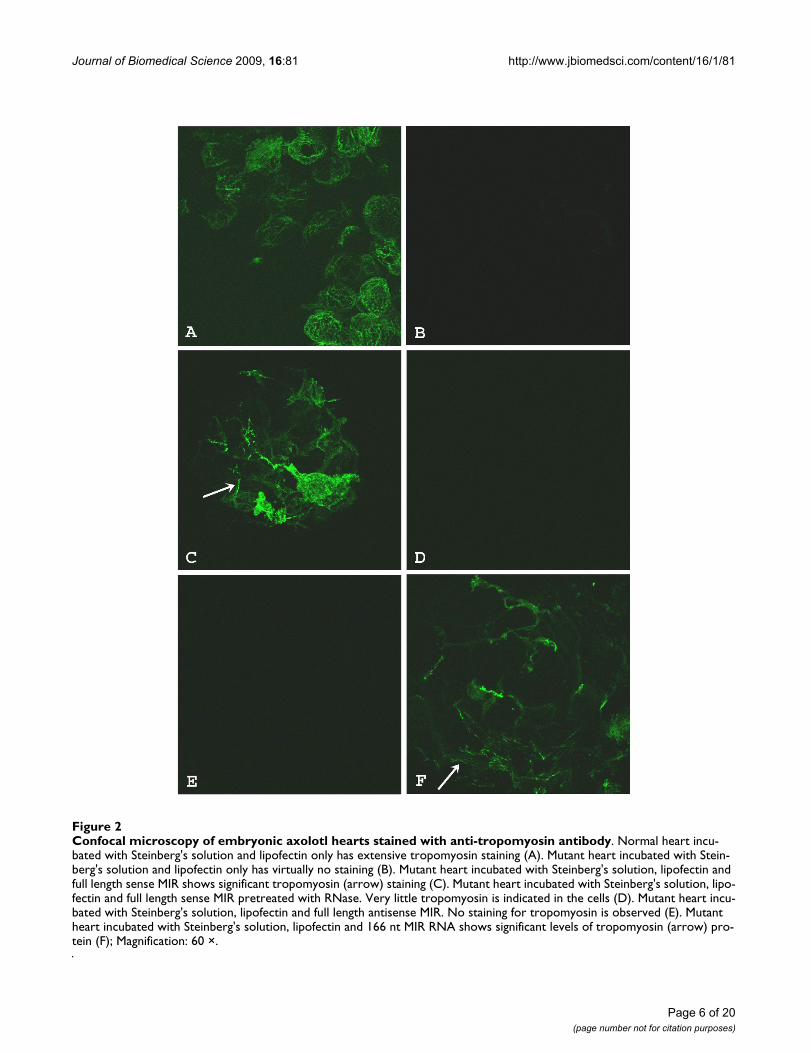
Full length MIR promotes tropomyosin protein expression in cardiac nonfunction mutant embryonic heartsIn vitro synthesized sense RNA from the full length (570nt) MIR gene showed significant rescuing ability of themutant hearts (Fig. 2). Three days after sense MIR transfec-tion, all of the 40 dissected mutant hearts showed positivetropomyosin staining (Fig. 2C) while antisense RNAtransfection showed only background levels of staining(Fig. 2D). Pretreatment of the MIR sense RNA with RNasebefore transfection into the mutant hearts totally abol-ished the activity of the sense RNA in promoting tropomy-osin expression (Fig. 2E). It is clear that the bioactivity ofpromoting tropomyosin expression in the mutant hearts
is due specifically to the sense MIR. These results are intotal agreement with our previous findings using the shortversion of the MIR (166 nt). The full length 570 nt RNAused for transfection in current experiments is at the samemolar concentration as the 166 nt RNA we used for previ-ous studies [7,11]. Since the original 166 nt RNA (partialsequence from MIR gene) is sufficient for the rescuing ofmutant hearts (Fig. 2F) [7,11], we believe that the 166 ntpartial sequence probably is the functional bioactive unitin the MIR gene.
MIR-promoted tropomyosin protein expression in the mutant hearts is not due to increased transcription levels or splicing pattern changes of tropomyosin genesTo determine whether MIR-promoted tropomyosin pro-tein expression in the mutant heart is due to regulation atthe transcriptional levels or changes in the splicing pat-terns of the TM transcripts, we proposed two hypotheses.
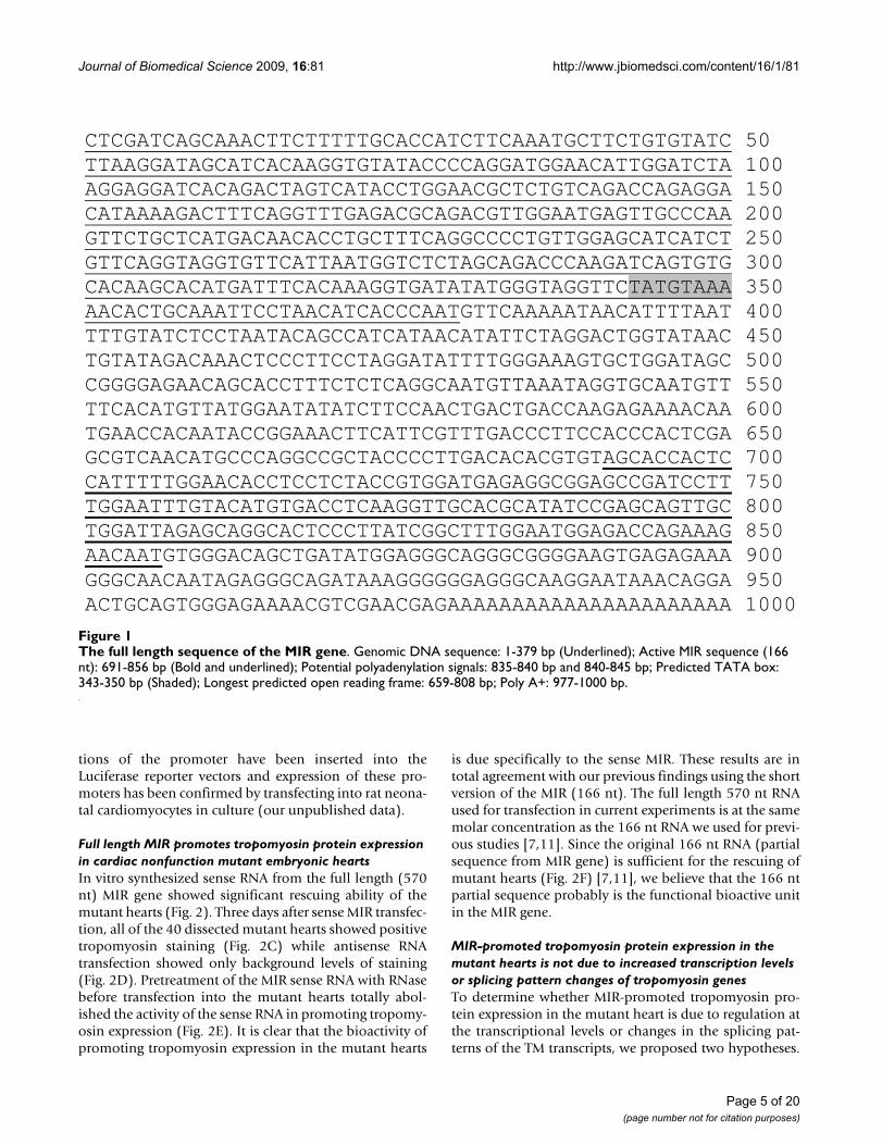
The full length sequence of the MIR geneFigure 1The full length sequence of the MIR gene. Genomic DNA sequence: 1-379 bp (Underlined); Active MIR sequence (166 nt): 691-856 bp (Bold and underlined); Potential polyadenylation signals: 835-840 bp and 840-845 bp; Predicted TATA box: 343-350 bp (Shaded); Longest predicted open reading frame: 659-808 bp; Poly A+: 977-1000 bp.
CTCGATCAGCAAACTTCTTTTTGCACCATCTTCAAATGCTTCTGTGTATC 50 TTAAGGATAGCATCACAAGGTGTATACCCCAGGATGGAACATTGGATCTA 100 AGGAGGATCACAGACTAGTCATACCTGGAACGCTCTGTCAGACCAGAGGA 150 CATAAAAGACTTTCAGGTTTGAGACGCAGACGTTGGAATGAGTTGCCCAA 200 GTTCTGCTCATGACAACACCTGCTTTCAGGCCCCTGTTGGAGCATCATCT 250 GTTCAGGTAGGTGTTCATTAATGGTCTCTAGCAGACCCAAGATCAGTGTG 300 CACAAGCACATGATTTCACAAAGGTGATATATGGGTAGGTTCTATGTAAA 350 AACACTGCAAATTCCTAACATCACCCAATGTTCAAAAATAACATTTTAAT 400 TTTGTATCTCCTAATACAGCCATCATAACATATTCTAGGACTGGTATAAC 450 TGTATAGACAAACTCCCTTCCTAGGATATTTTGGGAAAGTGCTGGATAGC 500 CGGGGAGAACAGCACCTTTCTCTCAGGCAATGTTAAATAGGTGCAATGTT 550 TTCACATGTTATGGAATATATCTTCCAACTGACTGACCAAGAGAAAACAA 600 TGAACCACAATACCGGAAACTTCATTCGTTTGACCCTTCCACCCACTCGA 650 GCGTCAACATGCCCAGGCCGCTACCCCTTGACACACGTGTAGCACCACTC 700 CATTTTTGGAACACCTCCTCTACCGTGGATGAGAGGCGGAGCCGATCCTT 750 TGGAATTTGTACATGTGACCTCAAGGTTGCACGCATATCCGAGCAGTTGC 800 TGGATTAGAGCAGGCACTCCCTTATCGGCTTTGGAATGGAGACCAGAAAG 850 AACAATGTGGGACAGCTGATATGGAGGGCAGGGCGGGGAAGTGAGAGAAA 900 GGGCAACAATAGAGGGCAGATAAAGGGGGGAGGGCAAGGAATAAACAGGA 950 ACTGCAGTGGGAGAAAACGTCGAACGAGAAAAAAAAAAAAAAAAAAAAAA 1000
Page 5 of 20(page number not for citation purposes)
Journal of Biomedical Science 2009, 16:81 http://www.jbiomedsci.com/content/16/1/81
Page 6 of 20(page number not for citation purposes)
Confocal microscopy of embryonic axolotl hearts stained with anti-tropomyosin antibodyFigure 2Confocal microscopy of embryonic axolotl hearts stained with anti-tropomyosin antibody. Normal heart incu-bated with Steinberg's solution and lipofectin only has extensive tropomyosin staining (A). Mutant heart incubated with Stein-berg's solution and lipofectin only has virtually no staining (B). Mutant heart incubated with Steinberg's solution, lipofectin and full length sense MIR shows significant tropomyosin (arrow) staining (C). Mutant heart incubated with Steinberg's solution, lipo-fectin and full length sense MIR pretreated with RNase. Very little tropomyosin is indicated in the cells (D). Mutant heart incu-bated with Steinberg's solution, lipofectin and full length antisense MIR. No staining for tropomyosin is observed (E). Mutant heart incubated with Steinberg's solution, lipofectin and 166 nt MIR RNA shows significant levels of tropomyosin (arrow) pro-tein (F); Magnification: 60 ×.
Journal of Biomedical Science 2009, 16:81 http://www.jbiomedsci.com/content/16/1/81
We tested those hypotheses by performing a series of real-time RT-PCR experiments.
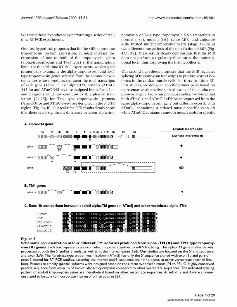
Our first hypothesis proposes that for the MIR to promotetropomyosin protein expression, it must increase theexpression of one or both of the tropomyosin genes(alpha-tropomyosin and TM4 type) at the transcriptionlevel. For the real-time RT-PCR experiments, we designedprimer pairs to amplify the alpha-tropomyosin and TM4type tropomyosin genes selected from the common exonsequences whose products represent the total transcriptsof each gene (Table 1). For alpha-TM, primers (ATmC-345-for and ATmC-345-rev) are designed in the Exon 3, 4and 5 regions which are common to all alpha-TM tran-scripts [24,25]; for TM4 type tropomyosin, primers(ATmC-3-for and ATmC-3-rev) are designed in the 3' UTRregion (Fig. 3A, B). Our real-time PCR results clearly showthat there is no significant difference between alpha-tro-
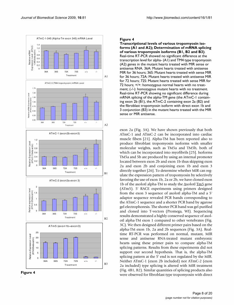
pomyosin or TM4 type tropomyosin RNA transcripts innormal (+/+), mutant (c/c), sense MIR- and antisenseMIR- treated mutant embryonic hearts (stage 37-38) attwo different time periods of the transfection of MIR (Fig.4A1, A2). These results clearly demonstrate that the MIRdoes not perform a regulatory function at the transcrip-tional level, thus disproving the first hypothesis.
Our second hypothesis proposes that the MIR regulatessplicing of tropomyosin transcripts to produce correct iso-forms in the cardiac muscle cells. For these real time RT-PCR studies, we designed specific primer pairs based onrepresentative alternative spliced exons of the alpha-tro-pomyosin gene. From our previous studies, we found thatboth ATmC-1 and ATmC-2 cDNAs are expressed from thesame alpha-tropomyosin gene but differ in exon 2, withATmC-1 containing a striated muscle specific exon 2bwhile ATmC-2 contains a smooth muscle isoform specific
Schematic representation of four different TM isoforms produced from alpha -TM (A) and TM4 type tropomyosin (B) genesFigure 3Schematic representation of four different TM isoforms produced from alpha -TM (A) and TM4 type tropomy-osin (B) genes. Each box represents an exon which is joined together by mRNA splicing. The alpha-TM gene is alternatively processed at both the 5' and/or 3' ends, as well as at the internal exons 6a/b. Our studies are focused on the 5' end sequence and exon 2a/b. The fibroblast type tropomyosin isoform (ATm5) has only the 5' sequence cloned with exon 1b and part of exon 3 cloned for RT-PCR studies, assuming the internal and 3' sequence are homologous to other vertebrates (dashed line box). Primers to amplify specific isoforms were designed based on the alternative spliced exons (P1 to P5). C. Highly conserved peptide sequence from exon 1b of axolotl alpha-tropomyosin compared to other vertebrate sequences. The indicated splicing pattern of axolotl tropomyosin genes are hypothetical based on other vertebrate sequences. ATmC-1, 2 and 3 were all dem-onstrated to be able to incorporate into myofibril structures [21].
Page 7 of 20(page number not for citation purposes)
Journal of Biomedical Science 2009, 16:81 http://www.jbiomedsci.com/content/16/1/81
exon 2a (Fig. 3A). We have shown previously that bothATmC-1 and ATmC-2 can be incorporated into cardiacmuscle fibers [21]. Alpha-TM has been reported also toproduce fibroblast tropomyosin isoforms with smallermolecular weights, such as TM5a and TM5b, both ofwhich can be incorporated into myofibrils [25]. IsoformsTM5a and 5b are produced by using an internal promoterlocated between exon 2b and exon 1b thus skipping exon2a and exon 2b and conjoining exon 1b and exon 3directly together [26]. To determine whether MIR can reg-ulate the expression pattern of tropomyosin by selectivelyfavoring the use of exon 1b, 2a or 2b, we have cloned exon1b of the axolotl alpha-TM to study the Axolotl Tm5 gene(ATm5). 5' RACE experiments using primers designedfrom the exon 3 sequence of axolotl alpha-TM and a 5'adaptor sequence revealed PCR bands corresponding tothe ATmC-1 sequence and a shorter PCR band by agarosegel electrophoresis. The shorter PCR band was gel purifiedand cloned into T-vectors (Promega, WI). Sequencingresults demonstrated a highly conserved sequence of axol-otl alpha-TM exon 1 compared to other vertebrates (Fig.3C). We then designed different primer pairs based on thealpha-TM exon 1b, 2a and 2b sequences (Fig. 3A). Real-time RT-PCR was performed on normal, mutant, MIRsense and antisense RNA-treated mutant embryonichearts using these primer pairs to compare alpha-TMsplicing patterns. Results from these experiments did notsupport our second hypothesis. That is, the alpha-TMsplicing pattern at the 5' end is not regulated by the MIR.Neither ATmC-1 (exon 2b included) nor ATmC-2 (exon2a included) type splicing is altered with MIR treatment(Fig. 4B1, B2). Similar quantities of splicing products alsowere observed for fibroblast type tropomyosin with directFigure 4
ATmC-1-345 (Alpha-Tm exon 345) mRNA Level
0
0.2
0.4
0.6
0.8
1
1.2
1.4
36A 36S 72A 72S +/+ -/-
Treatm ent
Rel
ativ
e m
RN
A
Exp
ress
ion
Lev
el
(AT
mC
-1-
345/
bet
a-ac
tin
)
A1
A2 A1
ATmC-3 (TM4 tropomyosin) mRNA Level
0.00
0.25
0.50
0.75
1.00
1.25
1.50
1.75
36A 36S 72A 72S +/+ -/
Treatm ent
Rel
ativ
e m
RN
A E
xpre
ssio
n L
evel
(A
Tm
C-3
/bet
a-ac
tin
)
-
A2
ATmC-1 (exon2b+exon3)
0
0.2
0.4
0.6
0.8
1
1.2
36A 36S 72A 72S +/+ -/-
Treatm ent
Rel
ativ
e E
xpre
ssio
n L
evel
(A
Tm
C-1
/bet
a-ac
tin
)
B1
ATmC-2 (exon2a+exon 3)
0
0.5
1
1.5
2
36A 36S 72A 72S +/+ -/-
Treatm ent
Rel
ativ
e ex
pre
ssio
n l
evel
(A
Tm
C-2
/bet
a-ac
tin
)
B2
ATm5 (exon1b+exon3)
0
5
10
15
20
25
36A 36S 72A 72S +/+ -/-
Treatm ent
Rel
ativ
e E
xpre
ssio
n L
evel
(A
Tm
5/b
eta-
acti
n)
B3
Transcriptional levels of various tropomyosin isoforms (A1 and A2); Determination of mRNA splicing of various tropo-myosin isoforms (B1, B2 and B3)Figure 4Transcriptional levels of various tropomyosin iso-forms (A1 and A2); Determination of mRNA splicing of various tropomyosin isoforms (B1, B2 and B3). Real-time RT-PCR showed no significant difference at the transcription level for alpha- (A1) and TM4 type tropomyosin (A2) genes in the mutant hearts treated with MIR sense or antisense RNA. 36A: Mutant hearts treated with antisense MIR for 36 hours; 36S: Mutant hearts treated with sense MIR for 36 hours; 72A: Mutant hearts treated with antisense MIR for 72 hours; 72S: Mutant hearts treated with sense MIR for 72 hours; +/+: homozygous normal hearts with no treat-ment; (-/-): homozygous mutant hearts with no treatment. Real-time RT-PCR showing no significant difference during mRNA splicing of the alpha-TM gene (the ATmC-1 contain-ing exon 2b (B1), the ATmC-2 containing exon 2a (B2) and the fibroblast tropomyosin isoform with direct exon 1b and 3 conjunction (B3) in the mutant hearts treated with the MIR sense or MIR antisense.
Page 8 of 20(page number not for citation purposes)
Journal of Biomedical Science 2009, 16:81 http://www.jbiomedsci.com/content/16/1/81
exons 1b and 3 conjunctions in all normal, mutant, MIRsense and antisense RNA treated hearts (Fig. 4B3).
Thus these studies reiterate that MIR-promoted tropomy-osin expression in the mutant hearts is not due toincreased transcription levels or splicing pattern changesin the tropomyosin genes.
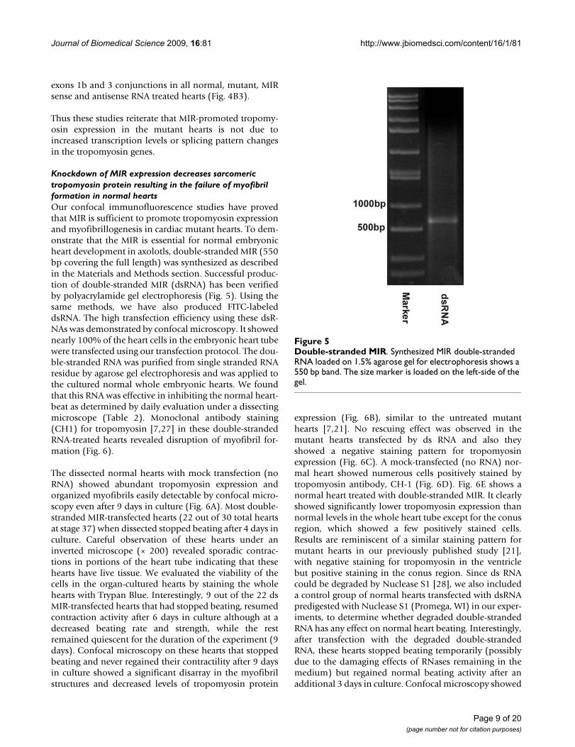
Knockdown of MIR expression decreases sarcomeric tropomyosin protein resulting in the failure of myofibril formation in normal heartsOur confocal immunofluorescence studies have provedthat MIR is sufficient to promote tropomyosin expressionand myofibrillogenesis in cardiac mutant hearts. To dem-onstrate that the MIR is essential for normal embryonicheart development in axolotls, double-stranded MIR (550bp covering the full length) was synthesized as describedin the Materials and Methods section. Successful produc-tion of double-stranded MIR (dsRNA) has been verifiedby polyacrylamide gel electrophoresis (Fig. 5). Using thesame methods, we have also produced FITC-labeleddsRNA. The high transfection efficiency using these dsR-NAs was demonstrated by confocal microscopy. It showednearly 100% of the heart cells in the embryonic heart tubewere transfected using our transfection protocol. The dou-ble-stranded RNA was purified from single stranded RNAresidue by agarose gel electrophoresis and was applied tothe cultured normal whole embryonic hearts. We foundthat this RNA was effective in inhibiting the normal heart-beat as determined by daily evaluation under a dissectingmicroscope (Table 2). Monoclonal antibody staining(CH1) for tropomyosin [7,27] in these double-strandedRNA-treated hearts revealed disruption of myofibril for-mation (Fig. 6).
The dissected normal hearts with mock transfection (noRNA) showed abundant tropomyosin expression andorganized myofibrils easily detectable by confocal micro-scopy even after 9 days in culture (Fig. 6A). Most double-stranded MIR-transfected hearts (22 out of 30 total heartsat stage 37) when dissected stopped beating after 4 days inculture. Careful observation of these hearts under aninverted microscope (× 200) revealed sporadic contrac-tions in portions of the heart tube indicating that thesehearts have live tissue. We evaluated the viability of thecells in the organ-cultured hearts by staining the wholehearts with Trypan Blue. Interestingly, 9 out of the 22 dsMIR-transfected hearts that had stopped beating, resumedcontraction activity after 6 days in culture although at adecreased beating rate and strength, while the restremained quiescent for the duration of the experiment (9days). Confocal microscopy on these hearts that stoppedbeating and never regained their contractility after 9 daysin culture showed a significant disarray in the myofibrilstructures and decreased levels of tropomyosin protein
expression (Fig. 6B), similar to the untreated mutanthearts [7,21]. No rescuing effect was observed in themutant hearts transfected by ds RNA and also theyshowed a negative staining pattern for tropomyosinexpression (Fig. 6C). A mock-transfected (no RNA) nor-mal heart showed numerous cells positively stained bytropomyosin antibody, CH-1 (Fig. 6D). Fig. 6E shows anormal heart treated with double-stranded MIR. It clearlyshowed significantly lower tropomyosin expression thannormal levels in the whole heart tube except for the conusregion, which showed a few positively stained cells.Results are reminiscent of a similar staining pattern formutant hearts in our previously published study [21],with negative staining for tropomyosin in the ventriclebut positive staining in the conus region. Since ds RNAcould be degraded by Nuclease S1 [28], we also includeda control group of normal hearts transfected with dsRNApredigested with Nuclease S1 (Promega, WI) in our exper-iments, to determine whether degraded double-strandedRNA has any effect on normal heart beating. Interestingly,after transfection with the degraded double-strandedRNA, these hearts stopped beating temporarily (possiblydue to the damaging effects of RNases remaining in themedium) but regained normal beating activity after anadditional 3 days in culture. Confocal microscopy showed
Double-stranded MIRFigure 5Double-stranded MIR. Synthesized MIR double-stranded RNA loaded on 1.5% agarose gel for electrophoresis shows a 550 bp band. The size marker is loaded on the left-side of the gel.
Page 9 of 20(page number not for citation purposes)
Journal of Biomedical Science 2009, 16:81 http://www.jbiomedsci.com/content/16/1/81
Page 10 of 20(page number not for citation purposes)
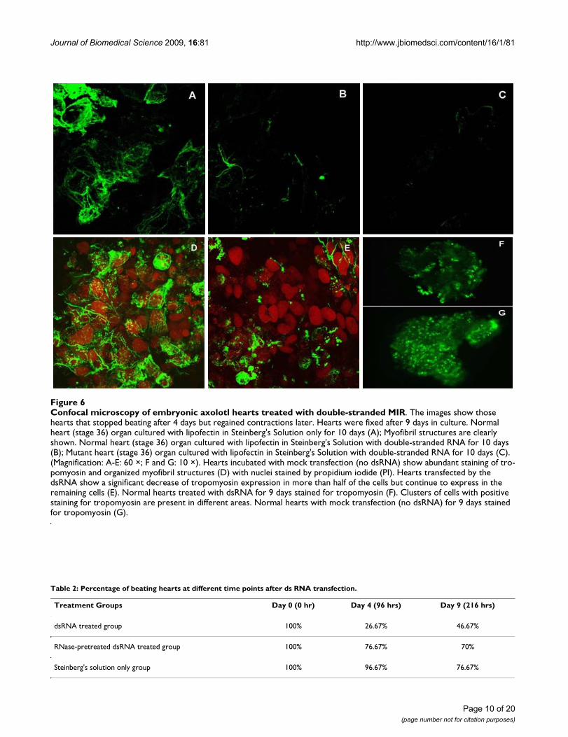
Confocal microscopy of embryonic axolotl hearts treated with double-stranded MIRFigure 6Confocal microscopy of embryonic axolotl hearts treated with double-stranded MIR. The images show those hearts that stopped beating after 4 days but regained contractions later. Hearts were fixed after 9 days in culture. Normal heart (stage 36) organ cultured with lipofectin in Steinberg's Solution only for 10 days (A); Myofibril structures are clearly shown. Normal heart (stage 36) organ cultured with lipofectin in Steinberg's Solution with double-stranded RNA for 10 days (B); Mutant heart (stage 36) organ cultured with lipofectin in Steinberg's Solution with double-stranded RNA for 10 days (C). (Magnification: A-E: 60 ×; F and G: 10 ×). Hearts incubated with mock transfection (no dsRNA) show abundant staining of tro-pomyosin and organized myofibril structures (D) with nuclei stained by propidium iodide (PI). Hearts transfected by the dsRNA show a significant decrease of tropomyosin expression in more than half of the cells but continue to express in the remaining cells (E). Normal hearts treated with dsRNA for 9 days stained for tropomyosin (F). Clusters of cells with positive staining for tropomyosin are present in different areas. Normal hearts with mock transfection (no dsRNA) for 9 days stained for tropomyosin (G).
Table 2: Percentage of beating hearts at different time points after ds RNA transfection.
Treatment Groups Day 0 (0 hr) Day 4 (96 hrs) Day 9 (216 hrs)
dsRNA treated group 100% 26.67% 46.67%
RNase-pretreated dsRNA treated group 100% 76.67% 70%
Steinberg's solution only group 100% 96.67% 76.67%
Journal of Biomedical Science 2009, 16:81 http://www.jbiomedsci.com/content/16/1/81
numerous positively-stained cells in these hearts whichappeared similar to untreated normal hearts after 9 daysin culture.
In the double-stranded MIR RNA treated normal heartsthat stopped beating after 3 or 4 days but regained con-tractions after 9 days, confocal microscopy studies stillrevealed decreased staining of tropomyosin protein (Fig.6F) compared to mock transfected cells (Fig. 6G). Organ-ized myofibrils with tropomyosin staining could be foundin a few sparsely distributed cells in the heart although theoverall numbers of stained cells were dramaticallydecreased in the dsRNA-treated hearts (Fig. 6E) comparedto the control normal hearts without dsRNA treatment(Fig. 6D). Since our whole-mount heart specimens areusually 150-200 μm in thickness, we used epifluorescencemicroscopy to view the fluorescence image of the wholeheart. Thus we were able to clearly observe the groups ofcells that show positive staining in the dsRNA-treatedwhole hearts (Fig. 6F, G).
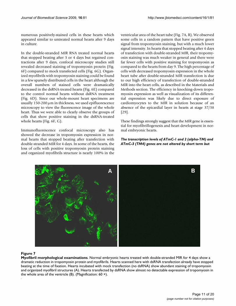
Immunofluorescence confocal microscopy also hasshowed the decrease in tropomyosin expression in nor-mal hearts that stopped beating after transfection withdouble-stranded MIR for 4 days. In some of the hearts, theloss of cells with positive tropomyosin protein stainingand organized myofibrils structure is nearly 100% in the
ventricular area of the heart tube (Fig. 7A, B). We observedsome cells in a random pattern that have positive greensignal from tropomyosin staining, but with a much lowersignal intensity. In hearts that stopped beating after 4 daysof transfection with double-stranded MIR, their tropomy-osin staining was much weaker in general and there werefar fewer cells with positive staining for tropomyosin ascompared to the hearts from day 9. The high percentage ofcells with decreased tropomyosin expression in the wholeheart tube after double-stranded MIR transfection is dueto our high efficiency of transfection of double-strandedMIR into the heart cells, as described in the Materials andMethods section. The efficiency in knocking-down tropo-myosin expression as well as visualization of its differen-tial expression was likely due to direct exposure ofcardiomyocytes to the MIR in solution because of anabsence of the epicardial layer in hearts at stage 37/38[29].
These findings strongly suggest that the MIR gene is essen-tial for myofibrillogenesis and heart development in nor-mal embryonic hearts.
The transcription levels of ATmC-1 and 2 (alpha-TM) and ATmC-3 (TM4) genes are not altered by short term but
Myofibril morphological examinationsFigure 7Myofibril morphological examinations. Normal embryonic hearts treated with double-stranded MIR for 4 days show a dramatic reduction in tropomyosin protein and myofibrils. Hearts scanned here with dsRNA transfection already have stopped beating at the time of fixation. Hearts incubated with mock transfection (no dsRNA) show abundant staining of tropomyosin and organized myofibril structures (A). Hearts transfected by dsRNA show almost no detectable expression of tropomyosin in the whole area of the ventricle (B). (Magnification: 60 ×).
Page 11 of 20(page number not for citation purposes)
Journal of Biomedical Science 2009, 16:81 http://www.jbiomedsci.com/content/16/1/81
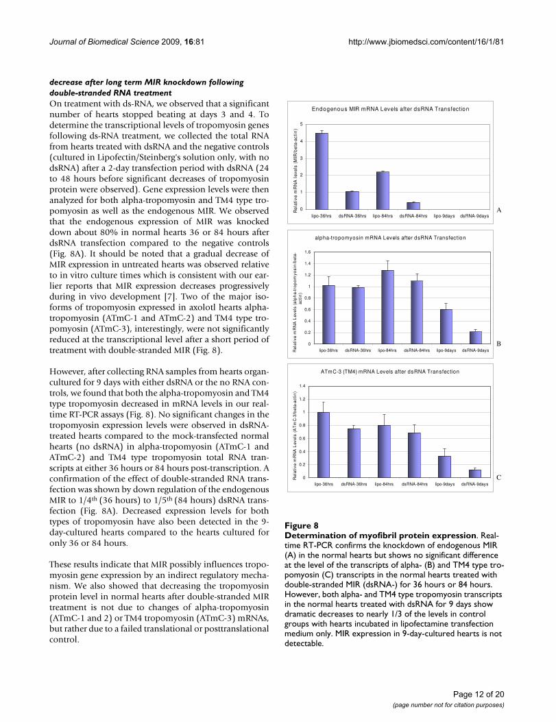
decrease after long term MIR knockdown following double-stranded RNA treatmentOn treatment with ds-RNA, we observed that a significantnumber of hearts stopped beating at days 3 and 4. Todetermine the transcriptional levels of tropomyosin genesfollowing ds-RNA treatment, we collected the total RNAfrom hearts treated with dsRNA and the negative controls(cultured in Lipofectin/Steinberg's solution only, with nodsRNA) after a 2-day transfection period with dsRNA (24to 48 hours before significant decreases of tropomyosinprotein were observed). Gene expression levels were thenanalyzed for both alpha-tropomyosin and TM4 type tro-pomyosin as well as the endogenous MIR. We observedthat the endogenous expression of MIR was knockeddown about 80% in normal hearts 36 or 84 hours afterdsRNA transfection compared to the negative controls(Fig. 8A). It should be noted that a gradual decrease ofMIR expression in untreated hearts was observed relativeto in vitro culture times which is consistent with our ear-lier reports that MIR expression decreases progressivelyduring in vivo development [7]. Two of the major iso-forms of tropomyosin expressed in axolotl hearts alpha-tropomyosin (ATmC-1 and ATmC-2) and TM4 type tro-pomyosin (ATmC-3), interestingly, were not significantlyreduced at the transcriptional level after a short period oftreatment with double-stranded MIR (Fig. 8).
However, after collecting RNA samples from hearts organ-cultured for 9 days with either dsRNA or the no RNA con-trols, we found that both the alpha-tropomyosin and TM4type tropomyosin decreased in mRNA levels in our real-time RT-PCR assays (Fig. 8). No significant changes in thetropomyosin expression levels were observed in dsRNA-treated hearts compared to the mock-transfected normalhearts (no dsRNA) in alpha-tropomyosin (ATmC-1 andATmC-2) and TM4 type tropomyosin total RNA tran-scripts at either 36 hours or 84 hours post-transcription. Aconfirmation of the effect of double-stranded RNA trans-fection was shown by down regulation of the endogenousMIR to 1/4th (36 hours) to 1/5th (84 hours) dsRNA trans-fection (Fig. 8A). Decreased expression levels for bothtypes of tropomyosin have also been detected in the 9-day-cultured hearts compared to the hearts cultured foronly 36 or 84 hours.
These results indicate that MIR possibly influences tropo-myosin gene expression by an indirect regulatory mecha-nism. We also showed that decreasing the tropomyosinprotein level in normal hearts after double-stranded MIRtreatment is not due to changes of alpha-tropomyosin(ATmC-1 and 2) or TM4 tropomyosin (ATmC-3) mRNAs,but rather due to a failed translational or posttranslationalcontrol.
Determination of myofibril protein expressionFigure 8Determination of myofibril protein expression. Real-time RT-PCR confirms the knockdown of endogenous MIR (A) in the normal hearts but shows no significant difference at the level of the transcripts of alpha- (B) and TM4 type tro-pomyosin (C) transcripts in the normal hearts treated with double-stranded MIR (dsRNA-) for 36 hours or 84 hours. However, both alpha- and TM4 type tropomyosin transcripts in the normal hearts treated with dsRNA for 9 days show dramatic decreases to nearly 1/3 of the levels in control groups with hearts incubated in lipofectamine transfection medium only. MIR expression in 9-day-cultured hearts is not detectable.
Endogenous MIR mRNA Levels after dsRNA Transfection
0
1
2
3
4
5
lipo-36hrs dsRNA-36hrs lipo-84hrs dsRNA-84hrs lipo-9days dsRNA-9days
Rel
ativ
e m
RN
A l
evel
s (M
IR/b
eta-
acti
n)
A
alpha-tropomyosin mRNA Levels after dsRNA Transfection
0
0.2
0.4
0.6
0.8
1
1.2
1.4
1.6
lipo-36hrs dsRNA-36hrs lipo-84hrs dsRNA-84hrs lipo-9days dsRNA-9daysRel
ativ
e m
RN
A L
evel
s (a
lph
a-tr
op
om
yosi
n/b
eta-
acti
n)
B
ATmC-3 (TM4) mRNA Levels after dsRNA Transfection
0
0.2
0.4
0.6
0.8
1
1.2
1.4
lipo-36hrs dsRNA-36hrs lipo-84hrs dsRNA-84hrs lipo-9days dsRNA-9days
Rel
ativ
e m
RN
A L
evel
s (A
Tm
C-3
/bet
a-ac
tin
)
C
Page 12 of 20(page number not for citation purposes)
Journal of Biomedical Science 2009, 16:81 http://www.jbiomedsci.com/content/16/1/81
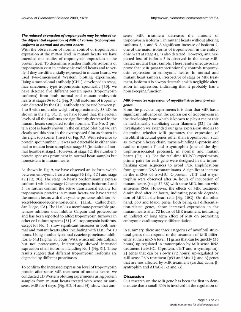
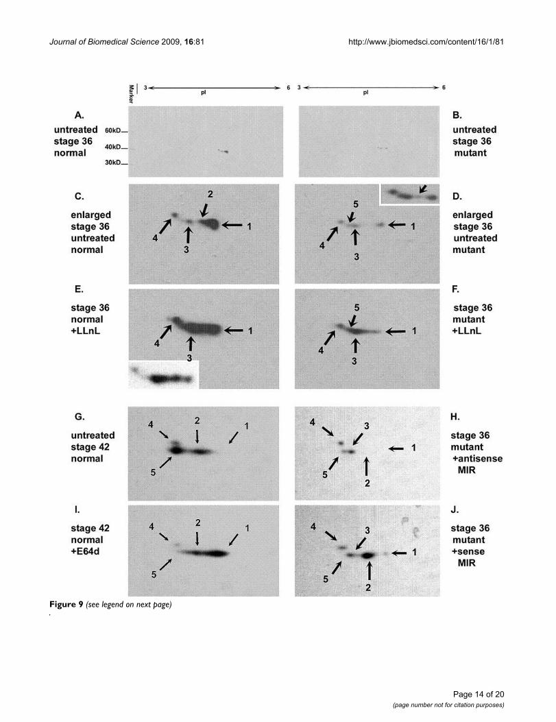
The reduced expression of tropomyosin may be related to the differential regulation of MIR of various tropomyosin isoforms in normal and mutant heartsWith the observation of normal control of tropomyosinexpression at the mRNA level in mutant hearts, we haveextended our studies of tropomyosin expression at theprotein level. To determine whether multiple isoforms oftropomyosin exist in embryonic axolotl hearts and to ver-ify if they are differentially expressed in mutant hearts, weused two-dimensional Western blotting experiments.Using a monoclonal antibody (CH1), developed to recog-nize sarcomeric type tropomyosin specifically [30], wehave detected five different protein spots (tropomyosinisoforms) from both normal and mutant embryonichearts at stages 36 to 42 (Fig. 9). All isoforms of tropomy-osin detected by the CH1 antibody are located between pI4 to 5 with molecular weight of approximately 38 kD. Asshown in the Fig. 9C, D, we have found that, the proteinlevels of all the isoforms are significantly decreased in themutant hearts compared to the normals. The No. 2 pro-tein spot is barely shown in the enlarged blot but we canclearly see this spot in the overexposed film as shown inthe right top corner (inset) of Fig. 9D. With reference toprotein spot number 5, it was not detectable in either nor-mal or mutant heart samples at stage 36 (initiation of nor-mal heartbeat stage). However, at stage 42, the number 5protein spot was prominent in normal heart samples butnonexistent in mutant hearts.
As shown in Fig. 9, we have observed an isoform switchbetween embryonic hearts at stage 36 (Fig. 9D) and stage42 (Fig. 9G). The stage 36 hearts predominantly expressisoform 1 while the stage 42 hearts express isoforms 2 and5. To further confirm the active translational activity fortropomyosin proteins in mutant hearts, we have treatedthe mutant hearts with the cysteine-protease inhibitor, N-acetyl-leucine-leucine-norleucinal (LLnL, Calbiochem,San Diego, CA). The LLnL is a membrane-permeable pro-teinase inhibitor that inhibits Calpain and proteosomeand has been reported to affect tropomyosin turnover inother cell culture systems [31]. All tropomyosin isoforms,except for No. 1, show significant increases in both nor-mal and mutant hearts after incubating with LLnL for 10hours. Using another lyosomal cysteine proteinase inhib-itor, E-64d (Sigma, St. Louis, WA), which inhibits Calpainbut not proteosome, interestingly showed increasedexpression of all isoforms including No.1 (Fig. 9I). Theseresults suggest that different tropomyosin isoforms aredegraded by different proteinases.
To confirm the increased expression level of tropomyosinprotein after sense MIR treatment of mutant hearts, weconducted 2D Western blotting experiments using proteinsamples from mutant hearts treated with sense or anti-sense MIR for 4 days. (Fig. 9D, H and 9J), show that anti-
sense MIR treatment decreases the amount oftropomyosin isoform 1 in mutant hearts without alteringisoforms 3, 4 and 5. A significant increase of isoform 2,one of the major isoforms of tropomyosin in the embry-onic heart at stage 42, is also detected. However, an unex-pected loss of isoform 5 is observed in the sense MIR-treated mutant heart sample. These results unequivocallyprove that MIR post-transcriptionally controls tropomy-osin expression in embryonic hearts. In normal andmutant heart samples, irrespective of stage or MIR treat-ment, isoform 4 is always detectable with negligible alter-ation in expression, indicating that it probably has ahousekeeping function.
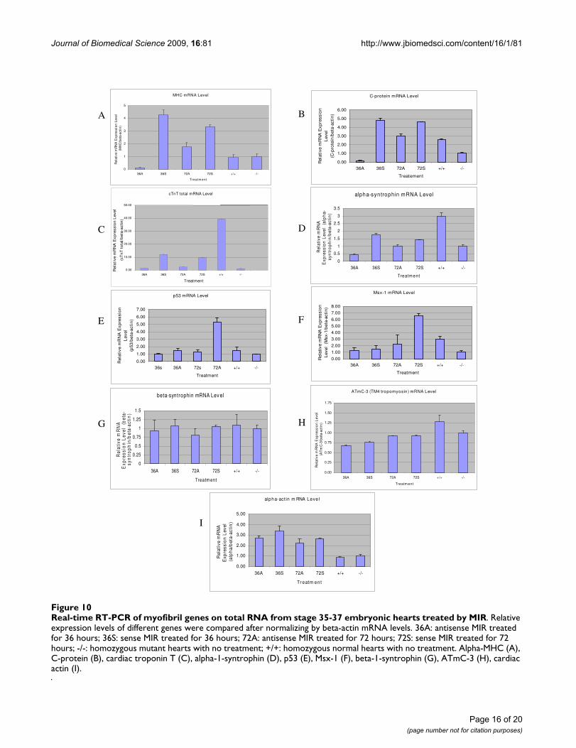
MIR promotes expression of myofibril structural protein genesFrom the previous experiments it is clear that MIR has asignificant influence on the expression of tropomyosin inthe developing heart which is known to play a major rolein mechanically stabilizing actin filaments [32]. In thisinvestigation we extended our gene expression studies todetermine whether MIR promotes the expression ofmyofibril structural genes other than tropomyosin, suchas, α-myosin heavy chain, myosin-binding C-protein andcardiac troponin T and α-syntrophin (one of the dys-trophin-associated proteins), in normal and mutanthearts (Fig. 10). For the real-time RT-PCR experiments,primer pairs for each gene were designed in the intron-flanking exon sequences to avoid PCR amplificationsfrom genomic DNA contaminants. A significant increasein the mRNA of α-MHC, C-protein, cTnT and α-syn-trophin were observed after 36 hours of incubation ofmutant hearts (stage 37-38) with sense MIR, but not withantisense RNA. However, the effects of MIR treatmentdiminished after 72 hours, indicating possible degrada-tion of MIR in the heart cells (Fig. 10G). On the otherhand, p53 and Msx-1 genes, both being cell differentia-tion-related genes, show increased expression in themutant hearts after 72 hours of MIR treatment, indicatingan indirect or long term effect of MIR on promotingembryonic cardiomyocyte differentiation.
In summary, there are three categories of myofibril struc-tural genes that respond to the treatment of MIR differ-ently at their mRNA level: 1) genes that can be quickly (36hours) up-regulated in transcription by MIR sense RNAtreatment (α-MHC, C-protein, cTnT and α-syntrophin);2) genes that can be slowly (72 hours) up-regulated byMIR sense RNA treatment (p53 and Msx-1); and 3) genesthat are not affected by MIR treatment (cardiac actin, β-syntrophin and ATmC-1, -2 and -3).
DiscussionOur research on the MIR gene has been the first to dem-onstrate that a small RNA is involved in the regulation of
Page 13 of 20(page number not for citation purposes)
Journal of Biomedical Science 2009, 16:81 http://www.jbiomedsci.com/content/16/1/81
Figure 9 (see legend on next page)
Page 14 of 20(page number not for citation purposes)
Journal of Biomedical Science 2009, 16:81 http://www.jbiomedsci.com/content/16/1/81
embryonic heart development in the cardiac mutant(gene c) of Ambystoma mexicanum. In the case of the MIRgene, since we now know that the MIR is a polyadenylatedRNA, there is reason to believe that it may functionthrough its unique secondary structure to promote myofi-brillogenesis in the mutant hearts. We favor this hypothe-sis for the following reasons: First, the RNA used forrescuing experiments (either 650 nt full length or 166 ntclone #4 RNA) was not processed for translation by cap-ping or polyadenylation before it was added to the heartcultures implying that it lacked necessary machinery fortranslation inside the cells. The possibility that the activeRNA rescues mutant hearts by providing mRNA that istranslated is unlikely. Second, although there are severalsmall open reading frames (ORF) predicted for the fulllength gene (the longest ORF is shown in Fig. 1), theshortest RNA we have shown to preserve bioactivity (140nt with 26 nt deleted from the 3' end of 166 nt Clone #4sequence shown with an underline in Fig. 1) does notcover the whole ORF [33]. Thus, the chance of this RNAbeing translated in the cells is virtually nonexistent.
Recently, there have been numerous tissue-specific andembryonic stem cell-expressed microRNAs cloned frommouse, some of which are heart specific [34,35]. Prior toour finding on the MIR [7,11] there had been no previ-ously reported non-coding RNAs important for heartdevelopment). In fact, the MIR gene appears to be the firstnon-coding RNA that has been reported to be involved inthe process of anterior endoderm inducing precardiacmesoderm to form functional cardiac muscle tissue [36].Further characterization is underway to rule out the possi-bility of MIR as being a typical miRNA and/or consider asa piRNA [37-39] or some other unique regulatory RNAmolecule.
One of the long term objectives of our studies is to findthe mammalian homolog of the MIR gene and studyrelated pathways in mammals, including human.
Although some of the noncoding RNAs, such as somemicro RNAs, are conserved between species, there are oth-ers that have many variations [40]. Based on sequencesimilarity, we have not yet identified the mammalian MIRhomolog gene from a database search although we havepreliminary data indicating that both sheep heart [7] andhuman heart (our unpublished data) contain functionalMIR homologs which are able to promote myofibrillogen-esis and rescue the mutant axolotl hearts. Recently, Mum-mery et al. [41] have indicated that there exists a verysimilar, probably identical, induction mechanism formouse heart development from signals secreted from cul-tured mouse visceral endoderm-like cells. This factor hasbeen found to be capable of promoting beating cardiomy-ocyte differentiation and beating in human embryonicstem cell cultures, indicating a non-protein factor secretedfrom mouse visceral endoderm cells, possibly the mousehomolog of our MIR gene. Thus, these results and findingsfrom our studies in the animal model, Ambystoma mexica-num, suggest that the relationship between endoderm andheart induction is genetically conserved across the verte-brate species.
We have previously demonstrated that the MIR (166 ntcore sequence) can bind to more than one protein. TheMIR gene and its binding proteins are apparently involvedin the regulation of tropomyosin expression [7]. Prior tocurrent studies, it was not clear whether the failure of tro-pomyosin expression in mutant hearts takes place at thetranscriptional level or the translational level and whichisoform(s) of tropomyosin is/are significantly reduced inthe mutant embryonic hearts. Our results in this studyprove unequivocally that the MIR and, perhaps, its bind-ing protein(s) work together to regulate tropomyosinexpression translationally or posttranslationally in themutant hearts since our studies did not detect transcrip-tional or splicing pattern differences in tropomyosingenes between normal and mutant embryonic hearts.Moreover, sequencing of the full-length cDNAs of both
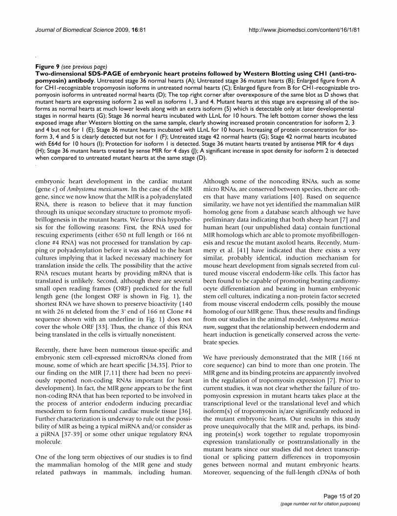
Two-dimensional SDS-PAGE of embryonic heart proteins followed by Western Blotting using CH1 (anti-tropomyosin) anti-bodyFigure 9 (see previous page)Two-dimensional SDS-PAGE of embryonic heart proteins followed by Western Blotting using CH1 (anti-tro-pomyosin) antibody. Untreated stage 36 normal hearts (A); Untreated stage 36 mutant hearts (B); Enlarged figure from A for CH1-recognizable tropomyosin isoforms in untreated normal hearts (C); Enlarged figure from B for CH1-recognizable tro-pomyosin isoforms in untreated normal hearts (D); The top right corner after overexposure of the same blot as D shows that mutant hearts are expressing isoform 2 as well as isoforms 1, 3 and 4. Mutant hearts at this stage are expressing all of the iso-forms as normal hearts at much lower levels along with an extra isoform (5) which is detectable only at later developmental stages in normal hearts (G); Stage 36 normal hearts incubated with LLnL for 10 hours. The left bottom corner shows the less exposed image after Western blotting on the same sample, clearly showing increased protein concentration for isoform 2, 3 and 4 but not for 1 (E); Stage 36 mutant hearts incubated with LLnL for 10 hours. Increasing of protein concentration for iso-form 3, 4 and 5 is clearly detected but not for 1 (F); Untreated stage 42 normal hearts (G); Stage 42 normal hearts incubated with E64d for 10 hours (I); Protection for isoform 1 is detected. Stage 36 mutant hearts treated by antisense MIR for 4 days (H); Stage 36 mutant hearts treated by sense MIR for 4 days (J); A significant increase in spot density for isoform 2 is detected when compared to untreated mutant hearts at the same stage (D).
Page 15 of 20(page number not for citation purposes)
Journal of Biomedical Science 2009, 16:81 http://www.jbiomedsci.com/content/16/1/81
Page 16 of 20(page number not for citation purposes)
Real-time RT-PCR of myofibril genes on total RNA from stage 35-37 embryonic hearts treated by MIRFigure 10Real-time RT-PCR of myofibril genes on total RNA from stage 35-37 embryonic hearts treated by MIR. Relative expression levels of different genes were compared after normalizing by beta-actin mRNA levels. 36A: antisense MIR treated for 36 hours; 36S: sense MIR treated for 36 hours; 72A: antisense MIR treated for 72 hours; 72S: sense MIR treated for 72 hours; -/-: homozygous mutant hearts with no treatment; +/+: homozygous normal hearts with no treatment. Alpha-MHC (A), C-protein (B), cardiac troponin T (C), alpha-1-syntrophin (D), p53 (E), Msx-1 (F), beta-1-syntrophin (G), ATmC-3 (H), cardiac actin (I).
I
C-protein mRNA Level
0.00
1.00
2.00
3.00
4.00
5.00
6.00
36A 36S 72A 72S +/+ -/-
Treatement
Rel
ativ
e m
RN
A E
xpre
ssio
n
Lev
el
(C-p
rote
in/b
eta-
acti
n)
beta-syntrophin mRNA Lev el
0
0.25
0.5
0.75
1
1.25
1.5
36A 36S 72A 72S +/+ -/-
Treatm ent
Rel
ativ
e m
RN
A
Exp
ress
ion
Lev
el
(bet
a-sy
ntr
op
hin
/bet
a-ac
tin
)
MHC mRNA Level
0
1
2
3
4
5
36A 36S 72A 72S +/+ -/-
Treatm ent
Rel
ativ
e m
RN
A E
xpre
ssio
n L
evel
(M
HC
/bet
a-ac
tin
)
A C E G
B D F H
alpha-syntroph in mRNA Level
0
0.5
1
1.5
2
2.5
3
3.5
36A 36S 72A 72S +/+ -/-
Treatm ent
Rel
ativ
e m
RN
A
Exp
ress
ion
Lev
el
(alp
ha-
syn
tro
ph
in/b
eta-
acti
n)
ATmC-3 (TM4 tropomyosin) mRNA Level
0.00
0.25
0.50
0.75
1.00
1.25
1.50
1.75
36A 36S 72A 72S +/+ -/-
Treatm ent
Rel
ativ
e m
RN
A E
xpre
ssio
n L
evel
(A
Tm
C-3
/bet
a-ac
tin
)
alpha-act in m RNA Leve l
0.00
1.00
2.00
3.00
4.00
5.00
36A 36S 72A 72S +/+ -/-
Tr eatm ent
Rel
ativ
e m
RN
A
Exp
ress
ion
Lev
el
(alp
ha/
bet
a-ac
tin
)
Msx-1 mRNA Level
0.00
1.00
2.00
3.00
4.00
5.00
6.00
7.00
8.00
36A 36S 72A 72S +/+ -/-
Treatment
Rel
ativ
e m
RN
A E
xpre
ssio
n
Lev
el (
Msx
-1/b
eta-
acti
n)
p53 mRNA Level
0.00
1.00
2.00
3.00
4.00
5.00
6.00
7.00
36s 36A 72s 72A +/+ -/-
Treatment
Rel
ativ
e m
RN
A E
xpre
ssio
n
Lev
el
(p53
/bet
a-ac
tin
)
0.00
10.00 20.00 30.00 40.00 50.00
36A 36S 72A 72S +/+ -/-
Treatment
cTnT total mRNA Level
Rel
ativ
e m
RN
A E
xpre
ssio
n L
evel
(cT
nT
to
tal/b
eta-
acti
n)
Journal of Biomedical Science 2009, 16:81 http://www.jbiomedsci.com/content/16/1/81
alpha-tropomyosin and ATmC-3 from the embryonichearts of both normal and mutant axolotls showed no dif-ferences reiterating the possible non-existence of splicevariants of these genes in mutant hearts.
In addition to its significance for studying the basic mech-anisms of myofibrillogenesis, irregularities in tropomy-osin expression have also been shown to have clinicalsignificance. There have been a number of reports indicat-ing that familial cardiomyopathy can be caused by muta-tions in the tropomyosin gene itself [42-44].Homozygous alpha-TM knockout mice are embryoniclethal [45]. Therefore, the cardiac mutant axolotl, with itsdeficiency in tropomyosin and its ability to be corrected,is potentially a very important model system for studyingregulation of tropomyosin isoforms. It is clear from ouranalyses of the cardiac lethal mutant axolotl, that gene 'c'does not control early heart morphogenesis since theheart appears to form normally at pre-heartbeat stages[46]. However, the expression of tropomyosin is signifi-cantly reduced at the heartbeat stage (stage 35) [47].Moreover, fewer isoforms of tropomyosin are detectablein embryonic mutant hearts by two-dimensional gel elec-trophoresis as compared to normal hearts [4]. This tropo-myosin deficiency is intriguing given the currentunderstanding of tropomyosin genetics and expression[16,17,25,44,48]. Despite these numerous studies, themechanisms of tropomyosin gene regulation in cardiactissues are not completely understood.
In addition to the identification of tissue specific tran-scription factors that regulate different tropomyosin genetranscription and specific intron/exon splicing regulatorysequences and factors that control alternative splicing,there are numerous recent findings that show both trans-lational and posttranslational (sorting and protein turno-ver) control on tropomyosin proteins. For example,Rethinasamy et al. [45] have shown that translational reg-ulation plays a major role in tropomyosin expression. Inheterozygous knockout mouse hearts with a 50% reduc-tion in cardiac muscle α-TM mRNA, no compensatoryincrease in transcript levels were found for striated muscleβ-TM or TM-30 isoforms. However, normal amounts ofstriated muscle α-TM protein are produced and integratedinto the myofibril, suggesting a mechanism for adjustingtranslational levels in functioning mouse hearts.
Besides the sorting mechanism for specific tropomyosinisoform subcellular localization, proteinase degradationis another important regulatory mechanism in the post-translational control of tropomyosin. In non-muscle nor-mal rat kidney (NRK) cells, it has been reported that largemolecular weight tropomyosin proteins are degradedfaster than smaller molecular weight tropomyosin pro-teins indicating different proteinases are involved in their
proteolysis [31]. The stress fiber component tropomyosindegradation in normal cells or under growth factor treat-ment can be blocked by LLnL, a proteinase inhibitor tolysosomal cathepsins B and L, cytoplasmic calpains I andII [49] and proteasomes. However, using inhibitors thatblock calpains but not proteasomes does not block TMdegradation [50]. These results indicate that striated mus-cle myofibril structural tropomyosins, stress-fiber highmolecular weight tropomyosins and small molecularweight non-muscle tropomyosins are degraded by differ-ent proteinases within the cell. In our studies using axolotlheart tissue, we have also shown that LLnL can preventdegradation of some striated muscle tropomyosin pro-teins (CH1 antibody recognizable) but not all, especiallyisoform number 1 that has increased expression alongwith heart development but has not been protected fromthe degradation by LLnL. Another Proteinase inhibitor, E-64d, inhibits Calpain activities but not proteosome activ-ites and thus protects a different subset of tropomyosinisoforms including isoform number 1 in the embryonichearts.
In our mutant axolotl system, the decreased translationallevel of tropomyosin protein, abnormal tropomyosinsorting and significantly higher rates of protein degrada-tion of the tropomyosin protein could all exist as a poten-tial foundation for this mutant phenotype. However,recent studies on mutant hearts with normal ATmC-1,ATmC-2 and ATmC-3 tropomyosin cDNA transfectedconstructs, clearly show reorganization of myofibril struc-tures, indicating the possible existence of a sorting mech-anism in mutant cardiomyocytes [20,51].
Two possible mechanisms by which MIR promotes tropo-myosin synthesis and myofibrillogenesis in the mutantaxolotl hearts might include:
1. A mechanism whereby the MIR enters the mutant heartcells, is transported into the nucleus, perhaps togetherwith its binding protein(s) [7], and promotes gene tran-scription of the various myofibril components. The MIRcould as well regulate RNA splicing processes for thesegenes as indicated in our studies on cardiac troponin T[23]. The myofibrillogenesis is thus promoted withincreased expression of building blocks of myofibrillarproteins. While the nascent myofibrillar structures mayincorporate tropomyosin proteins and protect them frombeing degraded in MIR treated hearts, it is possible thatthose proteins, existing in unprotected monomer form inthe untreated hearts, get degraded by unknown protein-ase(s).
2. A second possibility might be that MIR and its bindingproteins promote gene expression followed by translationof protein products that facilitate the translation of the
Page 17 of 20(page number not for citation purposes)
Journal of Biomedical Science 2009, 16:81 http://www.jbiomedsci.com/content/16/1/81
mRNA of tropomyosin and possibly others, thus provid-ing the essential building blocks for myofibrillogenesis. Itis very unlikely that MIR and its binding proteins directlypromote tropomyosin translation, since it takes 3-4 daysafter MIR transfection before increased tropomyosinexpression and myofibril formation is observed in themutant heart cells.
Recent studies show that short double-stranded RNAduplexes trigger post-transcriptional gene silencing andcan also induce epigenetic silencing of genes at the level oftranscription [52]. They can also interact at promoterregions and can activate or repress gene expression [53] oraffect translation without affecting transcription [54].
While studies on siRNAs and miRNAs were extensivelyconducted in a wide variety of systems, comparatively, noremarkable studies were reported on small RNAs espe-cially those that are similar to MIR. Recently, Makarev etal. [55] identified several miRNAs and small RNAs fromadult newt eye, 42 of which have no similarity withknown miRNAs or piRNAs. Our studies also clearly sug-gest that our MIR has no sequence homology with any ofthese known RNAs. Furthermore, none of the knownsmall RNAs, to our knowledge, have been shown to regu-late myofibrillogenesis similar to MIR. However, miRNAson the other hand have been implicated as regulators oftranscription as well as translation in a wide variety of sys-tems [54,56].
Although such reports indicate the regulatory role of dif-ferent small RNAs at the transcription and/or translationallevels, the underlying molecular mechanisms, however,are still poorly understood. Whatever the final mecha-nism of action of MIR turns out to be, it is very clear thatMIR promotes tropomyosin synthesis and myofibrillo-genesis in the mutant hearts and is essential for tropomy-osin expression and myofibrillogenesis in normal hearts.We are conducting several experiments to isolate func-tional mouse/human homologues of MIR for furthercharacterization. We assume also that MIR, or an MIR-type functional homologue, is involved in cardiomyocyteprecursor cell differentiation in all vertebrate species.
Competing interestsThe authors declare that they have no competing interests.
Authors' contributionsCZ designed and carried out many of the experiments inthe study. He cloned and sequenced the full length DNAfor the MIR and conducted the real-time RT-PCR experi-ments. PJ worked on the molecular biology and tissue cul-ture experiments contributing significantly to both areas.XH was involved in the planning of the experiments andthe immunofluorescent studies for tropomyosin in the
MIR rescued hearts. He also played a major role in writingthe manuscript for publication. GFS participated in theanalysis of the different tropomyosin isoforms in thedetermination of myofibril protein expression in theembryonic hearts. GA conducted and oversaw MIR rescueexperiments and performed many of the immunofluores-cent studies as well as real-time PCR studies. MPA pro-vided guidance in the selection and data presentation andanalysis. He also wrote significant portions of the manu-script. JW participated in the analysis of many of theimmunofluroescent images and participated in writingthe immunofluorescent sections of the manuscript. SLdesigned the mating protocols for the animal colony toobtain the time-staged embryos/tissues for the studies. Inaddition, she oversaw the preparations of the figures aswell as the text of the manuscript. DKD was involved inthe original design of the experiments for the study andparticipated in organizing the data to be included in themanuscript. In addition, he contributed significantly tothe writing of the manuscript for publication. LFL servedas the Principle Investigator on the study and the grantsthat supported the study. He oversaw all of the experi-ments in the study and coordinated the research activitiesof the study. He wrote/edited the entire manuscript and isthe communicating author of the paper. All authors readand approved the final manuscript.
AcknowledgementsThis study was supported by NIH grant HL061246 and by a Christine E. Lynn American Heart Association Grant-in-Aid to L.F.L. Some of the embryos used for this study were purchased from the Axolotl Colony at the University of Kentucky (NSF-DBI-0443496).
The content of this publication is solely the responsibility of the authors and does not necessarily represent the official views of the National Heart, Lung and Blood Institute or the National Institutes of Health.
References1. Humphrey RR: Genetic and experimental studies on a mutant
gene (c) determining absence of heart action in embryos ofthe Mexican axolotl (Ambystoma mexicanum). Dev Biol 1972,27:365-375.
2. Lemanski LF: Role of tropomyosin in actin filament formationin embryonic salamander heart cells. J Cell Biol 1979,82:227-238.
3. Moore PB, Lemanski LF: Quantitation of tropomyosin by radio-immunoassay in hearts of cardiac mutant axolotls,Ambystoma mexicanum. J Musc Res And Cell Motil 1982, 3:161-167.
4. Starr CM, Diaz JG, Lemanski LF: Analysis of actin and tropomy-osin in hearts of cardiac mutant axolotls by two-dimensionalgel electrophoresis, western blots, and immunofluorescentmicroscopy. J Morphol 1989, 201:1-10.
5. LaFrance SM, Lemanski LF: Imunofluorescent confocal analysisof tropomyosin in developing hearts of normal and cardiacmutant axolotls. Int J Devel Biol 1994, 38:695-700.
6. Zajdel RW, Zhu Y, Fransen ME, Lemanski LF: A primary cell cul-ture model for defective cardiac myofibrillogenesis in Mexi-can axolotl embryos. In Vitro Cell Dev Biol Anim 1997, 33:677-680.
7. Zhang C, Dube DK, Huang X, Zajdel RW, Bhatia R, Foster D, Leman-ski SL, Lemanski LF: A point mutation in bioactive RNA resultsin the failure of mutant heart correction in Mexican axolotls.Anat Embryol 2003, 206:495-506.
Page 18 of 20(page number not for citation purposes)
Journal of Biomedical Science 2009, 16:81 http://www.jbiomedsci.com/content/16/1/81
8. Lemanski LF, Mooseker MS, Peachey LD, Iyengar MR: Studies ofmuscle proteins in embryonic myocardial cells of cardiaclethal mutant mexican axolotls (Ambystoma mexicanum) byuse of heavy meromyosin binding and sodium dodecyl sul-fate polyacrylamide gel electrophoresis. J Cell Biol 1976,68:375-388.
9. Erginel-Unaltuna N, Dube DK, Lemanski LF: Protein synthesis dur-ing heart development in normal and cardiac mutant axol-otls. Axol Newsletter 1994, 23:48-60.
10. Ward SM, Dube DK, Fransen ME, Lemanski LF: Differentialexpression of C-protein isoforms in the developing heart ofnormal and cardiac lethal mutant axolotls (Ambystoma mex-icanum). Dev Dyn 1996, 205:93-103.
11. Lemanski LF, Nakatsugawa M, Bhatia R, Erginel-Unaltuna N, SpinnerBJ, Dube DK: A specific synthetic RNA promotes cardiacmyofibrillogenesis in the Mexican axolotl. Biochem Biophys ResCommun 1996, 229:974-981.
12. Hook J, Lemckert F, Qin H, Schevzov G, Gunning P: Gamma tropo-myosin gene products are required for embryonic develop-ment. Mol Cell Biol 2004, 24:2318-2323.
13. Vlahovich N, Schevzov G, Nair-Shaliker V, Ilkovski B, Artap ST, JoyaJE, Kee AJ, North KN, Gunning PW, Hardeman EC: Tropomyosin4 defines novel filaments in skeletal muscle associated withmuscle remodeling/regeneration in normal and diseasedmuscle. Cell Motil Cytoskeleton 2008, 65:73-85.
14. Zhao L, Zhao X, Tian T, Lu Q, Skrbo-Larssen N, Wu D, Kuang Z,Zheng X, Han Y, Yang S, Zhang C, Meng A: Heart-specific isoformof tropomyosin4 is essential for heartbeat in zebrafishembryos. Cardiovasc Res 2008, 80:200-208.
15. Grenklo S, Hillberg L, Rathje LZ, Pinaev G, Schutt CE, Lindberg U:Tropomyosin assembly intermediates in the control ofmicrofilament system turnover. Eur J Cell Biol 2008, 87:905-920.
16. Muthuchamy M, Boivin GP, Grupp IL, Wieczorek DF: Beta-tropo-myosin overexpression induces severe cardiac abnormali-ties. J Mol Cell Cardiol 1998, 30:1545-1557.
17. Hardy S, Theze N, Lepetit D, Allo MR, Thiebaud P: The Xenopuslaevis TM-4 gene encodes non-muscle and cardiac tropomy-osin isoforms through alternative splicing. Gene 1995,156:265-270.
18. Luque EA, Spinner BJ, Dube S, Dube DK, Lemanski LF: Differentialexpression of a novel isoform of alpha-tropomyosin in car-diac and skeletal muscle of the Mexican axolotl (Ambystomamexicanum). Gene 1997, 185:175-180.
19. Spinner BJ, Zajdel RW, McLean MD, Denz CR, Dube S, Mehta S,Choudhury A, Nakatsugawa M, Dobbins N, Lemanski LF, Dube DK:Characterization of a TM-4 type tropomyosin that is essen-tial for myofibrillogenesis and contractile activity in embry-onic hearts of the Mexican axolotl. J Cell Biochem 2002,85:747-761.
20. Zajdel RW, McLean MD, Lemanski SL, Muthuchamy M, WieczorekDF, Lemanski LF, Dube DK: Ectopic expression of tropomyosinpromotes myofibrillogenesis in mutant axolotl hearts. DevDyn 1998, 213:412-420.
21. Zajdel RW, McLean MD, Isitmangil G, Lemanski LF, Wieczorek DF,Dube DK: Alternation of cardiac myofibrillogenesis by lipo-some-mediated delivery of exogenous proteins and nucleicacids into whole embryonic hearts. Anat Embryol 2000,201:217-228.
22. Jia P, Zhang C, Huang XP, Poda M, Akbas F, Lemanski SL, Erginel-Unal-tuna N, Lemanski LF: A novel protein involoved in heart devel-opment in Ambystoma Mexicanum is localized in endoplasmicreticulum. J Biomed Sci 2008, 15:789-799.
23. Sferrazza GF, Zhang C, Jia P, Athauda G, Dube S, Lemanski SL, DubeDK, Lemanski LF: Role of Myofibril-Inducing RNA in cardiacTnT expression in developing Mexican axolotl. Biochem Bio-phys Res Com 2007, 357:32-37.
24. Cho YJ, Hitchcock-DeGregori SE: Relationship between alterna-tively spliced exons and functional domains in tropomyosin.Proc Natl Acad Sci USA 1991, 88:10153-10157.
25. Helfman DM, Berthier C, Grossman J, Leu M, Ehler E, Perriard E, Per-riard JC: Non-muscle tropomyosin-4 requires coexpressionwith other low molecular weight isoforms for binding to thinfilaments in cardiomyocytes. J Cell Sci 1999, 112:371-380.
26. Goodwin LO, Lees-Miller JP, Leonard MA, Cheley SB, Helfman DM:Four fibroblast tropomyosin isoforms are expressed from
the rat alpha-tropomyosin gene via alternative RNA splicingand the use of two promoters. J Biol Chem 1991, 266:8408-8415.
27. Lin JJ, Chou CS, Lin JL: Monoclonal antibodies against chickentropomyosin isoforms: production, characterization, andapplication. Hybridoma 1985, 4:223-242.
28. Vogt VM: Purification and further properties of single-strand-specific nuclease from Aspergillus oryzae. Eur J Biochem 1973,33:192-200.
29. Fransen MF, Lemanski LF: Epicardial development in the axolotl,Ambystoma mexicanum. Anat Rec 1990, 226:228-236.
30. Lin JJ, Lin JL: Assembly of different isoforms of actin and tropo-myosin into the skeletal tropomyosin-enriched microfila-ments during differentiation of muscle cells in vitro. J Cell Biol1986, 103:2173-2183.
31. Warren RH: TGF-alpha-induced breakdown of stress fibersand degradation of tropomyosin in NRK cells is blocked by aproteasome inhibitor. Exp Cell Res 1997, 236:294-303.
32. Ujfalusi Z, Vig A, Hild G, Nyitrai M: The effect of topomyosin onformin-bound actin filaments. Biophys J 2009, 96:162-168.
33. Lemanski LF, Zajdel RW, Nakasugawa M, Bhatia R, Spinner BJ,Fransen ME, Gaur AF, McLean MD, Lemanski SL, Dube DK: Molec-ular biology of heart development in the Mexican axolotl,Ambystoma mexicanum. Tsitologiia 1997, 39:918-927.
34. Lagos-Quintana M, Rauhut R, Yalcin A, Meyer J, Lendeckel W, TuschlT: Identification of tissue-specific microRNAs from mouse.Curr Biol 2002, 12:735-739.
35. Houbaviy HB, Murray MF, Sharp PA: Embryonic stem cell-specificMicroRNAs. Dev Cell 2003, 5:351-358.
36. Thum T, Catalucci D, Bauersachs J: MicroRNAs: novel regulatorsin cardiac development and disease. Cardiovasc Res 2008,79:562-570.
37. Zucker M: Mfold web server for nucleic acid folding andhybridization prediction. Nucleic Acids Res 2003, 31:3406-3415.
38. Grivna ST, Beyret E, Wang Z, Lin H: A novel class of small RNAsin mouse spermatogenic cells. Genes Dev 2006, 20:1709-1714.
39. O'Donnell KA, Boeke JD: Mighty piwis defend the germlineagainst genome intruders. Cell 2007, 129:37-44.
40. Sempere LF, Sokol NS, Dubrovsky EB, Berger EM, Ambros V: Tem-poral regulation of microRNA expression in Drosophila mel-anogaster mediated by hormonal signals and broad-Complex gene activity. Dev Biol 2003, 259:9-18.
41. Mummery C, Oostwaard DW, Doevendans P, Spijker R, Brink S vanden, Hassink R, Heyden M van der, Opthof T, Pera M, de la RiviereAB, Passier R, Tertoolen L: Differentiation of human embryonicstem cells to cardiomyocytes: role of coculture with visceralendoderm-like cells. Circulation 2003, 107:2733-2740.
42. Watkins H, Anan R, Coviello DA, Spirito P, Seidman JG, Seidman CE:A de novo mutation in alpha-tropomyosin that causes hyper-trophic cardiomyopathy. Circulation 1995, 91:2302-2305.
43. Bing W, Redwood CS, Purcell IF, Esposito G, Watkins H, Marston SB:Effects of two hypertrophic cardiomyopathy mutations inalpha-tropomyosin, Asp175Asn and Glu180Gly, on Ca2+regulation of thin filament motility. Biochem Biophys Res Com-mun 1997, 236:760-764.
44. Prabhakar R, Boivin GP, Grupp IL, Hoit B, Arteaga G, Solaro JR, Wiec-zorek DF: A familial hypertrophic cardiomyopathy alpha-tro-pomyosin mutation causes severe cardiac hypertrophy anddeath in mice. J Mol Cell Cardiol 2001, 33:1815-1828.
45. Rethinasamy PM, Muthuchamy M, Hewett T, Arteaga G, Solaro JR,Wieczorek DF: Molecular and physiological effects of alpha-tropomyosin ablation in the mouse. Circ Res 1998, 82:116-123.
46. Fransen MF, Lemanski LF: Myocardial cell relationships duringmorphogenesis in normal and cardiac lethal mutant axolotls,Ambystoma mexicanum. Am J Anat 1988, 183:245-257.
47. Lemanski LF, LaFrance SM, Erginel-Unaltuna N, Luque EA, Ward SM,Fransen ME, Mangiacapra FJ, Nakatsugawa M, Lemanski SL, CaponeRB, Goggins KJ, Nash BP, Bhatia R, Dube A, Gaur A, Zajdel RW, ZhuY, Spinner BJ, Pietras KM, Lemanski SF, Kovacs CP, VanArsdale X,Lemanski JL, Dube DK: The cardiac mutant gene c in axolotls:cellular, developmental, and molecular studies. Cell Mol BiolRes 1995, 41:293-305.
48. Gromak N, Rideau A, Southby J, Scadden AD, Gooding C, Hüttel-maier S, Singer RH, Smith CW: The PTB interacting proteinraver1 regulates alpha-tropomyosin alternative splicing.EMBO J 2003, 22:6356-6364.
Page 19 of 20(page number not for citation purposes)
Journal of Biomedical Science 2009, 16:81 http://www.jbiomedsci.com/content/16/1/81
Publish with BioMed Central and every scientist can read your work free of charge
"BioMed Central will be the most significant development for disseminating the results of biomedical research in our lifetime."
Sir Paul Nurse, Cancer Research UK
Your research papers will be:
available free of charge to the entire biomedical community
peer reviewed and published immediately upon acceptance
cited in PubMed and archived on PubMed Central
yours — you keep the copyright
Submit your manuscript here:http://www.biomedcentral.com/info/publishing_adv.asp
BioMedcentral
49. Sasaki T, Kishi M, Saito M, Tanaka T, Higuchi N, Kominami E, Katu-numa N, Murachi T: Inhibitory effect of di- and tripeptidyl alde-hydes on calpains and cathepsins. J Enzyme Inhib 1990,3:195-201.
50. Rock KL, Gramm C, Rothstein L, Clark K, Stein R, Dick L, Hwang D,Goldberg AL: Inhibitors of the proteasome block the degrada-tion of most cell proteins and the generation of peptides pre-sented on MHC class I molecules. Cell 1994, 78:761-771.
51. Denz CR, Narshi A, Zajdel RW, Dube DK: Expression of a novelcardiac-specific tropomyosin isoform in humans. Biochem Bio-phys Res Commun 2004, 320:1291-1297.
52. Papenfort K, Vogel J: Multiple target regulation by small non-coding RNAs rewires gene expression at the post-transcrip-tional level. Res Microbiol 2009, 160:278-287.
53. Janowski BA, Younger ST, Hardy DB, Ram R, Huffman KE, Corey DR:Activating gene expression in mammalian cells with pro-moter-targeted duplex RNAs. Nature Chem Biol 2007, 3:166-173.
54. Zhu S, Si M, Wu H, Mo Y: MicroRNA-21 Targets the TumorSuppressor Gene Tropomyosin 1 (TPM1). J Biol Chem 2007,282:14328-14336.
55. Makarev E, Spence JR, Del Rio-Tsonis K, Tsonis PA: Identificationof microRNAs and other small RNAs from the adult newteye. Mol Vision 2006, 12:1386-1391.
56. Luo X, Lin H, Pan Z, Xiao J, Zhang Y, Lu Y, Yang B, Wang Z: Down-regulation of miR-1/miR-133 Contributes to Re-expressionof Pacemaker Channel Genes HCN2 and HCN4 in Hyper-trophic Heart. J Biol Chem 2008, 283:20045-20052.
Page 20 of 20(page number not for citation purposes)