Embed Size (px)
Citation preview
Myocardial injury, necrosis and infarction
Harvey White Green Lane Cardiovascular Service and
Cardiovascular Research Unit Auckland City Hospital, Auckland,
New Zealand

Faculty Disclosure In accordance with the policy of the ESC the following presenter has indicated that they have a relationship, which in the context of their presentation, could be perceived as a real or apparent conflict of interest but do not consider that it will influence their presentation.
Research Grants
Sanofi Aventis; Eli Lilly; Medicines Company; NIH; Pfizer; Roche; Johnson & Johnson; Schering Plough; Merck Sharpe & Dohme; Astra Zeneca; GlaxoSmithKline; Daiichi Sankyo Pharma Development; Bristol-Myers Squibb
Advisory boards
Merck Sharpe & Dohme; Regado Biosciences
Faculty Disclosure ESC 2012
Definitions of injury, necrosis and infarction: Concise Oxford Dictionary
• Injury: Physical harm or damage
• Necrosis: Death of tissue caused by disease or injury
• Infarction: Dead tissue caused by an inadequate blood supply
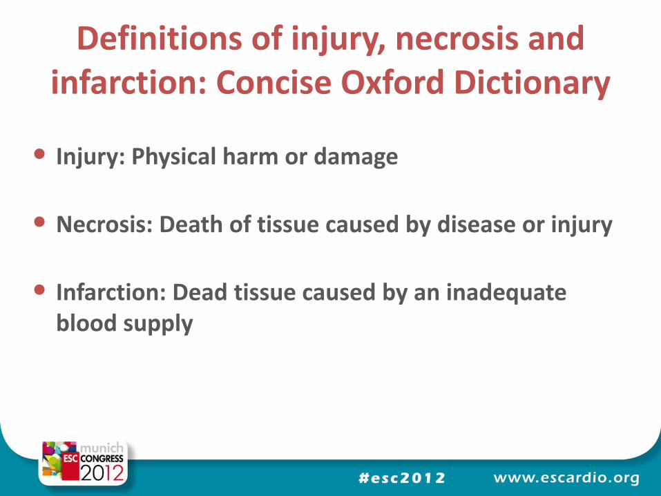
Definition of myocardial infarction
Acute myocardial
infarction is defined
as myocardial cell
death due to prolonged
myocardial ischemia
Pathology
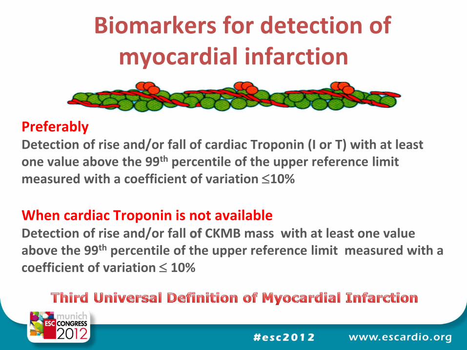
Biomarkers for detection of myocardial infarction
Preferably Detection of rise and/or fall of cardiac Troponin (I or T) with at least one value above the 99th percentile of the upper reference limit measured with a coefficient of variation 10%
When cardiac Troponin is not available Detection of rise and/or fall of CKMB mass with at least one value above the 99th percentile of the upper reference limit measured with a coefficient of variation 10%
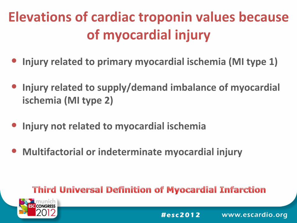
Elevations of cardiac troponin values because of myocardial injury
• Injury related to primary myocardial ischemia (MI type 1)
• Injury related to supply/demand imbalance of myocardial ischemia (MI type 2)
• Injury not related to myocardial ischemia
• Multifactorial or indeterminate myocardial injury
Myocardial injury: quote from the 3rd Universal definition document
“Small amounts of myocardial injury with necrosis may be detected which are associated with heart failure, renal failure, myocarditis, arrhythmias, pulmonary embolism or otherwise uneventful percutaneous or surgical coronary procedures. These should not be labelled as MI or a complication of the procedures, but rather as myocardial injury ”
Myocardial injury: quote from the 3rd Universal definition document
“It is recognized that the complexity of clinical circumstances may sometimes render it difficult to determine where individual cases my lie (with respect to myocardial injury vs. myocardial infarction).”
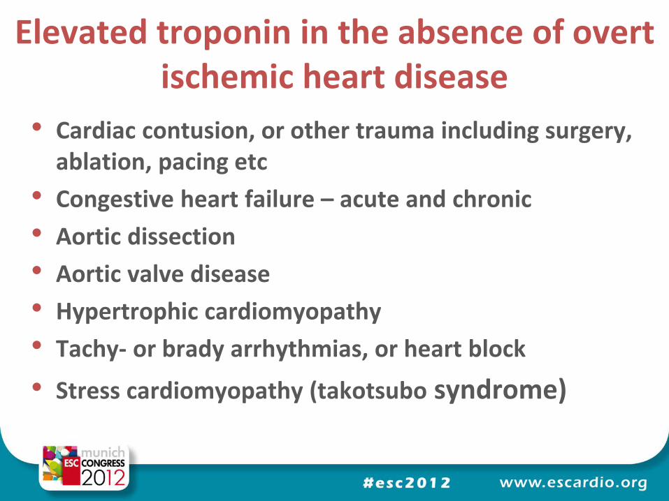
Elevated troponin in the absence of overt ischemic heart disease
• Cardiac contusion, or other trauma including surgery, ablation, pacing etc
• Congestive heart failure – acute and chronic
• Aortic dissection
• Aortic valve disease
• Hypertrophic cardiomyopathy
• Tachy- or brady arrhythmias, or heart block
• Stress cardiomyopathy (takotsubo syndrome)
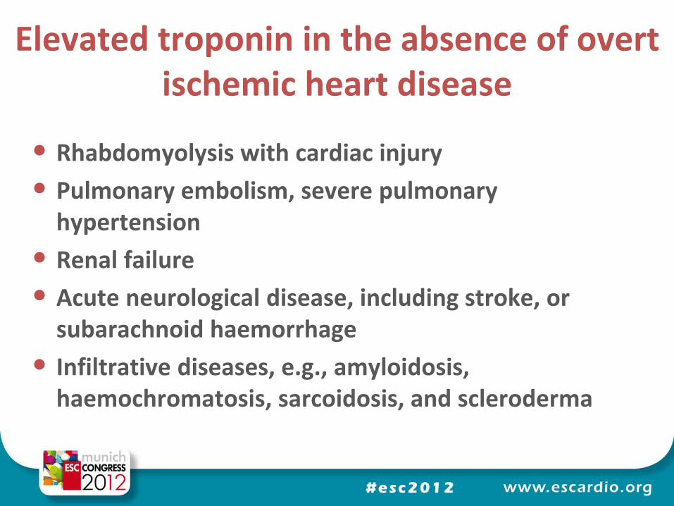
Elevated troponin in the absence of overt ischemic heart disease
• Rhabdomyolysis with cardiac injury
• Pulmonary embolism, severe pulmonary hypertension
• Renal failure
• Acute neurological disease, including stroke, or subarachnoid haemorrhage
• Infiltrative diseases, e.g., amyloidosis, haemochromatosis, sarcoidosis, and scleroderma
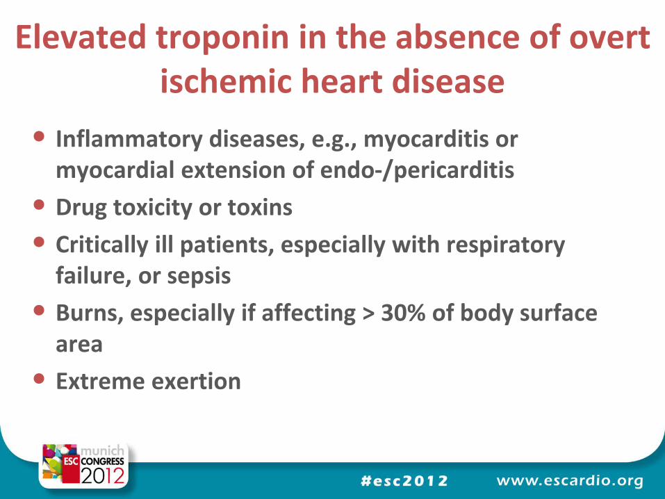
Elevated troponin in the absence of overt ischemic heart disease
• Inflammatory diseases, e.g., myocarditis or myocardial extension of endo-/pericarditis
• Drug toxicity or toxins
• Critically ill patients, especially with respiratory failure, or sepsis
• Burns, especially if affecting > 30% of body surface area
• Extreme exertion
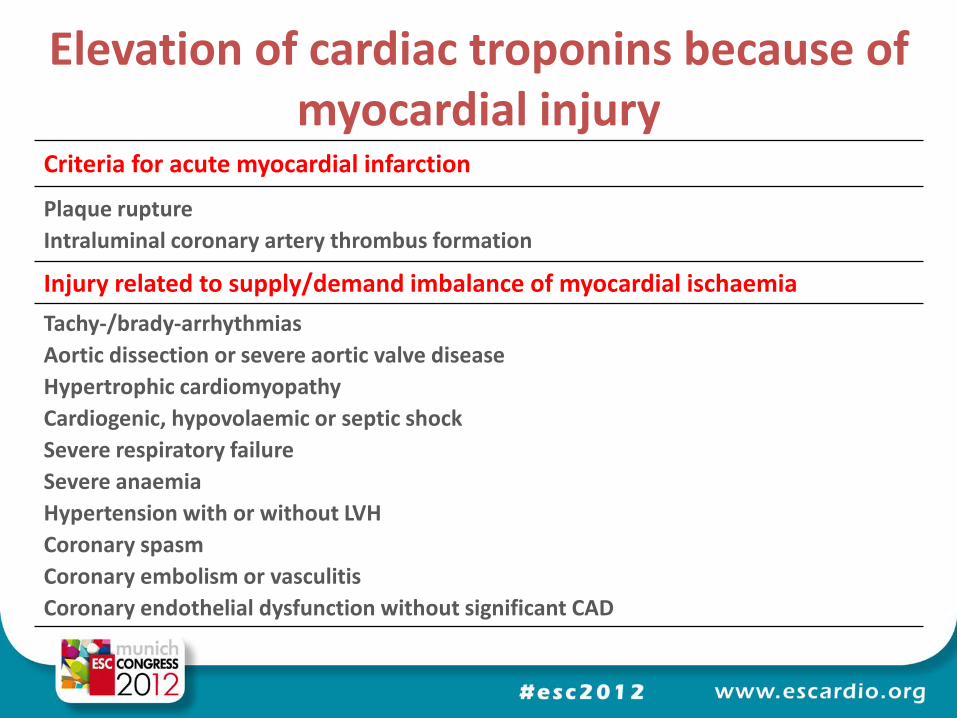
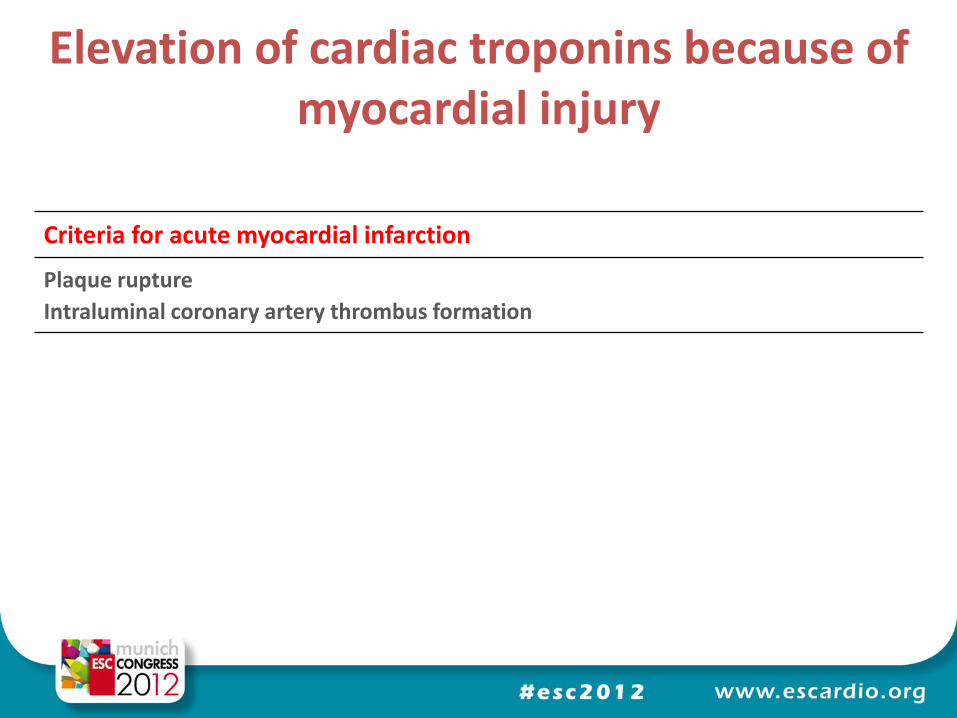
Elevation of cardiac troponins because of myocardial injury
Criteria for acute myocardial infarction
Plaque rupture
Intraluminal coronary artery thrombus formation
Injury related to supply/demand imbalance of myocardial ischaemia
Tachy-/brady-arrhythmias
Aortic dissection or severe aortic valve disease
Hypertrophic cardiomyopathy
Cardiogenic, hypovolaemic or septic shock
Severe respiratory failure
Severe anaemia
Hypertension with or without LVH
Coronary spasm
Coronary embolism or vasculitis
Coronary endothelial dysfunction without significant CAD
Elevation of cardiac troponins because of myocardial injury
Criteria for acute myocardial infarction
Plaque rupture
Intraluminal coronary artery thrombus formation
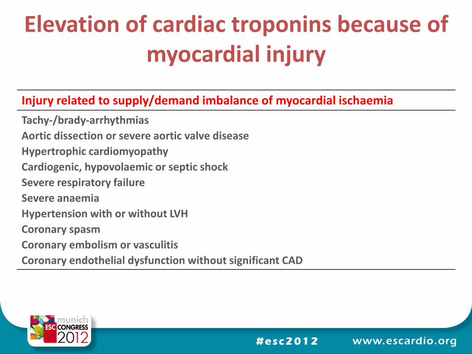
Elevation of cardiac troponins because of myocardial injury
Injury related to supply/demand imbalance of myocardial ischaemia
Tachy-/brady-arrhythmias
Aortic dissection or severe aortic valve disease
Hypertrophic cardiomyopathy
Cardiogenic, hypovolaemic or septic shock
Severe respiratory failure
Severe anaemia
Hypertension with or without LVH
Coronary spasm
Coronary embolism or vasculitis
Coronary endothelial dysfunction without significant CAD
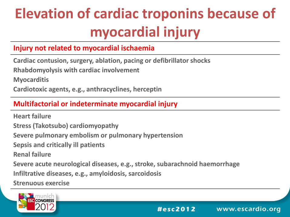
Elevation of cardiac troponins because of myocardial injury
Injury not related to myocardial ischaemia
Cardiac contusion, surgery, ablation, pacing or defibrillator shocks
Rhabdomyolysis with cardiac involvement
Myocarditis
Cardiotoxic agents, e.g., anthracyclines, herceptin
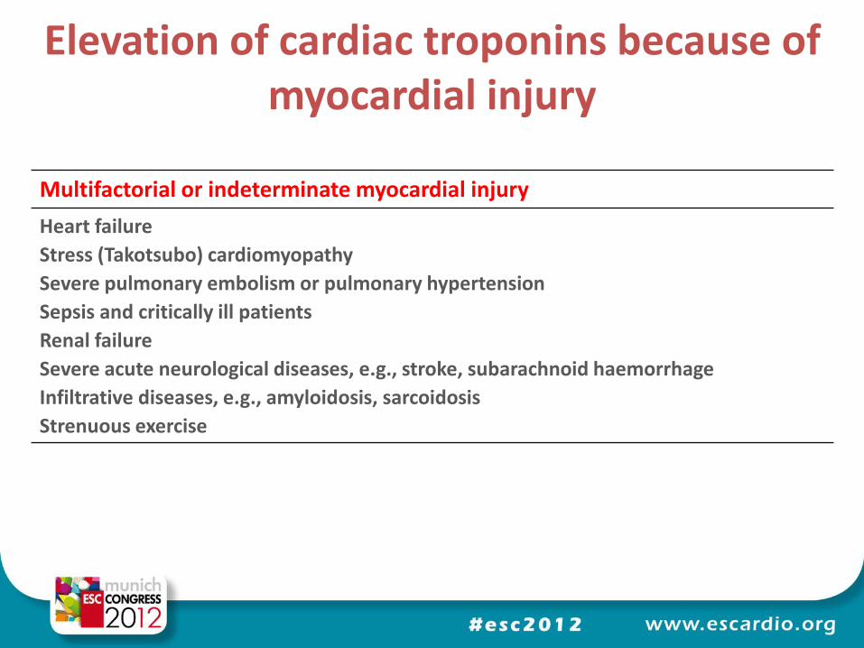
Multifactorial or indeterminate myocardial injury
Heart failure
Stress (Takotsubo) cardiomyopathy
Severe pulmonary embolism or pulmonary hypertension
Sepsis and critically ill patients
Renal failure
Severe acute neurological diseases, e.g., stroke, subarachnoid haemorrhage
Infiltrative diseases, e.g., amyloidosis, sarcoidosis
Strenuous exercise
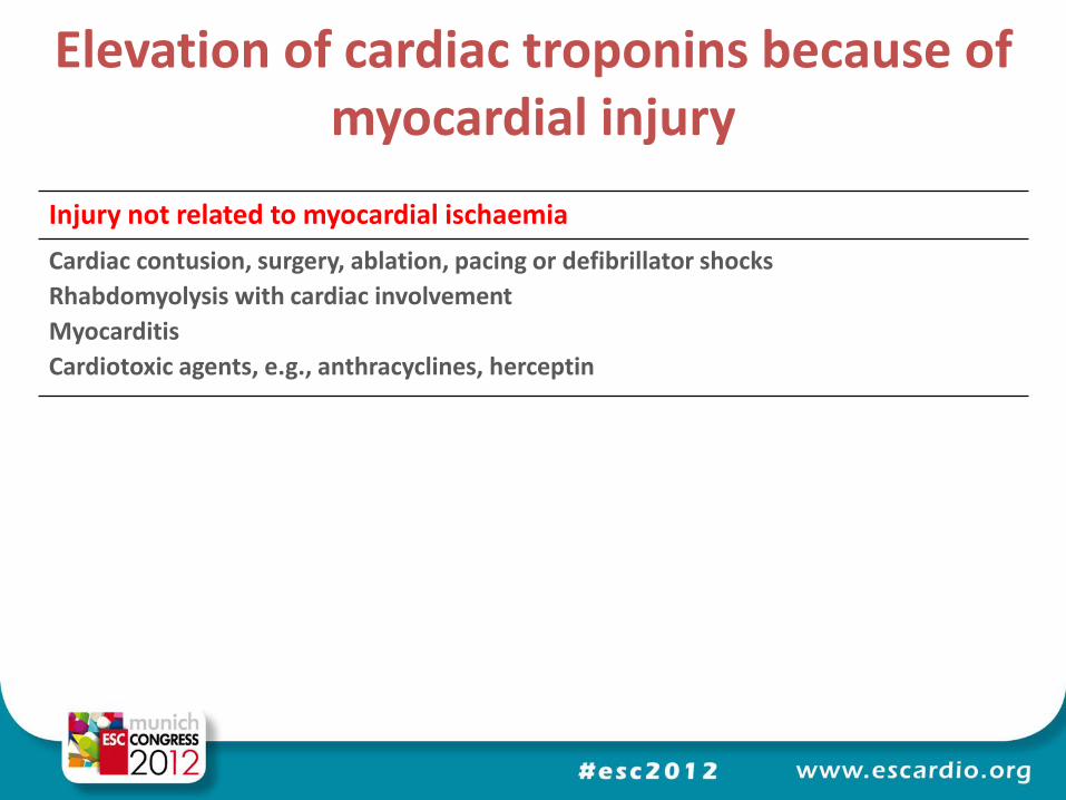
Elevation of cardiac troponins because of myocardial injury
Injury not related to myocardial ischaemia
Cardiac contusion, surgery, ablation, pacing or defibrillator shocks
Rhabdomyolysis with cardiac involvement
Myocarditis
Cardiotoxic agents, e.g., anthracyclines, herceptin
Elevation of cardiac troponins because of myocardial injury
Multifactorial or indeterminate myocardial injury
Heart failure
Stress (Takotsubo) cardiomyopathy
Severe pulmonary embolism or pulmonary hypertension
Sepsis and critically ill patients
Renal failure
Severe acute neurological diseases, e.g., stroke, subarachnoid haemorrhage
Infiltrative diseases, e.g., amyloidosis, sarcoidosis
Strenuous exercise
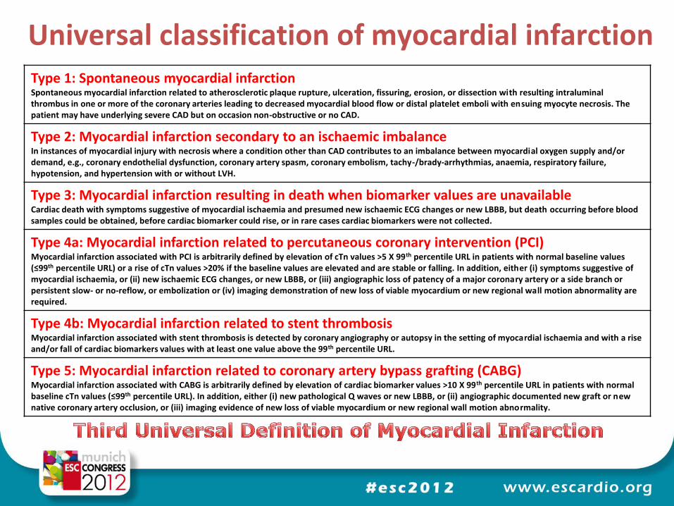
Universal classification of myocardial infarction Type 1: Spontaneous myocardial infarction Spontaneous myocardial infarction related to atherosclerotic plaque rupture, ulceration, fissuring, erosion, or dissection with resulting intraluminal thrombus in one or more of the coronary arteries leading to decreased myocardial blood flow or distal platelet emboli with ensuing myocyte necrosis. The patient may have underlying severe CAD but on occasion non-obstructive or no CAD.
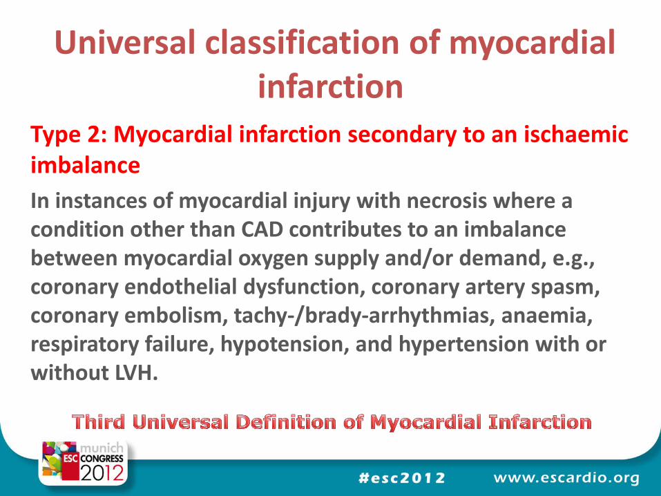
Type 2: Myocardial infarction secondary to an ischaemic imbalance In instances of myocardial injury with necrosis where a condition other than CAD contributes to an imbalance between myocardial oxygen supply and/or demand, e.g., coronary endothelial dysfunction, coronary artery spasm, coronary embolism, tachy-/brady-arrhythmias, anaemia, respiratory failure, hypotension, and hypertension with or without LVH.
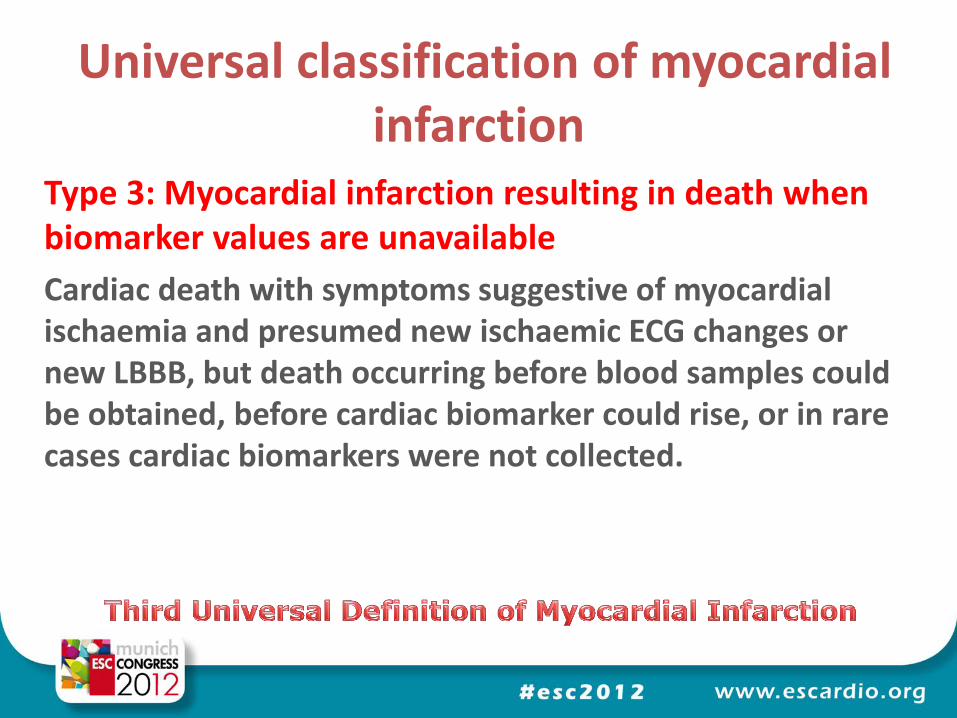
Type 3: Myocardial infarction resulting in death when biomarker values are unavailable
Cardiac death with symptoms suggestive of myocardial ischaemia and presumed new ischaemic ECG changes or new LBBB, but death occurring before blood samples could be obtained, before cardiac biomarker could rise, or in rare cases cardiac biomarkers were not collected.
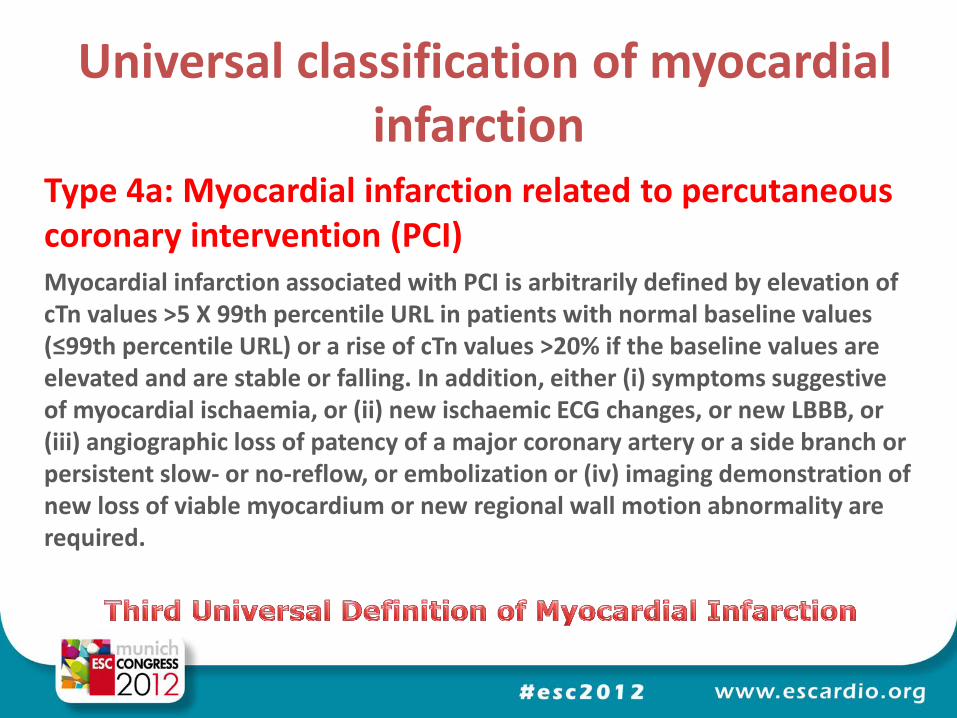
Type 4a: Myocardial infarction related to percutaneous coronary intervention (PCI) Myocardial infarction associated with PCI is arbitrarily defined by elevation of cTn values >5 X 99th percentile URL in patients with normal baseline values (≤99th percentile URL) or a rise of cTn values >20% if the baseline values are elevated and are stable or falling. In addition, either (i) symptoms suggestive of myocardial ischaemia, or (ii) new ischaemic ECG changes, or new LBBB, or (iii) angiographic loss of patency of a major coronary artery or a side branch or persistent slow- or no-reflow, or embolization or (iv) imaging demonstration of new loss of viable myocardium or new regional wall motion abnormality are required.
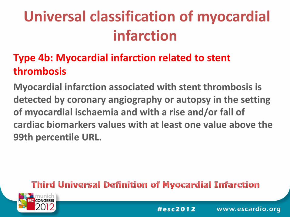
Type 4b: Myocardial infarction related to stent thrombosis
Myocardial infarction associated with stent thrombosis is detected by coronary angiography or autopsy in the setting of myocardial ischaemia and with a rise and/or fall of cardiac biomarkers values with at least one value above the 99th percentile URL.
Type 5: Myocardial infarction related to coronary artery bypass grafting (CABG)
Myocardial infarction associated with CABG is arbitrarily defined by elevation of cardiac biomarker values >10 X 99th percentile URL in patients with normal baseline cTn values (≤99th percentile URL). In addition, either (i) new pathological Q waves or new LBBB, or (ii) angiographic documented new graft or new native coronary artery occlusion, or (iii) imaging evidence of new loss of viable myocardium or new regional wall motion abnormality.
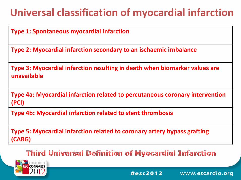
Universal classification of myocardial infarction
Type 1: Spontaneous myocardial infarction
Type 2: Myocardial infarction secondary to an ischaemic imbalance
Type 3: Myocardial infarction resulting in death when biomarker values are unavailable
Type 4a: Myocardial infarction related to percutaneous coronary intervention (PCI)
Type 4b: Myocardial infarction related to stent thrombosis
Type 5: Myocardial infarction related to coronary artery bypass grafting (CABG)
Universal classification of myocardial infarction
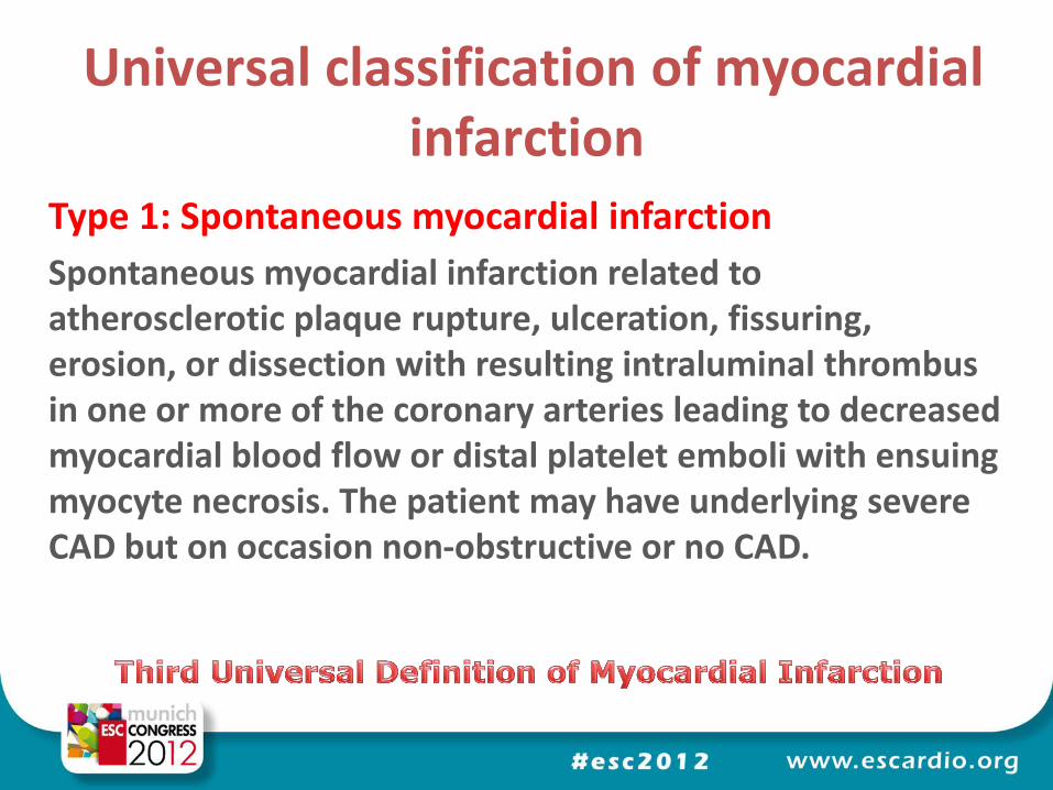
Type 1: Spontaneous myocardial infarction
Spontaneous myocardial infarction related to atherosclerotic plaque rupture, ulceration, fissuring, erosion, or dissection with resulting intraluminal thrombus in one or more of the coronary arteries leading to decreased myocardial blood flow or distal platelet emboli with ensuing myocyte necrosis. The patient may have underlying severe CAD but on occasion non-obstructive or no CAD.
Type 2: Myocardial infarction secondary to an ischaemic imbalance
In instances of myocardial injury with necrosis where a condition other than CAD contributes to an imbalance between myocardial oxygen supply and/or demand, e.g., coronary endothelial dysfunction, coronary artery spasm, coronary embolism, tachy-/brady-arrhythmias, anaemia, respiratory failure, hypotension, and hypertension with or without LVH.
Universal classification of myocardial infarction
Universal classification of myocardial infarction
Type 3: Myocardial infarction resulting in death when biomarker values are unavailable
Cardiac death with symptoms suggestive of myocardial ischaemia and presumed new ischaemic ECG changes or new LBBB, but death occurring before blood samples could be obtained, before cardiac biomarker could rise, or in rare cases cardiac biomarkers were not collected.
Universal classification of myocardial infarction
Type 4a: Myocardial infarction related to percutaneous coronary intervention (PCI)
Myocardial infarction associated with PCI is arbitrarily defined by elevation of cTn values >5 X 99th percentile URL in patients with normal baseline values (≤99th percentile URL) or a rise of cTn values >20% if the baseline values are elevated and are stable or falling. In addition, either (i) symptoms suggestive of myocardial ischaemia, or (ii) new ischaemic ECG changes, or new LBBB, or (iii) angiographic loss of patency of a major coronary artery or a side branch or persistent slow- or no-reflow, or embolization or (iv) imaging demonstration of new loss of viable myocardium or new regional wall motion abnormality are required.
Universal classification of myocardial infarction
Type 4b: Myocardial infarction related to stent thrombosis
Myocardial infarction associated with stent thrombosis is detected by coronary angiography or autopsy in the setting of myocardial ischaemia and with a rise and/or fall of cardiac biomarkers values with at least one value above the 99th percentile URL.
Universal classification of myocardial infarction
Type 5: Myocardial infarction related to coronary artery bypass grafting (CABG)
Myocardial infarction associated with CABG is arbitrarily defined by elevation of cardiac biomarker values >10 X 99th percentile URL in patients with normal baseline cTn values (≤99th percentile URL). In addition, either (i) new pathological Q waves or new LBBB, or (ii) angiographic documented new graft or new native coronary artery occlusion, or (iii) imaging evidence of new loss of viable myocardium or new regional wall motion abnormality.
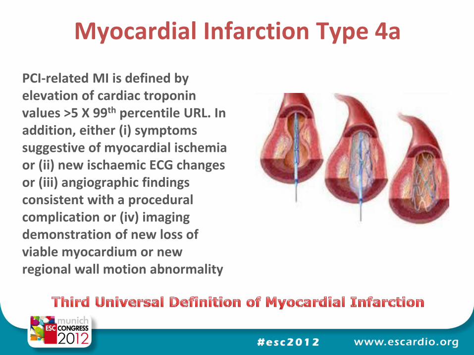
Myocardial Infarction Type 4a
PCI-related MI is defined by elevation of cardiac troponin values >5 X 99th percentile URL. In addition, either (i) symptoms suggestive of myocardial ischemia or (ii) new ischaemic ECG changes or (iii) angiographic findings consistent with a procedural complication or (iv) imaging demonstration of new loss of viable myocardium or new regional wall motion abnormality
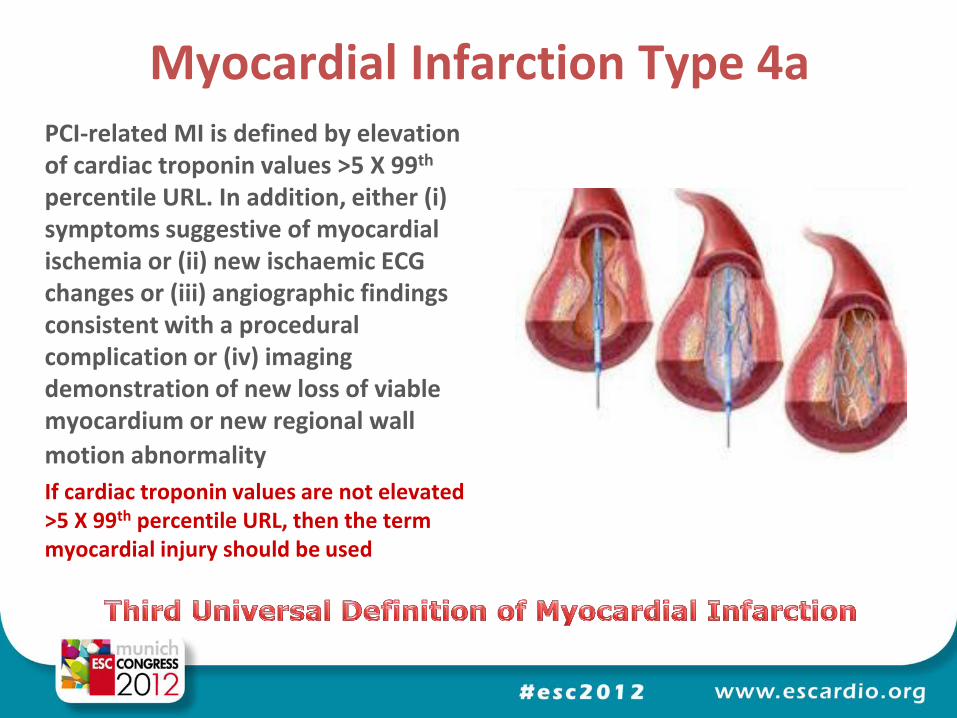
Myocardial Infarction Type 4a PCI-related MI is defined by elevation of cardiac troponin values >5 X 99th percentile URL. In addition, either (i) symptoms suggestive of myocardial ischemia or (ii) new ischaemic ECG changes or (iii) angiographic findings consistent with a procedural complication or (iv) imaging demonstration of new loss of viable myocardium or new regional wall
motion abnormality If cardiac troponin values are not elevated >5 X 99th percentile URL, then the term myocardial injury should be used
R
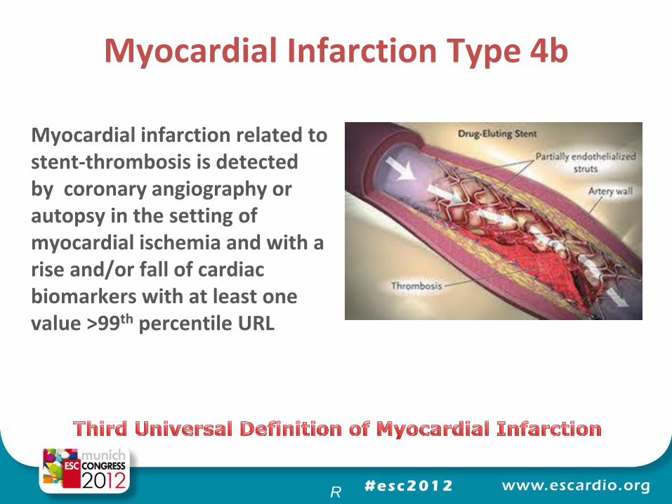
Myocardial Infarction Type 4b
Myocardial infarction related to stent-thrombosis is detected by coronary angiography or autopsy in the setting of myocardial ischemia and with a rise and/or fall of cardiac biomarkers with at least one value >99th percentile URL
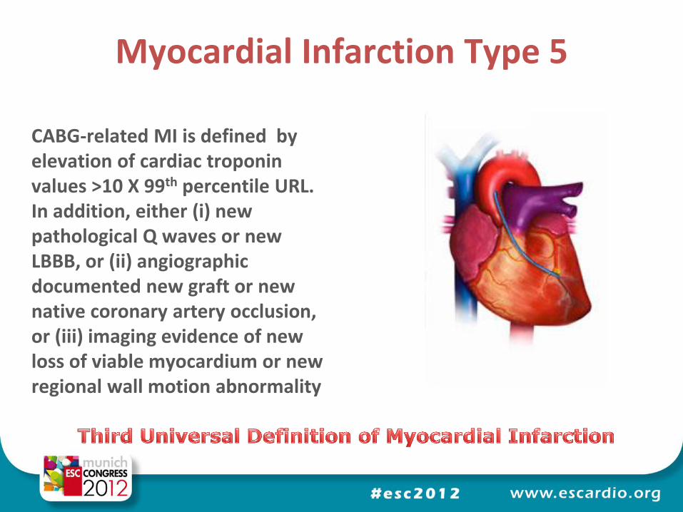
Myocardial Infarction Type 5
CABG-related MI is defined by elevation of cardiac troponin values >10 X 99th percentile URL. In addition, either (i) new pathological Q waves or new LBBB, or (ii) angiographic documented new graft or new native coronary artery occlusion, or (iii) imaging evidence of new loss of viable myocardium or new regional wall motion abnormality
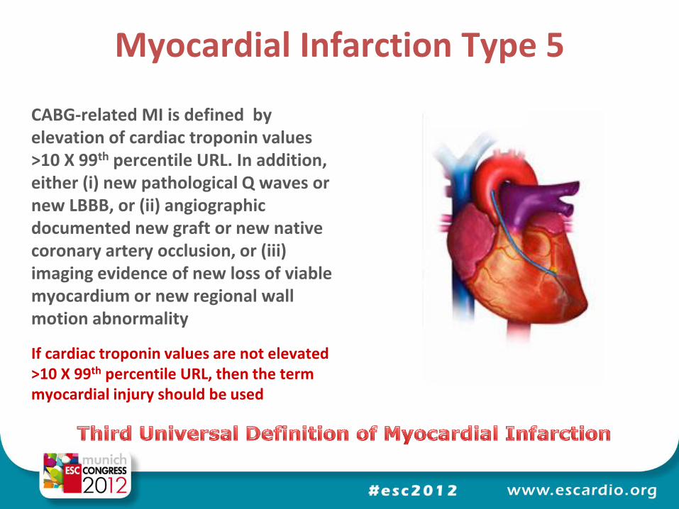
If cardiac troponin values are not elevated >10 X 99th percentile URL, then the term myocardial injury should be used
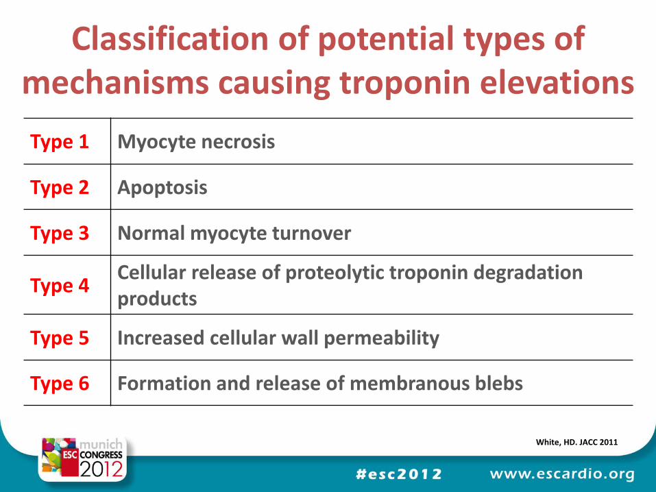
Type 1 Myocyte necrosis
Type 2 Apoptosis
Type 3 Normal myocyte turnover
Type 4 Cellular release of proteolytic troponin degradation products
Type 5 Increased cellular wall permeability
Type 6 Formation and release of membranous blebs
Classification of potential types of mechanisms causing troponin elevations
White, HD. JACC 2011
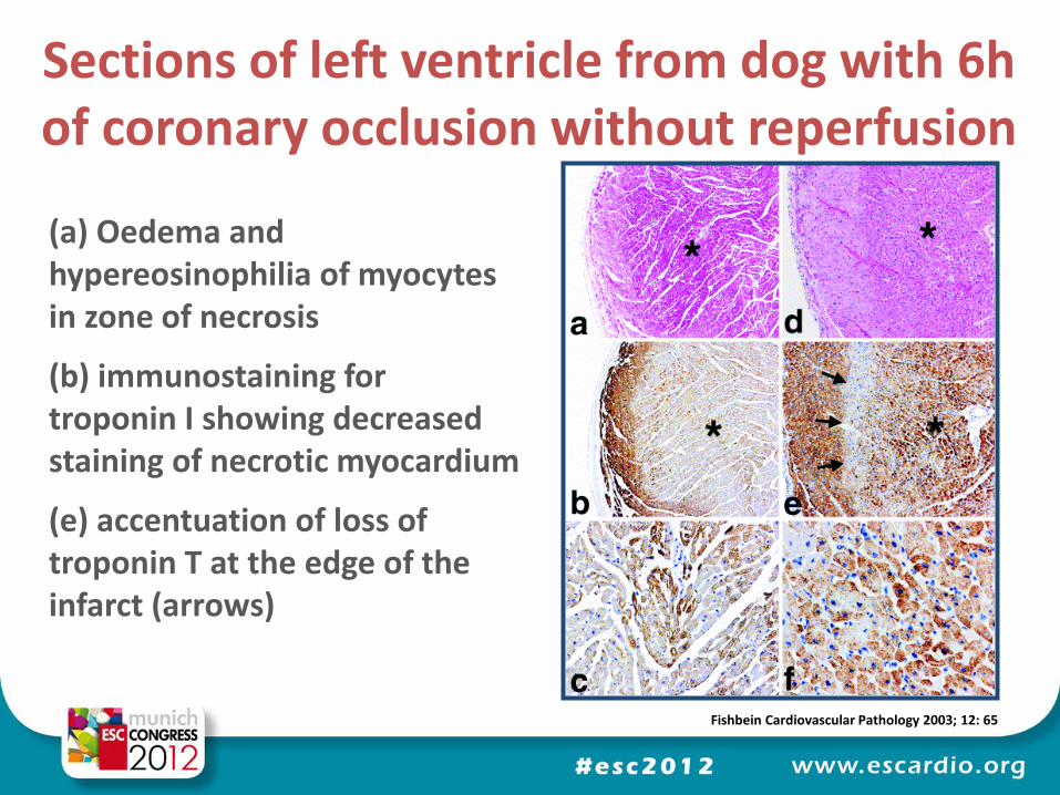
Sections of left ventricle from dog with 6h of coronary occlusion without reperfusion
Fishbein Cardiovascular Pathology 2003; 12: 65
(a) Oedema and hypereosinophilia of myocytes in zone of necrosis
(b) immunostaining for troponin I showing decreased staining of necrotic myocardium
(e) accentuation of loss of troponin T at the edge of the infarct (arrows)
• Apoptosis with preserved membrane integrity associated with activation of caspases that mediate the cleavage of structural proteins that may lead to the release of troponin
Type 2: Apoptosis
White, HD. JACC 2011
• Study of the integration of carbon-14 into the DNA of myocardial cells, generated by nuclear bomb testing, has shown that cardiac myocytes regenerate
• There is a decrease from 1% annual turnover at the age of 25 to 0.45% at the age of 75 with approximately 50% of cells exchanged during a normal life span
• Whether such low grade turnover results in release of troponin to the systemic circulation is unknown
Type 3: Normal myocyte cell turnover
White, HD. JACC 2011
• A potential cause of troponin release, without death of the cell and cellular membrane disruption, is the cellular release of proteolytic troponin degradation products
• Thus proteolysis to create small fragments could allow these to pass through a cellular membrane with normal membrane integrity
• Only 15 minutes of mild ischemia has been shown to cause development of troponin I degradation products
Type 4: Cellular release of proteolytic troponin degradation products
White, HD. JACC 2011
• Reversible injury to the cardiac myocyte membrane allowing permeability of troponins from the cytosol may occur due to myocardial stretch, or ischemia
• Simulation of stretch-responsive integrins has been shown to result in release of intact troponin from cultured viable cardiomyocytes without an increase in lactate production inferring that release may occur without ischemia or necrosis
• Also in a rat model increasing preload has been shown to be associated with release of troponin I, independent of ischemia
Type 5: Increased cellular wall permeability
White, HD. JACC 2011
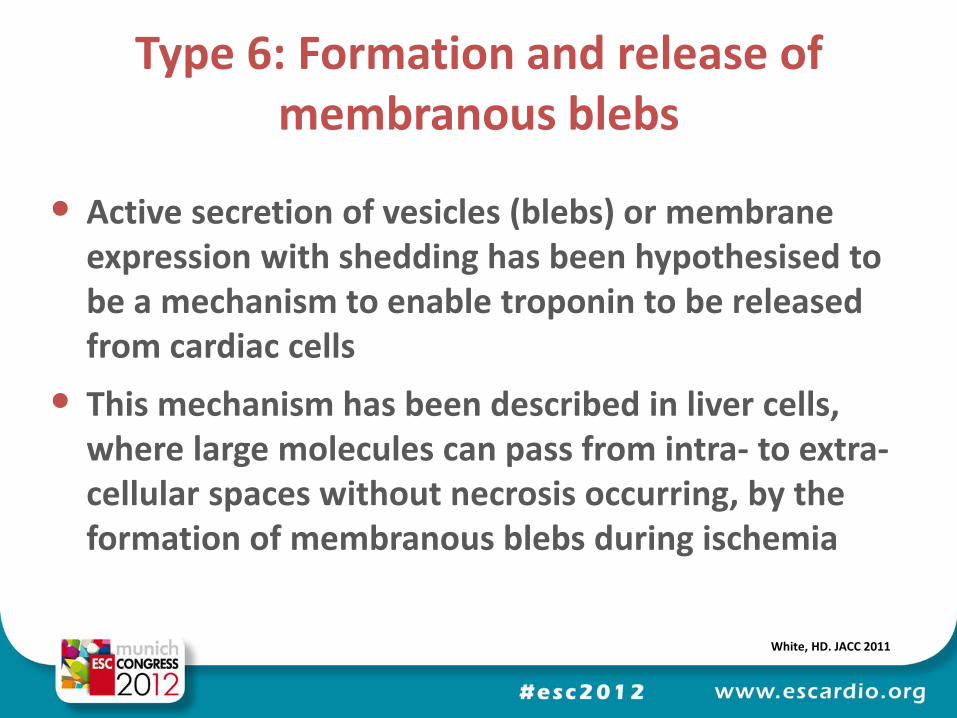
• Active secretion of vesicles (blebs) or membrane expression with shedding has been hypothesised to be a mechanism to enable troponin to be released from cardiac cells
• This mechanism has been described in liver cells, where large molecules can pass from intra- to extra-cellular spaces without necrosis occurring, by the formation of membranous blebs during ischemia
Type 6: Formation and release of membranous blebs
White, HD. JACC 2011

Microbleb formation of adult cultured myocytes
Schwartz et al. Am J Pathol 1984 in Hickman et al. Clin Chem 2010;411:318-23
Baseline 30 min of anoxia
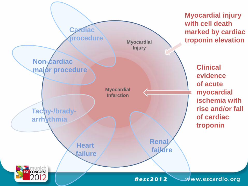
MIM
Myocardial injury
with cell death
marked by cardiac
troponin elevation
Cardiac
procedure
Tachy-/brady-
arrhythmia
Renal
failure
Heart
failure
Clinical
evidence
of acute
myocardial
ischemia with
rise and/or fall
of cardiac
troponin
Non-cardiac
major procedure
Myocardial
Injury
Myocardial
Infarction

Myocardial injury, necrosis and infarction
Harvey White Green Lane Cardiovascular Service and
Cardiovascular Research Unit Auckland City Hospital, Auckland,
New Zealand