Embed Size (px)
Citation preview
Myocardial Assessment with Cardiac CT: Ischemic HeartDisease and Beyond
Bryan C. Ramsey1 & Emilio Fentanes2 & Andrew D. Choi3 & Kelley R. Branch4& Dustin M. Thomas1
Published online: 2 June 2018#
AbstractPurpose of Review The aim of this review is to highlight recent advancements, current trends, and the expanding role for cardiacCT (CCT) in the evaluation of ischemic heart disease, nonischemic cardiomyopathies, and some specific congenital myocardialdisease states.Recent Findings CCT is a highly versatile imaging modality for the assessment of numerous cardiovascular disease states.Coronary CTangiography (CCTA) is now a well-established first-line imaging modality for the exclusion of significant coronaryartery disease (CAD); however, CCTA has modest positive predictive value and specificity for diagnosing obstructive CAD inaddition to limited capability to evaluate myocardial tissue characteristics.Summary CTP, when combined with CCTA, presents the potential for full functional and anatomic assessment with a singlemodality. CCT is a useful adjunct in select patients to both TTE and CMR in the evaluation of ventricular volumes and systolicfunction. Newer applications, such as dynamic CTP and DECT, are promising diagnostic tools offering the possibility of morequantitative assessment of ischemia. The superior spatial resolution and volumetric acquisition of CCT has an important role inthe diagnosis of other nonischemic causes of cardiomyopathies.
Keywords Cardiac CT . CT perfusion .Myocardial assessment . Cardiomyopathy . Dual-energy CT
Introduction
Cardiovascular disease remains the worldwide leading causeof morbidity and mortality accounting for up to 31% of alldeaths [1]. This trend continues to drive efforts to developadvanced detection and therapeutic modalities in hopes of
stemming this pattern. Increased focus on improved diagnos-tic techniques has fueled a rapid expansion in advanced car-diovascular imaging techniques over the last two decades.Cardiac CT (CCT), specifically coronary CT angiography(CCTA), has been well established for the evaluation of symp-tomatic patients with stable or acute chest pain and concern forcoronary artery disease (CAD) [2, 3]. Numerous studies havedemonstrated a very high negative predictive value (~ 99%)for the exclusion of CAD. Conversely, the positive predictivevalue of CCTA is modest (60–80% depending on the study) inpatients with a high pretest probability of obstructive CAD orthose with unfavorable conditions for high-quality imagingsuch as rapid heart rates and significant plaque calcifications[4]. The diagnostic power of gadolinium-enhanced cardiacmagnetic resonance (CMR) in the evaluation of ischemic heartdisease and cardiomyopathies has been well established and isthe preferred diagnostic test when the distinction betweenthese conditions is needed in a single study. Recent studieshave demonstrated similar shared characteristics in myocardi-al distribution and flux between iodinated contrast and gado-linium, particularly when iodinated contrast is coupled with
This article is part of the Topical Collection on Cardiac ComputedTomography
* Dustin M. [email protected]
1 Cardiology Division, Department of Medicine, San Antonio MilitaryMedical Center, San Antonio, TX, USA
2 Cardiology Division, Department of Medicine, Tripler ArmyMedical Center, Honolulu, HI, USA
3 Division of Cardiology, Department of Radiology, The GeorgeWashington University School of Medicine, Washington, DC, USA
4 Cardiology Division, University of Washington, Seattle, WA, USA
Current Cardiovascular Imaging Reports (2018) 11: 16https://doi.org/10.1007/s12410-018-9456-2
CARDIAC COMPUTED TOMOGRAPHY (M CHEEZUM AND B CHOW, SECTION EDITORS)
# The Author(s) 2018
X-ray photon attenuation profiles within the myocardium [5•].These findings have led to expanded applications of CCT inthe evaluation of ischemic heart disease and cardiomyopathies(references in comments) [6, 7, 8••].
CCT for Chamber Size and FunctionAssessment
Transthoracic echocardiography (TTE) is the most widelyavailable and commonly used technique for assessing cardiacstructure and function. However, TTE assessment may besuboptimal in certain subsets of patients, namely those withpoor imaging windows due to lung disease, obesity, chest walldefects, or overlying dressings in burn and post-surgical pa-tients. CMR imaging is a powerful adjunctive test in thesepatients and is the current gold standard for assessment ofcardiac volumes and systolic function. Compared with TTEand CMR, CCT has superior spatial resolution with decreasedbut comparable temporal resolution [9, 10]. Quantification ofventricular volumes and function requires acquisition of a fullcardiac cycle, or R-R interval, which requires retrospective,ECG-gated scanning in most scanner platforms. While earlystudies reported effective radiation doses of at least 10–14 mSv utilizing retrospective acquisition and 64-slice multi-detector CT (MDCT) scanner platforms, the latest genera-tion scanner platforms have achieved doses as low as3.8 mSv in select patients [11–13]. In head-to-head com-parison studies, CCT-derived ventricular volumes andejection fraction (EF) have excellent correlation withCMR and may be superior to both 2D and 3D echo [14•].When viewed in cine mode on a 3D workstation, CCT canbe used for the evaluation of regional wall motion changesin both the left ventricle (LV) and right ventricle (RV). Tooptimize acquisition and limit contrast exposure, contrastbolus injection should be tailored to the ventricle of inter-est. In LV-only imaging, scan triggering and injection pro-tocols similar to those utilized for CCTA can be utilized. Ifbiventricular assessment is needed, special attentionshould be paid to the contrast injection protocol to allowfor uniform contrast opacification of the chamber of inter-est while minimizing mixing and beam-hardening artifactscommon in the right heart. This typically requires atriphasic injection protocol utilizing a standard initial con-trast injection (4–6 mL/s) followed by a saline/contrastmixture (possibly at a lower injection rate of 2–3 mL/s)to maximize right-heart opacification and minimizingblood/contrast swirling, and completed with a saline bolus.CCT-derived RV measurements show excellent correlationwith CMR and can be especially useful in congenital heartdisease patients (such as tetralogy of Fallot) and in whomimplantable cardiac devices are already present [15].
Myocardial Imaging in Ischemic Heart Disease
Anatomy Versus Physiology in the Evaluation of CAD
Myocardial assessment in ischemic heart disease encompassesboth the anatomical assessment of the cardiac dimensions andstructure as well as indirectly assessing coronary artery steno-sis severity and CAD chronicity. There is a complex interac-tion between observed coronary anatomy (i.e., luminal steno-sis) and the presence of ischemia. Published data demonstratesthat a luminal stenosis ≥ 50% by CCTA correlates poorly withmyocardial ischemia by either single-photon emission com-puted tomography (SPECT) or positron emission tomography(PET) with positive predictive value (PPV) ranging from 29 to58% [16]. Conversely, ischemia is still present in up to 12% ofpatients with ≥ 50% stenosis [16]. The same is true for inva-sive coronary angiography (ICA). Furthermore, revasculariza-tion based on ICA stenosis alone does not reduce death ornonfatal MI compared with medical therapy [17].Physiologic assessment with invasive fractional flow reserve(iFFR) demonstrated that an intervention guided by vessel-specific ischemia for patients with indeterminate stenosis re-sulted in 33% less percutaneous coronary interventions and30% improvement in composite cardiovascular outcomes [18,19]. Given these robust data, many suggest that iFFR is thegold standard for ischemia assessment. The ongoingISCHEMIA trial (NCT01471522) will inform the discussionregarding outcomes with revascularization based solely onischemia. In the meantime, CCT with CCTA is positioned asthe single modality capable of simultaneously evaluating cor-onary artery anatomy and CAD burden and assessment ofphysiologic myocardial blood flow.
Multimodality Myocardial Imagingin Ischemic Heart Disease
The last decade has witnessed a shift in the diagnostic ap-proach for ischemic heart disease away from the utilizationof a single functional testing modality followed by ICA to apatient-centered multimodality approach. This approach takesinto account patient parameters, preferences, and radiationdose considerations to guide therapy. As such, providerstasked with the evaluation of ischemic heart disease need abaseline understanding of the strengths and limitations ofavailable modalities to allow for a multimodality imaging ap-proach to these patients.
Single-Photon Emission Computed Tomography
SPECT is a static imaging modality that leverages differentialdistribution and uptake of modest energy (70–120 keV) radio-tracers within the myocardium based on differences in
16 Page 2 of 16 Curr Cardiovasc Imaging Rep (2018) 11: 16
coronary blood flow and myocardial viability. SPECT imag-ing, compared to iFFR, has a sensitivity of 74% and specific-ity of 79% for the diagnosis of significant obstructive CAD[20]. Important limitations of SPECT imaging include diffi-culty in diagnosing high-risk CAD in the setting of balancedischemia (i.e., global low, but homogenous blood flow), poorspatial resolution and image quality in obese patients, andeffective radiation doses that average 12–15 mSv for stress-rest protocols [21, 22]. Obesity-related artifacts can be miti-gated with attenuation correction or prone imaging, thoughthese techniques can lead to artefactual perfusion defects thatrequire the reader to synthesize data from multiple acquisi-tions and can increase imaging time [23, 24]. Additionally,several academic centers have implemented protocols to re-duce radiation dose to include routine use of half-dose acqui-sitions resulting in 5–6 mSv doses [25]. The advantages ofSPECT imaging are the ability to perform testing in patientsthat can or cannot exercise, in virtually all heart rhythms, andin known CAD and prior coronary revascularization.Additionally, there is data demonstrating the ability ofSPECT to assess viability, albeit with significantly reducedsensitivity when compared to PET or CMR [26•, 27].Finally, dynamic SPECT techniques currently being validatedoffer the promise of quantifying myocardial blood flow utiliz-ing SPECT tracers [28].
Positron Emission Tomography
PET is a versatile nuclear imaging modality that detectshigh-energy (512 keV) photons that result from an anni-hilation interaction between a positron and a valence elec-tron. In addition to static perfusion data, the radiotracersRb-82 and 13N-ammonia can be used to quantify absolutecoronary blood flow and coronary flow reserve [29, 30].Viability assessment can also be performed utilizing theglucose analog fluorodeoxyglucose (FDG) by leveragingthe difference in metabolic properties between infarctedand hibernating tissues. When combined with anatomicCCT imaging (CAC and/or CCTA), the diagnostic perfor-mance of PET imaging for the diagnosis of CAD is great-ly increased with a reported sensitivity of 90% and spec-ificity of 95% [31]. The radiation cost of PET is modest at2–4 mSv with the primary limitation to more widespreaduse of this technology limited primarily by the cost, lim-ited scanner locations, limited available readers, and un-availability or expense of stress radiotracers.
Cardiac Magnetic Resonance
CMR is the gold standard for the assessment of cardiacstructure and function. Additionally, with emerging ap-plications such as T1 mapping, CMR is the best validat-ed noninvasive modality for tissue characterization. The
addition of intravenous gadolinium allows for both first-pass stress imaging, utilizing gradient echo sequences,for the assessment of myocardial ischemia [32, 33].Compared to SPECT and ICA, stress CMR assessmentof ischemia was found to have a sensitivity of 89% forboth and specificity of 76 and 87%, respectively [21,34–37]. Performance of late gadolinium enhancement(LGE) sequences provides information on the presenceand location of myocardial infarction, as well as robustprognostic information. Additionally, the transmural ex-tent of LGE uptake serves as a powerful tool in theevaluation of viability. Beyond the evaluation of ische-mic heart disease, mid-myocardial and/or epicardial up-take of LGE can also signal the presence of other infil-trative and inflammatory cardiomyopathies, such as sar-coidosis or idiopathic myocarditis. CMR with or withoutstress has its limitations. Notably, it is an expensive,time-consuming exam (often requiring 30–60 min), ispoorly tolerated in patients with severe claustrophobia,and requires multiple (sometimes prolonged) breathholds, and gadolinium should not be used in patientswith renal dysfunction (GFR < 30). Additionally, thepresence of ferrometallic materials within the myocardi-um can create signal voids and limit the diagnostic utilityof CMR even in those with MR conditional devices.
CCT in the Assessment of Ischemic HeartDisease
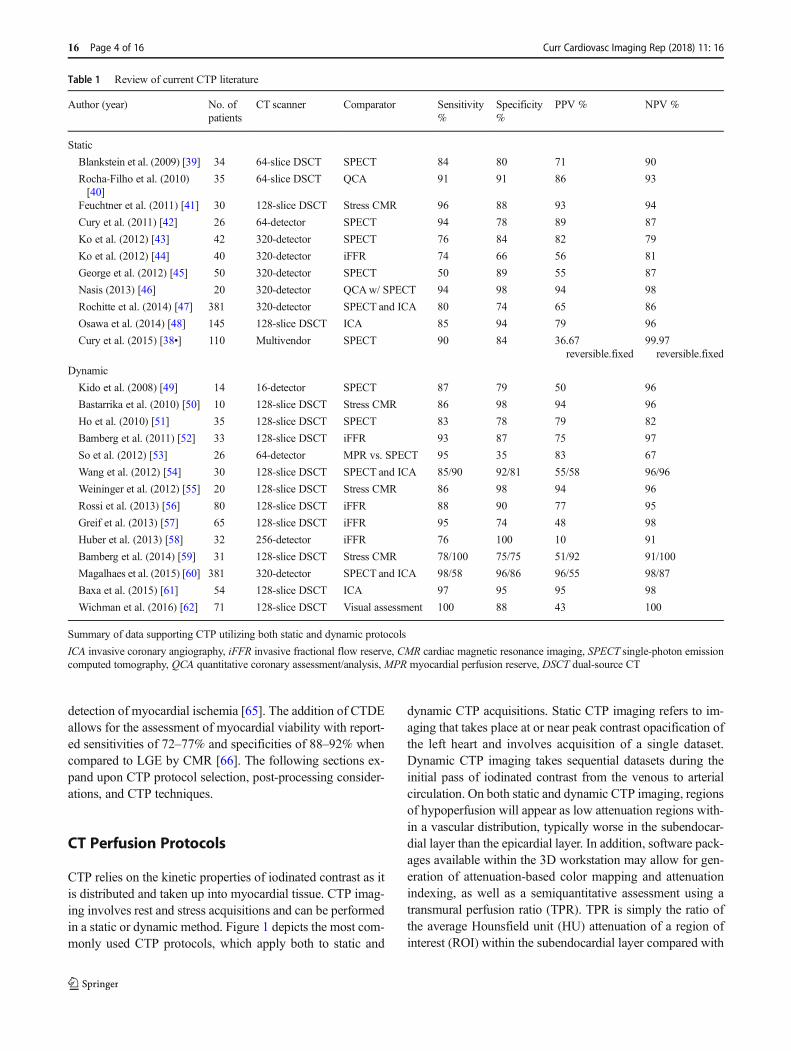
CCT is an emerging application with the potential to delivercoronary anatomy and functional significance in a single scan.Utilizing vasodilator stress agents, CCT is able to assess dif-ferences in myocardial distribution of iodinated contrast, atechnique referred to broadly as cardiac CT perfusion (CTP)[38•]. CTP protocols can differ based on the scanner platformbeing used, the information that is needed, and the desiredpatient throughput. Based on the protocol selected, the possi-bility exists to obtain detailed coronary anatomy (withCCTA), either first-pass (dynamic) or static stress perfusioninformation, stress and/or resting wall motion and EF, and CTdelayed enhancement (CTDE) for the detection of myocardialinfarction. Additionally, newer CT applications, such as dual-energy CT (DECT), show significant promise in the ability tofurther discriminate myocardial contrast uptake by leveragingthe differences in attenuation profiles between tissues andcontrast agents at different tube voltages. The accuracy ofstatic CTP imaging (Table 1) compared to SPECT forpredicting obstructive CAD on ICA is up to 96% sensitivityand 98% specificity, on a per vessel basis, with a PPV up to94% and a negative predictive value (NPV) up to 98% [39, 40,42, 43, 45–48, 63, 64]. CTP has a sensitivity and specificity of82 and 87% compared to stress CMR, respectively, for the
Curr Cardiovasc Imaging Rep (2018) 11: 16 Page 3 of 16 16
detection of myocardial ischemia [65]. The addition of CTDEallows for the assessment of myocardial viability with report-ed sensitivities of 72–77% and specificities of 88–92% whencompared to LGE by CMR [66]. The following sections ex-pand upon CTP protocol selection, post-processing consider-ations, and CTP techniques.
CT Perfusion Protocols
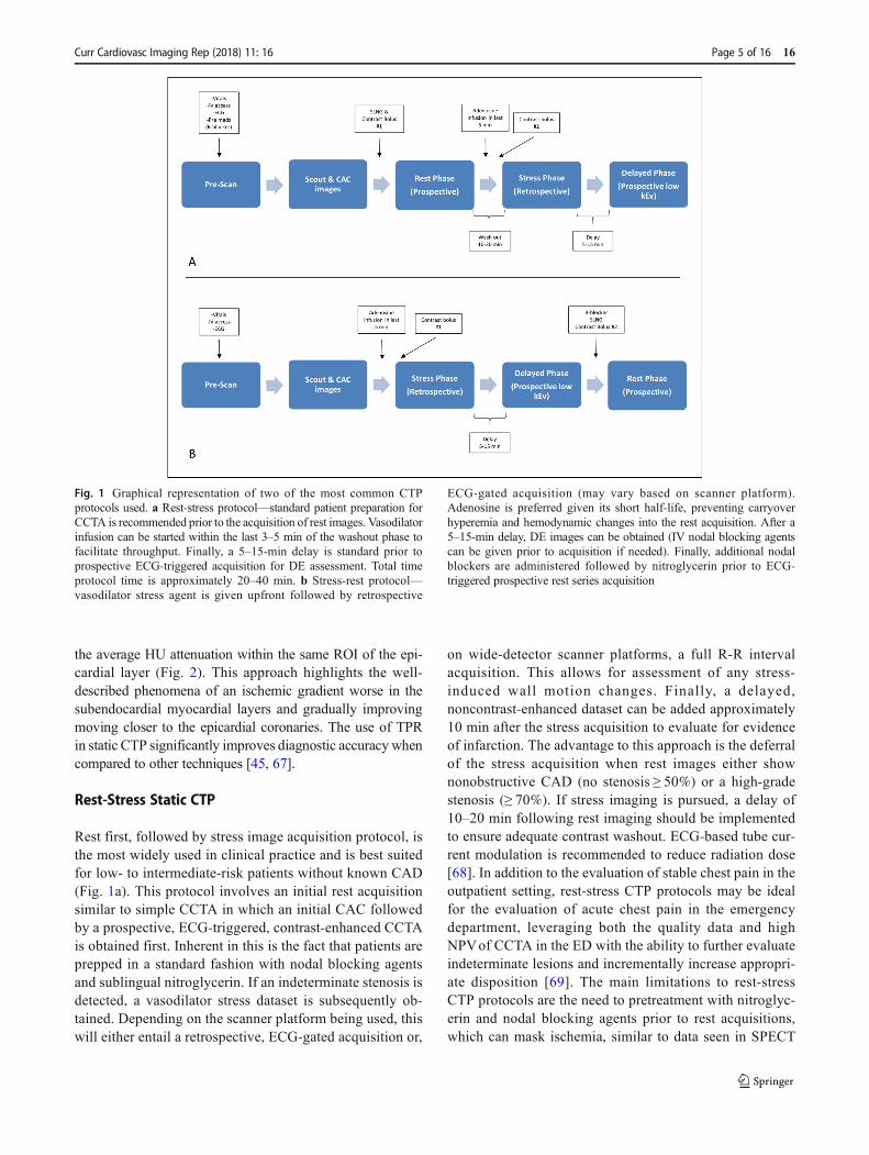
CTP relies on the kinetic properties of iodinated contrast as itis distributed and taken up into myocardial tissue. CTP imag-ing involves rest and stress acquisitions and can be performedin a static or dynamic method. Figure 1 depicts the most com-monly used CTP protocols, which apply both to static and
dynamic CTP acquisitions. Static CTP imaging refers to im-aging that takes place at or near peak contrast opacification ofthe left heart and involves acquisition of a single dataset.Dynamic CTP imaging takes sequential datasets during theinitial pass of iodinated contrast from the venous to arterialcirculation. On both static and dynamic CTP imaging, regionsof hypoperfusion will appear as low attenuation regions with-in a vascular distribution, typically worse in the subendocar-dial layer than the epicardial layer. In addition, software pack-ages available within the 3D workstation may allow for gen-eration of attenuation-based color mapping and attenuationindexing, as well as a semiquantitative assessment using atransmural perfusion ratio (TPR). TPR is simply the ratio ofthe average Hounsfield unit (HU) attenuation of a region ofinterest (ROI) within the subendocardial layer compared with
Table 1 Review of current CTP literature
Author (year) No. ofpatients
CT scanner Comparator Sensitivity%
Specificity%
PPV % NPV %
Static
Blankstein et al. (2009) [39] 34 64-slice DSCT SPECT 84 80 71 90
Rocha-Filho et al. (2010)[40]
35 64-slice DSCT QCA 91 91 86 93
Feuchtner et al. (2011) [41] 30 128-slice DSCT Stress CMR 96 88 93 94
Cury et al. (2011) [42] 26 64-detector SPECT 94 78 89 87
Ko et al. (2012) [43] 42 320-detector SPECT 76 84 82 79
Ko et al. (2012) [44] 40 320-detector iFFR 74 66 56 81
George et al. (2012) [45] 50 320-detector SPECT 50 89 55 87
Nasis (2013) [46] 20 320-detector QCAw/ SPECT 94 98 94 98
Rochitte et al. (2014) [47] 381 320-detector SPECT and ICA 80 74 65 86
Osawa et al. (2014) [48] 145 128-slice DSCT ICA 85 94 79 96
Cury et al. (2015) [38•] 110 Multivendor SPECT 90 84 36.67reversible.fixed
99.97reversible.fixed
Dynamic
Kido et al. (2008) [49] 14 16-detector SPECT 87 79 50 96
Bastarrika et al. (2010) [50] 10 128-slice DSCT Stress CMR 86 98 94 96
Ho et al. (2010) [51] 35 128-slice DSCT SPECT 83 78 79 82
Bamberg et al. (2011) [52] 33 128-slice DSCT iFFR 93 87 75 97
So et al. (2012) [53] 26 64-detector MPR vs. SPECT 95 35 83 67
Wang et al. (2012) [54] 30 128-slice DSCT SPECT and ICA 85/90 92/81 55/58 96/96
Weininger et al. (2012) [55] 20 128-slice DSCT Stress CMR 86 98 94 96
Rossi et al. (2013) [56] 80 128-slice DSCT iFFR 88 90 77 95
Greif et al. (2013) [57] 65 128-slice DSCT iFFR 95 74 48 98
Huber et al. (2013) [58] 32 256-detector iFFR 76 100 10 91
Bamberg et al. (2014) [59] 31 128-slice DSCT Stress CMR 78/100 75/75 51/92 91/100
Magalhaes et al. (2015) [60] 381 320-detector SPECT and ICA 98/58 96/86 96/55 98/87
Baxa et al. (2015) [61] 54 128-slice DSCT ICA 97 95 95 98
Wichman et al. (2016) [62] 71 128-slice DSCT Visual assessment 100 88 43 100
Summary of data supporting CTP utilizing both static and dynamic protocols
ICA invasive coronary angiography, iFFR invasive fractional flow reserve, CMR cardiac magnetic resonance imaging, SPECT single-photon emissioncomputed tomography, QCA quantitative coronary assessment/analysis, MPR myocardial perfusion reserve, DSCT dual-source CT
16 Page 4 of 16 Curr Cardiovasc Imaging Rep (2018) 11: 16
the average HU attenuation within the same ROI of the epi-cardial layer (Fig. 2). This approach highlights the well-described phenomena of an ischemic gradient worse in thesubendocardial myocardial layers and gradually improvingmoving closer to the epicardial coronaries. The use of TPRin static CTP significantly improves diagnostic accuracy whencompared to other techniques [45, 67].
Rest-Stress Static CTP
Rest first, followed by stress image acquisition protocol, isthe most widely used in clinical practice and is best suitedfor low- to intermediate-risk patients without known CAD(Fig. 1a). This protocol involves an initial rest acquisitionsimilar to simple CCTA in which an initial CAC followedby a prospective, ECG-triggered, contrast-enhanced CCTAis obtained first. Inherent in this is the fact that patients areprepped in a standard fashion with nodal blocking agentsand sublingual nitroglycerin. If an indeterminate stenosis isdetected, a vasodilator stress dataset is subsequently ob-tained. Depending on the scanner platform being used, thiswill either entail a retrospective, ECG-gated acquisition or,
on wide-detector scanner platforms, a full R-R intervalacquisition. This allows for assessment of any stress-induced wall motion changes. Finally, a delayed,noncontrast-enhanced dataset can be added approximately10 min after the stress acquisition to evaluate for evidenceof infarction. The advantage to this approach is the deferralof the stress acquisition when rest images either shownonobstructive CAD (no stenosis ≥ 50%) or a high-gradestenosis (≥ 70%). If stress imaging is pursued, a delay of10–20 min following rest imaging should be implementedto ensure adequate contrast washout. ECG-based tube cur-rent modulation is recommended to reduce radiation dose[68]. In addition to the evaluation of stable chest pain in theoutpatient setting, rest-stress CTP protocols may be idealfor the evaluation of acute chest pain in the emergencydepartment, leveraging both the quality data and highNPVof CCTA in the ED with the ability to further evaluateindeterminate lesions and incrementally increase appropri-ate disposition [69]. The main limitations to rest-stressCTP protocols are the need to pretreatment with nitroglyc-erin and nodal blocking agents prior to rest acquisitions,which can mask ischemia, similar to data seen in SPECT
Fig. 1 Graphical representation of two of the most common CTPprotocols used. a Rest-stress protocol—standard patient preparation forCCTA is recommended prior to the acquisition of rest images. Vasodilatorinfusion can be started within the last 3–5 min of the washout phase tofacilitate throughput. Finally, a 5–15-min delay is standard prior toprospective ECG-triggered acquisition for DE assessment. Total timeprotocol time is approximately 20–40 min. b Stress-rest protocol—vasodilator stress agent is given upfront followed by retrospective
ECG-gated acquisition (may vary based on scanner platform).Adenosine is preferred given its short half-life, preventing carryoverhyperemia and hemodynamic changes into the rest acquisition. After a5–15-min delay, DE images can be obtained (IV nodal blocking agentscan be given prior to acquisition if needed). Finally, additional nodalblockers are administered followed by nitroglycerin prior to ECG-triggered prospective rest series acquisition
Curr Cardiovasc Imaging Rep (2018) 11: 16 Page 5 of 16 16

imaging [70]. Additionally, residual circulating contrastfrom rest imaging can contaminate the stress acquisitionand hinder the diagnostic performance.
Stress-Rest Static CTP
Less commonly used when compared to rest-stress, stress-firstCTP is best suited for patients with intermediate to high pre-test risk known intermediate/indeterminate stenosis, or priorrevascularization where the assessment of ischemia in a par-ticular vascular territory is favored over coronary anatomy(Fig. 1b). When performing stress-first CTP, the pharmacoki-netics of the vasodilatory agents being usedmust be taken intoaccount. Dipyridamole, adenosine, or regadenoson can all beused and achieve hyperemia at various time periods followingadministration and sustain hyperemia for variable durations.Adenosine, owing to its rapid metabolism and thus rapid off-set with cessation of infusion, was used in a majority of thevalidation studies. Regadenoson is also a viable option and isthe preferred agent in SPECT and CMR due to ease of admin-istration and a low side effect profile. The limitation ofregadenoson stress-first CTP is to the persistence of heart rateelevation (30–40 min following regadenoson administration),making motion-free imaging of the coronaries challenging.Newer CT scanners can overcome the heart rate elevationassociated with regadenoson with the use of motion correctionsoftware and faster gantry rotation speeds allowing for stress-
only CTP and high-resolution coronary anatomy in a single,stress acquisition, mitigating the need for rest acquisition andthus conserving radiation dose.
Dynamic (First-Pass) CTP
Static imaging techniques, with or without stress acquisitions,are limited to single snapshots in time and do not providecomprehensive blood flow analysis. Historically, limitationsin scanner technology made static CTP the only viable meth-od. However, the latest generation 256- and 320-row detectorplatforms allow for imaging of the entire cardiac volume witha stationary table and a single gantry rotation. Additionally,second-generation dual-source CT (DSCT) can cover thissame volume utilizing a table shuttle method. The third-generation DSCT has increased z-axis coverage up to105 mm and, thus, can image the cardiac volume withoutthe need for table shuttling [50, 51, 55, 71]. This technologyallows for the performance of first-pass perfusion owing to theability of these newer generation scanners to acquire full car-diac datasets in short succession, termed dynamic CTP.Dynamic CTP allows for comparison of time-attenuation pro-files within myocardial segments, which facilitates directquantification of myocardial blood flow (MBF) [72]. MBFcalculation by dynamic CTP involves mathematic modelingderived from the deconvolution methods used in CMR [52,73]. In semiquantitative analysis, the time-attenuation curve
Fig. 2 The left-sided images depict a thick-slab three-chamber averageattenuation reconstruction (WW/WL 300/150) with a segment of theapical septal wall segment magnified to better demonstrate whereepicardial (epi) and subendocardial (endo) regions of interest (ROI)would be drawn. TPR is calculated by obtaining the average Hounsfieldunit (HU) attenuation from a ROI within the endo (HUendo) and dividingby the average HU derived from a ROI within the epi (HUepi) within thesame wall segment. A ratio < 1.0 is abnormal and ratios ≤ 0.75 are highly
suggestive of ischemia. The right-sided image represents availablepostprocessing application software available through various vendorsthat allow for semiautomated calculation of TPR throughout the entiremyocardium. Color overlay can be added to assist with visual assessmentof ischemia. In the presented image, there is evidence of ischemia in theLAD distribution. Of note, the apparent perfusion defect in theinferolateral wall segment represents a common artifact observed inCTP and not true ischemia in the left circumflex distribution
16 Page 6 of 16 Curr Cardiovasc Imaging Rep (2018) 11: 16
for a myocardial ROI is derived and a time-to-peak attenua-tion, attenuation upslope, and area under the curve are calcu-lated. This is the most commonly used semiquantitative meth-od as only the upslope time to peak attenuation is sampled,thus lowering effective radiation dose. Dynamic CTP valida-tion studies, utilizing 320-row MDCT and second-generationDSCT, have shown varying, but mostly positive results indetection of hemodynamically significant CAD when com-pared against ICA, CMR, and SPECT. Dynamic CTP (Table1) has demonstrated sensitivities ranging from 58 to 100%,specificities from 74 to 100%, NPV 82–100%, and PPV 43–100% [51, 56, 57, 60]. The biggest limitation of dynamic CTPis the relatively high radiation dose required (8.2 to 18.8 mSvin validation studies) [62, 73]. Dynamic CTP represents anemerging CCT application and further research is needed be-fore more widespread implementation is pursued.
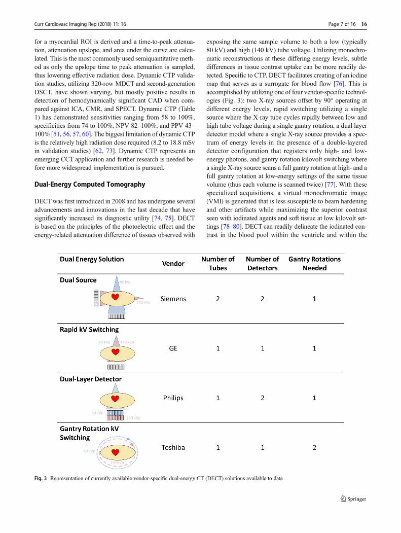
Dual-Energy Computed Tomography
DECTwas first introduced in 2008 and has undergone severaladvancements and innovations in the last decade that havesignificantly increased its diagnostic utility [74, 75]. DECTis based on the principles of the photoelectric effect and theenergy-related attenuation difference of tissues observed with
exposing the same sample volume to both a low (typically80 kV) and high (140 kV) tube voltage. Utilizing monochro-matic reconstructions at these differing energy levels, subtledifferences in tissue contrast uptake can be more readily de-tected. Specific to CTP, DECT facilitates creating of an iodinemap that serves as a surrogate for blood flow [76]. This isaccomplished by utilizing one of four vendor-specific technol-ogies (Fig. 3): two X-ray sources offset by 90° operating atdifferent energy levels, rapid switching utilizing a singlesource where the X-ray tube cycles rapidly between low andhigh tube voltage during a single gantry rotation, a dual layerdetector model where a single X-ray source provides a spec-trum of energy levels in the presence of a double-layereddetector configuration that registers only high- and low-energy photons, and gantry rotation kilovolt switching wherea single X-ray source scans a full gantry rotation at high- and afull gantry rotation at low-energy settings of the same tissuevolume (thus each volume is scanned twice) [77]. With thesespecialized acquisitions, a virtual monochromatic image(VMI) is generated that is less susceptible to beam hardeningand other artifacts while maximizing the superior contrastseen with iodinated agents and soft tissue at low kilovolt set-tings [78–80]. DECT can readily delineate the iodinated con-trast in the blood pool within the ventricle and within the
Fig. 3 Representation of currently available vendor-specific dual-energy CT (DECT) solutions available to date
Curr Cardiovasc Imaging Rep (2018) 11: 16 Page 7 of 16 16
vessels and absorbed by the myocardium and can then be usedto make color-coded maps, similar to SPECT images, thatdetail myocardial perfusion [76, 81]. Compared with SPECTand single-energy CTP, DECT protocols (Table 2) are ob-served to have a sensitivity of 82–94%, specificity of 71–94%, PPV of 53–91%, and NPV of 81–97% [84, 85].Historically, one of the main limitations to DECTwas the highrequired radiation dose and high contrast volume [86].However, subsequent advancements have shown that the useof ultralow-energy levels (40–50 kV) enhances iodine contrastdifferences and improves the accuracy of delayed enhance-ment imaging, particularly for the detection of scar [87].Several studies of DECT have achieved radiation doses of0.5 to 4.4 mSv, significantly reduced when compared to earlyDECTor SPECT [88, 89]. Additionally, no reduction in imagequality was observed despite reductions in contrast volumeapproaching 50% [90, 91]. Currently, DECT for myocardialperfusion is not routinely utilized in clinical practice as furtherstudy is ongoing to determine the optimal energy settings andto further investigate the various vendor-specific DECT solu-tions more thoroughly for cardiac imaging [92–94].
CTP Post-processing at the 3D Workstation
Post-processing of CTP datasets relies on the visual assess-ment of the ischemic myocardial segments in comparison tonormally perfused myocardium (Fig. 4). Multiplanerreformatted images allow for evaluation in the classic 17 seg-ment model view. Image display settings should be adjusted tothick MPR slabs (3–8 mm) and minimum intensity projection(MinIP) or average HU attenuation projection as opposed tomaximum intensity projection (MIP). This allows for moreready identification of ischemic segments. Finally, appropriatewindow width and level settings (200–300 and 100–150, re-spectively) should be utilized [39, 95]. These settings optimizethe displayed grayscale centering around the normal HU at-tenuation of the myocardium (average HU of 90–100) and thenarrow width accentuates ischemic or infarcted myocardiumranging from subzero HU to 30 HU [96, 97]. TPR (Fig. 2), asdiscussed above, is a semiquantitative assessment of perfusion
that measures the ratio of the average HU of the subendocar-dial to subepicardial tissue where a normal TPR has beendefined as above 1 and a ratio of 0.75 or less suggests ischemia[42]. The combination of DE-CCT with TPR compared toSPECT demonstrates a sensitivity of 86%, specificity of92%, positive predictive value of 92%, and negative predic-tive value of 85% for diagnosing clinically significant perfu-sion defects.
Limitations of CTP
Radiation dose, as mentioned above, continues to be a limita-tion to widespread implementation of CTP protocols. Newergeneration scanners and the possibility of single acquisitionCCTA and stress CTP hold promise for lowering radiationdose to levels more comparable to SPECT. Imaging artifacts,specifically beam hardening from the descending thoracic aor-ta, can affect interpretation of the inferolateral wall segmentsbymimicking a perfusion defect in that territory. Utilization ofbeta-blockers and nitrates, as is often required for acquisition
Table 2 Review of current literature supporting dual-energy CTP
Author (year) No. of patients CT scanner Comparator Sensitivity % Specificity % PPV % NPV %
Ruzsics et al. (2009) [74] 36 64-slice DSCT SPECT 92 93 83 97
Wang et al. (2011) [82] 31 64-slice DSCT Stress CMR 89 78 74 91
Ko et al. (2011) [83] 50 64-slice DSCT Stress CMR 89 78 74 91
Ko et al. (2012) [43] 45 64-slice DSCT ICA 89 74 80 85
Kim et al. (2014) [84] 50 128-slice DSCT Stress CMR 94 71 60 96
Summary of data supporting CTP utilizing both static and dynamic protocols
ICA invasive coronary angiography, CMR cardiac magnetic resonance imaging, DSCT dual-source CT
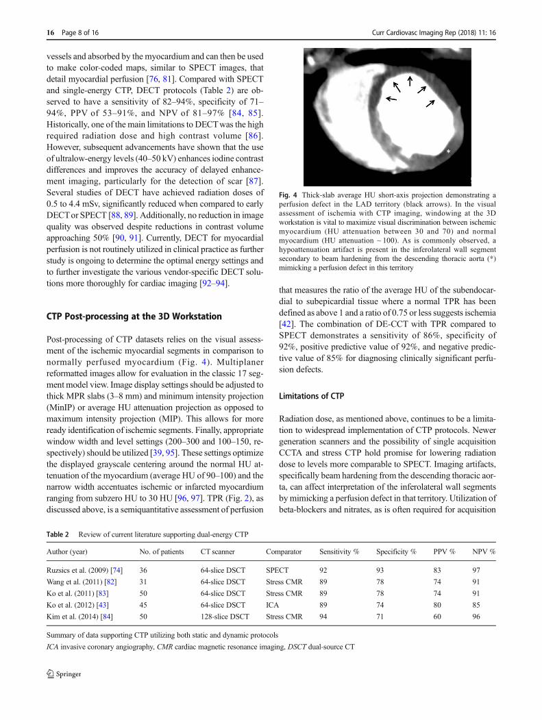
Fig. 4 Thick-slab average HU short-axis projection demonstrating aperfusion defect in the LAD territory (black arrows). In the visualassessment of ischemia with CTP imaging, windowing at the 3Dworkstation is vital to maximize visual discrimination between ischemicmyocardium (HU attenuation between 30 and 70) and normalmyocardium (HU attenuation ~ 100). As is commonly observed, ahypoattenuation artifact is present in the inferolateral wall segmentsecondary to beam hardening from the descending thoracic aorta (*)mimicking a perfusion defect in this territory
16 Page 8 of 16 Curr Cardiovasc Imaging Rep (2018) 11: 16
of CCTA data, reduces the sensitivity of CTP scans bymasking smaller, typically single-vessel, perfusion defects asshown in the SPECT literature [98, 99]. Finally, as summa-rized in Fig. 1a, a 10–20-min washout period is paramountwhen utilizing rest-stress acquisition protocols. Iodinated con-trast is slow to wash into (and subsequently out of) ischemicterritories. The presence of residual contrast in the myocardi-um at the time of the second contrast bolus injection narrowsthe attenuation profile differences between normal and ische-mic myocardium, thus reducing the sensitivity for detection ofischemic defects.
Infarct Assessment Utilizing CTDE
Over the last two decades, advancement in CMR with LGEhas revolutionized the assessment of myocardial fibrosis sec-ondary to infarction, infiltration, or inflammation. The abilityof CMR to assess these various tissue states is based on thepathologic effects on the tissues resulting in changes in tissuedensity and differential uptake of gadolinium. Iodinated con-trast has similar kinetics and distribution to gadoliniumallowing for the potential of DECT to detect infarction similarto CMR [100]. As mentioned above, CTDE involves the ac-quisition of a delayed, noncontrast-enhanced dataset obtainedapproximately 10 min after the last contrast-enhanced dataset.Similar to gadolinium imaging characteristics with CMR, in-farcted tissues will have a delayed washout for iodinated con-trast material and appear hyperenhancing [5•, 101]. Smallstudies have confirmed a correlation of 81–85% in the detec-tion of infarction compared to CMR [102, 103]. The prognos-tic importance of DE findings on CT was assessed in a smallstudy of 102 patients who showed a 19% rate of MACE at2 years. Based on these results, CTDE was identified as anindependent predictor of major adverse cardiovascular events(MACE) [104]. Utilization of ultralow kiloelectron volt set-tings can reduce artifact and accentuate smaller areas of resid-ual contrast uptake within the myocardium, thoughmore stud-ies are needed [87].
CCT in the Assessment of Nonischemicand Inheritable Cardiomyopathies
CCT can serve as an important adjunctive modality to TTEin patients with known or suspected cardiomyopathies, pri-marily in patients with claustrophobia, implantable cardiacdevices, and poor TTE windows. In the setting of newlydiagnosed heart failure with a reduced ejection fraction,CCTA is well validated to exclude significant CAD in pa-tients with low to intermediate pretest risk of CAD. Inpatients with reduced EF less than 35%, CCTA for theevaluation of CAD has a reported sensitivity of 98% and
specificity of 97% [105]. While a prospective, ECG-triggered protocol is routinely used to minimize patientradiation dose, full cardiac cycle imaging allows for theassessment of wall motion and facilitates ventricular volu-metric and EF assessment that correlate strongly withCMR [15, 106]. Several techniques including ECG-basedtube current modulation, low and ultralow kilovolt imag-ing, and iterative reconstruction have been used to reduceradiation dose in retrospective acquisition of images [107].When compared to TTE, SPECT, and CMR-based assess-ments, the CT-derived measurements correlate well with anobserved slight overestimation of LVEF. Specific to car-diomyopathies involving the RV, scan protocol changesto the contrast bolus injection may be necessary in orderto optimize RV opacification while minimizing blood-contrast mixing and beam-hardening artifacts. A triphasiccontrast injection protocol involving an initial 100% con-trast bolus at a rate between 4 and 6 mL/s followed by asaline/contrast mix at a lower rate (~ 2 mL/s) and terminat-ing with a saline bolus has been shown to provide optimalright-sided chamber opacification [108]. Table 3 highlightsCCT findings that can help to make a diagnosis. Asoutlined above, appropriate protocol selection is vital incardiomyopathies where regional wall motion, ventricularvolumes, or valve motion (SAM) is needed. As an exam-ple, Fig. 5 highlights the strengths of CCT in a patient withapical-variant hypertrophic cardiomyopathy. CCT allowsfor precise assessment of wall thickness and possible DEif appropriately protocoled. Additionally, the apicalaneurysm/pouch commonly encountered in apical-variantHCM is easily visualized, and though not present here,thrombus formation would be easily diagnosed.
Myocardial Assessment with Hybrid CardiacImaging (PET/CT)
PET when combined with CT has emerged as a powerfuldiagnostic modality, both in ischemic heart disease aswell as various inflammatory and infiltrative disease pro-cesses. PET imaging is commonly undertaken to assessthe metabolic activity of tissue utilizing the glucose an-alog 18F-fluorodeoxyglucose (FDG). FDG PET imaging,taking advantage of differences in glucose metabolismbetween normal myocytes and diseased myocytes, hasthe ability to detect hibernating myocardium in viabilitytesting and myocyte inflammation as seen in acute car-diac sarcoidosis [109]. Imaging these very different dis-ease states requires significant preimaging patient prepa-ration involving standardized protocols meant to manip-ulate the glucose substrate environment available tomyocytes [110].
Curr Cardiovasc Imaging Rep (2018) 11: 16 Page 9 of 16 16
Future Applications
While the utility of DE images has been discussed as it relatesto infarct detection in ischemic heart disease, iodine mappingwith single- or dual-energy CT can also be employed in theassessment of other cardiomyopathies where epicardial andmidmyocardial scar patterns are currently observed on CMRexclusively. CCT-based estimation of extracellular volume(ECV) by CCT may become a useful diagnostic and prognos-tic marker of myocardial remodeling similar to that observedwith T1 mapping by CMR [111–113]. Strain or deformation
imaging, a well-validated TTE for the early detection ofchemotherapy-induced cardiotoxicity, can also be calculatedon CCT using the velocity gradients between two points in themyocardium with comparable accuracy to that of TTE [114].
Conclusion
CCT in the form of CTP, particularly when combined withCCTA, is a powerful tool in the assessment of ischemicheart disease and, with newer generation scanner
Table 3 Common findings byCCT in cardiomyopathies Cardiomyopathy CCT findings
Dilated nonischemic cardiomyopathy(NICM)
• Global systolic dysfunction
• Dilated ventricle
• Apical tenting of MV leaflets
• Hypertrabeculation not meeting LVNC criteria
• Absence of significant CAD
Hypertrophic cardiomyopathy (HCM) • Asymmetric hypertrophy of basal interventricular septum or apex
• Wall segment > 15 mm at end-diastole (> 25 mm with HTN)
• SAM of the MVon cine imaging
• Patchy or diffuse midmyocardial DCE
Myocarditis/myopericarditis • Global or regional HK
• ± Pericardial effusion
• Midmyocardial or epicardial DCE
Sarcoidosis • Patchy uptake of DCE
• Global or regional WMA in noncoronary distribution
• Focal wall thickening (acute) or wall thinning (chronic)
Amyloidosis • Diffusely increased myocardial wall thickening
• Biatrial enlargement
• Diffuse subendocardial (but can have transmural) DCE
LV noncompaction • Increased ratio of noncompacted to compacted myocardium > 2.2 inend-diastole
• Involvement of > 2 segments apical to papillary muscles
• NC mass of LV > 20–25% total LV mass
• NC mass > 15 g/m2
• LV crypt thrombus
Arrhythmogenic RV cardiomyopathy(ARVC)
• Excessive mural fat content, particularly within the RV
• Regional RV WMA
• RVaneurysm
• RV dilation (EDV > 110 mL/m2 males/> 100 mL/m2 females)
• RV systolic dysfunction (RVEF < 40%)
Stress-induced cardiomyopathy(Takotsubo)
• Hyperdynamic basal wall segments
• Akinetic/dyskinetic apical segments
• Absence of DCE (i.e., no evidence of infarct)
• SAM
List of the most commonly encountered cardiomyopathies and their correlating findings on cardiac computedtomography (CCT)
MV mitral valve, LVNC left ventricular noncompaction, CAD coronary artery disease, HTN hypertension, SAMsystolic anterior motion,DCE delayed contrast enhancement,WMAwall motion abnormality, NC noncompacted,LV left ventricle, RV right ventricle, EDV end-diastolic volume, RVEF right ventricular ejection fraction
16 Page 10 of 16 Curr Cardiovasc Imaging Rep (2018) 11: 16
platforms, presents the potential for full functional andanatomic assessment with a single contrast injection andlow radiation dose dataset acquisition. CCT is a usefuladjunct to both TTE and CMR in the evaluation of ven-tricular volumes and systolic function, particularly in pa-tients with implantable cardiac devices or severe claustro-phobia. Newer applications of CCT, namely dynamic CTPand DECT, are promising diagnostic tools offering thepossibility of more quantitative assessment of ischemiathan offered by static perfusion imaging. Finally, givenits superior spatial resolution and volumetric acquisition,CCT has an important role in the diagnosis of othernonischemic causes of cardiomyopathies most notablyLVNC, ARVC, and HCM.
Funding This research received no grant from any funding agency in thepublic, commercial, or not-for-profit sectors. The opinions and assertionscontained herein are the authors alone and do not constitute endorsementby the U.S. Army Medical Department, the U.S. Army Office of theSurgeon General, the Department of the Army, or the United StatesGovernment.
Compliance with Ethical Standards
Conflict of Interest BC Ramsey, E Fentanes, AD Choi, and DMThomas all declare no conflicts of interest.
KR Branch reports grants from Astellas, outside of the submittedwork.
Human and Animal Rights and Informed Consent All studies by theauthors involving animal and/or human subjects were performed afterapproval by the appropriate institutional review boards. When required,written informed consent was obtained from all participants.
Open Access This article is distributed under the terms of the CreativeCommons At t r ibut ion 4 .0 In te rna t ional License (h t tp : / /creativecommons.org/licenses/by/4.0/), which permits unrestricted use,distribution, and reproduction in any medium, provided you give appro-priate credit to the original author(s) and the source, provide a link to theCreative Commons license, and indicate if changes were made.
References
Papers of particular interest, published recently, have beenhighlighted as:• Of importance•• Of major importance
1. Roth GA, Huffman MD, Moran AE, Feigin V, Mensah GA,Naghavi M, et al. Global and regional patterns in cardiovascularmortality from 1990 to 2013. Circulation. 2015;132(17):1667–78.https://doi.org/10.1161/circulationaha.114.008720.
2. Taylor AJ, Cerqueira M, Hodgson JM, Mark D, Min J, O'Gara P,et al. ACCF/SCCT/ACR/AHA/ASE/ASNC/NASCI/SCAI/SCMR 2010 appropriate use criteria for cardiac computed tomog-raphy. A report of the American College of CardiologyFoundation Appropriate Use Criteria Task Force, the Society ofCardiovascular Computed Tomography, the American College ofRadiology, the American Heart Association, the American Societyof Echocardiography, the American Society of NuclearCardiology, the North American Society for CardiovascularImaging, the Society for Cardiovascular Angiography andInterventions, and the Society for Cardiovascular MagneticResonance. J Am Coll Cardiol. 2010;56(22):1864–94. https://doi.org/10.1016/j.jacc.2010.07.005.
3. Hendel RC, Patel MR, Kramer CM, PoonM, Hendel RC, Carr JC,et al. ACCF/ACR/SCCT/SCMR/ASNC/NASCI/SCAI/SIR 2006appropriateness criteria for cardiac computed tomography and car-diac magnetic resonance imaging: a report of the AmericanCollege of Cardiology Foundation Quality Strategic DirectionsCommittee Appropriateness Criteria Working Group, AmericanCollege of Radiology, Society of Cardiovascular ComputedTomography, Society for Cardiovascular Magnetic Resonance,American Society of Nuclear Cardiology, North AmericanSociety for Cardiac Imaging, Society for CardiovascularAngiography and Interventions, and Society of InterventionalRadiology. J Am Coll Cardiol. 2006;48(7):1475–97. https://doi.org/10.1016/j.jacc.2006.07.003.
4. Meijboom WB, Meijs MF, Schuijf JD, Cramer MJ, Mollet NR,vanMieghemCA, et al. Diagnostic accuracy of 64-slice computedtomography coronary angiography: a prospective, multicenter,multivendor study. J Am Coll Cardiol. 2008;52(25):2135–44.https://doi.org/10.1016/j.jacc.2008.08.058.
5.• Gerber BL, Belge B, Legros GJ, Lim P, Poncelet A, Pasquet A,Gisellu G, Coche E, Vanoverschelde JL Characterization of acuteand chronic myocardial infarcts by multidetector computed to-mography: comparison with contrast-enhanced magnetic reso-nance. Circulation. 2006;113(6):823–33. doi:https://doi.org/10.1161/circulationaha.104.529511. A sentinel paper inestablishing CCT imaging parameters for assessment ofinfarction.
6. Budoff MJ, Nakazato R, Mancini GB, Gransar H, Leipsic J,Berman DS, et al. CT angiography for the prediction of hemody-namic significance in intermediate and severe lesions: head-to-head comparison with quantitative coronary angiography using
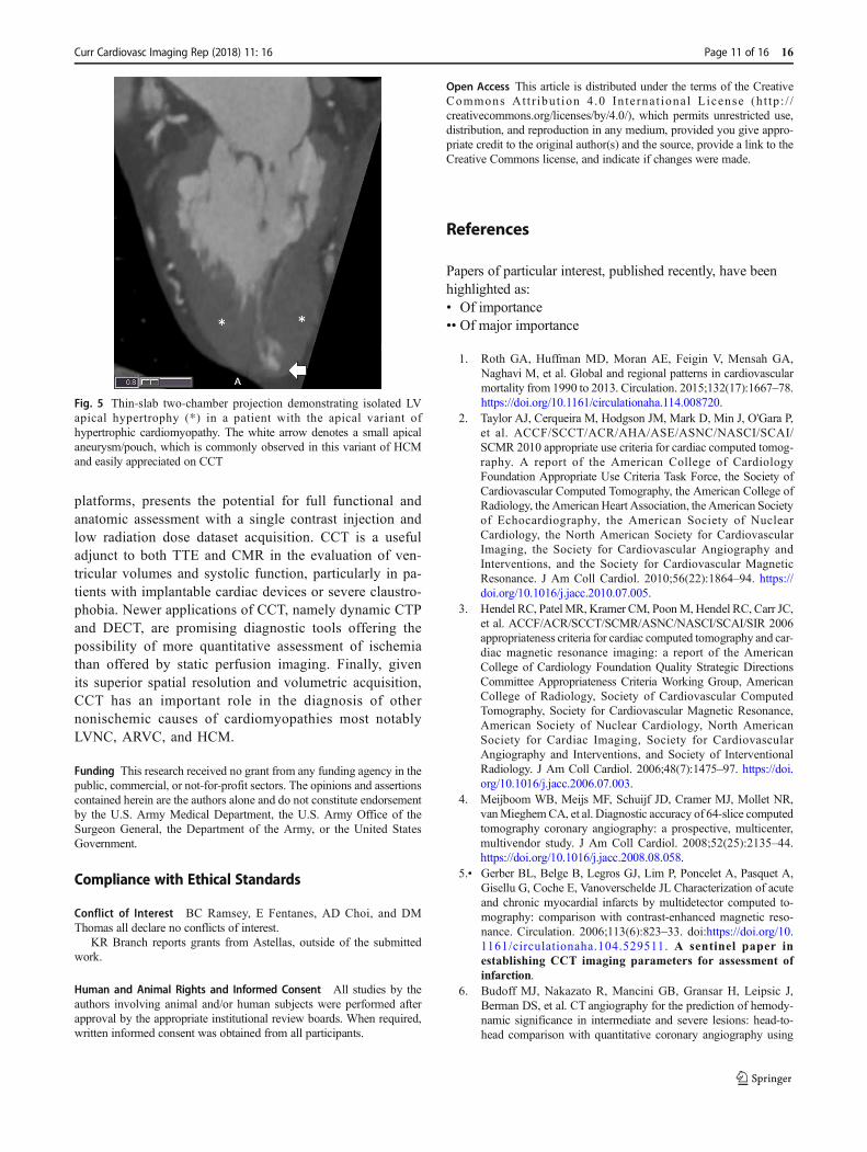
Fig. 5 Thin-slab two-chamber projection demonstrating isolated LVapical hypertrophy (*) in a patient with the apical variant ofhypertrophic cardiomyopathy. The white arrow denotes a small apicalaneurysm/pouch, which is commonly observed in this variant of HCMand easily appreciated on CCT
Curr Cardiovasc Imaging Rep (2018) 11: 16 Page 11 of 16 16
fractional flow reserve as the reference standard. JACCCardiovasc Imaging. 2016;9:559–64. https://doi.org/10.1016/j.jcmg.2015.08.021.
7. Budoff MJ, Li D, Kazerooni EA, Thomas GS, Mieres JH, ShawLJ. Diagnostic accuracy of noninvasive 64-row computed tomo-graphic coronary angiography (CCTA) comparedwith myocardialperfusion imaging (MPI): the PICTURE study, a prospective mul-ticenter trial. Acad Radiol. 2017;24(1):22–9. https://doi.org/10.1016/j.acra.2016.09.008.
8.•• Pelgrim GJ, Dorrius M, Xie X, den Dekker MA, Schoepf UJ,Henzler T, et al. The dream of a one-stop-shop: meta-analysis onmyocardial perfusion CT. Eur J Radiol. 2015;84(12):2411–20.https://doi.org/10.1016/j.ejrad.2014.12.032. Meta-analysisoutlining results of multiple prospective CTP trials
9. Schuleri KH, George RT, Lardo AC. Applications of cardiac mul-tidetector CT beyond coronary angiography. Nat Rev Cardiol.2009;6(11):699–710. https://doi.org/10.1038/nrcardio.2009.172.
10. Budoff MJ, Dowe D, Jollis JG, Gitter M, Sutherland J, HalamertE, et al. Diagnostic performance of 64-multidetector row coronarycomputed tomographic angiography for evaluation of coronaryartery stenosis in individuals without known coronary artery dis-ease: results from the prospective multicenter ACCURACY(Assessment by Coronary Computed Tomographic Angiographyof Individuals Undergoing Invasive Coronary Angiography) trial.J Am Coll Cardiol. 2008;52(21):1724–32. https://doi.org/10.1016/j.jacc.2008.07.031.
11. Kim SM, Kim YN, Choe YH. Adenosine-stress dynamic myocar-dial perfusion imaging using 128-slice dual-source CT: optimiza-tion of the CT protocol to reduce the radiation dose. Int JCardiovasc Imaging. 2013;29(4):875–84. https://doi.org/10.1007/s10554-012-0138-x.
12. Fujita M, Kitagawa K, Ito T, Shiraishi Y, Kurobe Y, Nagata M, etal. Dose reduction in dynamic CT stress myocardial perfusionimaging: comparison of 80-kV/370-mAs and 100-kV/300-mAsprotocols. Eur Radiol. 2014;24(3):748–55. https://doi.org/10.1007/s00330-013-3063-z.
13. Jakobs TF, Becker CR, Ohnesorge B, Flohr T, Suess C, SchoepfUJ, et al. Multislice helical CTof the heart with retrospective ECGgating: reduction of radiation exposure by ECG-controlled tubecurrent modulation. Eur Radiol. 2002;12(5):1081–6. https://doi.org/10.1007/s00330-001-1278-x.
14.• Greupner J, Zimmermann E, Grohmann A, Dubel HP, Althoff TF,Borges AC, et al. Head-to-head comparison of left ventricularfunction assessment with 64-row computed tomography, biplaneleft cineventriculography, and both 2- and 3-dimensional transtho-racic echocardiography: comparison with magnetic resonance im-aging as the reference standard. J Am Coll Cardiol. 2012;59(21):1897–907. https://doi.org/10.1016/j.jacc.2012.01.046.Prospective, multimodality assessment which demonstratedthe accuracy and precision of CCT for ventricular volumesand EF assessment compared with the gold standard, CMR
15. Raman SV, ShahM,McCarthy B, Garcia A, Ferketich AK.Multi-detector row cardiac computed tomography accurately quantifiesright and left ventricular size and function compared with cardiacmagnetic resonance. AmHeart J. 2006;151(3):736–44. https://doi.org/10.1016/j.ahj.2005.04.029.
16. Blankstein R, Di Carli MF. Integration of coronary anatomy andmyocardial perfusion imaging. Nat Rev Cardiol. 2010;7(4):226–36. https://doi.org/10.1038/nrcardio.2010.15.
17. Boden WE, O’Rourke RA, Teo KK, Hartigan PM, Maron DJ,Kostuk WJ, et al. Optimal medical therapy with or without PCIfor stable coronary disease. N Engl J Med. 2007;356(15):1503–16. https://doi.org/10.1056/NEJMoa070829.
18. Tonino PA, De Bruyne B, Pijls NH, Siebert U, Ikeno F, van’t VeerM, et al. Fractional flow reserve versus angiography for guiding
percutaneous coronary intervention. N Engl J Med. 2009;360(3):213–24. https://doi.org/10.1056/NEJMoa0807611.
19. De Bruyne B, FearonWF, Pijls NH, Barbato E, Tonino P, Piroth Z,et al. Fractional flow reserve-guided PCI for stable coronary arterydisease. N Engl J Med. 2014;371(13):1208–17. https://doi.org/10.1056/NEJMoa1408758.
20. TakxRA, Blomberg BA, El Aidi H, Habets J, de Jong PA, Nagel Eet al. Diagnostic accuracy of stress myocardial perfusion imagingcompared to invasive coronary angiography with fractional flowreserve meta-analysis. Circ Cardiovasc Imaging. 2015;8(1).https://doi.org/10.1161/circimaging.114.002666.
21. Thompson RC, O’Keefe JH, McGhie AI, Bybee KA, ThompsonEC, Bateman TM. Reduction of SPECTMPI radiation dose usingcontemporary protocols and technology. JACC CardiovascImaging. 2018;11(2 Pt 1):282–3. https://doi.org/10.1016/j.jcmg.2017.03.008.
22. Carpeggiani C, Picano E, Brambilla M, Michelassi C, Knuuti J,Kauffman P, et al. Variability of radiation doses of cardiac diag-nostic imaging tests: the RADIO-EVINCI study (RADIationdOsesubproject of the EVINCI study). BMC Cardiovasc Disord.2017;17(1):63. https://doi.org/10.1186/s12872-017-0474-9.
23. Huang JY, Huang CK, Yen RF, Wu HY, Tu YK, Cheng MF, et al.Diagnostic performance of attenuation-corrected myocardial per-fusion imaging for coronary artery disease: a systematic reviewand meta-analysis. Journal of Nuclear Medicine: official publica-tion, Society of Nuclear Medicine. 2016;57(12):1893–8. https://doi.org/10.2967/jnumed.115.171462.
24. Worden NE, Lindower PD, Burns TL, Chatterjee K, Weiss RM. Asecond look with prone SPECT myocardial perfusion imagingreduces the need for angiography in patients at low risk for cardiacdeath or MI. J Nucl Cardiol. 2015;22(1):115–22. https://doi.org/10.1007/s12350-014-9934-0.
25. Nakazato R, Berman DS, Hayes SW, Fish M, Padgett R, Xu Y, etal. Myocardial perfusion imaging with a solid-state camera: sim-ulation of a very low dose imaging protocol. Journal of NuclearMedicine: official publication, Society of Nuclear Medicine.2013;54(3):373–9. https://doi.org/10.2967/jnumed.112.110601.
26.•• Wolk MJ, Bailey SR, Doherty JU, Douglas PS, Hendel RC,Kramer CM, et al. ACCF/AHA/ASE/ASNC/HFSA/HRS/SCAI/SCCT/SCMR/STS 2013 multimodality appropriate use criteriafor the detection and risk assessment of stable ischemic heartdisease: a report of the American College of CardiologyFoundation Appropriate Use Criteria Task Force, AmericanHeart Association, American Society of Echocardiography,American Society of Nuclear Cardiology, Heart Failure Societyof America, Heart Rhythm Society, Society for CardiovascularAngiography and Interventions, Society of CardiovascularComputed Tomography, Society for Cardiovascular MagneticResonance, and Society of Thoracic Surgeons. J Am CollCardiol. 2014;63(4):380–406. https://doi.org/10.1016/j.jacc.2013.11.009. Multimodality imaging guidelines endorsed byall pertinent cardiovascular and imaging societies pertainingto the evaluation of stable ischemic heart disease
27. Udelson JE, Coleman PS, Metherall J, Pandian NG, Gomez AR,Griffith JL, et al. Predicting recovery of severe regional ventriculardysfunction. Comparison of resting scintigraphy with 201Tl and99mTc-sestamibi. Circulation. 1994;89(6):2552–61.
28. Agostini D, Roule V, Nganoa C, RothN, Baavour R, Parienti JJ, etal. First validation of myocardial flow reserve assessed by dynam-ic (99m)Tc-sestamibi CZT-SPECT camera: head to head compar-ison with (15)O-water PET and fractional flow reserve in patientswith suspected coronary artery disease. The WATERDAY study.Eur J Nucl Med Mol Imaging. 2018; https://doi.org/10.1007/s00259-018-3958-7.
29. Alessio AM, Bassingthwaighte JB, Glenny R, Caldwell JH.Validation of an axially distributed model for quantification of
16 Page 12 of 16 Curr Cardiovasc Imaging Rep (2018) 11: 16
myocardial blood flow using (1)(3)N-ammonia PET. J NuclCardiol. 2013;20(1):64–75. https://doi.org/10.1007/s12350-012-9632-8.
30. Gullberg GT, Shrestha UM, Seo Y. Dynamic cardiac PET imag-ing: technological improvements advancing future cardiac health.J Nucl Cardiol. 2018; https://doi.org/10.1007/s12350-018-1201-3.
31. Mc Ardle BA, Dowsley TF, de Kemp RA, Wells GA, BeanlandsRS. Does rubidium-82 PET have superior accuracy to SPECTperfusion imaging for the diagnosis of obstructive coronary dis-ease?: a systematic review and meta-analysis. J Am Coll Cardiol.2012;60(18):1828–37. https://doi.org/10.1016/j.jacc.2012.07.038.
32. Hamon M, Fau G, Nee G, Ehtisham J, Morello R, Hamon M.Meta-analysis of the diagnostic performance of stress perfusioncardiovascular magnetic resonance for detection of coronary ar-tery disease. J Cardiovasc Magn Reson. 2010;12:29. https://doi.org/10.1186/1532-429X-12-29.
33. Greenwood JP, Maredia N, Younger JF, Brown JM, Nixon J,Everett CC, et al. Cardiovascular magnetic resonance and single-photon emission computed tomography for diagnosis of coronaryheart disease (CE-MARC): a prospective trial. Lancet.2012;379(9814):453–60. https://doi.org/10.1016/s0140-6736(11)61335-4.
34. Jaarsma C, Leiner T, Bekkers SC, Crijns HJ,Wildberger JE, NagelE, et al. Diagnostic performance of noninvasive myocardial per-fusion imaging using single-photon emission computed tomogra-phy, cardiac magnetic resonance, and positron emission tomogra-phy imaging for the detection of obstructive coronary artery dis-ease: a meta-analysis. J Am Coll Cardiol. 2012;59(19):1719–28.https://doi.org/10.1016/j.jacc.2011.12.040.
35. Schwitter J, Wacker CM, van RossumAC, Lombardi M, Al-SaadiN, Ahlstrom H, et al. MR-IMPACT: comparison of perfusion-cardiac magnetic resonance with single-photon emission comput-ed tomography for the detection of coronary artery disease in amulticentre, multivendor, randomized trial. Eur Heart J.2008;29(4):480–9. https://doi.org/10.1093/eurheartj/ehm617.
36. Greenwood JP, Motwani M, Maredia N, Brown JM, Everett CC,Nixon J, et al. Comparison of cardiovascular magnetic resonanceand single-photon emission computed tomography in womenwithsuspected coronary artery disease from the Clinical Evaluation ofMagnetic Resonance Imaging in Coronary Heart Disease (CE-MARC) trial. Circulation. 2014;129(10):1129–38. https://doi.org/10.1161/circulationaha.112.000071.
37. Schwitter J, Wacker CM, Wilke N, Al-Saadi N, Sauer E, HuettleK, et al. MR-IMPACT II: magnetic resonance imaging for myo-cardial perfusion assessment in coronary artery disease trial:perfusion-cardiac magnetic resonance vs. single-photon emissioncomputed tomography for the detection of coronary artery disease:a comparative multicentre, multivendor trial. Eur Heart J.2013;34(10):775–81. https://doi.org/10.1093/eurheartj/ehs022.
38.• Cury RC, Kitt TM, Feaheny K, Blankstein R, Ghoshhajra BB,Budoff MJ, et al. A randomized, multicenter, multivendor studyof myocardial perfusion imaging with regadenoson CT perfusionvs single photon emission CT. J Cardiovasc Comput Tomogr.2015;9(2):103–12.e1-2. https://doi.org/10.1016/j.jcct.2015.01.002. Multivendor analysis of CTP accuracy when comparedto SPECTutilizing a regadenoson stress protocol
39. Blankstein R, Shturman LD, Rogers IS, Rocha-Filho JA, OkadaDR, Sarwar A, et al. Adenosine-induced stress myocardial perfu-sion imaging using dual-source cardiac computed tomography. JAmColl Cardiol. 2009;54(12):1072–84. https://doi.org/10.1016/j.jacc.2009.06.014.
40. Rocha-Filho JA, Blankstein R, Shturman LD, Bezerra HG, OkadaDR, Rogers IS, et al. Incremental value of adenosine-inducedstress myocardial perfusion imaging with dual-source CT at
cardiac CT angiography. Radiology. 2010;254(2):410–9. https://doi.org/10.1148/radiol.09091014.
41. Feuchtner G, Goetti R, Plass A, Wieser M, Scheffel H, Wyss C, etal. Adenosine stress high-pitch 128-slice dual-source myocardialcomputed tomography perfusion for imaging of reversible myo-cardial ischemia: comparison with magnetic resonance imaging.Circ Cardiovasc Imaging. 2011;4(5):540–9. https://doi.org/10.1161/circimaging.110.961250.
42. Cury RC, Magalhaes TA, Paladino AT, Shiozaki AA, Perini M,Senra T, et al. Dipyridamole stress and rest transmural myocardialperfusion ratio evaluation by 64 detector-row computed tomogra-phy. J Cardiovasc Comput Tomogr. 2011;5(6):443–8. https://doi.org/10.1016/j.jcct.2011.10.012.
43. Ko BS, Cameron JD, Meredith IT, Leung M, Antonis PR, NasisA, et al. Computed tomography stress myocardial perfusion im-aging in patients considered for revascularization: a comparisonwith fractional flow reserve. Eur Heart J. 2012;33(1):67–77.https://doi.org/10.1093/eurheartj/ehr268.
44. Ko SM, Choi JW, Hwang HK, Song MG, Shin JK, Chee HK.Diagnostic performance of combined noninvasive anatomic andfunctional assessment with dual-source CTand adenosine-inducedstress dual-energy CT for detection of significant coronary steno-sis. AJR Am J Roentgenol. 2012;198(3):512–20. https://doi.org/10.2214/ajr.11.7029.
45. George RT, Arbab-Zadeh A, Miller JM, Vavere AL, Bengel FM,Lardo AC, et al. Computed tomography myocardial perfusionimaging with 320-row detector computed tomography accuratelydetects myocardial ischemia in patients with obstructive coronaryartery disease. Circ Cardiovasc Imaging. 2012;5(3):333–40.https://doi.org/10.1161/circimaging.111.969303.
46. Nasis A, Ko BS, Leung MC, Antonis PR, Nandurkar D, WongDT, et al. Diagnostic accuracy of combined coronary angiographyand adenosine stress myocardial perfusion imaging using 320-detector computed tomography: pilot study. Eur Radiol.2013;23(7):1812–21. https://doi.org/10.1007/s00330-013-2788-z.
47. Rochitte CE, George RT, Chen MY, Arbab-Zadeh A, Dewey M,Miller JM, et al. Computed tomography angiography and perfu-sion to assess coronary artery stenosis causing perfusion defectsby single photon emission computed tomography: the CORE320study. Eur Heart J. 2014;35(17):1120–30. https://doi.org/10.1093/eurheartj/eht488.
48. Osawa K,Miyoshi T, KoyamaY, HashimotoK, Sato S, NakamuraK, et al. Additional diagnostic value of first-pass myocardial per-fusion imaging without stress when combined with 64-row detec-tor coronary CT angiography in patients with coronary artery dis-ease. Heart. 2014;100(13):1008–15. https://doi.org/10.1136/heartjnl-2013-305468.
49. Kido T, Kurata A, Higashino H, Inoue Y, Kanza RE, Okayama H,et al. Quantification of regional myocardial blood flow using first-pass multidetector-row computed tomography and adenosine tri-phosphate in coronary artery disease. Circ J. 2008;72(7):1086–91.
50. Bastarrika G, Ramos-Duran L, Rosenblum MA, Kang DK, RoweGW, Schoepf UJ. Adenosine-stress dynamic myocardial CT per-fusion imaging: initial clinical experience. Investig Radiol.2010 ;45 (6 ) : 306–13 . h t t p s : / / do i . o rg / 10 . 1097 /RLI .0b013e3181dfa2f2.
51. Ho KT, Chua KC, Klotz E, Panknin C. Stress and rest dynamicmyocardial perfusion imaging by evaluation of complete time-attenuation curves with dual-source CT. JACC CardiovascImaging. 2010;3(8):811–20. https://doi.org/10.1016/j.jcmg.2010.05.009.
52. Bamberg F, Becker A, Schwarz F, Marcus RP, Greif M, vonZiegler F, et al. Detection of hemodynamically significant coro-nary artery stenosis: incremental diagnostic value of dynamic CT-based myocardial perfusion imaging. Radiology. 2011;260(3):689–98. https://doi.org/10.1148/radiol.11110638.
Curr Cardiovasc Imaging Rep (2018) 11: 16 Page 13 of 16 16
53. So A,Wisenberg G, IslamA,Amann J, RomanoW, Brown J, et al.Non-invasive assessment of functionally relevant coronary arterystenoses with quantitative CT perfusion: preliminary clinical ex-periences. Eur Radiol. 2012;22(1):39–50. https://doi.org/10.1007/s00330-011-2260-x.
54. Wang Y, Qin L, Shi X, Zeng Y, Jing H, Schoepf UJ, et al.Adenosine-stress dynamic myocardial perfusion imaging withsecond-generation dual-source CT: comparison with conventionalcatheter coronary angiography and SPECT nuclear myocardialperfusion imaging. AJR Am J Roentgenol. 2012;198(3):521–9.https://doi.org/10.2214/ajr.11.7830.
55. Weininger M, Schoepf UJ, Ramachandra A, Fink C, Rowe GW,Costello P, et al. Adenosine-stress dynamic real-time myocardialperfusion CT and adenosine-stress first-pass dual-energy myocar-dial perfusion CT for the assessment of acute chest pain: initialresults. Eur J Radiol. 2012;81(12):3703–10. https://doi.org/10.1016/j.ejrad.2010.11.022.
56. Rossi A, Uitterdijk A, Dijkshoorn M, Klotz E, Dharampal A, vanStraten M, et al. Quantification of myocardial blood flow byadenosine-stress CT perfusion imaging in pigs during various de-grees of stenosis correlates well with coronary artery blood flowand fractional flow reserve. Eur Heart J Cardiovasc Imaging.2013;14(4):331–8. https://doi.org/10.1093/ehjci/jes150.
57. Greif M, von Ziegler F, Bamberg F, Tittus J, Schwarz F,D’Anastasi M, et al. CT stress perfusion imaging for detectionof haemodynamically relevant coronary stenosis as defined byFFR. Heart. 2013;99(14):1004–11. https://doi.org/10.1136/heartjnl-2013-303794.
58. Huber AM, Leber V, Gramer BM, Muenzel D, Leber A, Rieber J,et al. Myocardium: dynamic versus single-shot CT perfusion im-aging. Radiology. 2013;269(2):378–86. https://doi.org/10.1148/radiol.13121441.
59. Bamberg F, Marcus RP, Becker A, Hildebrandt K, Bauner K,Schwarz F, et al. Dynamic myocardial CT perfusion imaging forevaluation of myocardial ischemia as determined byMR imaging.JACC Cardiovasc Imaging. 2014;7(3):267–77. https://doi.org/10.1016/j.jcmg.2013.06.008.
60. Magalhaes TA, Kishi S, George RT, Arbab-Zadeh A, Vavere AL,Cox C, et al. Combined coronary angiography and myocardialperfusion by computed tomography in the identification of flow-limiting stenosis—the CORE320 study: an integrated analysis ofCTcoronary angiography and myocardial perfusion. J CardiovascComput Tomogr. 2015;9(5):438–45. https://doi.org/10.1016/j.jcct.2015.03.004.
61. Baxa J, Hromadka M, Sedivy J, Stepankova L, Molacek J,Schmidt B, et al. Regadenoson-stress dynamic myocardial perfu-sion improves diagnostic performance of CT angiography in as-sessment of intermediate coronary artery stenosis in asymptomaticpatients. Biomed Res Int. 2015;2015:105629–7. https://doi.org/10.1155/2015/105629.
62. Wichmann JL, Meinel FG, Schoepf UJ, Varga-Szemes A,Muscogiuri G, Cannao PM, et al. Semiautomated global quanti-fication of left ventricular myocardial perfusion at stress dynamicCT: diagnostic accuracy for detection of territorial myocardialperfusion deficits compared to visual assessment. Acad Radiol.2016;23(4):429–37. https://doi.org/10.1016/j.acra.2015.12.005.
63. Kachenoura N, Gaspar T, Lodato JA, Bardo DM, Newby B, GipsS, et al. Combined assessment of coronary anatomy and myocar-dial perfusion using multidetector computed tomography for theevaluation of coronary artery disease. Am J Cardiol.2009;103(11):1487–94. https://doi.org/10.1016/j.amjcard.2009.02.005.
64. George RT, Arbab-Zadeh A, Miller JM, Kitagawa K, Chang HJ,Bluemke DA, et al. Adenosine stress 64- and 256-row detectorcomputed tomography angiography and perfusion imaging: a pilotstudy evaluating the transmural extent of perfusion abnormalities
to predict atherosclerosis causing myocardial ischemia. CircCardiovasc Imaging. 2009;2(3):174–82. https://doi.org/10.1161/circimaging.108.813766.
65. Tanabe Y, Kido T, Uetani T, Kurata A, Kono T, Ogimoto A, et al.Differentiation of myocardial ischemia and infarction assessed bydynamic computed tomography perfusion imaging and compari-son with cardiac magnetic resonance and single-photon emissioncomputed tomography. Eur Radiol. 2016;26(11):3790–801.https://doi.org/10.1007/s00330-016-4238-1.
66. Cury RC, Magalhaes TA, Borges AC, Shiozaki AA, Lemos PA,Junior JS, et al. Dipyridamole stress and rest myocardial perfusionby 64-detector row computed tomography in patients withsuspected coronary artery disease. Am J Cardiol. 2010;106(3):310–5. https://doi.org/10.1016/j.amjcard.2010.03.025.
67. Mahnken AH, Lautenschlager S, Fritz D, Koos R, Scheuering M.Perfusion weighted color maps for enhanced visualization ofmyo-cardial infarction by MSCT: preliminary experience. Int JCardiovasc Imaging. 2008;24(8):883–90. https://doi.org/10.1007/s10554-008-9318-0.
68. Carrascosa P, Capunay C. Myocardial CT perfusion imaging forischemia detection. Cardiovasc Diagn Ther. 2017;7(2):112–28.https://doi.org/10.21037/cdt.2017.04.07.
69. Thomas DM, Larson CW, Cheezum MK, Villines TC, BranchKR, Blankstein R, et al. Rest-only myocardial CT perfusion inacute chest pain. South Med J. 2015;108(11):688–94. https://doi.org/10.14423/smj.0000000000000372.
70. Zoghbi GJ, Dorfman TA, Iskandrian AE. The effects of medica-tions on myocardial perfusion. J Am Coll Cardiol. 2008;52(6):401–16. https://doi.org/10.1016/j.jacc.2008.04.035.
71. Hsiao EM, Rybicki FJ, Steigner M. CT coronary angiography:256-slice and 320-detector row scanners. Curr Cardiol Rep.2010;12(1):68–75. https://doi.org/10.1007/s11886-009-0075-z.
72. Ebersberger U, Marcus RP, Schoepf UJ, Lo GG, Wang Y, BlankeP, et al. Dynamic CT myocardial perfusion imaging: performanceof 3D semi-automated evaluation software. Eur Radiol.2014;24(1):191–9. https://doi.org/10.1007/s00330-013-2997-5.
73. Bastarrika G, Ramos-Duran L, Schoepf UJ, Rosenblum MA,Abro JA, Brothers RL, et al. Adenosine-stress dynamic myocar-dial volume perfusion imaging with second generation dual-source computed tomography: concepts and first experiences. JCardiovasc Comput Tomogr. 2010;4(2):127–35. https://doi.org/10.1016/j.jcct.2010.01.015.
74. Ruzsics B, Schwarz F, Schoepf UJ, Lee YS, Bastarrika G,Chiaramida SA, et al. Comparison of dual-energy computed to-mography of the heart with single photon emission computedtomography for assessment of coronary artery stenosis and ofthe myocardial blood supply. Am J Cardiol. 2009;104(3):318–26. https://doi.org/10.1016/j.amjcard.2009.03.051.
75. Ruzsics B, Lee H, Powers ER, Flohr TG, Costello P, Schoepf UJ.Images in cardiovascular medicine. Myocardial ischemia diag-nosed by dual-energy computed tomography: correlation withsingle-photon emission computed tomography. Circulation.2008;117(9):1244–5. https://doi.org/10.1161/circulationaha.107.745711.
76. Koonce JD, Vliegenthart R, Schoepf UJ, Schmidt B, WahlquistAE, Nietert PJ, et al. Accuracy of dual-energy computed tomog-raphy for the measurement of iodine concentration using cardiacCT protocols: validation in a phantom model. Eur Radiol.2014;24(2):512–8. https://doi.org/10.1007/s00330-013-3040-6.
77. Danad I, Fayad ZA, Willemink MJ, Min JK. New applications ofcardiac computed tomography: dual-energy, spectral, and molec-ular CT imaging. JACC Cardiovasc Imaging. 2015;8(6):710–23.https://doi.org/10.1016/j.jcmg.2015.03.005.
78. Scheske JA, O’Brien JM, Earls JP, Min JK, LaBounty TM, CuryRC, et al. Coronary artery imaging with single-source rapid
16 Page 14 of 16 Curr Cardiovasc Imaging Rep (2018) 11: 16
kilovolt peak-switching dual-energy CT. Radiology. 2013;268(3):702–9. https://doi.org/10.1148/radiol.13121901.
79. Yu L, Christner JA, Leng S, Wang J, Fletcher JG, McColloughCH. Virtual monochromatic imaging in dual-source dual-energyCT: radiation dose and image quality. Med Phys. 2011;38(12):6371–9. https://doi.org/10.1118/1.3658568.
80. So A, Hsieh J, Narayanan S, Thibault JB, Imai Y, Dutta S, et al.Dual-energy CT and its potential use for quantitative myocardialCT perfusion. J Cardiovasc Comput Tomogr. 2012;6(5):308–17.https://doi.org/10.1016/j.jcct.2012.07.002.
81. Kang DK, Schoepf UJ, Bastarrika G, Nance JW Jr, Abro JA,Ruzsics B. Dual-energy computed tomography for integrative im-aging of coronary artery disease: principles and clinical applica-tions. Semin Ultrasound CT MR. 2010;31(4):276–91. https://doi.org/10.1053/j.sult.2010.05.004.
82. Wang R, Yu W, Wang Y, He Y, Yang L, Bi T, et al. Incrementalvalue of dual-energy CT to coronary CT angiography for the de-tection of significant coronary stenosis: comparison with quanti-tative coronary angiography and single photon emission comput-ed tomography. Int J Cardiovasc Imaging. 2011;27(5):647–56.https://doi.org/10.1007/s10554-011-9881-7.
83. Ko SM, Choi JW, SongMG, Shin JK, Chee HK, Chung HW, et al.Myocardial perfusion imaging using adenosine-induced stressdual-energy computed tomography of the heart: comparison withcardiac magnetic resonance imaging and conventional coronaryangiography. Eur Radiol. 2011;21(1):26–35. https://doi.org/10.1007/s00330-010-1897-1.
84. Kim SM, Chang SA, Shin W, Choe YH. Dual-energy CT perfu-sion during pharmacologic stress for the assessment of myocardialperfusion defects using a second-generation dual-source CT: acomparison with cardiac magnetic resonance imaging. J ComputAssist Tomogr. 2014;38(1):44–52. https://doi.org/10.1097/RCT.0b013e3182a77626.
85. Ko SM, Park JH, Hwang HK, Song MG. Direct comparison ofstress- and rest-dual-energy computed tomography for detectionof myocardial perfusion defect. Int J Cardiovasc Imaging.2014;30(Suppl 1):41–53. https://doi.org/10.1007/s10554-014-0410-3.
86. Albrecht MH, Trommer J, Wichmann JL, Scholtz JE, Martin SS,Lehnert T, et al. Comprehensive comparison of virtualmonoenergetic and linearly blended reconstruction techniques inthird-generation dual-source dual-energy computed tomographyangiography of the thorax and abdomen. Investig Radiol.2 016 ; 5 1 ( 9 ) : 5 82–90 . h t t p s : / / d o i . o r g / 1 0 . 1 097 / r l i .0000000000000272.
87. Rodriguez-Granillo GA, Carrascosa P, Cipriano S, de Zan M,Deviggiano A, Capunay C, et al. Myocardial signal density levelsand beam-hardening artifact attenuation using dual-energy com-puted tomography. Clin Imaging. 2015;39(5):809–14. https://doi.org/10.1016/j.clinimag.2015.04.007.
88. Meinel FG, De Cecco CN, Schoepf UJ, Nance JW Jr, SilvermanJR, Flowers BA, et al. First-arterial-pass dual-energy CT for as-sessment of myocardial blood supply: do we need rest, stress, anddelayed acquisition? Comparison with SPECT. Radiology.2014;270(3):708–16. https://doi.org/10.1148/radiol.13131183.
89. Bettencourt N, Ferreira ND, Leite D, Carvalho M, Ferreira WDS,Schuster A, et al. CAD detection in patients with intermediate-high pre-test probability: low-dose CT delayed enhancement de-tects ischemic myocardial scar with moderate accuracy but doesnot improve performance of a stress-rest CT perfusion protocol.JACC Cardiovasc Imaging. 2013;6(10):1062–71. https://doi.org/10.1016/j.jcmg.2013.04.013.
90. Carrascosa P, Capunay C, Rodriguez-Granillo GA, Deviggiano A,Vallejos J, Leipsic JA. Substantial iodine volume load reduction inCT angiography with dual-energy imaging: insights from a pilot
randomized study. Int J Cardiovasc Imaging. 2014;30(8):1613–20. https://doi.org/10.1007/s10554-014-0501-1.
91. Carrascosa P, Leipsic JA, Capunay C, Deviggiano A, Vallejos J,Goldsmit A, et al. Monochromatic image reconstruction by dualenergy imaging allows half iodine load computed tomographycoronary angiography. Eur J Radiol. 2015;84(10):1915–20.https://doi.org/10.1016/j.ejrad.2015.06.019.
92. Secchi F, De Cecco CN, Spearman JV, Silverman JR,Ebersberger U, Sardanelli F, et al. Monoenergetic extrapola-tion of cardiac dual energy CT for artifact reduction. ActaRadiol (Stockholm, Sweden : 1987). 2015;56(4):413–8.https://doi.org/10.1177/0284185114527867.
93. YamadaM, JinzakiM, Kuribayashi S, Imanishi N, Funato K, AisoS. Beam-hardening correction for virtual monochromatic imagingof myocardial perfusion via fast-switching dual-kVp 64-slicecomputed tomography: a pilot study using a human heart speci-men. Circ J. 2012;76(7):1799–801.
94. So A, Lee TY, Imai Y, Narayanan S, Hsieh J, Kramer J, et al.Quantitative myocardial perfusion imaging using rapid kVpswitch dual-energy CT: preliminary experience. J CardiovascComput Tomogr. 2011;5(6):430–42. https://doi.org/10.1016/j.jcct.2011.10.008.
95. Rogers IS, Cury RC, Blankstein R, Shapiro MD, Nieman K,Hoffmann U, et al. Comparison of postprocessing techniques forthe detection of perfusion defects by cardiac computed tomogra-phy in patients presenting with acute ST-segment elevation myo-cardial infarction. J Cardiovasc Comput Tomogr. 2010;4(4):258–66. https://doi.org/10.1016/j.jcct.2010.04.003.
96. Stanton CL, Haramati LB, Berko NS, Travin MI, Jain VR, JacobiAH, et al. Normal myocardial perfusion on 64-detector restingcardiac CT. J Cardiovasc Comput Tomogr. 2011;5(1):52–60.https://doi.org/10.1016/j.jcct.2010.11.003.
97. Nieman K, Cury RC, Ferencik M, Nomura CH, Abbara S,Hoffmann U, et al. Differentiation of recent and chronicmyocardial infarction by cardiac computed tomography.Am J Cardiol. 2006;98(3):303–8. https://doi.org/10.1016/j.amjcard.2006.01.101.
98. Mahmarian JJ, Fenimore NL, Marks GF, Francis MJ,Morales-Ballejo H, Verani MS, et al. Transdermal nitro-glycerin patch therapy reduces the extent of exercise-induced myocardial ischemia: results of a double-blind,placebo-controlled trial using quantitative thallium-201 to-mography. J Am Coll Cardiol. 1994;24(1):25–32.
99. Reyes E, Stirrup J, Roughton M, D’Souza S, Underwood SR,Anagnostopoulos C. Attenuation of adenosine-induced myocardi-al perfusion heterogeneity by atenolol and other cardioselectivebeta-adrenoceptor blockers: a crossover myocardial perfusion im-aging study. J Nucl Med. 2010;51(7):1036–43. https://doi.org/10.2967/jnumed.109.073411.
100. Saeed M, Bremerich J, Wendland MF, Wyttenbach R, WeinmannHJ, Higgins CB. Reperfused myocardial infarction as seen withuse of necrosis-specific versus standard extracellular MR contrastmedia in rats. Radiology. 1999;213(1):247–57. https://doi.org/10.1148/radiology.213.1.r99se30247.
101. Wang J, Xiang B, Lin HY, Liu H, Freed D, Arora RC, et al.Differential MR delayed enhancement patterns of chronic myo-cardial infarction between extracellular and intravascular contrastmedia. PLoS One. 2015;10(3):e0121326. https://doi.org/10.1371/journal.pone.0121326.
102. Wang R, Zhang Z, Xu L, Ma Q, He Y, Lu D, et al. Low doseprospective ECG-gated delayed enhanced dual-source computedtomography in reperfused acute myocardial infarction comparisonwith cardiac magnetic resonance. Eur J Radiol. 2011;80(2):326–30. https://doi.org/10.1016/j.ejrad.2010.01.007.
103. Jacquier A, Boussel L, Amabile N, Bartoli JM, Douek P, MoulinG, et al. Multidetector computed tomography in reperfused acute
Curr Cardiovasc Imaging Rep (2018) 11: 16 Page 15 of 16 16
myocardial infarction. Assessment of infarct size and no-reflow incomparison with cardiac magnetic resonance imaging. InvestigRadiol. 2008;43(11):773–81. https://doi.org/10.1097/RLI.0b013e318181c8dd.
104. Sato A, Nozato T, Hikita H, AkiyamaD, Nishina H, Hoshi T, et al.Prognostic value of myocardial contrast delayed enhancementwith 64-slice multidetector computed tomography after acutemyocardial infarction. J Am Coll Cardiol. 2012;59(8):730–8.https://doi.org/10.1016/j.jacc.2011.10.890.
105. Andreini D, Pontone G, Pepi M, Ballerini G, Bartorelli AL,Magini A, et al. Diagnostic accuracy of multidetector computedtomography coronary angiography in patients with dilated cardio-myopathy. J Am Coll Cardiol. 2007;49(20):2044–50. https://doi.org/10.1016/j.jacc.2007.01.086.
106. Guo YK, Gao HL, Zhang XC, Wang QL, Yang ZG, Ma ES.Accuracy and reproducibility of assessing right ventricular func-tion with 64-section multi-detector row CT: comparison withmagnetic resonance imaging. Int J Cardiol. 2010;139(3):254–62.https://doi.org/10.1016/j.ijcard.2008.10.031.
107. Halliburton SS, Abbara S, Chen MY, Gentry R, Mahesh M, RaffGL, et al. SCCT guidelines on radiation dose and dose-optimization strategies in cardiovascular CT. J CardiovascComput Tomogr. 2011;5(4):198–224. https://doi.org/10.1016/j.jcct.2011.06.001.
108. Lu JG, Lv B, Chen XB, Tang X, Jiang SL, Dai RP.What is the bestcontrast injection protocol for 64-row multi-detector cardiac com-puted tomography? Eur J Radiol. 2010;75(2):159–65. https://doi.org/10.1016/j.ejrad.2009.04.035.
109. Skali H, Schulman AR, Dorbala S. 18F-FDG PET/CT for theassessment of myocardial sarcoidosis. Curr Cardiol Rep.2013;15(4). https://doi.org/10.1007/s11886-013-0370-6.
110. Bokhari S, Shahzad R, Castano A, Maurer MS. Nuclear imagingmodalities for cardiac amyloidosis. J Nucl Cardiol. 2014;21(1):175–84. https://doi.org/10.1007/s12350-013-9803-2.
111. Lee HJ, Im DJ, Youn JC, Chang S, Suh YJ, Hong YJ, et al.Myocardial extracellular volume fraction with dual-energy equi-librium contrast-enhanced cardiac CT in nonischemic cardiomy-opathy: a prospective comparison with cardiac MR imaging.Radiology. 2016;280(1):49–57. https://doi.org/10.1148/radiol.2016151289.
112. Kellman P, Wilson JR, Xue H, Ugander M, Arai AE. Extracellularvolume fraction mapping in the myocardium, part 1: evaluation ofan automated method. J Cardiovasc Magnetic Resonance: officialjournal of the Society for Cardiovascular Magn Reson. 2012;14:63. https://doi.org/10.1186/1532-429x-14-63.
113. Nacif MS, Kawel N, Lee JJ, Chen X, Yao J, Zavodni A, et al.Interstitial myocardial fibrosis assessed as extracellular volumefraction with low-radiation-dose cardiac CT. Radiology.2012;264(3):876–83. https://doi.org/10.1148/radiol.12112458.
114. Buss SJ, Schulz F, Mereles D, Hosch W, Galuschky C,Schummers G, et al. Quantitative analysis of left ventricular strainusing cardiac computed tomography. Eur J Radiol. 2014;83(3):e123–30. https://doi.org/10.1016/j.ejrad.2013.11.026.
16 Page 16 of 16 Curr Cardiovasc Imaging Rep (2018) 11: 16