Embed Size (px)
Citation preview
Saudi Journal of Ophthalmology (2015) 29, 85–88
Case Report
Myelinated retinal nerve fibers (MRNF) – Dilemmas relatedto their influence on visual function
Peer review under responsibilityof Saudi Ophthalmological Society,King Saud University Production and hosting by Elsevier
Access this article onlinwww.saudiophthaljournwww.sciencedirect.com
Received 24 April 2012; received in revised form 18 August 2013; accepted 8 September 2013; available online 21 September 2013.
a Department of Ophthalmology, Poznan City Hospital, Poland of Ophthalmology, ul. Szwajcarska 3, 61-285 Poznan, Polandb Department of Ophthalmology, University of Warmia and Mazury, ul. _Zołnierska 14C, Olsztyn, Poland
⇑ Corresponding author at: Department of Ophthalmology, Poznan City Hospital, Poland of Ophthalmology, ul. Szwajcarska 3, 61-285 Poznan,Tel.: +48 61 8739169.e-mail address: [email protected] (A. Grzybowski).
Andrzej Grzybowski a,b,⇑, Iwona Winiarczyk a
Abstract
Myelinated nerve fibers (MNF) occur in less than 1% of the population, however, they might be responsible for diagnosticdilemmas in cases with visual loss. The case report of an aged pseudophakic patient with visual deterioration in the right eyeand MNF in both eyes is presented. The documentation provided by the patient proved recent several examinations of both fundi,and all of them were described as normal. Physical examination revealed the posterior capsule opacification in the right eye, whitelesions on the retina of the right eye around the optic disk, and in the left eye – the peripheral, which could correspond to themyelinated fibers. Although visual field changes and OCTs corresponded to the NMF, it turned out, however, that visual acuityloss was in fact caused by PCO and was reversed by the YAG capsulotomy procedure. This case shows some problems relatedto MNF diagnosis and evaluation of their influence on visual function.
Keywords: Myelinated nerve fibers, Visual loss, Visual field defects, OCT
� 2013 Production and hosting by Elsevier B.V. on behalf of Saudi Ophthalmological Society, King Saud University.http://dx.doi.org/10.1016/j.sjopt.2013.09.002
Introduction
The first description of myelinated retinal nerve fibers(MRNF) was given in 1856 the German pathologist RudolfVirchow based on eye preparations. He wrote that ‘‘retinawas white, very thick and wrinkled. Macula was normal andnear the optic disc, though more deeply situated, were thick,opaque, chalk-white spots, which spread around the disc inthe shape of a star, so that when I wanted to draw the linebetween the disc and macula on each side of the two hadthe same divergence. In the other eye, I found, without muchsurprise, in the same place, the ring around the disc with awidth of 2–2, 5 mm, regressing towards the outside’’.1 Todaythis developmental anomaly occurs in less than 1% of thepopulation.2
MRNF may be congenital or acquired in nature. It is usuallyasymptomatic. It presents as white or greyish white spotswith jagged edges, in conjunction with the optic disk, aroundit, or not related. It is a rare case of family inheritance.3
Case report
The patient, aged 66, was admitted because of the dete-rioration of vision in the right eye, which appeared in the last2–3 months. The patient was pseudophakic in both eyes for3 years. There were no other eye diseases or eye injuries inthe history.
The documentation provided by the patient proved recentseveral examinations of both fundi, and all of them were
e:al.com
Poland.
86 A. Grzybowski, I. Winiarczyk
described as normal. The corrected visual acuity in the righteye was 0.3, in the left eye �0.6.
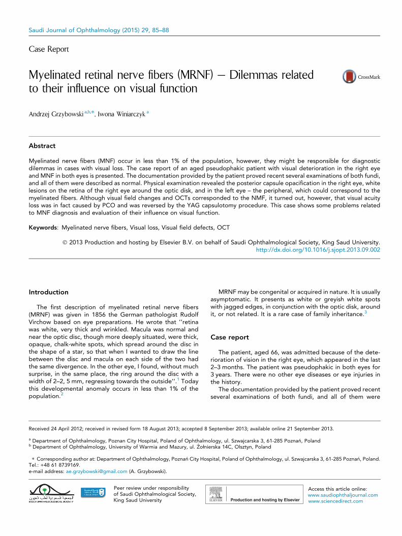
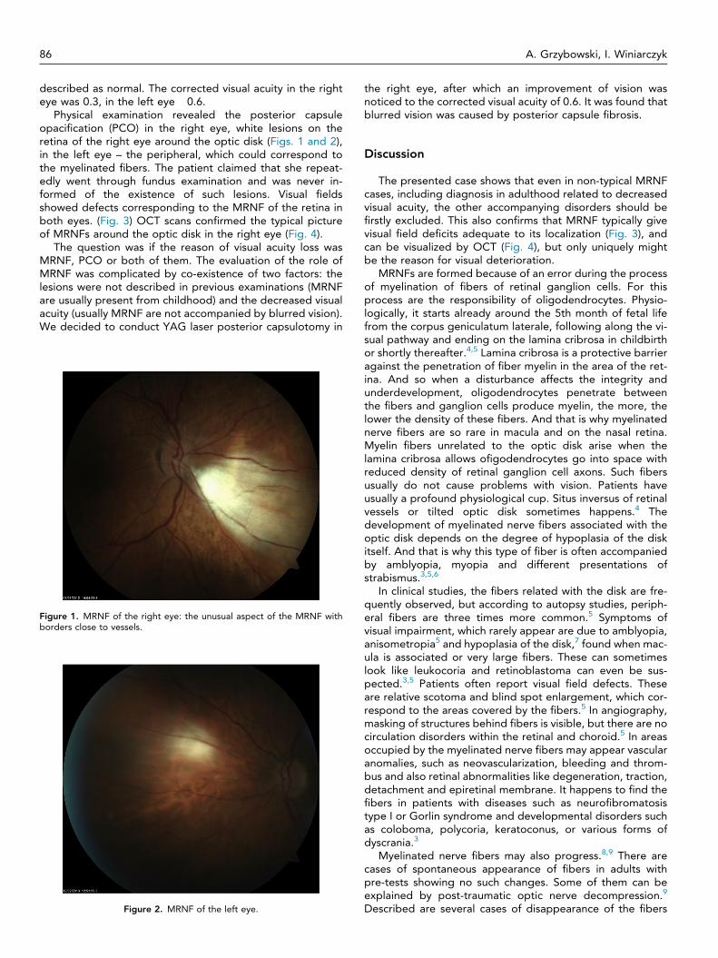
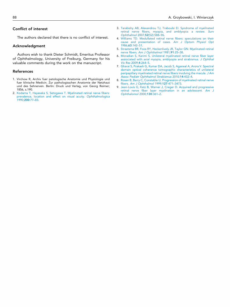
Physical examination revealed the posterior capsuleopacification (PCO) in the right eye, white lesions on theretina of the right eye around the optic disk (Figs. 1 and 2),in the left eye – the peripheral, which could correspond tothe myelinated fibers. The patient claimed that she repeat-edly went through fundus examination and was never in-formed of the existence of such lesions. Visual fieldsshowed defects corresponding to the MRNF of the retina inboth eyes. (Fig. 3) OCT scans confirmed the typical pictureof MRNFs around the optic disk in the right eye (Fig. 4).
The question was if the reason of visual acuity loss wasMRNF, PCO or both of them. The evaluation of the role ofMRNF was complicated by co-existence of two factors: thelesions were not described in previous examinations (MRNFare usually present from childhood) and the decreased visualacuity (usually MRNF are not accompanied by blurred vision).We decided to conduct YAG laser posterior capsulotomy in
Figure 1. MRNF of the right eye: the unusual aspect of the MRNF withborders close to vessels.
Figure 2. MRNF of the left eye.
the right eye, after which an improvement of vision wasnoticed to the corrected visual acuity of 0.6. It was found thatblurred vision was caused by posterior capsule fibrosis.
Discussion
The presented case shows that even in non-typical MRNFcases, including diagnosis in adulthood related to decreasedvisual acuity, the other accompanying disorders should befirstly excluded. This also confirms that MRNF typically givevisual field deficits adequate to its localization (Fig. 3), andcan be visualized by OCT (Fig. 4), but only uniquely mightbe the reason for visual deterioration.
MRNFs are formed because of an error during the processof myelination of fibers of retinal ganglion cells. For thisprocess are the responsibility of oligodendrocytes. Physio-logically, it starts already around the 5th month of fetal lifefrom the corpus geniculatum laterale, following along the vi-sual pathway and ending on the lamina cribrosa in childbirthor shortly thereafter.4,5 Lamina cribrosa is a protective barrieragainst the penetration of fiber myelin in the area of the ret-ina. And so when a disturbance affects the integrity andunderdevelopment, oligodendrocytes penetrate betweenthe fibers and ganglion cells produce myelin, the more, thelower the density of these fibers. And that is why myelinatednerve fibers are so rare in macula and on the nasal retina.Myelin fibers unrelated to the optic disk arise when thelamina cribrosa allows ofigodendrocytes go into space withreduced density of retinal ganglion cell axons. Such fibersusually do not cause problems with vision. Patients haveusually a profound physiological cup. Situs inversus of retinalvessels or tilted optic disk sometimes happens.4 Thedevelopment of myelinated nerve fibers associated with theoptic disk depends on the degree of hypoplasia of the diskitself. And that is why this type of fiber is often accompaniedby amblyopia, myopia and different presentations ofstrabismus.3,5,6
In clinical studies, the fibers related with the disk are fre-quently observed, but according to autopsy studies, periph-eral fibers are three times more common.5 Symptoms ofvisual impairment, which rarely appear are due to amblyopia,anisometropia5 and hypoplasia of the disk,7 found when mac-ula is associated or very large fibers. These can sometimeslook like leukocoria and retinoblastoma can even be sus-pected.3,5 Patients often report visual field defects. Theseare relative scotoma and blind spot enlargement, which cor-respond to the areas covered by the fibers.5 In angiography,masking of structures behind fibers is visible, but there are nocirculation disorders within the retinal and choroid.5 In areasoccupied by the myelinated nerve fibers may appear vascularanomalies, such as neovascularization, bleeding and throm-bus and also retinal abnormalities like degeneration, traction,detachment and epiretinal membrane. It happens to find thefibers in patients with diseases such as neurofibromatosistype I or Gorlin syndrome and developmental disorders suchas coloboma, polycoria, keratoconus, or various forms ofdyscrania.3
Myelinated nerve fibers may also progress.8,9 There arecases of spontaneous appearance of fibers in adults withpre-tests showing no such changes. Some of them can beexplained by post-traumatic optic nerve decompression.9
Described are several cases of disappearance of the fibers
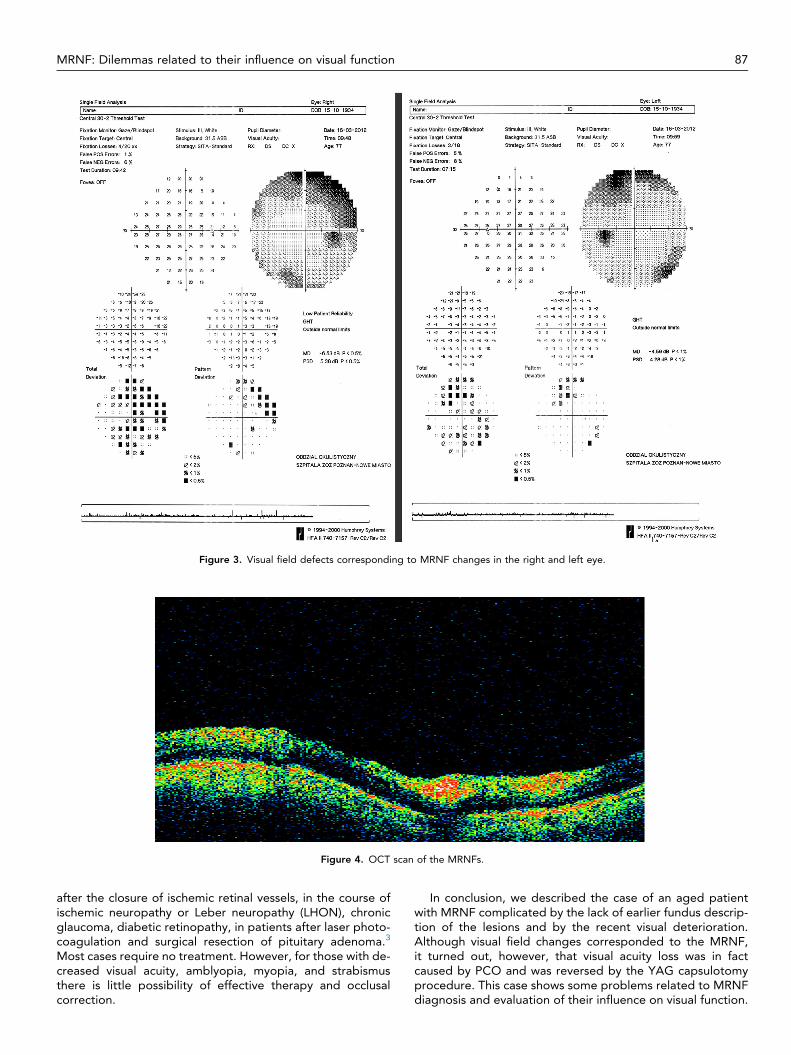
Figure 3. Visual field defects corresponding to MRNF changes in the right and left eye.
Figure 4. OCT scan of the MRNFs.
MRNF: Dilemmas related to their influence on visual function 87
after the closure of ischemic retinal vessels, in the course ofischemic neuropathy or Leber neuropathy (LHON), chronicglaucoma, diabetic retinopathy, in patients after laser photo-coagulation and surgical resection of pituitary adenoma.3
Most cases require no treatment. However, for those with de-creased visual acuity, amblyopia, myopia, and strabismusthere is little possibility of effective therapy and occlusalcorrection.
In conclusion, we described the case of an aged patientwith MRNF complicated by the lack of earlier fundus descrip-tion of the lesions and by the recent visual deterioration.Although visual field changes corresponded to the MRNF,it turned out, however, that visual acuity loss was in factcaused by PCO and was reversed by the YAG capsulotomyprocedure. This case shows some problems related to MRNFdiagnosis and evaluation of their influence on visual function.
88 A. Grzybowski, I. Winiarczyk
Conflict of interest
The authors declared that there is no conflict of interest.
Acknowledgment
Authors wish to thank Dieter Schmidt, Emeritus Professorof Ophthalmology, University of Freiburg, Germany for hisvaluable comments during the work on the manuscript.
References
1. Virchow R, Archiv fuer patologische Anatomie und Physiologie undfuer klinische Medicin. Zur pathologisschen Anatomie der Netzhautund des Sehnerven. Berlin: Druck und Verlag, von Georg Reimer;1856, s.190.
2. Kodama T, Hayasaka S, Setogawa T. Myelinated retinal nerve fibers:prevalence, location and effect on visual acuity. Ophthalmologica1990;200:77–83.
3. Tarabishy AB, Alexandrou TJ, Traboulsi EI. Syndrome of myelinatedretinal nerve fibers, myopia, and amblyopia: a review. SurvOphthalmol 2007;52(52):588–96.
4. Williams TD. Medullated retinal nerve fibers: speculations on theircause and presentation of cases. Am J Optom Physiol Opt1986;63:142–51.
5. Straatsma BR, Foos RY, Heckenlively JR, Taylor GN. Myelinated retinalnerve fibers. Am J Ophthalmol 1981;91:25–38.
6. Moradian S, Karimi S. Unilateral myelinated retinal nerve fiber layerassiociated with axial myopia, amblyopia and strabismus. J OphthalVis Res 2009;4:264–5.
7. Gharai S, Prakash G, Kumar DA, Jacob S, Agarwal A, Arora V. Spectraldomain optical coherence tomographic characteristics of unilateralperipapillary myelinated retinal nerve fibers involving the macula. J AmAssoc Pediatr Ophthalmol Strabismus 2010;14:432–4.
8. Rosen B, Barry C, Constable IJ. Progression of myelinated retinal nervefibers. Am J Ophthalmol 1999;127:471–3473.
9. Jean-Louis G, Katz B, Warner J, Creger D. Acquired and progressiveretinal nerve fiber layer myalination in an adolescent. Am JOphthalomol 2000;130:361–2.