Embed Size (px)
Citation preview
Myelin-associated glycoprotein genemutation causes Pelizaeus-Merzbacherdisease-like disorder
Alexander Lossos,1,* Nimrod Elazar,2,* Israela Lerer,3,* Ora Schueler-Furman,4
Yakov Fellig,5 Benjamin Glick,6 Bat-El Zimmerman,3 Haim Azulay,5 Shlomo Dotan,7
Sharon Goldberg,7 John M. Gomori,8 Penina Ponger,1 J. P. Newman,1 Hodaifah Marreed,3
Andreas J. Steck,9 Nicole Schaeren-Wiemers,9 Nofar Mor,2 Michal Harel,10 Tamar Geiger,10
Yael Eshed-Eisenbach,2 Vardiella Meiner3,* and Elior Peles2
*These authors contributed equally to this work.
Pelizaeus-Merzbacher disease is an X-linked hypomyelinating leukodystrophy caused by mutations or rearrangements in PLP1. It
presents in infancy with nystagmus, jerky head movements, hypotonia and developmental delay evolving into spastic tetraplegia
with optic atrophy and variable movement disorders. A clinically similar phenotype caused by recessive mutations in GJC2 is
known as Pelizaeus-Merzbacher-like disease. Both genes encode proteins associated with myelin. We describe three siblings of a
consanguineous family manifesting the typical infantile-onset Pelizaeus-Merzbacher disease-like phenotype slowly evolving into a
form of complicated hereditary spastic paraplegia with mental retardation, dysarthria, optic atrophy and peripheral neuropathy in
adulthood. Magnetic resonance imaging and spectroscopy were consistent with a demyelinating leukodystrophy. Using genetic
linkage and exome sequencing, we identified a homozygous missense c.399C4G; p.S133R mutation in MAG. This gene, previ-
ously associated with hereditary spastic paraplegia, encodes myelin-associated glycoprotein, which is involved in myelin mainten-
ance and glia-axon interaction. This mutation is predicted to destabilize the protein and affect its tertiary structure. Examination of
the sural nerve biopsy sample obtained in childhood in the oldest sibling revealed complete absence of myelin-associated glyco-
protein accompanied by ill-formed onion-bulb structures and a relatively thin myelin sheath of the affected axons.
Immunofluorescence, cell surface labelling, biochemical analysis and mass spectrometry-based proteomics studies in a variety of
cell types demonstrated a devastating effect of the mutation on post-translational processing, steady state expression and sub-
cellular localization of myelin-associated glycoprotein. In contrast to the wild-type protein, the p.S133R mutant was retained in the
endoplasmic reticulum and was subjected to endoplasmic reticulum-associated protein degradation by the proteasome. Our find-
ings identify involvement of myelin-associated glycoprotein in this family with a disorder affecting the central and peripheral
nervous system, and suggest that loss of the protein function is responsible for the unique clinical phenotype.
1 Department of Neurology and Agnes Ginges Centre for Human Neurogenetics, Hebrew University-Hadassah Medical Centre,Jerusalem, Israel
2 Department of Molecular Cell Biology, Weizmann Institute of Science, Rehovot, Israel3 Department of Genetics and Metabolic Diseases, Hebrew University-Hadassah Medical Centre, Jerusalem, Israel4 Department of Microbiology and Molecular Genetics, Institute for Medical Research Israel-Canada, Faculty of Medicine, Hebrew
University, Jerusalem, Israel5 Department of Pathology, Hebrew University-Hadassah Medical Centre, Jerusalem, Israel6 Paediatric Neuromuscular Service, Alyn Paediatric Rehabilitation Centre, Jerusalem, Israel7 Department of Ophthalmology, Hebrew University-Hadassah Medical Centre, Jerusalem, Israel
doi:10.1093/brain/awv204 BRAIN 2015: Page 1 of 16 | 1
Received April 2, 2015. Revised May 21, 2015. Accepted May 27, 2015.
� The Author (2015). Published by Oxford University Press on behalf of the Guarantors of Brain. All rights reserved.
For Permissions, please email: [email protected]
Brain Advance Access published July 15, 2015by guest on July 29, 2015
Dow
nloaded from
8 Department of Radiology, Hebrew University-Hadassah Medical Centre, Jerusalem, Israel9 Department of Biomedicine, University Hospital Basel, University of Basel, Switzerland
10 Department of Human Molecular Genetics and Biochemistry, Sackler Faculty of Medicine, Tel Aviv University, Tel Aviv, Israel
Correspondence to: Vardiella Meiner, MD,
Department of Genetics and Metabolic Diseases,
Hebrew University-Hadassah Medical Centre,
PO Box 12000 Jerusalem,
91120 Israel
E-mail: [email protected]
Correspondence may also be addressed to: Alexander Lossos,
MD, Department of Neurology,
Hebrew University-Hadassah Medical Centre,
PO Box 12000 Jerusalem,
91120 Israel
E-mail: [email protected]
Elior Peles,
Department of Molecular Cell Biology,
Weizmann Institute of Science,
Rehovot, Israel
E-mail: [email protected]
Keywords: hereditary spastic paraplegia; MAG; Pelizaeus-Merzbacher-like disease
Abbreviations: (E)GFP = (enhanced) green fluorescent protein; ERAD = endoplasmic reticulum-associated protein degradation;HLD = hypomyelinating leukodystrophy; HSP = hereditary spastic paraplegia, Ig = immunoglobulin; PMD = Pelizaeus-Merzbacherdisease; PMLD = Pelizaeus-Merzbacher-like disease
IntroductionPelizaeus-Merzbacher disease (PMD; OMIM #312080) is
the prototype hypomyelinating leukodystrophy (HLD1) clas-
sically presenting in infancy with nystagmus, jerky head
movements, hypotonia and developmental delay evolving
into spastic tetraplegia with optic atrophy and variable
movement disorders (Hobson and Garbern, 2012). It is an
X-linked disorder caused by mutations or rearrangements in
the PLP1 gene encoding two alternatively spliced isoforms of
an integral membrane proteolipid protein (PLP/DM20), a
major component of the CNS and a minor one of the per-
ipheral nervous system (PNS) compact myelin (Han et al.,
2013). A clinically similar phenotype associated with hypo-
myelination but not linked to PLP1 is termed Pelizaeus-
Merzbacher-like disease (PMLD) (Hobson and Garbern,
2012) and classified with a genetically heterogeneous group
of HLD (Pouwels et al., 2014). PMLD1 (OMIM #608804),
classified as HLD2, accounts for 8% of such patients
(Henneke et al., 2008). It is caused by recessive mutations
in GJC2, encoding gap junction protein gamma 2 or con-
nexin 47 expressed in oligodendrocytes and involved in
oligodendrocyte-astrocyte channelling and in the mainten-
ance of myelin (Orthmann-Murphy et al., 2009).
Additional disorders resembling PMD are associated with
delayed myelination, as in SLC16A2-related dysfunction of
monocarboxylate thyroid hormone transporter 8 (Vaurs-
Barriere et al., 2009) (OMIM #300523), or with hypomye-
lination secondary to a neuronal or axonal process, as in a
presumed AIMP1-related dysregulation of the neurofila-
ment network in HLD3 (Feinstein et al., 2010; Pouwels
et al., 2014) (OMIM #260600), in HSPD1-related
compromise of the mitochondrial chaperonin HSP60 in
HLD4 (Magen et al., 2008; Pouwels et al., 2014)
(OMIM #612233), and occasionally in TUBB4A-related
tubulin beta-4A dysfunction in HLD6 (Miyatake et al.,
2014; Shimojima et al., 2015) (OMIM #612438).
We report the clinical, molecular and cell biological data
of a multiplex family with early PMD-like disorder slowly
evolving into a form of hereditary spastic paraplegia (HSP)
associated with demyelination due to a homozygous loss-
of-function mutation in MAG, the gene previously reported
in HSP (Novarino et al., 2014). Encoded protein, known as
myelin-associated glycoprotein MAG, is a cell adhesion
molecule that belongs to the immunoglobulin (Ig) super-
family of proteins enriched in myelinating glial cells and
involved in glia-axon interactions.
Materials and methods
Patients
Since 2009, we have evaluated three affected siblings of a con-sanguineous family of Palestinian Arab origin (Fig. 1A). Of theliving clinically unaffected family members, 13 were availablefor examination. The family was registered in the IsraeliHSP Database in 2009. Clinical diagnosis and a prospective
2 | BRAIN 2015: Page 2 of 16 A. Lossos et al.
by guest on July 29, 2015D
ownloaded from
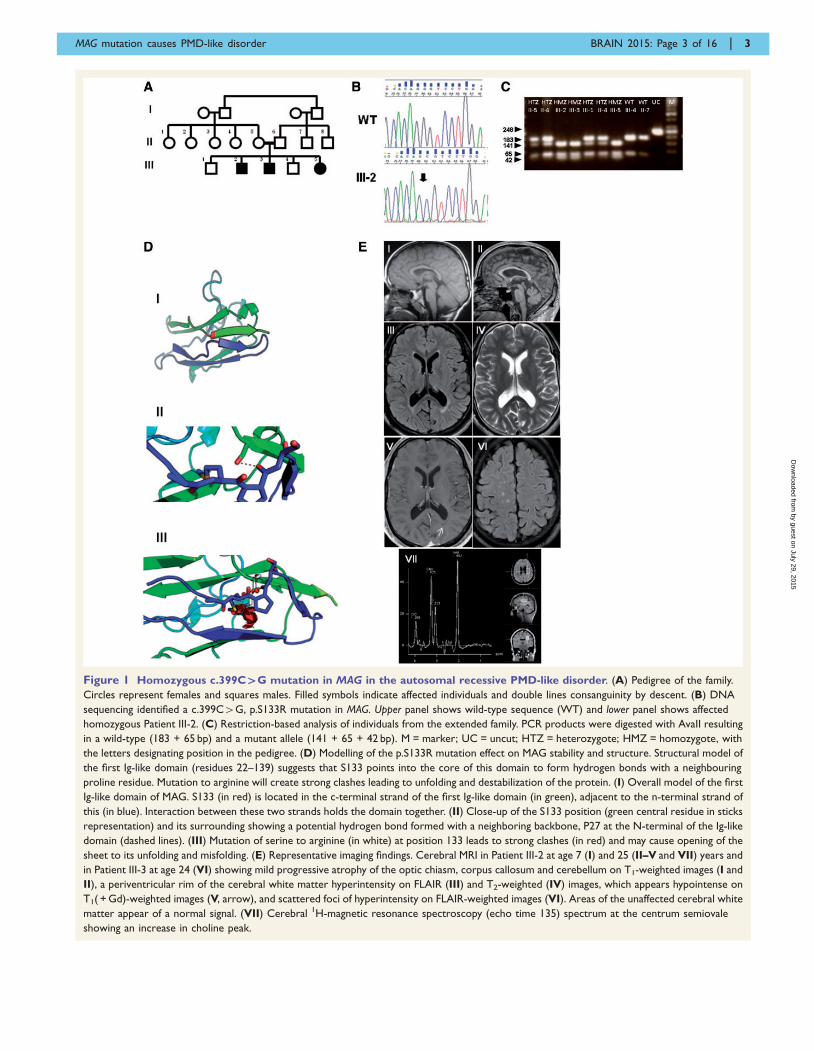
Figure 1 Homozygous c.399C`G mutation in MAG in the autosomal recessive PMD-like disorder. (A) Pedigree of the family.
Circles represent females and squares males. Filled symbols indicate affected individuals and double lines consanguinity by descent. (B) DNA
sequencing identified a c.399C4G, p.S133R mutation in MAG. Upper panel shows wild-type sequence (WT) and lower panel shows affected
homozygous Patient III-2. (C) Restriction-based analysis of individuals from the extended family. PCR products were digested with AvaII resulting
in a wild-type (183 + 65 bp) and a mutant allele (141 + 65 + 42 bp). M = marker; UC = uncut; HTZ = heterozygote; HMZ = homozygote, with
the letters designating position in the pedigree. (D) Modelling of the p.S133R mutation effect on MAG stability and structure. Structural model of
the first Ig-like domain (residues 22–139) suggests that S133 points into the core of this domain to form hydrogen bonds with a neighbouring
proline residue. Mutation to arginine will create strong clashes leading to unfolding and destabilization of the protein. (I) Overall model of the first
Ig-like domain of MAG. S133 (in red) is located in the c-terminal strand of the first Ig-like domain (in green), adjacent to the n-terminal strand of
this (in blue). Interaction between these two strands holds the domain together. (II) Close-up of the S133 position (green central residue in sticks
representation) and its surrounding showing a potential hydrogen bond formed with a neighboring backbone, P27 at the N-terminal of the Ig-like
domain (dashed lines). (III) Mutation of serine to arginine (in white) at position 133 leads to strong clashes (in red) and may cause opening of the
sheet to its unfolding and misfolding. (E) Representative imaging findings. Cerebral MRI in Patient III-2 at age 7 (I) and 25 (II–V and VII) years and
in Patient III-3 at age 24 (VI) showing mild progressive atrophy of the optic chiasm, corpus callosum and cerebellum on T1-weighted images (I and
II), a periventricular rim of the cerebral white matter hyperintensity on FLAIR (III) and T2-weighted (IV) images, which appears hypointense on
T1( + Gd)-weighted images (V, arrow), and scattered foci of hyperintensity on FLAIR-weighted images (VI). Areas of the unaffected cerebral white
matter appear of a normal signal. (VII) Cerebral 1H-magnetic resonance spectroscopy (echo time 135) spectrum at the centrum semiovale
showing an increase in choline peak.
MAG mutation causes PMD-like disorder BRAIN 2015: Page 3 of 16 | 3
by guest on July 29, 2015D
ownloaded from
follow-up were performed using the clinical chart (http://spatax.wordpress.com/downloads/) developed by the interna-tional SPATAX (spastic paraplegia and ataxia) network (co-ordinator: A. Durr, MD, PhD). For cognitive evaluation andassessment of the intelligence quotient we used Mini-MentalState Examination (Folstein et al., 1975) and the Test for Non-Verbal Intelligence (TONI-2), a language-free tool for obtain-ing predicted non-verbal intelligence scores (www.agsnet.com/).The study was approved by the institutional and national reviewboards. Informed consent was obtained prior to enrolment fromall the participants.
Autozygosity mapping andlinkage analysis
Because of the parental consanguinity, we determined sharedhomozygous regions using genome-wide linkage analysis withthe Affymetrix� Gene-Chip Human Mapping 250K Nsp Array.Data handling, evaluation and the statistical analysis were per-formed using HomozygosityMapper (Seelow et al., 2009). Toassess and confirm segregation with the phenotype, we used se-lected short tandem repeat (STR) markers for genotyping theremaining family members (details available upon request).
Whole exome sequencing
For whole exome sequencing, DNA obtained from Patient III-2was fragmented, end paired, adenylated and ligated to adap-ters. Exome capture and sequencing was performed with theAgilent SureSelect 38 Mb All Exon Hybridization Array. TheSureSelect protocol was used to prepare libraries for paired-end sequencing on an Illumina HiSeq 2000 platform withmean depth coverage of 30� . The sequenced reads werealigned, and variant calling was performed with the October2011 release of DNAnexus software with the human genomeassembly hg19 (GRCh37) as reference. The raw list of variantswas filtered to exclude variants present in the dbSNP129, inthe ‘HapMap’ and in the ‘1000 Genomes’ databases. Rigorousfiltering based on global (minor allele frequency) MAF 5 1%,predicted functional consequence, and sequence conservationleft us with sequence variants of interest that were validatedand verified using Sanger sequencing and restriction fragmentlength polymorphism (RFLP) analysis.
Mutation analysis
For the MAG c.399C4G; p.S133R mutation (NM_002361.3),PCR product (248 bp; amplified by forward 5’-GAAACTGCACCCTCCTGCT-3’ and reverse 5’-CAAATCAGCACCTCCCAGATC-3’) was digested with AvaII (Promega) as per manufac-turer’s protocol and run on NuSieve� agarose (3:1%) gel lead-ing to the detection of a normal and a mutant allele(183 + 65 bp and 141 + 65 + 42 bp, respectively).
For the PLP1 c.594C4A variant (NM_000533.3), we usedAlwNI restriction enzyme analysis with the primersPLP1EX5F-5’-TGGTTTTAATGTCTGGCACA-3’ and PLP1EX5R-5’-CTCATAATCACCACCCTCCTT-3’. PLP1 cDNAwas analysed with the primers PLP1-290F-5’-AGGCAGATCTTTGGCGACTA-3’ and PLP1-800R-5’-GTGAGCAGGGAAACCAGTGT-3’. PCR products were run on agarose geland visualized under UV illumination.
Expression of wild-type andmutant MAG
Human MAG was cloned by reverse transcription-PCR fromtotal RNA of human femoralis cDNA and was subcloned intopb-actin-ECGFP exchanging the ECGFP sequence (pb-actin-hSMAG), or into the same vector upstream of the EGFP(pb-actin-hSMAG-GFP) (Erb et al., 2003). c.399C4G muta-tion was generated by PCR on pb-actin-hSMAG as template.A forward primer (5’-CTCCATCTCCAGCCTCGGG-3’) and areverse primer (5’-GGTGTTGACGATATCCAGGACCCTGTGCTCTGAGAAGGTGTAC-3’) containing the mutation anda neighbouring endogenous EcoRV site were used. AHindIII–EcoRV-digested PCR fragment was ligated into thetemplate plasmid in the same site to generate pb-actin-hSMAG-S133R. The pb-actin-hSMAG-S133R-GFP wasgenerated by replacing the HindIII–EcoRV fragment ofhSMAG-GFP with the one obtained from pb-actin-hSMAG-S133R. Plasmids containing myc-tagged versions of wild-typeand mutant MAG were made by PCR cloning of the corres-ponding genes into pMX vector (Addgene). The latter plasmidwas also used to generate viral vectors containing GFP-taggedproteins. The open reading frame of all constructs was con-firmed by DNA sequencing. Transfection of HEK-293T, COS7and Schwann cells were done using CaPO4, LipofectamineTM
(Invitrogen), and LipofectamineTM 2000 reagent (Invitrogen),respectively. Lentiviral stocks were generated by CaPO4-transfection of phoenix packaging cells (Invitrogen). RatSchwann cells and oligodendrocyte precursor cells were cul-tured as described (Eshed et al., 2005).
Antibodies and immunofluorescencelabelling
We used mouse monoclonal antibody (MAb) D3A2G5 againstthe extracellular domain of human MAG (Burger et al., 1990),a monoclonal anti-mouse MAG (clone MAb 513, EMDMillipore), rabbit polyclonal anti-MAG (H-300; Santa CruzBiotechnologies), and polyclonal anti-L-MAG (Heape et al.,1999, kindly obtained from Dr A. Heape, University ofOulu, Finland). For immunohistochemical staining of thesural nerve sample, we used anti-MAG antibody againstamino acids 1–300 mapping near the N-terminus of humanMAG (H-300); sc-15324, Santa Cruz Biotechnology Inc, 1:50,and NF-Neurofilament Protein, Clone 2F11, Dako, 1:500.Other antibodies included mouse anti-calnexin (MAb3126;EMD Millipore), mouse monoclonal antibody to vesicle dock-ing protein p115 (kindly provided by Dr S. Lev, WeizmannInstitute, Israel), rabbit anti-VapB (K-16; Santa CruzBiotechnologies), and rabbit polyclonal antibody to Caspr(Peles et al., 1997). Fluorophore-coupled secondary antibodiesincluded 488- coupled anti-rabbit and mouse IgG (Invitrogen),Cy3-coupled anti-rabbit and anti-mouse (JacksonLaboratories). Immunofluorescence labelling was carried outessentially as described (Spiegel et al., 2007). Fluorescenceimages were obtained using an Axioskop2 microscopeequipped with an ApoTom imaging system (Carl Zeiss) fittedwith a Hamamatsu ORCA-ER CCD camera. Images wereacquired and processed using the Zen2012 (Carl Zeiss) andPhotoshop software (Adobe).
4 | BRAIN 2015: Page 4 of 16 A. Lossos et al.
by guest on July 29, 2015D
ownloaded from
Structural protein modelling
Structural model of MAG and the corresponding MAG-S133Rmutant was generated using the HHpred tool (Soding, 2005),to identify structural templates for homology modelling, andthe I-Tasser tool (Roy et al., 2010), to create the model using alonger domain definition than predicted earlier by other tools(May et al., 1998). The structure of a similar protein siaload-hesin (Protein Data Bank code 1qfo) with a 26% sequenceidentity and 66% sequence similarity (May et al., 1998)served as the basis for this model.
Immunoprecipitation and westernblot analysis
Immunoprecipitation, sodium dodecyl sulphate-polyacrylamidegel electrophoresis (SDS-PAGE) and western blotting weredone as described (Spiegel et al., 2007) with the exceptionthat the chemiluminescence signal was detected using theChemiDoc MP System (Bio-Rad). Deglycosylation and remov-ing of high mannose structures was achieved by incubating thedenatured immunocomplexes with endoglycosidase H (NewEngland Biolabs) for 1 h at 37�C. Co-immunoprecipitation ofendogenously expressed proteins was performed using HEK-293T cells either incubated with dimethyl sulphoxide or incu-bated with the indicated amount of bortezomib overnight.Cells were solubilized in TritonTM X-100 containing buffer,incubated with GFP-Trap�-Agarose (ChromoTek) followedby western blot analysis (Peles et al., 1997). When indicated,protein synthesis was stopped by the addition of cyclohexa-mide to a final concentration of 50 mg/ml for various times.Cells were then washed in phosphate-buffered saline, lysedand total protein lysates were subjected to western blot ana-lysis. Cell surface biotinylation was done as described (Gollanet al., 2003).
Proteomics sample preparationand mass spectrometry analysis
Cells were lysed with 1% NP40, 150 mM NaCl in 50 mM TrisHCl pH 7.6 buffer supplemented with protease inhibitors.Protein from each sample (5 mg) was mixed with GFP-Trap�-Agarose (ChromoTek) for 2 h at 4�C. Samples werewashed three times with the same buffer containing 0.05%NP40 followed by three washes without NP40. Proteinswere reduced with 1 mM dithiothreitol in 2 M urea and sub-sequently alkylated with 5 mM iodoacetamide. On-bead pro-tein digestion was performed with sequencing grade-modifiedtrypsin (Promega) and peptides were acidified with trifluoroa-cetic acid (TFA) followed by purification on C18 (stageTips).Liquid chromatography-mass spectrometry (LC-MS/MS) ana-lysis was performed on the EASY-nLC1000 UHPLC system(Thermo Scientific) coupled to the Q-Exactive Plus mass spec-trometers (Thermo Scientific) via the EASY-SprayTM ionizationsource. Peptides were loaded onto 50 cm long EASY-SprayTM
PepMap columns (Thermo Scientific) with 140-min gradientusing buffer A (0.1% formic acid) and separated using a 7–28% buffer B (80% acetonitrile, 0.1% formic acid). All massspectrometry measurements were done in a data-dependentmode using a top-10 method. Raw mass spectrometry fileswere analysed with MaxQuant version 1.5.0.36 and the
Andromeda search engine integrated into the same version(Cox et al., 2011). MS/MS spectra were searched against theUniprotKB database. Quantification was performed using thelabel-free algorithm MaxLFQ (Cox et al., 2014).
All bioinformatics analyses were performed on log2 LFQintensity values. Data analysis was performed after filteringfor valid values in at least two samples in one group (emptyvector/wild-type/S133R mutant). Statistical tests and calcula-tions were done using the Perseus program. To identify poten-tial interactors, Student’s t-test was performed between emptyvector triplicate and either wild-type or mutant triplicates withpermutation-based false discovery rate (FDR) 50.05 andS0 = 1.5 and resulted in a list of 1010 proteins with higherintensities in the MAG samples. A second Student’s t-test(FDR = 0.05, S0 = 1.5) was performed on these proteins to dis-tinguish between the wild-type and mutant binders.Hierarchical clustering was done after z-score normalizationof the proteins, and was based on Euclidean distances betweenaverages. Protein networks were constructed in the string data-base and visualized in Cytoscape. Fisher exact tests were donewith a Benjamini–Hochberg FDR threshold of 0.02.
Results
Patients manifest complicatedhereditary spastic paraplegia
The main clinical findings at initial examination and the
available documented medical history are summarized in
Table 1. In short, the patients were born after uneventful
pregnancies and labour and, except for Patient III-2 who
had transverse limb defect affecting phalanges in three fin-
gers of his right hand, none had dysmorphic features. Head
circumferences were within normal limits. Early develop-
mental and speech delay were noticed soon after birth
with initiation of supported gait at age 4–5 years and
slow but progressive deterioration to walker and wheel-
chair over a period of 15 years, despite physiotherapy.
Borderline-to-mild mental retardation was documented in
all three siblings. They attended special school, obtained
basic reading and writing skills, and achieved partial inde-
pendence in basic daily activities. Based on the combination
of early ocular and neurological findings (Table 1), initial
diagnosis of PMD was raised by age 5 years (B.G.).
On examination, patients had spastic dysarthria and
moderate-to-severe spastic paraparesis with hyporeflexia
in the arms and knees, reduced (Patient III-5) or absent
(Patients III-2 and III-3) ankle reflexes and bilateral exten-
sor plantar response. Muscle tone in the arms was only
mildly increased and was associated with dysmetria.
Reduced deep tendon reflexes together with distal leg atro-
phy and distal vibration sensory loss suggested an asso-
ciated peripheral neuropathy. Patients displayed mild
psychomotor slowing, intact short-term verbal memory,
full orientation in all domains, and scored in the range of
mild (Patient III-5) to borderline (Patients III-2 and III-3)
mental retardation.
MAG mutation causes PMD-like disorder BRAIN 2015: Page 5 of 16 | 5
by guest on July 29, 2015D
ownloaded from
Formal neuro-ophthalmological examination (Table 1)
revealed similar findings in all three siblings, including hori-
zontal nystagmus and optic nerve atrophy with reduced
visual acuity, visual field defect, and thinning of the peri-
papillary retinal ganglion cell fibres (Supplementary
Table 1). In addition, all had hypermetropia and astigma-
tism, and Patient III-2 had primary open angle glaucoma
well-controlled with locally applied medications. Recorded
visual-evoked potentials were of significantly low amplitude
and of prolonged latency, more in the older siblings
(Supplementary Table 1). Brainstem auditory-evoked re-
sponses were of delayed latency but a recognizable wave
pattern in all three siblings.
Over a period of 4 years, all three siblings were available
for follow-up examinations, showing a very slow progres-
sion of their functional motor disability and of the clinical
findings.
Metabolic screening and EEG were normal in Patient III-2.
Peripheral nerve conduction study (Supplementary Table 1)
demonstrated in the elder siblings, prolonged distal motor
latencies with normal or borderline compound motor
action potentials and reduced motor and sensory velocities,
indicating motor and sensory peripheral neuropathy. There
were no temporal dispersion or conduction blocks.
Cerebral MRI (Supplementary Table 1) demonstrated bi-
lateral optic nerve atrophy accompanied by mild corpus
callosum and cerebellar atrophy. In addition, there was a
rim of predominantly posterior cerebral periventricular and
subtle pontine white matter T2/FLAIR-hyperintensity, and
rare scattered foci of subcortical white matter hyperinten-
sity (Fig. 1E). Areas of unaffected cerebral white matter
appeared of a normal signal, suggesting a normal myelin-
ation pattern. Mild ventricular dilatation was also noted,
likely the result of white matter volume loss. Although
these findings were similar in all three siblings, they
tended to be more pronounced in the older ones.
Furthermore, comparison with a study from 18 years ear-
lier in Patient III-2 revealed slow progression of all findings.
Single volume 1H-magnetic resonance spectroscopy ob-
tained with PRESS acquisition at the centrum semiovale
(Fig. 1E) revealed elevated ratios of choline to creatine
and to N-acetyl aspartate at a long (135 ms) echo time,
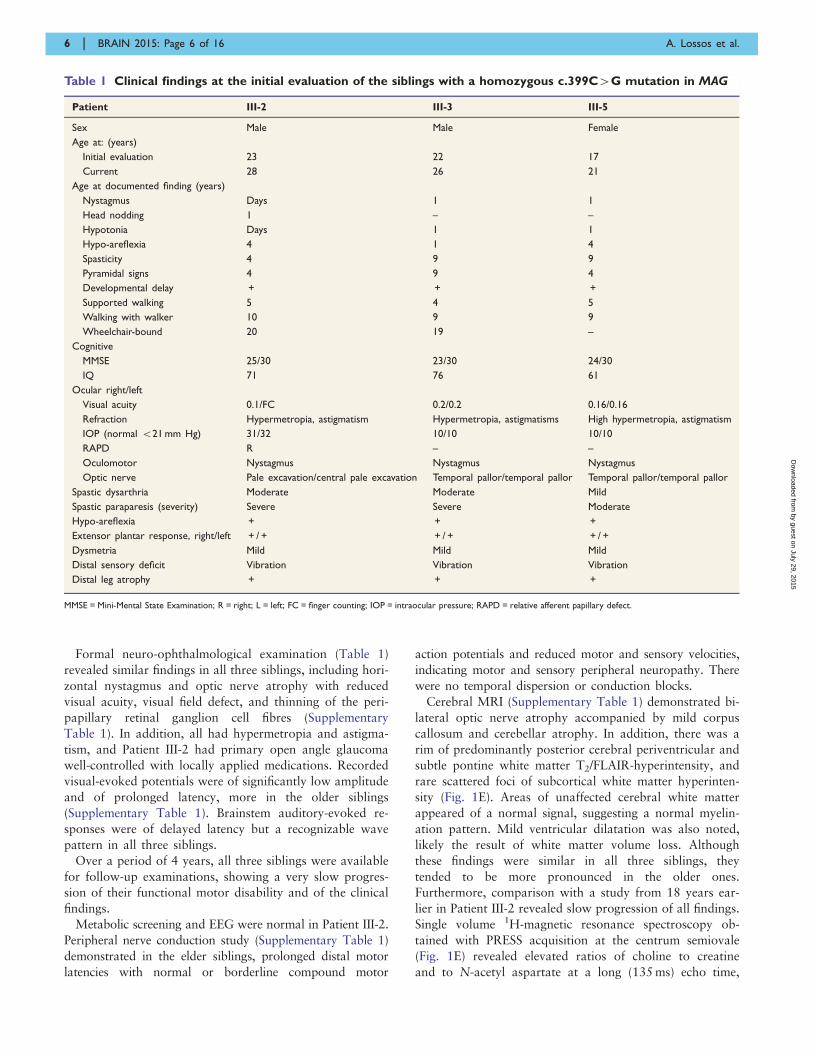
Table 1 Clinical findings at the initial evaluation of the siblings with a homozygous c.399C4G mutation in MAG
Patient III-2 III-3 III-5
Sex Male Male Female
Age at: (years)
Initial evaluation 23 22 17
Current 28 26 21
Age at documented finding (years)
Nystagmus Days 1 1
Head nodding 1 – –
Hypotonia Days 1 1
Hypo-areflexia 4 1 4
Spasticity 4 9 9
Pyramidal signs 4 9 4
Developmental delay + + +
Supported walking 5 4 5
Walking with walker 10 9 9
Wheelchair-bound 20 19 –
Cognitive
MMSE 25/30 23/30 24/30
IQ 71 76 61
Ocular right/left
Visual acuity 0.1/FC 0.2/0.2 0.16/0.16
Refraction Hypermetropia, astigmatism Hypermetropia, astigmatisms High hypermetropia, astigmatism
IOP (normal 521 mm Hg) 31/32 10/10 10/10
RAPD R – –
Oculomotor Nystagmus Nystagmus Nystagmus
Optic nerve Pale excavation/central pale excavation Temporal pallor/temporal pallor Temporal pallor/temporal pallor
Spastic dysarthria Moderate Moderate Mild
Spastic paraparesis (severity) Severe Severe Moderate
Hypo-areflexia + + +
Extensor plantar response, right/left + / + + / + + / +
Dysmetria Mild Mild Mild
Distal sensory deficit Vibration Vibration Vibration
Distal leg atrophy + + +
MMSE = Mini-Mental State Examination; R = right; L = left; FC = finger counting; IOP = intraocular pressure; RAPD = relative afferent papillary defect.
6 | BRAIN 2015: Page 6 of 16 A. Lossos et al.
by guest on July 29, 2015D
ownloaded from
particularly in the older siblings (Supplementary Table 1).
Taken together, these findings were consistent with a
slowly progressive leukodystrophy of a demyelinating
rather than a hypomyelinating pattern (Bizzi et al., 2008).
At age 9 years, Patient III-2 was evaluated elsewhere. Left
sural nerve biopsy performed at that time was interpreted
as normal. Revision of paraffin embedded sections, epoxy
resin embedded semi-thin sections and electron microscopy
(Fig. 2) did not reveal significant alterations, except for rare
ill-formed onion-bulb structures and apparent minimal loss
of axon-glia interaction in rare myelinated axons, although
traction or tearing artefact cannot not be excluded. There
was no ‘wide-spaced’ myelin formation and no apparent
decrease in axonal density. Although subtle, these features
may indicate minimal demyelination.
Molecular analysis identifiesc.399C4G; p.S133R mutation in MAG
Autozygosity mapping in Patients III-2 and III-3 (Fig. 1A)
yielded four shared homozygous regions encompassing
the following chromosomal regions: chr11:47993662–
50299865, chr11:50300221–56027727, chr19:13552763–
35976171, and chr18: 45 667 057–67599516 (build
hg19). To narrow these candidate regions, we performed
segregation analysis using polymorphic microsatellites cor-
responding to these shared loci, which ruled out chromo-
some 18 and chromosome 11 regions as potential
candidate options. We remained with a single 21 Mb
homozygous region corresponding to chromosome 19
(15244658–35976171) encompassing 234 protein-coding
genes and a total of 1350 exons.
In light of this large number of genes, we elected to per-
form whole exome sequencing in Patient III-2. The number
of called variants [insertion/deletions (Indels) and single
nucleotide variations (SNVs)] was 108 357. Of these vari-
ants, 878 were homozygous located in the linked non-
excluded region on chromosome 19. Filtering this group re-
sulted in 190 coding variants that were all common and
frequently found in the published and in-house variant data-
bases (EVS, 1000 genomes, HapMap) except for a unique
variant in the MAG gene on chromosome 19:35786868. A
silent variant rs2301600 in this codon is a common SNP;
however, affected individuals in this family carried a mis-
sense change c.399C4G, p.S133R (Fig. 1B). In silico ana-
lyses with several pathogenicity prediction programs
predicted the amino acid substitution to be tolerated by
SIFT yet damaging to the protein function using PolyPhen-
2 and MutationTaster. Restriction-based analysis demon-
strated perfect segregation in the extended family and only
the three patients were homozygotes for the mutation (Fig.
1C). This variant was never reported in the database gener-
ated by the Exome Aggregation Consortium (ExAC),
Cambridge, MA (http://exac.broadinstitute.org) (December,
2014). Serine at position 133 is moderately conserved all
the way down to the zebrafish in which a similar polar
neutral amino acid is found. Screening 192 anonymous
chromosomes from a similar ethnic background did not
reveal any carrier for the mutation.
Because of a PMD-like phenotype, we extensively
searched for potential variants in the PLP1 gene. A single
variant on chromosome X, 103 042 867 predicted to cause
a silent change (c.594C4A; p.Gly198Gly), was further
analysed to assess potential splicing effect. This variant
did not segregate with the phenotype and, although
shared by the two affected brothers Patients III-2 and
III-3, was not detected in their affected sister, Patient
III-5. Furthermore, PLP1 cDNA did not differ in size be-
tween the three patients and normal controls, and we con-
cluded that the disease phenotype is not influenced by this
variant. We did not identify potential disease-causing vari-
ants in the HLD-causing genes AIMP1, GJC2, HSPD1,
TUBB4A, FAM126A and RARS.
p.S133R mutation is predictedto affect the structure of MAG
The p.S133R is located at the end of the first of five Ig-like
domains (Letunic et al., 2012). To evaluate the structural
importance of the affected S133 residue, we generated a
structural model of 118 residues (22–139) showing that
S133 points into the protein core to form hydrogen
bonds with the backbone of residues at the beginning of
the Ig-like domain (Fig. 1D). This interaction is also
observed in the solved homologue structures and could ex-
plain the rather unusual burial of a polar residue in the
core. Position 133 is located within the last strand of the
Ig-like domain and is inserted between two additional
strands to form a beta-sheet, including a bulge of different
sizes just prior to 133 (Fig. 1D).
The p.S133R mutation results in the replacement of a
small serine with a much larger arginine, thereby disrupting
this domain and leading to a significant destabilization of
the protein (Fig. 1D). In addition, this mutation could also
affect glycosylation, as it is located in the vicinity of the
R118, a sialic acid binding site in the same domain.
p.S133R mutation affects thepost-translational processingand folding of MAG
To further examine consequences of the mutation on the
biochemical characteristics of MAG, we prepared constructs
that direct the expression of human MAG or human MAG
carrying the S133R mutation (MAG-S133R). In addition, we
cloned human MAG or MAG-S133R upstream of EGFP or
a myc-tag. Western blot analysis of COS7 cells transfected
with the wild-type constructs revealed the presence of mul-
tiple MAG or MAG-GFP glycoforms, whereas MAG or
MAG-GFP containing the S133R mutation appeared as a
single band (Fig. 3A), indicating that the substitution of
serine at position 133 to arginine affected the post-
MAG mutation causes PMD-like disorder BRAIN 2015: Page 7 of 16 | 7
by guest on July 29, 2015D
ownloaded from
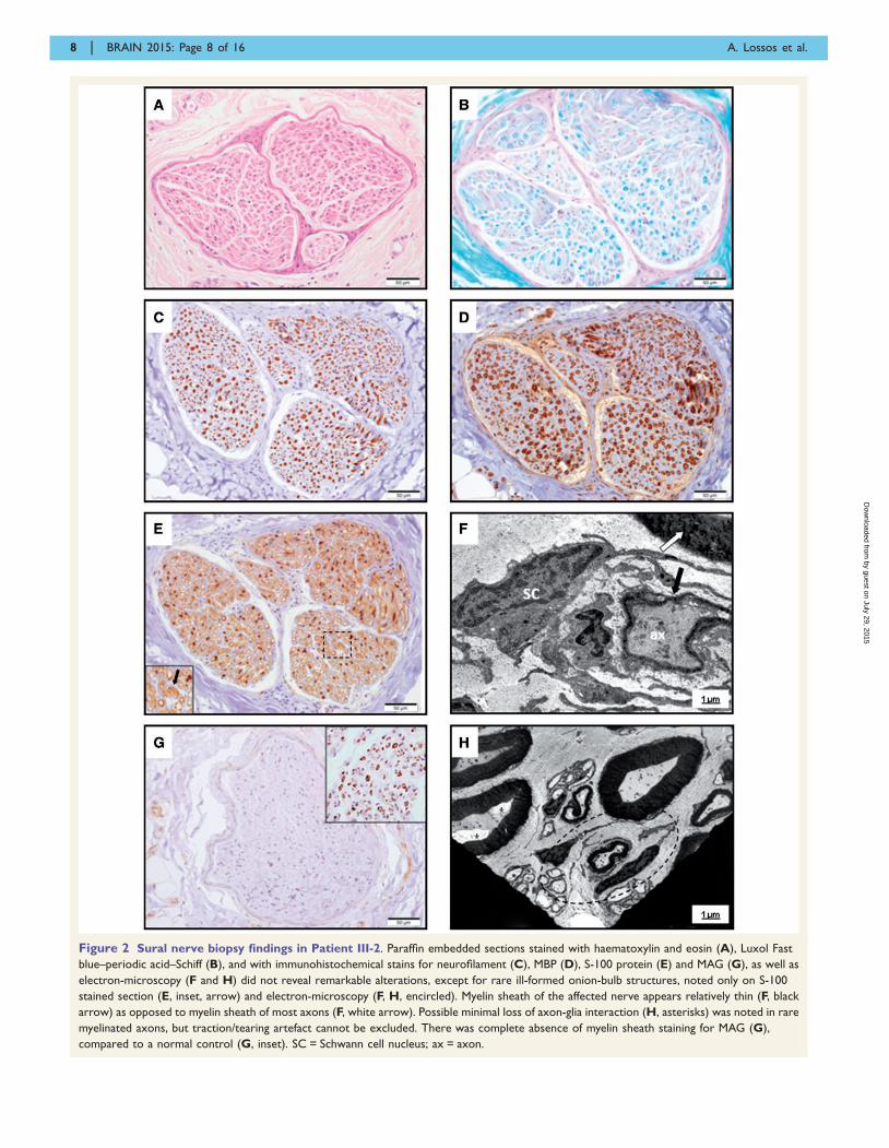
Figure 2 Sural nerve biopsy findings in Patient III-2. Paraffin embedded sections stained with haematoxylin and eosin (A), Luxol Fast
blue–periodic acid–Schiff (B), and with immunohistochemical stains for neurofilament (C), MBP (D), S-100 protein (E) and MAG (G), as well as
electron-microscopy (F and H) did not reveal remarkable alterations, except for rare ill-formed onion-bulb structures, noted only on S-100
stained section (E, inset, arrow) and electron-microscopy (F, H, encircled). Myelin sheath of the affected nerve appears relatively thin (F, black
arrow) as opposed to myelin sheath of most axons (F, white arrow). Possible minimal loss of axon-glia interaction (H, asterisks) was noted in rare
myelinated axons, but traction/tearing artefact cannot be excluded. There was complete absence of myelin sheath staining for MAG (G),
compared to a normal control (G, inset). SC = Schwann cell nucleus; ax = axon.
8 | BRAIN 2015: Page 8 of 16 A. Lossos et al.
by guest on July 29, 2015D
ownloaded from
translational processing of MAG. Further analysis using
endoglycosidase H (EndoH) revealed that while the wild-
type protein contained highly processed complex
oligosaccharides, MAG-S133R only contained immature
asparagine-linked mannose-rich oligosaccharides (Fig. 3B).
The presence of high mannose glycans in glycoproteins is
an indication of endoplasmic reticulum retention and protein
folding abnormalities (Helenius and Aebi, 2004).
Immunolabelling of COS7 cells transfected with wild-type
or mutant MAG using four different antibodies to MAG
revealed that in contrast to monoclonal antibody (Mab)
D3A2G5, which is directed to the extracellular portion of
human MAG (Burger et al., 1990), or polyclonal antibodies
against the intracellular domain (Heape et al., 1999), MAb
513 did not detect the mutant MAG-S133R (Fig. 3C). Given
that the latter antibody recognizes a conformational epitope
(Meyer-Franke et al., 1995), these results further support the
detrimental effect of the S133R mutation on folding of
MAG. These results are in line with previous observations
that N-linked glycosylation of MAG plays a role in its
proper folding (Tropak et al., 1997).
MAG-S133R mutant is trappedin the endoplasmic reticulum
Whereas wild-type transfected COS7 cells showed
MAG present on the entire cell, MAG-S133R mutant was
concentrated around the nucleus, suggesting that it does
not reach the cell surface (Fig. 3C). To further determine
whether this is the case, we immunolabelled live transfected
COS7 cells using the MAb D3A2G5 (Fig. 4A). Unlike wild-
type MAG, which was detected on the cell surface, MAG-
S133R could only be detected in detergent-permeabilized
cells. Similar distribution was detected in Schwann cells
expressing MAG-GFP and MAG-S133R-GFP (Fig. 4B).
Analysis of Schwann cells co-expressing MAG-MYC or
MAG-S133R-MYC with farnesylated GFP that marks the
plasma membrane (Rhee et al., 2006) showed similar dis-
tribution (Fig. 4C). In addition, cell surface biotinylation of
HEK293T cells expressing MAG-GFP and MAG-S133R-
GFP using a membrane-impermeable reagent demonstrated
that only wild-type MAG was detectable on the cell surface
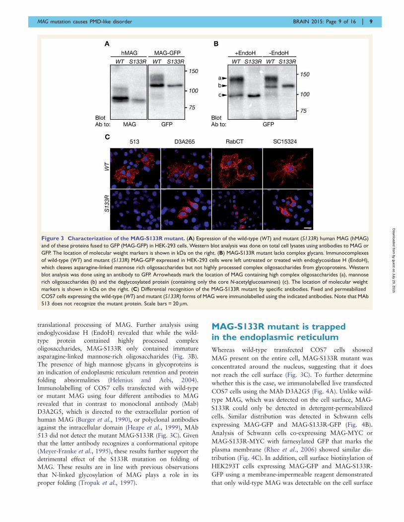
Figure 3 Characterization of the MAG-S133R mutant. (A) Expression of the wild-type (WT) and mutant (S133R) human MAG (hMAG)
and of these proteins fused to GFP (MAG-GFP) in HEK-293 cells. Western blot analysis was done on total cell lysates using antibodies to MAG or
GFP. The location of molecular weight markers is shown in kDa on the right. (B) MAG-S133R mutant lacks complex glycans. Immunocomplexes
of wild-type (WT) and mutant (S133R) MAG-GFP expressed in HEK-293 cells were left untreated or treated with endoglycosidase H (EndoH),
which cleaves asparagine-linked mannose rich oligosaccharides but not highly processed complex oligosaccharides from glycoproteins. Western
blot analysis was done using an antibody to GFP. Arrowheads mark the location of MAG containing high complex oligosaccharides (a), mannose
rich oligosaccharides (b) and the deglycosylated protein (containing only the core N-acetylglucosamines) (c). The location of molecular weight
markers is shown in kDa on the right. (C) Differential recognition of the MAG-S133R mutant by specific antibodies. Fixed and permeabilized
COS7 cells expressing the wild-type (WT) and mutant (S133R) forms of MAG were immunolabelled using the indicated antibodies. Note that MAb
513 does not recognize the mutant protein. Scale bars = 20 mm.
MAG mutation causes PMD-like disorder BRAIN 2015: Page 9 of 16 | 9
by guest on July 29, 2015D
ownloaded from
(Fig. 4D). Double immunolabelling of HeLa cells express-
ing wild-type or MAG-S133R using antibodies to MAG
and to endoplasmic reticulum protein calnexin (encoded
by CANX) (Ou et al., 1993) or to the Golgi-associated
protein p115 (encoded by USO1) (Waters et al., 1992)
showed that MAG-S133R is confined to the endoplasmic
reticulum but does not co-localized with p115 (Fig. 5A and
B). The mutant, but not the wild-type protein, co-localized
with the endoplasmic reticulum protein vesicle-associated
membrane protein-associated protein B/C (encoded by
VAPB) (Peretti et al., 2008) in both transfected COS7
(Fig. 5C) and rat Schwann cells (Fig. 5F). MAG-S133R
also co-localized with CASPR, contactin-associated protein
(encoded by CNTNAP1) (Fig. 5D), a transmembrane pro-
tein that requires the presence of contactin to exit the endo-
plasmic reticulum (Faivre-Sarrailh et al., 2000; Gollan
et al., 2003). In addition, we found that when expressed
in rat oligodendrocytes, MAG-S133R-GFP was also con-
fined to the endoplasmic reticulum (Fig. 5E).
Immunoprecipitation of MAG-GFP and MAG-S133R-GFP
from HEK293T cells revealed that unlike MAG, MAG-
S133R co-immunoprecipitated with endoplasmic reticu-
lum-resident glycan calnexin and endoplasmic reticulum
chaperone 78 kDa glucose-regulated protein (Bip, encoded
by HSPA5) (Gething, 1999) (Fig. 5G). These results dem-
onstrate that MAG-S133R does not reach the cell surface
and is stuck in the endoplasmic reticulum, further confirm-
ing the detrimental effect of the p.S133R mutation on the
folding and processing of MAG.
MAG-S133R undergoesproteasome-dependent degradation
Endoplasmic reticulum accumulation of terminally mis-
folded proteins leads to their degradation (Meusser et al.,
2005). To examine whether endoplasmic reticulum-retained
MAG-S133R is subject to degradation, we determined the
half-life of MAG-MYC and MAG-S133R-MYC proteins in
HEK293T cells using cyclohexamide chase assay (Fig. 6A
and B). MAG-S133R-MYC displayed significantly higher
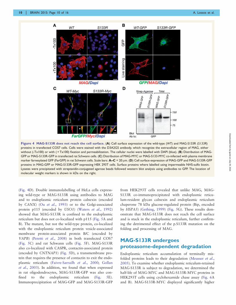
Figure 4 MAG-S133R does not reach the cell surface. (A) Cell surface expression of the wild-type (WT) and MAG-S133R (S133R)
proteins in transfected COS7 cells. Cells were stained with the D3A2G5 antibody, which recognize the extracellular region of MAG, either
without (-Tx100) or with ( + Tx100) fixation and permeabilization. The cellular nuclei were labelled with DAPI (blue). (B) Distribution of MAG-
GFP or MAG-S133R-GFP in transfected rat Schwann cells. (C) Distribution of MAG-MYC or MAG-S133-MYC co-infected with plasma membrane
marker farnesylated GFP (FarGFP) in rat Schwann cells. Scale bars: A–C = 20mm. (D) Cell surface expression of MAG-GFP and MAG-S133R-GFP
proteins in MAG-GFP or MAG-S133R-GFP expressing HEK 293T cells. Surface proteins where labelled using impermeable NHS-sulfo biotin.
Lysates were precipitated with streptavidin-conjugated agarose beads followed western blot analysis using antibodies to GFP. The location of
molecular weight markers is shown in kDa on the right.
10 | BRAIN 2015: Page 10 of 16 A. Lossos et al.
by guest on July 29, 2015D
ownloaded from
degradation rate with a half-life of �2 h compared to a
very stable MAG-MYC. Degradation rate of MAG-S133R
was reduced by the addition of proteasome inhibitor
bortezomib (Fig. 6C). In bortezomib-treated cells, MAG-
S133R-GFP, but not MAG-GFP, was polyubiquitinated,
indicating that it is destined to the proteasome
for degradation (Fig. 6E). In addition, the fact that
polyubiquitination of MAG-S133R-GFP could only be de-
tected after proteasomal inhibition, further indicates that
the mutant protein is rapidly degraded (Altier et al.,
2011). Immunoprecipitation of MAG-GFP and MAG-
S133R-GFP in HEK293T cells showed that as opposed to
wild-type protein, MAG-S133R was associated with com-
ponents of the endoplasmic reticulum-associated protein
degradation (ERAD) machinery, such as P97 (OS9, SEL1)
(Christianson et al., 2008) (Fig. 6D).
To determine what intracellular proteins are associated
with the wild-type and the mutant protein, we subjected
MAG-GFP and MAG-S133R-GFP immunocomplexes to
high resolution mass spectrometry-based proteomic ana-
lysis. Statistical tests showed clear differences between inter-
actors of wild-type MAG-GFP and MAG-S133R-GFP
(Student t-test, FDR = 0.05, S0 = 1.5). Wild-type MAG
was mainly associated with proteins related to the Golgi
apparatus and protein glycosylation, consistent with its
localization at the plasma membrane and its post-
translational N-glycosylation modifications (Fig. 7). In
contrast, MAG-S133R was associated with a number of
endoplasmic reticulum processing proteins, such as BiP,
calnexin, calreticulin, PDIA3, and PDIA4, with proteins
related to protein folding, such as the chaperonin-contain-
ing T-complex (Horwich and Willison, 1993), and with
COP1 coating proteins, responsible for the retrograde
transport from Golgi back to the endoplasmic reticulum
(Spang and Schekman, 1998) (Fig. 7A and B). Finally,
the mutant protein was associated with a number of
ERAD proteins, such as P97, OS9, SEL1 and EDEM3, as
well as with proteasomal proteins, such as PSMC1,
PSMC4, PSMD1 and PSMD3 (Fig. 7B). Collectively, our
results demonstrate that the p.S133R mutation affects the
post-translational processing and folding of MAG, leading
to its retention in the endoplasmic reticulum with subse-
quent ERAD-mediated proteasomal degradation.
Based on these results, we immunohistochemically
stained sural nerve biopsy sample of Patient III-2 for
MAG showing complete absence of myelin sheath staining
(Fig. 2G).
DiscussionUsing genetic linkage, exome sequencing and cellular ex-
pression studies, we identified a homozygous missense
c.399 C4G; p.S133R mutation in MAG in a consanguin-
eous multiplex family with PMD-like disorder. Affected sib-
lings presented the typical for PMD developmental and
motor delay of infantile-onset with early nystagmus and
hypotonia slowly evolving into a form of complicated
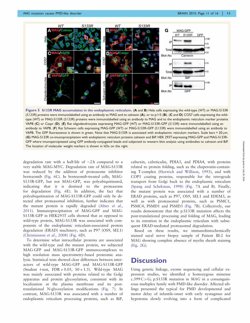
Figure 5 S133R MAG accumulates in the endoplasmic reticulum. (A and B) Hela cells expressing the wild-type (WT) or MAG-S133R
(S133R) proteins were immunolabelled using an antibody to MAG and to calnexin (A), or to p115 (B). (C and D) COS7 cells expressing the wild-
type (WT) or MAG-S133R (S133R) proteins were immunolabelled using an antibody to MAG and to the endoplasmic reticulum marker proteins
VAPB (C) or Caspr (D). (E) Rat oligodendrocytes expressing MAG-GFP (WT) or MAG-S133R-GFP (S133R) were immunolabelled using an
antibody to VAPB. (F) Rat Schwann cells expressing MAG-GFP (WT) or MAG-S133R-GFP (S133R) were immunolabelled using an antibody to
VAPB. The GFP fluorescence is shown in green. Note that MAG-S133R is associated with endoplasmic reticulum markers. Scale bars = 20 mm.
(G) MAG-S133R co-imunoprecipitation with endoplasmic reticulum proteins calnexin and BiP. HEK 293Texpressing MAG-GFP and MAG-S133R-
GFP where imunopercipitated using GFP antibody-conjugated beads and subjected to western blot analysis using antibodies to calnexin and BiP.
The location of molecular weight markers is shown in kDa on the right.
MAG mutation causes PMD-like disorder BRAIN 2015: Page 11 of 16 | 11
by guest on July 29, 2015D
ownloaded from
HSP with mental retardation, dysarthria, optic atrophy and
peripheral neuropathy in adulthood. The mild variability
observed among the siblings likely results from age-related
progression of the underlying process. Although HLD1,
HLD2 and HLD4 also exhibit PMD-like and HSP pheno-
types, the combination of clinical and imaging findings
clearly distinguishes the disorder in this family from other
forms (Supplementary Table 2).
As the PMD-like phenotype is associated with myelin-
related genes PLP1 and GJC2 (Pouwels et al., 2014), in-
volvement of MAG in this family may not be surprising.
The encoded protein MAG is a quantitatively minor
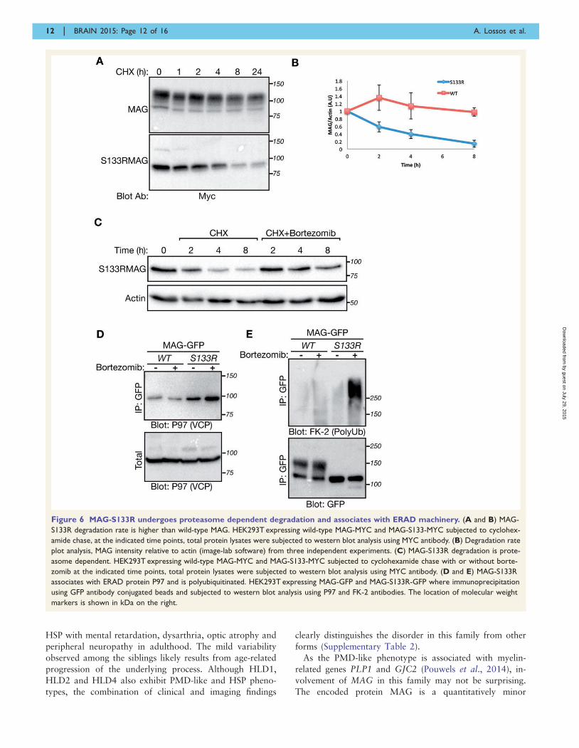
Figure 6 MAG-S133R undergoes proteasome dependent degradation and associates with ERAD machinery. (A and B) MAG-
S133R degradation rate is higher than wild-type MAG. HEK293T expressing wild-type MAG-MYC and MAG-S133-MYC subjected to cyclohex-
amide chase, at the indicated time points, total protein lysates were subjected to western blot analysis using MYC antibody. (B) Degradation rate
plot analysis, MAG intensity relative to actin (image-lab software) from three independent experiments. (C) MAG-S133R degradation is prote-
asome dependent. HEK293T expressing wild-type MAG-MYC and MAG-S133-MYC subjected to cyclohexamide chase with or without borte-
zomib at the indicated time points, total protein lysates were subjected to western blot analysis using MYC antibody. (D and E) MAG-S133R
associates with ERAD protein P97 and is polyubiquitinated. HEK293T expressing MAG-GFP and MAG-S133R-GFP where immunoprecipitation
using GFP antibody conjugated beads and subjected to western blot analysis using P97 and FK-2 antibodies. The location of molecular weight
markers is shown in kDa on the right.
12 | BRAIN 2015: Page 12 of 16 A. Lossos et al.
by guest on July 29, 2015D
ownloaded from
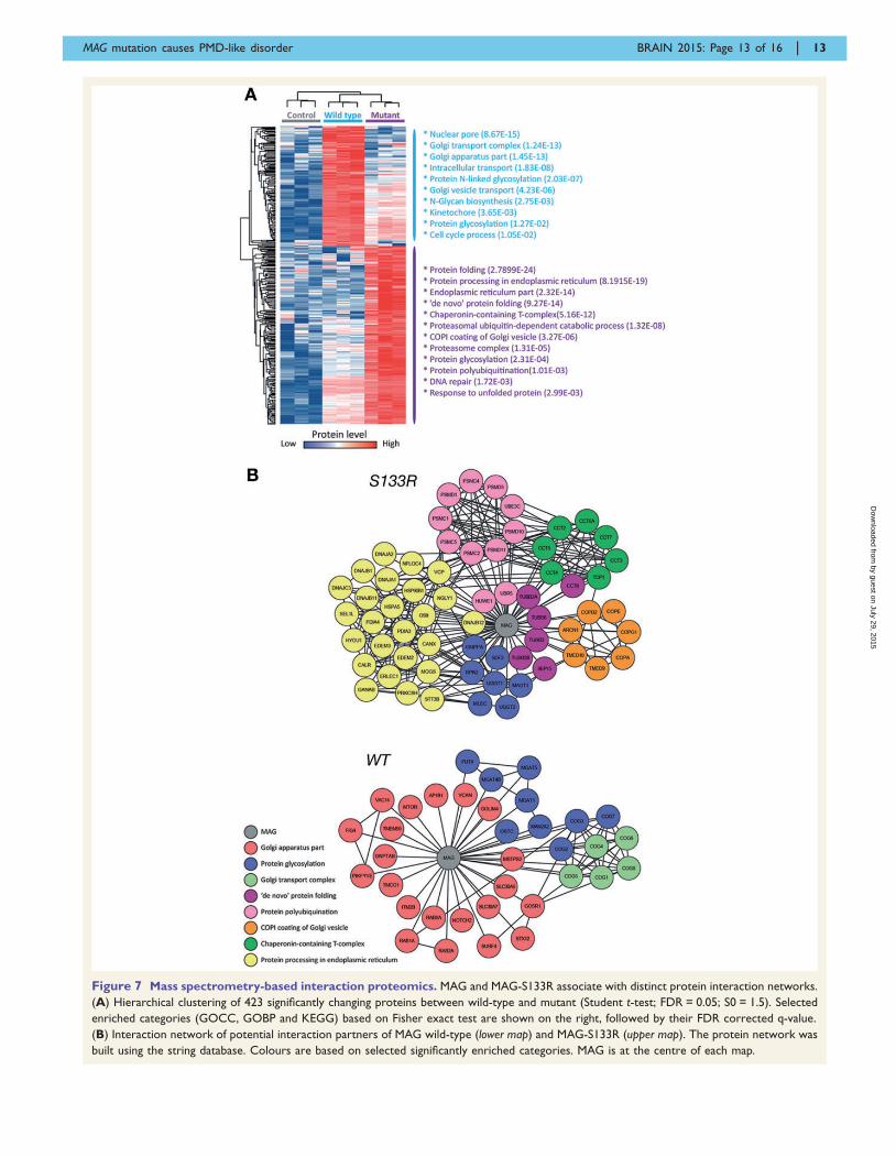
Figure 7 Mass spectrometry-based interaction proteomics. MAG and MAG-S133R associate with distinct protein interaction networks.
(A) Hierarchical clustering of 423 significantly changing proteins between wild-type and mutant (Student t-test; FDR = 0.05; S0 = 1.5). Selected
enriched categories (GOCC, GOBP and KEGG) based on Fisher exact test are shown on the right, followed by their FDR corrected q-value.
(B) Interaction network of potential interaction partners of MAG wild-type (lower map) and MAG-S133R (upper map). The protein network was
built using the string database. Colours are based on selected significantly enriched categories. MAG is at the centre of each map.
MAG mutation causes PMD-like disorder BRAIN 2015: Page 13 of 16 | 13
by guest on July 29, 2015D
ownloaded from
component of myelin and is expressed solely by oligo-
dendrocytes and Schwann cells (Quarles, 2007), consistent
with the combined CNS and PNS involvement in our pa-
tients. It is an adhesion molecule in the periaxonal layer of
mature non-compact myelin membrane facing the axon.
Based on its location and the fact that its expression cor-
relates with the initiation of myelination (Quarles, 2007),
MAG is thought to mediate interactions between myelinat-
ing glia and their underlying axons (Lopez, 2014).
MAG is a highly glycosylated transmembrane protein con-
taining an extracellular segment with five tandem Ig-like do-
mains (Quarles, 2007). The c.399C4G mutation identified
in this family involves the first Ig-like domain and leads to a
single amino acid substitution p.S133R, predicted to desta-
bilize the protein, affect its glycosylation and cause defective
folding. Accordingly, our studies demonstrate that the
MAG-S133R mutant contains mannose-rich oligosaccharides
instead of highly processed complex oligosaccharides, indi-
cative of its endoplasmic reticulum retention (Helenius and
Aebi, 2004). In line with the notion that N-linked glycosyla-
tion is important for proper folding (Tropak et al., 1997),
MAG-S133R is not recognized by MAb 513, which recog-
nizes conformational epitope (Meyer-Franke et al., 1995) of
the wild-type protein. The MAG-S133R mutant is present in
the endoplasmic reticulum in a variety of transfected cells,
co-immunoprecipitating with the endoplasmic reticulum
chaperones calnexin and calreticulin, known to increase
the efficiency of glycoprotein folding and to ensure that mis-
folded proteins are retained in the endoplasmic reticulum
(Rajagopalan et al., 1994). The mutant MAG also associates
with the endoplasmic reticulum protein BiP (GRP78), which
facilitates protein folding and regulates the unfolded protein
response. Mass spectrometry-based proteomics corroborates
our findings that MAG-S133R is retained in the endoplasmic
reticulum. Furthermore, MAG-S133R interacts with proteins
of the COP1 family, responsible for the retrograde transport
of proteins from Golgi to endoplasmic reticulum, which
likely prevents mutated MAG to proceed along the secretory
pathway.
Cyclohexamide experiments reveal that MAG-S133R
undergoes rapid degradation and removal, compared to
the stable wild-type MAG. Moreover, this rapid degradation
is proteasome-dependent. Consistent with this observation,
MAG-S133R, but not the wild-type MAG, is polyubiquiti-
nated and is associated with ERAD machinery proteins P97,
OS9, SEL1 and EDEM3. Moreover, MAG-S133R directly
associates with proteasomal subunits representative of the
proteasome-dependent nature of degradation. It is also im-
portant to note that MAG-S133R does not induce the acti-
vation of endoplasmic reticulum stress mediators PERK,
IRE1 and ATF6 endoplasmic reticulum. Collectively, our re-
sults suggest that the c.399C4G; p.S133R mutation in
MAG leads to ERAD-mediated loss of the protein, which
corresponds to the immunohistochemical absence of MAG
in the sural nerve biopsy specimen of Patient III-2 (Fig. 2G).
Loss of tissue immmunoreactivity for MAG also charac-
terizes genetically manipulated knockout mice (Li et al.,
1994; Montag et al., 1994). When examined by the age
of 4 months, affected animals show surprisingly mild clin-
ical and pathological alterations in the CNS and PNS, dis-
play normal peripheral nerve conduction, and are able to
generate mature myelin. Delayed CNS myelination, redun-
dant or disrupted compact myelin, oligodendroglial periax-
onal area and cytoplasmic collar changes (Montag et al.,
1994), tremor and impaired coordination (Li et al., 1994)
are among the reported alterations at this stage (Lopez,
2014). Normal MRI myelination pattern in our patients
in childhood (Patient III-2) and in adulthood is consistent
with these findings and still leaves a possibility of delayed
myelination in infancy to explain their early PMD-like
manifestations.
At a later age, additional alterations are described in
MAG knockout animals, including degeneration of axons
and myelin in the PNS accompanied by the formation
of onion bulb-like structures (Fruttiger et al., 1995).
Peripheral nerve conduction studies show reduced velocities
with a less significantly decreased compound muscle action
potential amplitudes (Weiss et al., 2001). These abnormal-
ities qualitatively resemble electron microscopic findings in
Patient III-2 (Fig. 2) and an apparent age-related worsening
of the peripheral nerve conduction findings between sib-
lings, Patients III-5, III-3 and III-2 (Supplementary Table 1).
Long-term alterations in the CNS in MAG knockout ani-
mals include axonal degeneration (Nguyen et al., 2009) and
dying-back oligodendrogliopathy, which possibly represents
initial stages of a slow demyelinating process (Lassmann
et al., 1997). Clinical and electrophysiological signs of the
CNS involvement in our patients in adulthood accompanied
by the cerebral MRI and 1H-magnetic resonance spectros-
copy abnormalities (Supplementary Table 1 and Fig. 1E) are
also indicative of a demyelinating form of leukodystrophy.
Taken together, our findings confirm a slowly progressive
involvement of the PNS and CNS in this family with
c.399C4G mutation in MAG and are consistent with the
suggested function of MAG in a long-term maintenance and
integrity of myelin and axons (Lopez, 2014).
Recently, another mutation in MAG has been reported in
a monumental study of multiple HSP families (Novarino
et al., 2014). The two affected sisters carrying the
c.1288T4G; p.C430G mutation in the core of the fifth Ig-
like domain, manifested early-onset HSP associated with nys-
tagmus, cerebellar signs, amyotrophy, sensory loss and poor
school achievements, akin to the late clinical phenotype in
our patients. Although their MRI was tabulated as normal,
no details, 1H-magnetic resonance spectroscopy or nerve
conduction results were available for comparison.
In summary, we provide the first conclusive evidence for
the involvement of MAG in a combined CNS and PNS
disorder manifesting early PMD-like clinical phenotype
later evolving into a complicated HSP. Given autosomal
recessive inheritance in this family, we postulate that the
underlying mechanism of the p.S133R mutation is related
to the ERAD-mediated loss of MAG function. Because
MAG is developmentally regulated through alternative
14 | BRAIN 2015: Page 14 of 16 A. Lossos et al.
by guest on July 29, 2015D
ownloaded from
mRNA splicing of the cytoplasmic domain generating short
and long isoforms differentially expressed in the PNS and
CNS, it is conceivable that MAG is responsible for a
broader range of neurogenetic disorders.
Supplementary materialSupplementary material is available at Brain online.
AcknowledgementsWe thank Sima Lev and Tony Heap for supplying
antibodies.
FundingThis work was supported in part by the NIH (NS50220 to
E.P.), the Israel Science Foundation, the Dr. Miriam and
Sheldon G. Adelson Medical Research Foundation, the
Israeli MOH grant (#5914), the Israeli MOH/ERA-Net
(#4800), the Karl Kahane Foundation, and the Agnes
Ginges stipend (to B-EA). O.S-F. was supported by the
Israel Science Foundation, founded by the Israel Academy
of Science and Humanities (#319/11), the United States-
Israel Binational Science Foundation (#2009418), and a
starting grant from the European Research Council
(#310873). E.P. is the Incumbent of the Hanna Hertz
Professorial Chair for Multiple Sclerosis and Neuroscience.
ReferencesAltier C, Garcia-Caballero A, Simms B, You H, Chen L, Walcher J,
et al. The Cavbeta subunit prevents RFP2-mediated ubiquitination
and proteasomal degradation of L-type channels. Nat Neurosci
2011; 14: 173–80.
Bizzi A, Castelli G, Bugiani M, Barker PB, Herskovits EH, Danesi U,
et al. Classification of childhood white matter disorders using proton
MR spectroscopic imaging. AJNR Am J Neuroradiol 2008; 29:
1270–5.
Burger D, Simon M, Perruisseau G, Steck AJ. The epitope(s) recog-
nized by HNK-1 antibody and IgM paraprotein in neuropathy is
present on several N-linked oligosaccharide structure on human
P0 and myelin-associated glycoprotein. J Neurochem 1990; 54:
1569–75.
Christianson JC, Shaler TA, Tyler RE, Kopito RR. OS-9 and GRP94
deliver mutant alpha 1-antitrypsin to the Hrd1-SEL1L ubiquitin
ligase for ERAD. Nat Cell Biol 2008; 10: 272–82.Cox J, Neuhauser N, Michalski A, Scheltema RA, Olsen JV, Mann M.
Andromeda: a peptide search engine integrated into the MaxQuant
environment. J Proteome Res 2011; 10: 1794–805.
Cox J, Hein MY, Luber CA, Paron I, Nagaraj N, Mann M. Accurate
proteome-wide label-free quantification by delayed normalization
and maximal peptide ration extraction, termed MaxLFQ. Mol Cell
Proteomics 2014; 13: 2513–26.
Erb M, Steck AJ, Nave KA, Schaeren-Wiemers N. Differential expres-
sion of L- and S-MAG upon cAMP stimulated differentiation in
oligodendroglial cell. J Neurosci Res 2003; 71: 326–37.
Eshed Y, Feiberg K, Poliak S, Sabanay H, Sarig-Nadir O, Spiegel I,
et al. Gliomedin mediates Schwann cell-axon interaction and the
molecular assembly of the nodes of Ranvier. Neuron 2005; 47:
215–29.
Faivre-Sarrailh C, Gauthier F, Denisenko-Nehrbass N, Le Bivic A,
Rougon G, Girault JA. The glycosylphosphatidyl inositol-anchored
adhesion molecule F3/contactin is required for surface transport of
paranodin/contactin-associated protein (caspr). J Cell Biol 2000;
149: 491–502.
Feinstein M, Markus B, Noyman I, Shalev H, Flusser H, Shelef I, et al.
Pelizaeus-Merzbacher-like disease caused by AIMP1/p43 homozy-
gous mutation. Am J Hum Genet 2010; 87: 820–8.
Folstein MF, Folstein SE, McHugh PR. “Mini-mental state”. A prac-
tical method for grading the cognitive state of patients for the clin-
ician. J Psychiatr Res 1975; 12,189–98.
Fruttiger M, Montag D, Schachner M, Martini R. Crucial role for the
myelin-associated glycoprotein in the maintenance of axon-myelin
integrity. Eur J Neurosci 1995; 7: 511–5.Gething MJ. Role and regulation of the ER chaperon BiP. Semin Cell
Dev Biol 1999; 10: 465–72.Gollan L, Salomon D, Salzer JL, Peles E. Caspr regulates the process-
ing of contactin and inhibits its binding to neurofascin. J Cell Biol
2003; 163: 1213–8.
Han H, Myllykoski M, Ruskamo S, Wang C, Kursula P. Myelin-spe-
cific proteins: a structurally diverse group of membrane-interacting
molecules. Biofactors 2013; 39: 233–41.
Heape AM, Lehto VP, Kursula P. The expression of recombinant large
myelin-associated glycoprotein cytoplasmic domain and the purifica-
tion of native myelin-associated glycoprotein from rat brain and
peripheral nerve. Protein Expr Purif 1999; 15: 349–61.
Helenius A, Aebi M. Roles of N-linked glycans in the endoplasmic
reticulum. Annu Rev Biochem 2004; 73: 1019–49.
Henneke M, Combes P, Diekmann S, Bertini E, Brockmann K, Burlina
AP, et al. GJA12 mutations are a rare cause of Pelizaeus-
Merzbacher-like disease. Neurology 2008; 70: 748–54.
Hobson GM, Garbern JY. Pelizaeus-Merzbacher disease, Pelizaeus-
Merzbacher-like disease 1, and related disorders. Semin Neurol
2012; 32: 62–7.
Horwich AL, Willison KR. Protein folding in the cell: functions of two
families of molecular chaperon, hsp 60 and TF55-TCP1. Phil Trans
R Soc Lond Biol Sci 1993; 339: 313–25; discussion 25–6.
Lassmann H, Bartsch U, Montag D, Schachner M. Dying-back oligo-
dendrogliopathy: a late sequel of myelin-associated glycoprotein de-
ficiency. Glia 1997; 19: 104–10.
Letunic I, Doerks T, Bork P. SMART 7: recent updates to the protein
domain annotation resource. Nucleic Acids Res 2012; 40: D302–5.Li C, Tropak MB, Gerlai R, Clapoff S, Abramow-Newerly W, Trapp
B, et al. Myelination in the absence of myelin-associated glycopro-
tein. Nature 1994; 369: 747–50.
Lopez PHH. Role of myelin-associated glycoprotein (siglec-4A) in the
nervous system. Adv Neurobiol 2014; 9: 245–62.
Magen D, Georgopoulos C, Bross P, Ang D, Segev Y, Goldsher D,
et al. Mitochondrial Hsp60 chaperonopathy causes an autosomal-
recessive neurodegenerative disorder linked to brain hypomyelina-
tion and leukodystrophy. Am J Hum Genet 2008; 83: 30–42.
May AP, Robinson RC, Vinson M, Crocker PR, Jones EY. Crystal
structure of the N-terminal domain of sialoadhesin in complex with
3’ sialyllactose at 1.85 A resolution. Mol Cell 1998; 1: 719–28.
Meyer-Franke A, Tropak MB, Roder JC, Fischer P, Beyreuther K,
Probstmeier R, et al. Functional topography of myelin-associated
glycoprotein. II. Mapping of domains on molecular fragments.
J Neurosci Res 1995; 41: 311–23.
Meusser B, Hirsh C, Jarosch E, Sommer T. ERAD: the long road to
destruction. Nat Cell Biol 2005; 7: 766–72.
Miyatake S, Osaka H, Shiina M, Sasaki M, Takanashi J, Haginoya K,
et al. Expanding the phenotypic spectrum of TUBB4A-associated
hypomyelinating leukodystrophies. Neurology 2014; 82: 2230–37.
MAG mutation causes PMD-like disorder BRAIN 2015: Page 15 of 16 | 15
by guest on July 29, 2015D
ownloaded from
Montag D, Giese KP, Bartsch U, Martini R, Lang Y, Bluthmann H,et al. Mice deficient for the myelin-associated glycoprotein show
subtle abnormalities in myelin. Neuron 1994; 13: 229–46.
Nguyen T, Mehta NR, Conant K, Kim K-J, Jones M, Calabresi PA,
et al. Axonal protective effects of the myelin-associated glycoprotein.J Neurosci 2009; 29: 630–7.
Novarino G, Fenstermaker AG, Zaki MS, et al. Exome sequencing
links corticospinal motor neuron disease to common neurodegenera-
tive disorders. Science 2014; 343: 506–11.Orthmann-Murphy JL, Salsano E, Abrams CK, Bizzi A, Uziel G,
Freidin MM, et al. Hereditary spastic paraplegia is a novel pheno-
type for GJA12/GJC2 mutations. Brain 2009; 132: 426–38.Ou WJ, Cameron PH, Thomas DY, Bergeron JJ. Association of fold-
ing intermediates of glycoproteins with calnexin during protein mat-
uration. Nature 1993; 364: 771–6.
Peles E, Nativ M, Lustig M, Grumet M, Schilling J, Martinez R, et al.Identification of a novel contactin-associated transmembraine recep-
tor with multiple domains implicated in protein-protein interactions.
Embo J 1997; 16: 978–88.
Peretti D, Dahan N, Shimoni E, Hirschberg K, Lev S. Coordinatedlipid transfer between the endoplasmic reticulum and the Golgi com-
plex requires the VAP proteins and is essential for Golgi-mediated
transport. Mol Biol Cell 2008; 19: 3871–84.
Pouwels PJW, Vanderver A, Bernard G, Wolf NI, Dreha-KulczewksiSF, Deoni SCL, et al. Hypomyelinating leukodystrophies:
translational research progress and prospects. Ann Neurol 2014;
76: 5–19.Quarles RH. Myelin-associated glycoprotein (MAG): past, present and
beyond. J Neurochem 2007; 100: 1431–48.
Rajagopalan S, Xu Y, Brenner MB. Retention of unassembled compo-
nents of integral membrane proteins by calnexin. Science 1994; 263:387–90.
Rhee JM, Pirity MK, Lackan CS, Long JZ, Kondoh G, Takeda J, et al.In vivo imaging and differential localization of lipid-modified GFP-
variant fusions in embryonic stem cells and mice. Genesis 2006; 44:
202–18.
Roy A, Kucukural A, Zhang Y. I-TASSER: a unified platform forautomated protein structure and function prediction. Nat Protoc
2010; 5: 725–38.
Shimojima K, Okumura A, Ikeno M, Nishimura A, Saito A, Saitsu H,
et al. A de novo TURBB4A mutation in a patient with hypomyeli-nation mimicking Pelizaeus-Merzbacher disease. Brain Dev 2015;
37: 281–5. 10.1016/j.braindev.2014.05.004
Seelow D, Schuelke M, Hildebrandt F, Nurnberg P.HomozygosityMapper-an interactive approach to homozygosity
mapping. Nucleic Acids Res 2009; 37: W593–9.
Soding J. Protein homology detection by HMM-HMM comparison.
Bioinformatics 2005; 21: 951–60.Spang A, Schekman R. Reconstitution of retrograde transport from
Golgi to the ER in vitro. J Cell Biol 1998; 143: 589–99.
Spiegel I, Adamsky K, Eshed Y, Milo R, Sabanay H, Sarig-Nadir O,
et al. A central role for Necl4 (SynCAM4) in Schwann cell-axoninteraction and myelination. Nat Neurosci 2007; 10: 861–9.
Tropak MB, Roder JC. Regulation of myelin-associated glycoprotein
binding by sialylated cis-ligands. J Neurochem 1997; 68: 1753–63.
Vaurs-Barriere C, Deville M, Sarret C, Giraud G, Des Portes V, Prats-Vinas J-M, et al. Pelizaeus-Merzbacher-like disease presentation of
MCT8 mutated male subjects. Ann Neurol 2009; 65: 114–8.
Waters MG, Clary DO, Rothman JE. A novel 115-kD peripheralmembrane protein is required for intercisternal transport in the
Golgi stack. J Cell Biol 1992; 118: 1015–26.
Weiss MD, Luciano CA, Quarles RH. Nerve conduction abnormalities
in aging mice deficient for myelin-associated glycoprotein. MuscleNerve 2001; 24: 1380–7.
16 | BRAIN 2015: Page 16 of 16 A. Lossos et al.
by guest on July 29, 2015D
ownloaded from