Embed Size (px)
DESCRIPTION
Mycology – Yeast. Student Lab Division of Medical Technology Carol Larson MSEd, MT(ASCP). Basic Characteristics. Unicellular Generally normal flora. Basic Characteristics. Asexual reproduction Blastoconidia Pseudohyphae Arthroconidia. Direct Examination. - PowerPoint PPT Presentation
Citation preview

Mycology –Yeast
Student Lab
Division of Medical Technology
Carol Larson MSEd, MT(ASCP)

Basic Characteristics
• Unicellular
• Generally normal flora

Basic Characteristics
• Asexual reproduction– Blastoconidia– Pseudohyphae– Arthroconidia

Direct Examination
• Observe for reproductive structures
• Gram stain– Gram positive
• India ink stain– Capsules

Growth Requirements
• Growth in 2-3 days
• Temperature– 25-37ºC– Best at 30ºC

Colony Morphology
• Appearance– Similar to bacteria colony
• Texture– Glabrous– May be filamentous
• Pigmentation
• Confuse with Staphylococcus

Yeast ID Methods
• Germ Tube Production– First test performed when yeast– Procedure– Interpretation

Yeast ID Methods
• Cornmeal-Tween 80 agar– Procedure– Observe microscopically for
• Chlamydospores• Blastoconidia• Pseudohyphae• Arthrospores

Yeast ID Methods
• Niger Seed Agar (Birdseed agar)– Procedure– Observe for brown pigment caused by
phenol oxidase activity– Cryptococcus
neoformans

Yeast ID Methods
• Urease– Procedure– Positive:
• Cryptococcus sp. (rapid)• Trichosporon sp.• Rhodotorula sp.
– Negative:• Candida sp.• Geotrichum sp.

Yeast ID Methods
• Carbohydrate assimilation– Utilize carbohydrate as sole source of
carbon in the presence of oxygen– Positive = growth
• Carbohydrate fermentation– Utilize a carbohydrate anaerobically– Positive = gas
Yeast ID Methods
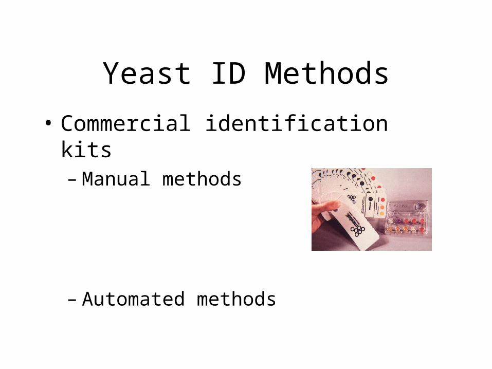
• Commercial identification kits– Manual methods
– Automated methods
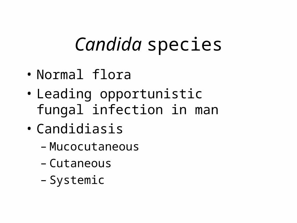
Candida species
• Normal flora
• Leading opportunistic fungal infection in man
• Candidiasis– Mucocutaneous– Cutaneous– Systemic

Factors predisposing to infection
• Immunocompromised
• Malignancy
• Prolonged antibiotic therapy
• Lupus, Diabetes
• Pregnancy and use of birth control pills
• Age
• Damaged skin barrier

Candida albicans
• #1 fungal isolate in laboratory
• Germ tube: positive
• CMT: clustered blastoconidia at septa of pseudohyphae, terminal chlamydospores
• CHO: sucrose positive

Candida albicans

Candida stellatoidea
• Germ tube: positive
• CMT: clustered blastoconidia at septa of pseudohyphae, terminal chlamydospores
• CHO: sucrose negative

Candida tropicalis
• Germ tube: negative
• CMT: sparse single or short-chained blastoconidia anywhere along pseudohyphae, rare chlamydospores
• CHO: sucrose positive

Candida glabrata
• Causes UTI
• Germ tube: negative
• CMT: only blastoconidia, no pseudohyphae
• CHO: only glucose and trehalose positive

Candida krusei
• Germ tube: negative
• CMT: tree-like branching of abundant blastoconidia from the septa of elongated pseudohyphae (“cross-matchsticks” appearance)

Candida pseudotropicalis
• Germ tube: negative
• CMT: branched pseudohyphae with chains of elongated blastoconidia – logs in a stream arrangement of broken up blastoconidia positioned parallel to each other

Candida parapsilosis
• Germ tube: negative
• CMT: few single or small clustered blastoconidia at or between septa of thin curved pseudohyphae. Sometimes giant pseudohyphae may be observed

Cryptococcus neoformans
• Cryptococcosis– Pulmonary– Meningitis
• Pigeon droppings

Cryptococcus neoformans
• Gram stain
• India ink stain

Cryptococcus neoformans
• Germ tube: negative
• CMT: large, round blastoconidia, no pseudohyphae
• Urease: positive (within 3 hours)

Cryptococcus neoformans
• Niger seed agar: brown pigment

Cryptococcus neoformans
• Cryptococcal antigen test
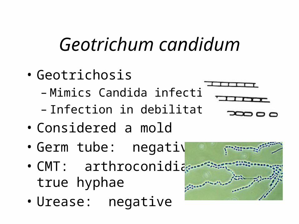
Geotrichum candidum
• Geotrichosis– Mimics Candida infections– Infection in debilitated people
• Considered a mold
• Germ tube: negative
• CMT: arthroconidia andtrue hyphae
• Urease: negative

Trichosporon beigelii
• Causes White Piedra
• Germ tube: negative
• CMT: arthroconidia, true hyphae, rare blastoconidia
• Urease: positive

Rhodotorula species
• Saprophytic yeast found in soil
• Rarely a pathogen
• Often encapsulated
• Urease: positive
• Colony pigmentation: reddish-orange

Saccharomyces species
• Rarely a pathogen
• Ascospores (acid fast positive)

Malassezia furfur
• Causes Tinea Versicolor
• Septicemia in patients receiving intravenous lipid therapy
• KOH: clusters of thick-walled, round budding yeast and short, straight or angular mycelial fragments (spaghetti and meatball appearance)
• Lipophilic

In Summary …
• Key characteristics of yeast
• Identification methods– Germ tube– CMT– Biochemical tests
• Clinically significant yeast– Candida albicans– Cryptococcus neoformans


![Mycology - Dr. Hashemi.ppt - iacld.ir · A yeastA yeast--like dematiaceus fungus, ... Erythrasma Definition: ... Mycology - Dr. Hashemi.ppt [Compatibility Mode] Author: bathaei](https://img.pdfslide.us/doc/110x75/5ad803fa7f8b9ab8378cd5eb/mycology-dr-iacldir-yeasta-yeast-like-dematiaceus-fungus-erythrasma.jpg)