Embed Size (px)
Citation preview

MYCOLOGYScience for studying of fungi
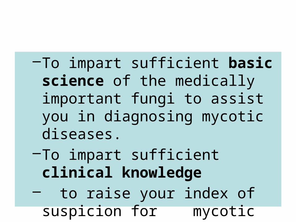
–To impart sufficient basic science of the medically important fungi to assist you in diagnosing mycotic diseases.
–To impart sufficient clinical knowledge
– to raise your index of suspicion for mycotic diseases.
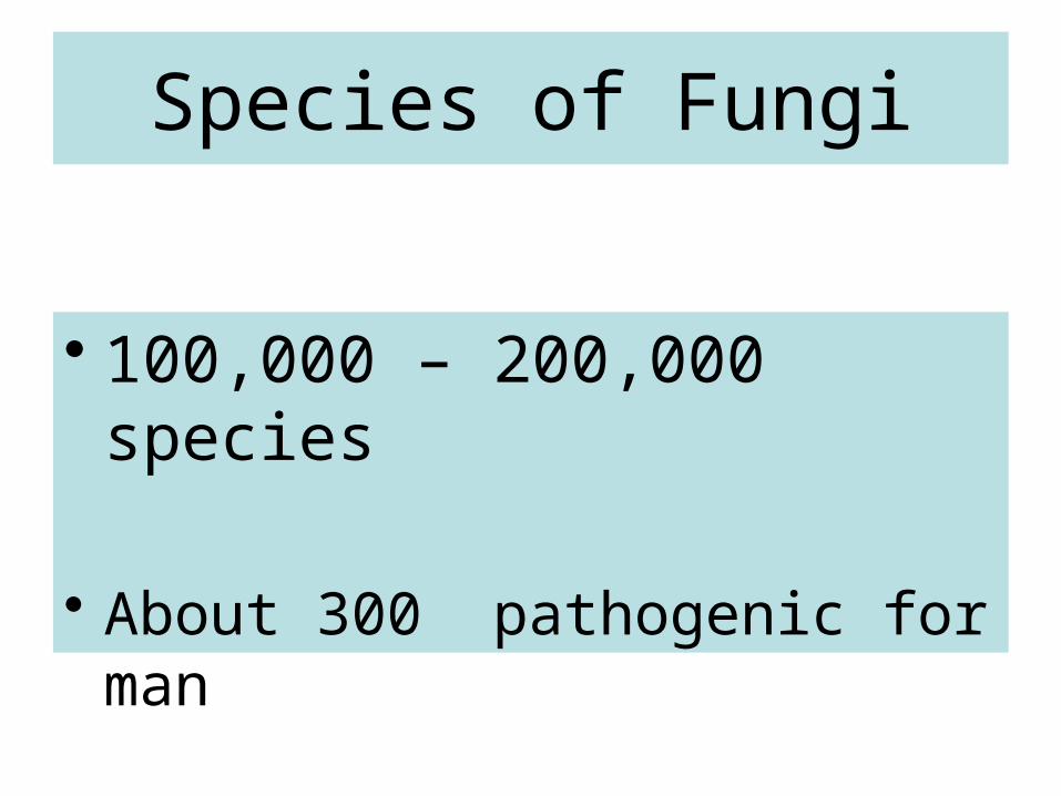
Species of Fungi
• 100,000 – 200,000 species
• About 300 pathogenic for man
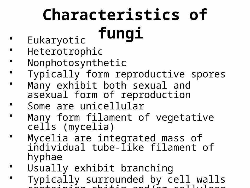
Characteristics of fungi • Eukaryotic• Heterotrophic• Nonphotosynthetic• Typically form reproductive spores• Many exhibit both sexual and asexual form of
reproduction • Some are unicellular• Many form filament of vegetative cells
(mycelia)• Mycelia are integrated mass of individual tube-
like filament of hyphae• Usually exhibit branching• Typically surrounded by cell walls containing
chitin and/or cellulose
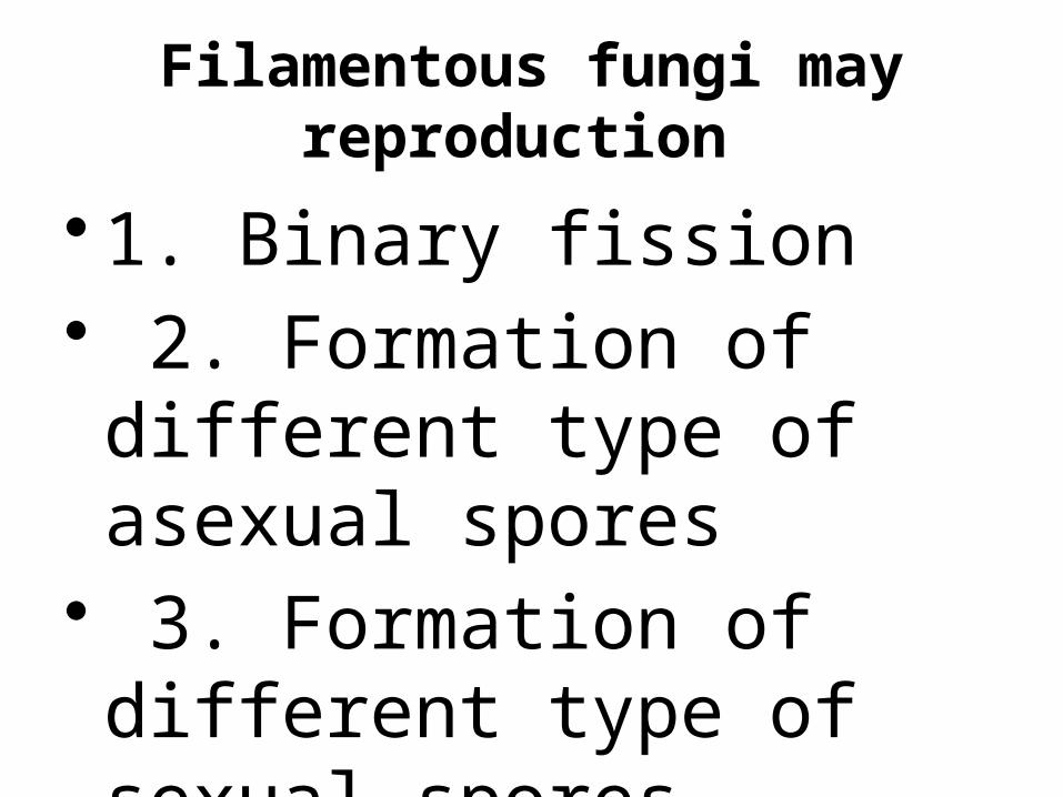
Filamentous fungi may reproduction
• 1. Binary fission• 2. Formation of different type of asexual spores
• 3. Formation of different type of sexual spores
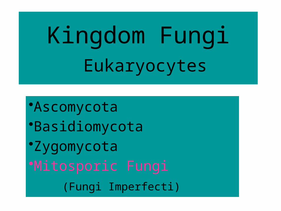
Kingdom Fungi Eukaryocytes
•Ascomycota•Basidiomycota•Zygomycota•Mitosporic Fungi
(Fungi Imperfecti)
CLASSIFICATION • 1.Phycomycetes where sexual spores are thick walled resting
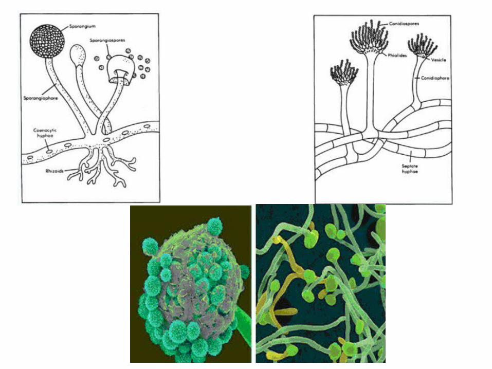
structures resulting from sexual fusion. The hyphae are usually non-septate and asexual spores (sporarigiospores) are formed inside a sac called sporangium. Examples are the genera Mucor and Rhizopus.
• 2. Ascomycetes: sexual fusion results in formation of a sac (ascus) containing either 4 or 8 ascospores. Most commonly asexual spores are borne externally on tips of hyphae and are called conidia. Examples are the genera Aspergillus (Figure 1.4) and Penicillium.
• 3. Basidiomycetes : sexual fusion results in formation of a club shaped structure called basidium with four spores borne externally. Asexual spores are most commonly conida.
• 4.Deuteromycetes (Fungi Imperfecti) are mostly similar to ascomycetes but a sexual stage is not demonstrated.
MORPHOLOGY • A. Yeasts are unicellular organisms that reproduce
by budding or fission. Each yeast cell usually contains a single nucleus.
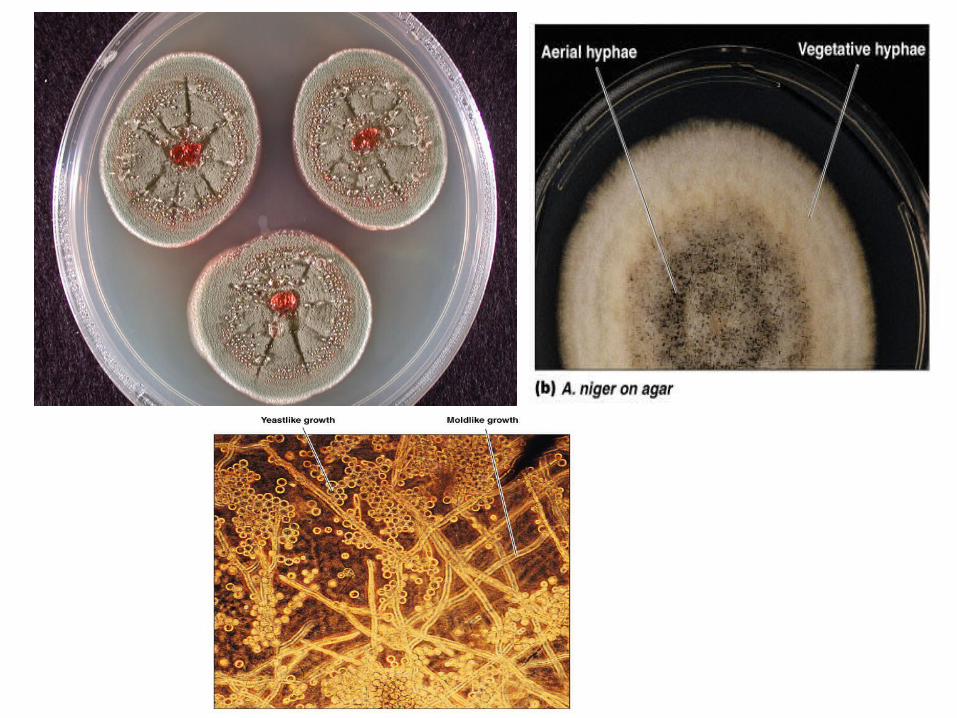
• B. Filamentous fungi (hyphae) are multicellular structures with branching, tubular cells.Hyphae without septa are coenocytic (nonseptate). Masses of hyphae are called mycelia; the terms "hyphae" and "mycelia" are commonly used interchangeably.
• C. Dimorphic fungi occur both as yeasts and as mycelia, depending on the environmental conditions:
• 1. In the parasitic or pathogenic form (seen in tissue, in exudates, or in cultures on en riched medium incubated at 37C), dimorphic fungi are yeasts.
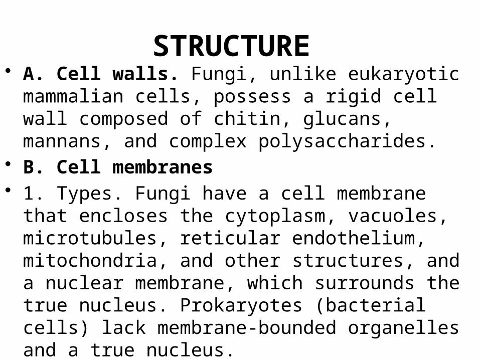
STRUCTURE • A. Cell walls. Fungi, unlike eukaryotic mammalian
cells, possess a rigid cell wall com posed of chitin, glucans, mannans, and complex polysaccharides.
• B. Cell membranes• 1. Types. Fungi have a cell membrane that encloses
the cytoplasm, vacuoles, microtubules, reticular endothelium, mitochondria, and other structures, and a nuclear membrane, which surrounds the true nucleus. Prokaryotes (bacterial cells) lack mem brane-bounded organelles and a true nucleus.
• Composition. Fungal membranes contain ergosterol
PATHOGENESlS
• The establishment of mycotic infection usually depends on the size of the inoculum and the resistance of the host. The severity of infection depends mostly on the host response to the organism
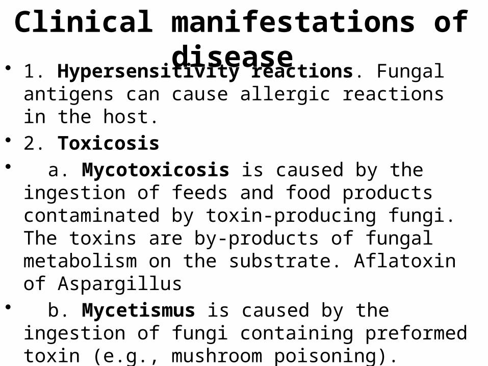
Clinical manifestations of disease • 1. Hypersensitivity reactions. Fungal antigens can
cause allergic reactions in the host.• 2. Toxicosis• a. Mycotoxicosis is caused by the ingestion of
feeds and food products contaminated by toxin-producing fungi. The toxins are by-products of fungal metabolism on the substrate. Aflatoxin of Aspargillus
• b. Mycetismus is caused by the ingestion of fungi containing preformed toxin (e.g., mushroom poisoning).
• 3. Infection
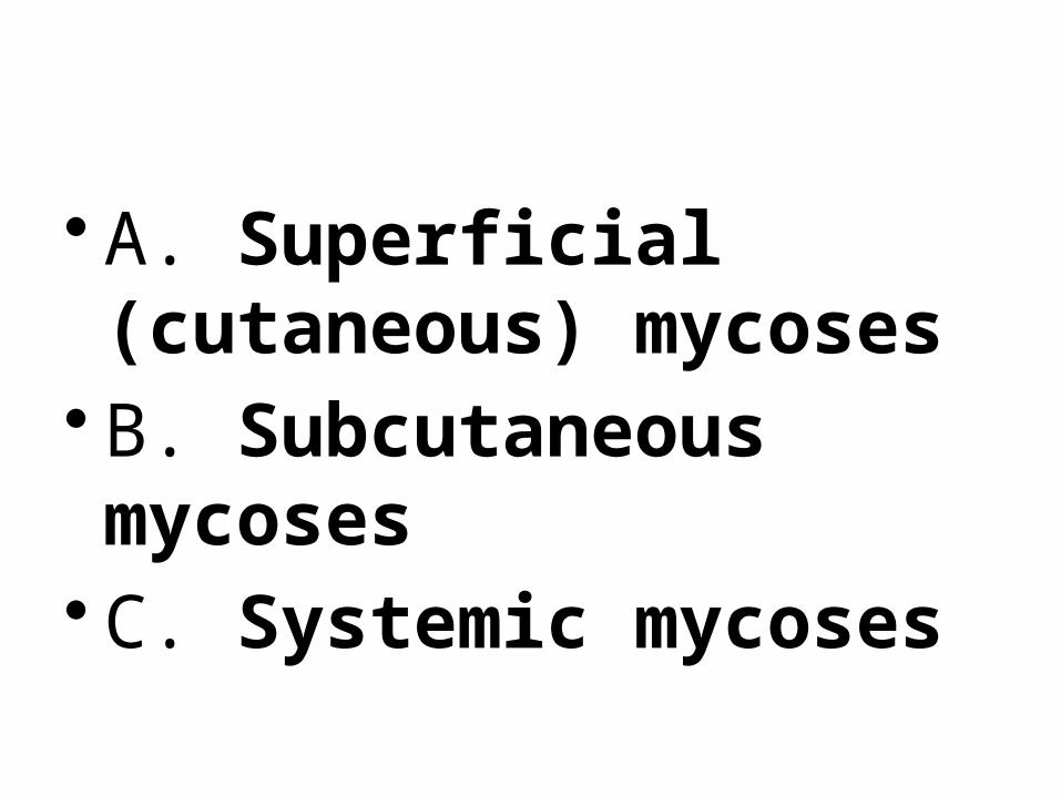
• A. Superficial (cutaneous) mycoses
• B. Subcutaneous mycoses
• C. Systemic mycoses
A. Superficial (cutaneous) mycoses
• Superficial (cutaneous) mycoses are confined to the outer layers of the skin, nail, or hair. The fungi involved are called dermatophytes.
• Tinea nigra, dark braun patches (Selnium Sulfide)
B. Subcutaneous mycoses
• Subcutaneous mycoses are confined to the subcutaneous tissue and only rarely spread systemically. They usually form deep, ulcerated skin lesions and have a protracted clinical course. The infection commonly involves the lower extremities because the causative organisms are soil saprophytes that are introduced through trauma to the feet or legs.
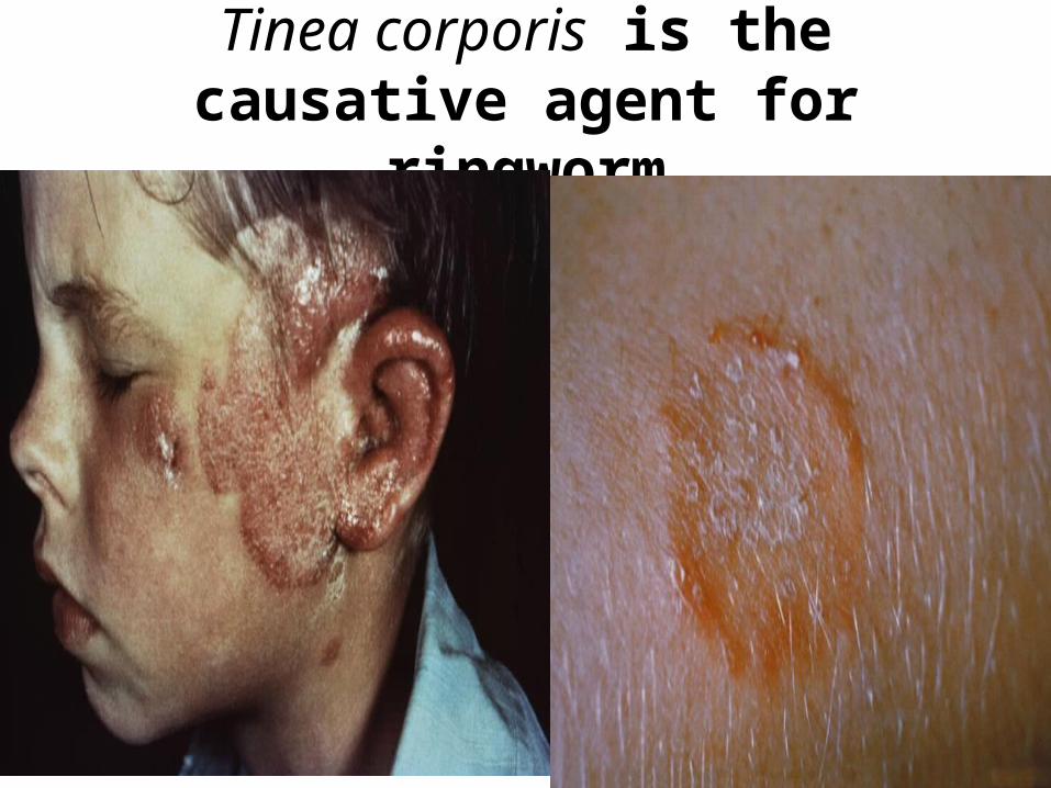
Tinea corporis is the causative agent for ringworm

Tinea crusis•is the causative agent for groin and scrotum

Tinea pedis is the causative agent for athlete's foot

Tinea capitis is the causative agent for scalp
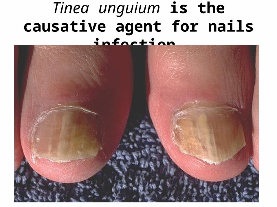
Tinea unguium is the causative agent for nails infection
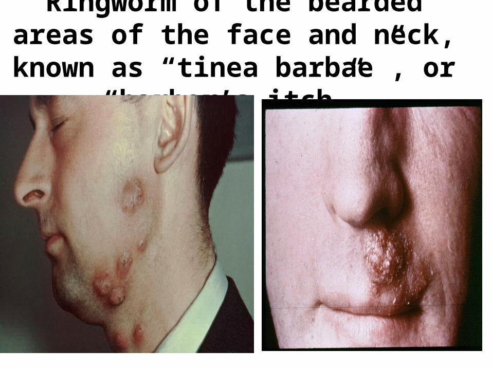
Ringworm of the bearded areas of the face and neck, known as “tinea
barbae”, or “barber’s itch”.
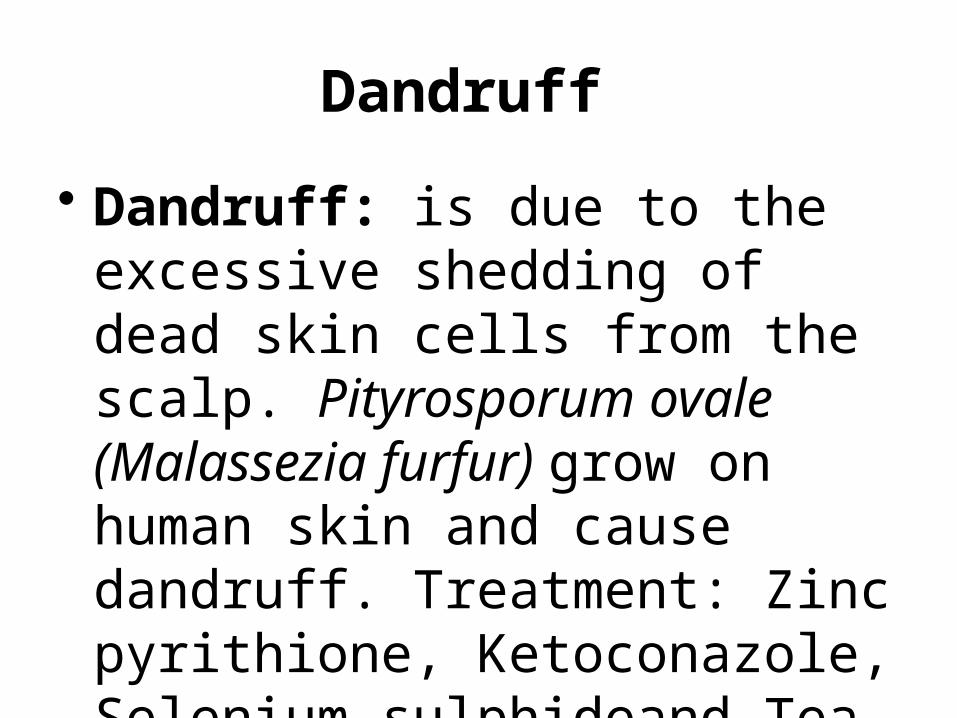
Dandruff
• Dandruff: is due to the excessive shedding of dead skin cells from the scalp. Pityrosporum ovale (Malassezia furfur) grow on human skin and cause dandruff. Treatment: Zinc pyrithione, Ketoconazole, Selenium sulphideand Tea Tree oil.
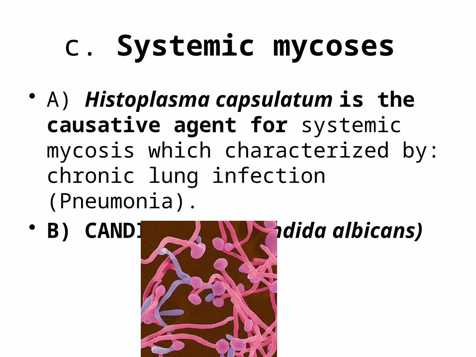
c. Systemic mycoses
• A) Histoplasma capsulatum is the causative agent for systemic mycosis which characterized by: chronic lung infection (Pneumonia).
• B) CANDIDIASIS (Candida albicans)
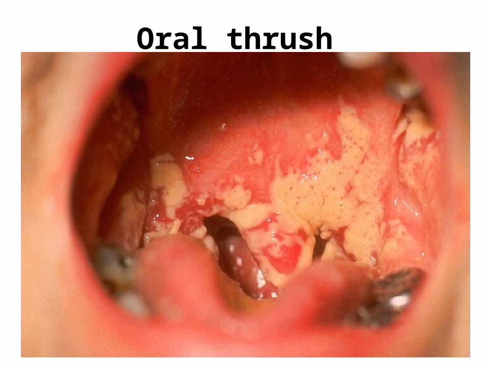
Oral thrush
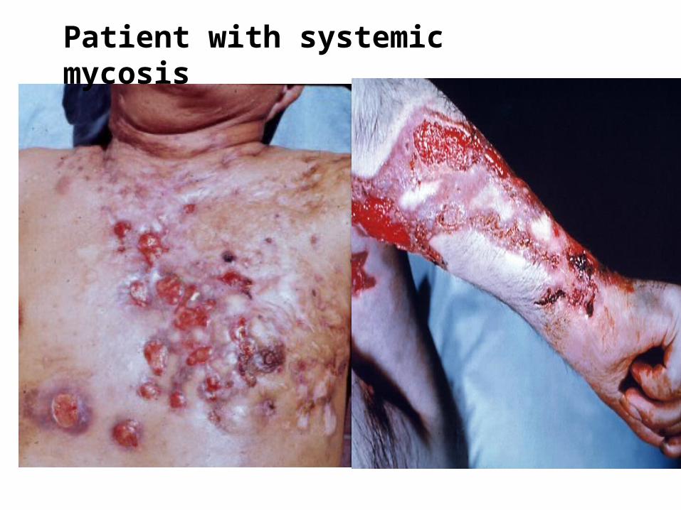
Patient with systemic mycosis