Embed Size (px)
Citation preview
R EV I EW AR T I C L E
Mycobacterium tuberculosis: success through dormancy
Martin Gengenbacher & Stefan H.E. Kaufmann
Department of Immunology, Max Planck Institute for Infection Biology, Berlin, Germany
Correspondence: Stefan H.E. Kaufmann,
Department of Immunology, Max Planck
Institute for Infection Biology, Chariteplatz 1,
10117 Berlin, Germany. Tel.: +49 30 28460
500; fax: +49 30 28460 501; e-mail:
Received 5 August 2011; revised 22
December 2011; accepted 31 January 2012.
Final version published online 8 March 2012.
DOI: 10.1111/j.1574-6976.2012.00331.x
Editor: Christoph Dehio
Keywords
granuloma; persistence; host immune
response; latency.
Abstract
Tuberculosis (TB) remains a major health threat, killing nearly 2 million
individuals around this globe, annually. The only vaccine, developed almost
a century ago, provides limited protection only during childhood. After dec-
ades without the introduction of new antibiotics, several candidates are cur-
rently undergoing clinical investigation. Curing TB requires prolonged
combination of chemotherapy with several drugs. Moreover, monitoring the
success of therapy is questionable owing to the lack of reliable biomarkers.
To substantially improve the situation, a detailed understanding of the cross-
talk between human host and the pathogen Mycobacterium tuberculosis (Mtb)
is vital. Principally, the enormous success of Mtb is based on three capaci-
ties: first, reprogramming of macrophages after primary infection/phago-
cytosis to prevent its own destruction; second, initiating the formation of
well-organized granulomas, comprising different immune cells to create a
confined environment for the host–pathogen standoff; third, the capability to
shut down its own central metabolism, terminate replication, and thereby
transit into a stage of dormancy rendering itself extremely resistant to host
defense and drug treatment. Here, we review the molecular mechanisms
underlying these processes, draw conclusions in a working model of myco-
bacterial dormancy, and highlight gaps in our understanding to be addressed
in future research.
Introduction
The etiology of tuberculosis (TB) – one of the most dev-
astating diseases of humankind – was first elucidated by
Robert Koch (1843–1910) in 1882 (Koch, 1882, 1932; Ka-
ufmann & Winau, 2005). Koch developed a specific stain-
ing method based on methylene blue combined with
brown counterstaining of host tissues with vesuvin for
the causative agent Mycobacterium tuberculosis (Mtb),
which allowed the visualization of bacteria not only in
cultures, but also in tissues (Box 1). Precisely, 130 years
down the road, his diagnostic method is still in use virtu-
ally unchanged. Potent antibiotics have been discovered,
and public health systems improved significantly in many
parts of the world since Koch’s times. Nevertheless, Mtb
remains as deadly as it was, claiming nearly 2 million
lives annually and exploiting an estimated 2 billion as res-
ervoir of latently Mtb-infected (LTBI) individuals (Dye
et al., 1999; Yew & Leung, 2008). These figures were cal-
culated when our globe hosted about 6 billion people. In
2011, when we reached the 7 billion mark, about 2.3 bil-
lion LTBI individuals appear more likely. Currently, up
to 9 million new cases of TB arise each year, more than
ever before (Dye & Williams, 2010). Most cases are not
because of new infections but through the reactivation of
dormant Mtb residing in LTBI hosts (Fig. 1).
ª 2012 Federation of European Microbiological Societies FEMS Microbiol Rev 36 (2012) 514–532Published by Blackwell Publishing Ltd. All rights reserved
MIC
ROBI
OLO
GY
REV
IEW
S
Box 1. Profile of Mtb
The bacillus causing TB in humans belongs to the
genus Mycobacterium that includes several other
obligate human pathogens, most importantly Myco-
bacterium leprae (leprosy), Mycobacterium africanum
(TB-like symptoms, lower pathogenicity), and Myco-
bacterium bovis (primarily TB in cattle). The family of
pathogenic mycobacteria arose from soil-dwelling
ancestors. They most likely became pathogens to
animal and human hosts during domestication of
animals about 10 000 years ago (Smith et al., 2009). At
37 °C and under optimal availability of oxygen and
nutrients, a single Mtb organism has a generation time
of 18–24 h and forms a white to light-yellow colony
on agar within 3–4 weeks. The aerobic-to-facultative
anaerobe, Gram-positive pathogen is surrounded by an
impermeable and thick cell wall/capsule that is made
of peptidoglycans, polysaccharides, unusual glycolipids,
and lipids mainly consisting of long-chain fatty acids,
such as mycolic acid. Unlike many other bacteria, Mtb
does not form spores but has the capacity to become
dormant – a nonreplicating state characterized by low
metabolic activity and phenotypic drug resistance. Note
that phenotypic drug resistance is related to a specific
physiologic state and independent from genetic muta-
tions. Mtb is typically visualized by Ziehl–Neelsen
(acid-fast) staining and appears as a rod-shaped red
bacillus. The GC-rich (65.6%) 4.4-Mbp genome of Mtb
is one of the biggest among the bacteria and encodes
about 4000 predicted proteins (http://genolist.pasteur.
fr/TubercuList/, Cole et al., 1998; integrated platform
for TB research: http://www.tbdb.org/, Reddy et al.,
2009). M. bovis Bacillus Calmette–Guerin (BCG) (atten-
uated form of M. bovis) and Mycobacterium smegmatis
are nonpathogenic and therefore common surrogates for
Mtb in research. The former is used as vaccine in children
with partial success. Mtb is typically diagnosed by
microscopy in the sputum of active TB patients. A
regime of several drugs is available to effectively cure the
disease by 6–9 months of combination therapy. Incom-
plete treatment or noncompliance of patients often leads
to drug-resistant Mtb, which is conferred by genetic
mutations.
Primary infection can: (1) progress toward active dis-
ease; (2) be contained as latent infection; (3) be eradi-
cated by the host’s immune system. Less than 10% of
infected individuals develop active TB during their life-
time. It is impossible to predict who will contain latent
infection throughout their lifetime and remain healthy,
and who will develop active TB at some point. How-
ever, the risk of active disease is increased in immuno-
compromising situations such as during antitumor
necrosis factor therapy of patients with chronic inflam-
matory diseases, by diabetes/obesity or by co-infection
with human immunodeficiency virus (HIV)/acquired
immunodeficiency syndrome (AIDS) (Barry et al.,
2009). Other risk factors include alcoholism and poor
nutrition. Multiple factors are likely involved in defin-
ing overall risk of TB, and the genetic makeup of both
host and pathogen plays a decisive role. Thus, biomar-
kers that would allow prognosis of TB reactivation in
healthy individuals with LTBI would be of tremendous
value.
Today, intervention measures for controlling TB are
available. We have at hand numerous drugs to cure the
disease, diagnostics to identify patients, and a vaccine to
prevent severe forms of childhood TB. Yet, these mea-
sures are insufficient for the following reasons:
(1) Diagnosis of TB in low-income countries is correct in
only an estimated half of all TB cases.
(2) Treatment for TB requires 3–4 drugs given for
6 months or longer. Frequently, compliance is poor and
premature termination of drug therapy often results in
emergence of resistant strains. Today, an estimated 50
million individuals harbor multidrug-resistant Mtb of
whom 500 000 fall ill annually. Even worse, strains of vir-
tually untreatable extensively drug-resistant (XDR) Mtb
are on the rise, and XDR-TB has already been notified in
58 countries. Even totally drug-resistant-TB has been
described (Velayati et al., 2009).
(3) The current vaccine, M. bovis BCG, protects against
severe forms of childhood TB, but fails to protect against
adult pulmonary TB, which has become the most preva-
lent form of the disease today. Hence, BCG does not
impact the transmission of Mtb.
Many definitions in infectious disease research are diffi-
cult to apply in TB. Needless to say, the etiologic agent
Mtb is a true pathogen and not an opportunistic micro-
organism, even though active disease only develops in the
minority of infections. Virulence describes the capability
of the pathogen to cause disease in quantitative terms.
High virulence, therefore, is often related to marked
severity of disease and vice versa. Yet, the decisive survival
factor of Mtb is its capacity to persist in the host for long
periods of time, both during noncontagious LTBI and
contagious active TB before it is spread. To further
complicate the situation, pathogenicity of TB is largely
influenced by the host immune response. Hence, a dis-
cussion of the disease without regard for the host would
remain incomplete. Although our review of the molecular
mechanisms of TB is oriented toward the pathogen’s
perspective, we will consider host influences where
appropriate.
FEMS Microbiol Rev 36 (2012) 514–532 ª 2012 Federation of European Microbiological SocietiesPublished by Blackwell Publishing Ltd. All rights reserved
Mycobacterium tuberculosis: success through dormancy 515
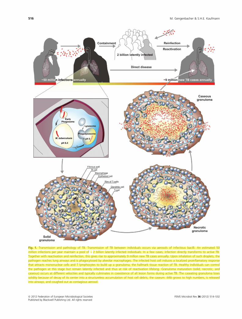
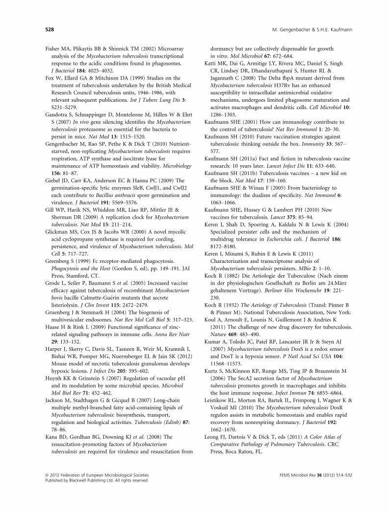
Fig. 1. Transmission and pathology of TB. Transmission of TB between individuals occurs via aerosols of infectious bacilli. An estimated 50
million infections per year maintain a pool of ~ 2 billion latently infected individuals. In a few cases, infection directly transforms to active TB.
Together with reactivation and reinfection, this gives rise to approximately 9 million new TB cases annually. Upon inhalation of such droplets, the
pathogen reaches lung airways and is phagocytosed by alveolar macrophages. The infected host cell induces a localized proinflamatory response
that attracts mononuclear cells and T lymphocytes to build up a granuloma, the hallmark tissue reaction of TB. Healthy individuals can control
the pathogen at this stage but remain latently infected and thus at risk of reactivation lifelong. Granuloma maturation (solid, necrotic, and
caseous) occurs at different velocities and typically culminates in coexistence of all lesion forms during active TB. The caseating granuloma loses
solidity because of decay of its center into a structureless accumulation of host cell debris, the caseum. Mtb grows to high numbers, is released
into airways, and coughed out as contagious aerosol.
ª 2012 Federation of European Microbiological Societies FEMS Microbiol Rev 36 (2012) 514–532Published by Blackwell Publishing Ltd. All rights reserved
516 M. Gengenbacher & S.H.E. Kaufmann
Infection of the alveolar macrophage
Macrophages operate as prime defense cells against
microbial intruders (Nathan & Shiloh, 2000; Liu & Mod-
lin, 2008; Deretic et al., 2009). These microorganisms are
ingested by phagocytosis, a process consisting of mem-
brane invaginations finally culminating in phagosome for-
mation (Aderem & Underhill, 1999). This organelle is a
part of the intracellular trafficking and transport system
and the site to which the entire arsenal of host defense is
targeted (Schekman, 1994; Rothman & Wieland, 1996;
Gruenberg & Stenmark, 2004). Microorganisms captured
in the phagosome experience increasing acidification,
reactive oxygen and nitrogen species (ROS and RNS),
hydrolytic enzymes, and cationic antimicrobial peptides
(CAMPs). Acidic pH inside the maturing phagosome
activates enzymes that degrade bacterial lipids and pro-
teins (Huynh & Grinstein, 2007). Simultaneously micro-
bial metabolism is suppressed by such conditions. ROS
and RNS generated by the phagosomal enzymes NADPH
phagocyte oxidase and inducible nitric oxide synthase
(iNOS) damage captured microorganisms by modification
of their DNA, lipids, thiols, tyrosine side chains, and
active centers of metal-dependent proteins (Fang, 2004).
Further damage of ingested pathogens is incurred by
CAMPs via permeabilization of their cell membrane (Pur-
dy & Russell, 2007). It must be kept in mind that the
responses in mice, one of the most common model
organisms, might be different from humans with respect
to iNOS, ROS, and RNS. The final steps of bacterial
destruction and clearance require phagolysosome fusion.
All of the described destruction pathways are influenced
by the host’s immune status. Macrophage activation
via cytokines, notably, interferon-gamma (IFN-c), for
instance, allows these host cells to control their intracellu-
lar predators (Cooper et al., 1993; North & Jung, 2004).
Phagosomal content can then be further processed
toward the antigen presentation pathway (Wolf & Ploegh,
1995; Pieters, 1997). Components of Mtb, notably
secreted proteins, are processed by macrophages and den-
dritic cells (DCs). Resulting peptides are loaded on the
gene products of the major histocompatibility complex,
and in this way, T cells are instructed to allow for an
appropriate adaptive immune response (Amigorena et al.,
1994; Tulp et al., 1994; West et al., 1994). Thus, T lym-
phocytes are critical for the control of Mtb during latent
infection. Failure of T cells to maintain protective immu-
nity promotes reactivation of TB.
Humans become infected with Mtb by inhaling minute
aerosol droplets carrying a small number of bacteria
(Kaufmann, 2001) (Fig. 1). At the site of infection, the
lung, Mtb bacilli are phagocytosed by alveolar macro-
phages. These cells are programmed to combat microbial
intruders and to ultimately destroy them. However, Mtb
manages to escape eradication by macrophages and sur-
vives within these cells (Armstrong & Hart, 1975; Kauf-
mann, 2001; Russell, 2001). The unique composition of
the mycobacterial cell wall and envelope likely enables the
tubercle bacillus to enter macrophages by employing mul-
tiple receptors such as Fc-, complement-, or mannose
receptors and the DC-specific intercellular adhesion mole-
cule-3-grabbing nonintegrin (DC-SIGN) (Brennan &
Nikaido, 1995; Ernst, 1998; Greenberg, 1999; Cambi
et al., 2005). Some receptors allow silent entry (CR), and
others induce defense mechanisms (FcR). Once inside the
resting macrophage, Mtb impairs phagosome maturation
(Vergne et al., 2003, 2005; Walburger et al., 2004; Robin-
son et al., 2007; Axelrod et al., 2008; Katti et al., 2008;
Russell et al., 2009). Principally, phagosome maturation is
a highly complex process in which the phagosome har-
boring a particle other than Mtb constantly interacts with
the recycling endosome, secretory organelles, multivesicu-
lar bodies, and the endoplasmic reticulum (Desjardins,
2009). Following the oxidative burst, the phagosome gets
acidified, a process lasting between 10 and 20 min. Acidi-
fication is mediated by proton pumps that reduce the
neutral pH to an acidic pH of ca. 5.0 (Yates et al., 2005).
Mtb arrests this process and therefore also downstream
events (Sturgill-Koszycki et al., 1994). In contrast to Mtb-
containing phagosomes, those harboring inert particles go
on to fuse with the lysosome within an hour to 1.5 h.
Acidification of the organelle is arrested by Mtb at pH
6.4, which is near the neutral pH and is significantly
higher than in the terminal phagolysosome at pH 4.5–5.0(Sturgill-Koszycki et al., 1996; MacMicking et al., 2003;
Yates et al., 2005). IFN-c activation of macrophages pro-
motes delivery of Mtb into the mature phagolysosome, as
shown by colocalization with green fluorescent bacilli,
and probably remains viable in this extremely hostile
environment (Via et al., 1998). Some Mtb mutants that
fail to prevent phagosome maturation do survive in the
resting macrophage, while others are attenuated (Pethe
et al., 2004; MacGurn & Cox, 2007). An alternative strat-
egy of Mtb has been established more recently: its egress
from the phagosome into the cytosol of macrophages
(van der Wel et al., 2007; Behar et al., 2010). Additional
host defense mechanisms include apoptosis and auto-
phagy (Behar et al., 2010; Deretic, 2010; Levine et al.,
2011). Apoptosis is a highly regulated process mediated
by host mechanisms which likely contributes to host pro-
tection. In contrast, necrosis is driven by exogenous
insults which might benefit the pathogen rather than the
host. Studies on experimental TB in mice have provided
evidence of such an association (Pan et al., 2005). Viru-
lent Mtb inhibit apoptosis by a number of antiapoptotic
genes, and more recent evidence suggests a role of prosta-
FEMS Microbiol Rev 36 (2012) 514–532 ª 2012 Federation of European Microbiological SocietiesPublished by Blackwell Publishing Ltd. All rights reserved
Mycobacterium tuberculosis: success through dormancy 517
glandin E2 in this mechanism (Behar et al., 2010). Auto-
phagy is an essential mechanism for host cell integrity
that can also serve as defense mechanism against bacterial
pathogens (Deretic, 2010; Levine et al., 2011). In TB, con-
tribution of autophagy to protection has been described
(Alonso et al., 2007). In sum, the intracellular survival
stratagem of tubercle bacilli comprises not only active
manipulation of host defense mechanisms to neutralize
and counteract a highly aggressive armamentarium of
activated macrophages but also robust resistance against
assault.
Adaptation of Mtb to the intracellularenvironment of macrophages
Mtb is shielded from the environment by a robust cell
wall (Box 1). Upon phagocytosis by host cells, Mtb expe-
riences drastic environmental changes and therefore has
to realign its metabolism to assure survival. Genome-wide
microarray techniques to study Mtb’s transcriptional
response to this transition have provided deeper insights
into the nature of the phagosomal environment, which
was suggested to be nitrosative, oxidative, low in oxygen
tension, and limited in nutrients (Schnappinger et al.,
2003). Additionally, the pathogen upregulated genes
involved in lipid metabolism confirming previous evi-
dence that lipids are critical for virulence of Mtb (McKin-
ney et al., 2000; Movahedzadeh et al., 2004; Brzostek
et al., 2007; Chang et al., 2009; Nesbitt et al., 2010). Iso-
citrate lyase (Icl) was identified as gate enzyme of the gly-
oxylate shunt, a short-cut of the tricarboxylic acid (TCA)
cycle, bypassing the steps of carbon loss by CO2 forma-
tion. The glyoxylate cycle is mobilized in Mtb growing on
fatty acids as exclusive carbon source and also during
chronic infection in mice, suggesting that lipids are acces-
sible as nutrients in vivo (McKinney et al., 2000). Meta-
bolic pathways that are relevant during infection are
shown in Fig. 2.
Although the exact nature of carbon sources utilized
during infection remains elusive, Mtb has been shown to
metabolize host-derived cholesterol (Pandey & Sassetti,
2008). Disruption of mce4 encoding a cholesterol trans-
porter results in the failure of Mtb to maintain chronic
infection in mice while retaining full virulence during the
acute phase comparable to observations made for an Icl-
deficient mutant (McKinney et al., 2000; Mohn et al.,
2008; Pandey & Sassetti, 2008). Recent data demonstrate
that Mtb residing in phagosomes of macrophages utilizes
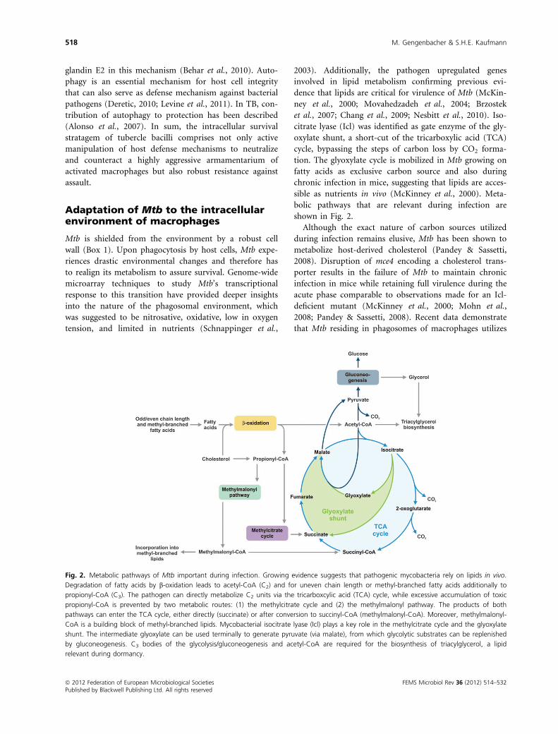
Fig. 2. Metabolic pathways of Mtb important during infection. Growing evidence suggests that pathogenic mycobacteria rely on lipids in vivo.
Degradation of fatty acids by b-oxidation leads to acetyl-CoA (C2) and for uneven chain length or methyl-branched fatty acids additionally to
propionyl-CoA (C3). The pathogen can directly metabolize C2 units via the tricarboxcylic acid (TCA) cycle, while excessive accumulation of toxic
propionyl-CoA is prevented by two metabolic routes: (1) the methylcitrate cycle and (2) the methylmalonyl pathway. The products of both
pathways can enter the TCA cycle, either directly (succinate) or after conversion to succinyl-CoA (methylmalonyl-CoA). Moreover, methylmalonyl-
CoA is a building block of methyl-branched lipids. Mycobacterial isocitrate lyase (Icl) plays a key role in the methylcitrate cycle and the glyoxylate
shunt. The intermediate glyoxylate can be used terminally to generate pyruvate (via malate), from which glycolytic substrates can be replenished
by gluconeogenesis. C3 bodies of the glycolysis/gluconeogenesis and acetyl-CoA are required for the biosynthesis of triacylglycerol, a lipid
relevant during dormancy.
ª 2012 Federation of European Microbiological Societies FEMS Microbiol Rev 36 (2012) 514–532Published by Blackwell Publishing Ltd. All rights reserved
518 M. Gengenbacher & S.H.E. Kaufmann
triacylglycerol from the host cell to be stored in the form
of intracellular lipid droplets (Daniel et al., 2011).
Catabolism of cholesterol, odd-chain fatty acids, methyl-
branched fatty acids, and amino acids funnels into propi-
onyl-CoA, a C3 intermediate, which is toxic in excess
(Savvi et al., 2008; Yang et al., 2009). However, propio-
nyl-CoA toxicity can be avoided by condensing the C3
body with oxaloacetate to form succinate and pyruvate by
the 2-methylcitrate cycle (Fig. 2). Intriguingly, both
mycobacterial enzymes Icl-1 and Icl-2 can act as 2-meth-
ylcitrate lyase (Munoz-Elias et al., 2006). Thus, mycobac-
terial Icl plays a critical dual role during infection (1) to
bypass carbon loss using the glyoxylate shunt under lim-
ited nutrient availability and (2) to prevent the excessive
accumulation of toxic propionyl-CoA. Recent studies
carried out with steady-state chemostat cultures have
demonstrated the importance of Icl in slow-growing Mtb
metabolizing glycerol as main carbon source. Under such
conditions, anapleuric reactions prevailed while the
pathogen was also capable of carbon dioxide fixation as
demonstrated by isotope flux (Beste et al., 2011). Propio-
nyl-CoA metabolization can also be performed by the
methylmalonyl-CoA pathway ending up in methylmalo-
nyl-CoA, which can either be converted into succinyl-
CoA by a vitamin B12-dependent mutase or directly
incorporated into methyl-branched fatty acids (Savvi
et al., 2008) (Fig. 2). These fatty acids are found among
the large family of unique mycobacterial lipids which
build up the pathogen’s cell envelope (Brennan &
Nikaido, 1995; Jackson et al., 2007). Typically, the loss
of cell wall components leads to decreased virulence
(Glickman et al., 2000; Makinoshima & Glickman, 2005).
In conclusion, propionyl-CoA detoxification is extremely
critical for Mtb in vivo. The asymmetric cleavage of isoci-
trate by Icl produces glyoxylate, which is converted into
malate and succinate. The TCA cycle intermediate malate
can be used to generate pyruvate and further to replenish
the pool of glycolytic intermediates by gluconeogenesis
(Fig. 2). Such intermediates are required to produce the
essential building blocks of proteins, DNA, and the cell
wall. Gluconeogenesis is critical throughout TB infec-
tion in mice and, thus, might be relevant in dormancy
(Marrero et al., 2010).
Mtb tolerates low pH (i.e. 4.5–5.4) inside the phago-
lysosome of INF-c-activated macrophages (Schaible et al.,
1998; Via et al., 1998). The lack of complete acidification
in Mtb-infected resting macrophages is likely – at least in
part – caused by the exclusion of the phagosomal proton
ATPase and by the secretion of mycobacterial urease, an
enzyme producing neutralizing ammonia from urea (Rey-
rat et al., 1995; Sturgill-Koszycki et al., 1996; Grode et al.,
2005). Yet, Mtb is exposed to acidic conditions in vivo as
evidenced by the fact that the first-line TB drug pyrazina-
mide kills the pathogen only at low pH in vitro (Zhang &
Mitchison, 2003). In further support of this notion, acid-
sensitive Mtb mutants are attenuated in mice, and tran-
scriptional analysis of the bacillus residing inside activated
macrophages revealed upregulation of pH-responsive
genes (Buchmeier et al., 2000; Raynaud et al., 2002;
Rohde et al., 2007; Vandal et al., 2008). Interestingly,
most acid-sensitive Mtb mutants show defects in genes
associated with cell wall biogenesis (Vandal et al., 2009).
This unique lipid-rich permeability barrier has been sug-
gested to provide effective protection to protons more
than 100 years ago (Metchnikoff, 1905). It is therefore
not surprising that acid-sensitive mutants also confer
hypersensitivity to detergents or lipophilic antibiotics
owing to increased cell wall permeability (Vandal et al.,
2008). Although a pH sensing adenylate cyclase (Rv1264)
has been described in Mtb, it turned out to be nonessen-
tial in vivo (Tews et al., 2005; Dittrich et al., 2006). More
recently, the membrane-associated serine protease
Rv3671c has been characterized. Even though the precise
functions of the protein remain elusive, the authors ele-
gantly showed that an Rv3671c mutant failed to maintain
a neutral intrabacterial pH in acidic culture medium as
well as in the late (acidic) phagolysosome of activated
macrophages (Vandal et al., 2008). Moreover, the mutant
was attenuated in mice suggesting that acid resistance is
critical for the virulence of the tubercle bacillus. A recent
study demonstrated that aprABC, a locus unique to path-
ogenic mycobacteria, is involved in adaptation of Mtb to
the phagosomal low pH environment. Disruption of
aprABC conferred a defect in intracellular growth of the
pathogen and influenced lipid abundance of intracellular
stores and cell wall (Abramovitch et al., 2011). In sum,
Mtb is resistant to elevated acidic stress in the late phago-
lysosome compartment of macrophages at conditions that
are lethal for many other microbial pathogens (Huynh &
Grinstein, 2007).
The defense repertoire that any bacterial intruder expe-
riences in the phagosome of an activated macrophage is
not limited to low pH but also includes ROS and RNS.
The enzyme phagocyte oxidase (NOX2) transfers elec-
trons from cytosolic NADPH to phagosomal oxygen to
form superoxide anions. These highly reactive anions dis-
mutate into hydrogen peroxide and finally produce toxic
hydroxyl radicals, which are members of the ROS family
(Bedard & Krause, 2007). Accordingly, this reaction is
often referred to as ‘oxidative burst’. In monocytes and
neutrophils, chlorination further adds to the toxicity of
ROS (Bedard & Krause, 2007). RNS are produced by
iNOS, an enzyme that generates nitrate and nitrite. The
latter intermediate reacts at low pH to nitrous acid
that forms nitric oxide and nitrogen dioxide, two highly
reactive radicals (Nathan & Shiloh, 2000). Finally, the two
FEMS Microbiol Rev 36 (2012) 514–532 ª 2012 Federation of European Microbiological SocietiesPublished by Blackwell Publishing Ltd. All rights reserved
Mycobacterium tuberculosis: success through dormancy 519
molecules, nitric oxide and superoxide, form the toxic
peroxynitrite (Beckman et al., 1990; Bogdan, 2001;
Nathan & Ehrt, 2004). To avert toxicity caused by ROS
and NOS, Mtb follows a dual strategy of detoxification
and damage repair.
The mycobacterial enzyme catalase peroxidase encoded
by katG converts hydrogen peroxide into water and oxy-
gen. Accordingly, a katG loss-of-function Mtb mutant
showed hypersensitivity to hydrogen peroxide in vitro
(Ng et al., 2004). In NOX2-deficient mice, the mutant
was fully virulent while a unique phenotype of transient
attenuation was reported in iNOS�/� mice: Initial growth
was followed by rapid decline and a lag phase of about 6
–8 weeks characterized by stable bacterial burden in
lungs, after which replicative activity was resumed. How-
ever, the molecular mechanisms of the observed phenom-
ena remain unclear. Several other mycobacterial genes
have been implicated in ROS and RNS detoxification (in-
depth reviewed by Ehrt & Schnappinger, 2009):
• sodA and sodC, both encoding a superoxide dismutase
(Dussurget et al., 2001; Edwards et al., 2001; Pidding-
ton et al., 2001; Sassetti & Rubin, 2003).
• secA2, part of an accessory export system (Braunstein
et al., 2003; Kurtz et al., 2006; Rigel & Braunstein,
2008).
• mshA, required for mycothiol biosynthesis (Vilcheze
et al., 2008).
• cysH, involved in sulfur assimilation (Senaratne et al.,
2006).
• ahpC, ahpD, dalT, lpd, encoding the multifunctional
NADH-dependent peroxidase and peroxynitrite reduc-
tase (Shi & Ehrt, 2006; Bryk et al., 2008).
Although the underlying molecular mechanisms require
further elucidation, it is clear that Mtb employs a large
variety of counterstrategies to detoxify ROS and RNS.
The second principal strategy to oppose the effects of
highly reactive intermediates includes repair of the dam-
age caused or degradation of affected biomolecules and
their rapid replacement via de novo synthesis. Protea-
somes are multiprotein complexes that degrade peptides
by multiple proteolyic activities (Etlinger & Goldberg,
1977; Lowe et al., 1995). In Mtb, two putative accessory
factors PafA and Mpa are involved in coupling the pro-
karyotic ubiquitin-like protein Pup to peptides that are
destined for turnover and recognition of Pup-tagged pro-
teins, respectively (Pearce et al., 2008; Striebel et al.,
2009; Sutter et al., 2009). Mpa is an ATPase likely
involved in unfolding and delivery of proteins into the
proteolytic core of proteasomes (Wang et al., 2009).
Mutations in the respective genes mpa and pafA increase
the sensitivity of the tubercle bacillus to RNS (Darwin,
2009). Disruption of the mpa gene reduced virulence of
Mtb in wild-type mice, which was less profound in
iNOS�/� mice. Conditional silencing of the essential
genes prcB and prcA, which encode proteolytic core pro-
teins of the proteasome, not only conferred sensitivity to
RNS but also impaired the survival of Mtb during chronic
infection in mice (Gandotra et al., 2007). In sum, protea-
some-mediated protein turnover becomes critical during
RNS-related stress. Two different but not mutually exclu-
sive scenarios or combinations could explain these results:
(1) degradation of a transcriptional repressor that con-
trols expression of proteins required for the synthesis of
antioxidants; (2) removal of irreversibly damaged proteins
with toxic potential (Darwin, 2009).
More recently, transcriptional profiling of Mtb and
human macrophages during infection provided evidence
for heavy metal poisoning (Tailleux et al., 2008; Botella
et al., 2011). In particular, ctpC encoding a putative zinc
efflux pump in mycobacteria was strongly induced upon
infection, suggesting exposure of the bacillus to zinc ions
inside the phagosomal compartment. Indeed, disruption
of the ctpC gene rendered Mtb hypersensitive to zinc.
While infection progresses, zinc ions are quickly released
from stores inside host cells and translocated to the
phagosome. Thus, zinc poisoning is likely exploited by
macrophages to destroy Mtb. Indeed, zinc is known to be
of particular importance for the immune system playing
multiple roles, for instance, in defense and signaling
(Rink & Gabriel, 2000; Haase & Rink, 2009). Although
not yet directly shown, accumulating evidence suggests
that Mtb counteracts the stressor zinc by expression of an
appropriate efflux pump (Botella et al., 2011).
Inside the mature phagolysosome, Mtb experiences the
permeabilizing properties of CAMPs (Purdy & Russell,
2007). The positively charged CAMPs – cathelicidin, hep-
cidin, and ubiquitin-related peptides – gain their bacteri-
cidal activity by disrupting the negatively charged
bacterial cell wall (Alonso et al., 2007; Liu et al., 2007;
Sow et al., 2007). Microorganisms lower their affinity to
CAMPs by reduction of their negative surface charge (Pe-
schel & Sahl, 2006). The Mtb plasma membrane contains
a positively charged lipid consisting of phosphatidyl glyc-
erol linked to lysine moieties. Its generation requires the
lysine transferase LysX (Maloney et al., 2009). Sensitivity
to positively charged antibiotics and a specific CAMP of
neutrophils was increased in an Mtb lysX loss-of-function
mutant. Virulence of this mutant was lowered in mice
and guinea pigs, demonstrating a critical role of LysX in
vivo (Maloney et al., 2009). The unique impermeable cell
envelope of Mtb represents a physical barrier for CAMPs.
Expression of the major porin of M. smegmatis, MspA, in
Mtb, which does not encode an ortholog, increases
membrane permeability (Mailaender et al., 2004). Con-
comitantly such strains become more sensitive to ubiqu-
ª 2012 Federation of European Microbiological Societies FEMS Microbiol Rev 36 (2012) 514–532Published by Blackwell Publishing Ltd. All rights reserved
520 M. Gengenbacher & S.H.E. Kaufmann
itin-derived CAMPs (Purdy et al., 2009). Probably, the
most obvious strategy of pathogenic mycobacteria to pro-
tect themselves from phagosomal assault is to translocate
from the hostile organelle to the cytosol of host cells.
Such behavior has been suggested for Mtb and M. leprae
(van der Wel et al., 2007). If true, this could have deep
implications, not only for the fate of bacilli in host cells,
but also for granuloma formation. Although the host
macrophage has developed a remarkable arsenal of bacte-
ricidal mechanisms over millennia of coevolution, Mtb
has learned to survive in this hostile intracellular environ-
ment. As a result, the coevolutionary tie shifted to the tis-
sue level, namely to the formation of a remarkable self-
organizing capsular structure for bacterial containment,
the granuloma (Reece & Kaufmann, 2011).
Existence of Mtb inside granulomas
Infected macrophages migrate and thereby transport Mtb
from the airways into pulmonary tissue sites. There, an
inflammatory focus is formed comprised of infected mac-
rophages and freshly immigrant monocytes (Fig. 1). The
primary lesion matures into a granuloma, the hallmark of
TB, while some infected cells disseminate to seed secondary
lesions in the lung. In the majority of individuals, the path-
ogen is controlled at this stage by the immune system and
does not spread further: LTBI is established. The solid
granuloma is not only the site of Mtb containment during
latency but also the source of tissue damage at the early
stage of disease (Reece & Kaufmann, 2011). In other words,
it is the histological correlate of both protection and
pathology. In humans, the granuloma shows high plasticity
and three major types can be distinguished. These are (1)
solid granulomas which contain Mtb; (2) necrotic granulo-
mas typical for early stages of active TB; (3) caseous granu-
lomas during end-stage or severe TB. These different stages
are not distinct entities but form a continuum.
Solid granulomas prevail during LTBI. These highly
structured tissue reactions are comprised of mononuclear
phagocytes of different developmental stages, DCs, as well
as T and B lymphocytes. Although T lymphocytes are the
critical mediators of protection in TB, B lymphocytes are
also abundant. Often, lymphocytes form an outer ring
whereas in the central parts, mononuclear phagocytes,
fibroblasts, and DCs predominate. The solid granuloma is
typically encircled by a fibrotic wall that separates it from
surrounding tissue. The burden of Mtb inside solid gran-
ulomas is low. Most likely these bacilli are in a stage of
dormancy, characterized by low metabolic activity with
non- to low-replicating persistence. Often, such bacteria
are difficult to grow under normal culture conditions and
therefore have been termed viable but not culturable
(VBNC).
The necrotic granuloma remains well structured, but
the center becomes increasingly necrotic, that is, com-
posed of solid cell detritus which is often hypoxic. Later,
Mtb organisms can be resuscitated: they start replicating
and become metabolically active.
In caseous granlomas, the center becomes liquefied
leading to cavity formation. The structure of these granu-
lomas wanes. Evidence has been presented for a harmful
role of polymorphic neutrophilic granuloyctes (Lowe
et al., 2011). In cavitary lesions, high oxygen content is
reestablished. Moreover, the caseous material provides a
fertile source of nutrient-promoting growth of the patho-
gen up to some trillion organisms. Finally, Mtb finds
access to blood capillaries and the alveolar space paving
the way not only for dissemination to other organs but
also for transmission to other individuals.
During active TB, different stages of granulomas coex-
ist and provide a multitude of diverse microenvironments
to which the pathogen has to adapt. Hence, the bacillus is
found in different areas of the granulomas, intracellularly
within the rim of host mononuclear phagocytes, some
DCs, and perhaps fibroblasts as well as extracellularly in
the caseous center consisting of host cell debris. It has
been proposed that unfavorable conditions inside the
granuloma, such as nutrient limitation and low oxygen
tension, trigger the metabolic downshift of subpopula-
tions of Mtb to dormancy. As most TB drugs target func-
tions essential for growth, they fail to eradicate
nonreplicating bacilli. This could explain the prolonged
treatment time required to cure disease. In vitro models
of dormancy have been developed to study nonreplicating
persistence of Mtb. As early as 1933, Loebel et al.
observed that carbon starvation terminates the growth of
the tubercle bacillus and causes a drastic drop in respira-
tion, indicating a low metabolic rate. More recent work
confirmed that nutrient-starved nonreplicating bacilli
undergo a global downregulation of metabolic genes,
including those involved in respiration (Betts et al.,
2002). Such bacilli are extremely tolerant to TB drugs
(Xie et al., 2005). However, nutrient-starved Mtb remains
sensitive to inhibition of NADH dehydrogenase 2, a sin-
gle protein enzyme that can serve as alternate entry point
of electrons into the electron transport chain but lacks
proton translocation capacity (Xie et al., 2005; Yano
et al., 2006; Teh et al., 2007; Gengenbacher et al., 2010).
Other characteristics of the pathogen analyzed in this
in vitro dormancy model include significantly reduced
intracellular ATP levels and very low but continuous
respiration. The glyoxylate shunt enzyme Icl is essential
for the survival of nutrient-starved nonreplicating Mtb
(Gengenbacher et al., 2010). Promotion of the glyoxylate
cycle under limited nutrient access is in the best interest
of the bacillus, as no carbon is lost by CO2-formation in
FEMS Microbiol Rev 36 (2012) 514–532 ª 2012 Federation of European Microbiological SocietiesPublished by Blackwell Publishing Ltd. All rights reserved
Mycobacterium tuberculosis: success through dormancy 521
contrast to the citrate cycle. Icl is required for the mainte-
nance of a chronic infection in the mouse model of TB,
suggesting a relevant role of nutrient limitation in vivo
(McKinney et al., 2000).
The influence of oxygen shortage on Mtb has been
extensively studied. In 1996, Wayne and coworkers intro-
duced an Mtb-in vitro dormancy model based on gradual
oxygen depletion (Wayne & Hayes, 1996). The pathogen
passes through two phases of declining metabolic activity
to dormancy and phenotypic drug resistance. More
detailed physiological characterization revealed reduction
of intracellular ATP in hypoxic nonreplicating bacilli and
sensitivity of the pathogen to further depletion, observa-
tions which later on were also made for nutrient-starved
nonreplicating Mtb (Rao et al., 2008; Gengenbacher et al.,
2010). Even though both models generate quiescent
organisms by contrary conditions – carbon depletion in
an oxygen-rich environment vs. oxygen starvation in a
nutrient-rich medium – physiological overlaps identified
could qualify as core features of dormancy. In line with
the general upregulation of lipid metabolism genes during
oxygen starvation of mycobacteria, hypoxic nonreplicating
BCG accumulates neutral lipid triacylglycerol to form vis-
ible intracellular droplets. Importantly, such lipid stores
are required for regrowth of the hypoxic nonreplicating
organism in nutrient-rich medium (Low et al., 2009).
Accumulation of triacylglycerol droplets might therefore
be important during dormancy and could be useful for
the identification of dormant mycobacteria.
On the genetic level, the adaption to changes in oxygen
availability is mediated by the DosS/DosT-DosR regula-
tory complex that controls roughly 50 genes (Boon &
Dick, 2002; Park et al., 2003). In other words, the DosR
regulon governs metabolic shift of Mtb from aerobic to
anaerobic functioning, ensures survival of the bacillus
during hypoxia-induced in vitro dormancy, and controls
reversal to replication upon reexposure to oxygen (Rustad
et al., 2009; Leistikow et al., 2010). Furthermore, dosR
responds to nitric oxide and carbon monoxide (Kumar
et al., 2007). Yet, disruption of the dosR gene only slightly
affects the survival of the pathogen in different animal
models, such as mouse, guinea pig, or rabbit; the molecu-
lar basis of this finding remains unclear (Converse et al.,
2009). The role of hypoxia in vivo was impressively
analyzed by Via et al. (2008), who introduced the
hypoxia-activated compound pimonidazole to different
experimental animal species and directly measured oxygen
tension in granulomas of guinea pigs, rabbits, and non-
human primates. This study revealed that low oxygen
pressure could restrict the growth of aerobic to micro-
aerophilic Mtb in the hypoxic core of necrotic and solid
granulomas. Note that TB histopathology of non-human
primates most closely resembles active TB in humans
(Leong et al., 2011). The characteristic continuum of
granulomatous lesions in human TB is rarely reflected in
small animal models. Guinea pigs show lesions of differ-
ent types. As they are extraordinarily susceptible to Mtb,
caseous lesions predominate. Mice, the most widely used
experimental animals for research on immunology and
infection, develop nonhypoxic ill-structured lesions,
and distinct stages are not observed. To capitalize on the
wealth of information available from the mouse model,
mice that develop human-like TB pathology would be of
great value. Recently, a mouse model that mimics the dif-
ferent stages of granulomas similar to human TB has
been introduced. The iNOS-deficient mouse mutant
infected with Mtb developed well-structured solid granu-
lomas, which controlled the pathogen at low to inter-
mediate load. Neutralization of IFN-c led to granuloma
necrosis with hypoxic areas, followed by massive case-
ation. In this model, cathepsin G (CatG) was identified as
critical effector molecule of both protection and pathol-
ogy. CatG activity, in turn, was controlled by serpin b3
and fine-tuning of this control mechanism seems to
decide whether pathology or protection prevails (Reece
et al., 2010). Production of RNS by the iNOS system rep-
resents a vital antimicrobial defense mechanism, but
because of its strict oxygen dependence, it is likely insuffi-
ciently active in hypoxic areas of granulomas. In another
murine model of TB, the sst1 locus of mice has been
demonstrated to prevent formation of necrotic lesions.
The intracellular pathogen resistance 1-protein encoded
within this locus possesses the ability to direct infected
macrophages to undergo apoptosis rather than necrosis
(Pan et al., 2005). Such necrotic lesions have recently
been shown to be hypoxic (Harper et al., 2012). Whether
‘human-like’ mouse models have potential for broad
application in TB research has yet to be determined. The
widespread assumption that chronic TB infection is
caused by a rather static equilibrium of slow or nonrepli-
cating bacilli has recently been questioned. Authors engi-
neered Mtb to harbor an instable plasmid that was lost
during cell division. This replication clock used to study
TB infection in mice has provided evidence for active
replication of Mtb, not only during the acute stage, but
also throughout the chronic phase of infection (Gill et al.,
2009).
In vitro models aim at reproducing impacts of the host
environment on Mtb. Thus, the pathogen has been
debarred from iron or phosphate and exposed to low
concentrations of nitric oxide to mimic either the phago-
some of host macrophages or the necrotic center of gran-
ulomas (Fisher et al., 2002; Ohno et al., 2003; Rifat et al.,
2009). Other studies in M. bovis BCG have combined dif-
ferent stressors in one model system to better represent
the microenvironment of mycobacteria in vivo (Bryk
ª 2012 Federation of European Microbiological Societies FEMS Microbiol Rev 36 (2012) 514–532Published by Blackwell Publishing Ltd. All rights reserved
522 M. Gengenbacher & S.H.E. Kaufmann
et al., 2008). Most recently, drug-tolerant persister bacilli
have been isolated from in vitro cultures of Mtb by D-
cycloserine treatment. A few persisters were found during
lag and early exponential phases, while they made up to
1% in late exponential and stationary phases. The global
transcriptome of such persisters was then profiled and
compared to the transcriptomes obtained from various
in vitro dormancy models. Authors identified a set of five
genes upregulated in all models that probably represents
a core dormancy response (Keren et al., 2011). These
were: acr2, encoding a heat shock protein; the transcrip-
tional regulator gene Rv1152; pdhA, the gene of a putative
pyruvate dehydrogenase subunit; a hypothetical protein
encoded by Rv2517c; and lat, encoding an L-lysine-epsi-
lon aminotransferase. To date, an unusually high number
of 65 toxin–antitoxin (TA) loci were identified in the
genome of Mtb. A TA module produces a toxin that is
detoxified by its respective antitoxin. In E. coli, a number
of TA modules are relevant in dormancy (Keren et al.,
2004; Vazquez-Laslop et al., 2006). Interestingly, in the
recent transcriptome study of Keren et al., 10 TA loci
were overexpressed in Mtb persisters, suggesting their
importance in survival without replication. Altogether,
in vitro dormancy models of Mtb suggest that stress-
related genes and alternative pathways are upregulated,
while the genes of central metabolic routes, including
glycolysis, TCA cycle, energy production and respiration,
are downregulated. The specific roles of distinct genes
such as TA modules remain to be elucidated.
Reactivation and resuscitation
In the human host, Mtb persisting in a dormant stage
causes LTBI without clinical disease. While Mtb is well
equipped for persistence in the host, the term ‘persister’ is
used for those Mtb organisms that are phenotypically resis-
tant to drugs although they are in fact genetically suscepti-
ble to these antibiotics. The reason for this is probably
transformation of bacteria into a nonreplicating stage with
low-to-absent metabolic activity – the precise conditions of
dormancy. Principally, this feature underlies the so-called
Cornell model of Mtb persistence in mice. In this model,
animals infected with Mtb are treated with the drugs pyraz-
inamide and isoniazid to reduce bacterial load to a level,
which is nondetectable by culture. At first sight, drug treat-
ment achieves sterile eradication of the pathogen, but after
termination of drug treatment, some bacilli recover and
grow to high abundance causing reactivation of TB
(McCune et al., 1966). Prolonged culture of stationary-
phase Mtb can generate bacteria that fail to grow to visible
colonies on agar. Regrowth of these VBNC organisms was
only supported in spent media taken from exponentially
growing Mtb (Shleeva et al., 2002). VBNC bacilli in spu-
tum of patients with TB could lead to false-negative results
of diagnostics, as those organisms are ‘invisible’ in standard
cultures (Mukamolova et al., 2010). A very recent report
showed that M. smegmatis (Box 1) generates cell-to-cell
heterogeneity by asymmetric growth in combination with
time-controlled cell division. Most importantly, distinct
subpopulations showed different susceptibility to antibiot-
ics (Aldridge et al., 2011).
Accumulating evidence suggests that regrowth of dor-
mant Mtb is initiated by resuscitation, which is the rees-
tablishment of metabolic and replicative activity.
Resuscitation has been studied at the molecular level in
Micrococcus luteus where a so-called resuscitation-promot-
ing factor (Rpf) has been shown to induce resuscitation.
Rpf orthologs of Mtb possess a conserved domain with
putative lysozyme activity and therefore might cleave the
peptidoglycan network that makes up the cell wall (Co-
hen-Gonsaud et al., 2005). Similarly, germination of
spores of Bacillus anthracis begins with hydrolysis of the
cell wall (Giebel et al., 2009). Recent studies in Bacillus
subtilis have revealed a signaling cascade that is initiated
by peptidoglycan degradation products (Shah et al.,
2008). Altogether, regrowth could be initiated through a
cell wall hydrolysis step, but further steps involved are
thus far unknown. The genome of Mtb comprises five rpf
genes, and it has been claimed that such genes can facili-
tate recovery of Mtb in sputum of patients with active TB
or in freeze-dried M. bovis BCG (Wu et al., 2008; Muka-
molova et al., 2010). As Mtb possesses several rpf genes,
redundancy likely exists. As a result, only multideletion
mutants, not single knockouts of rpf genes, show
impaired resuscitation in vitro and attenuation in mice
(Kana et al., 2008). In the mouse, chronic progression of
disease characterized by high bacterial burden is observed,
while reactivation in humans develops from minute
bacterial load of LTBI. Hence, mice are probably not a
suitable model to study reactivation of TB.
Evidence for coexistence of different Mtb stages in
infected individuals is increasing. Thus, probably only a
few dormant bacilli coexist in face of a large number of
metabolically active replicating organisms during active
TB. The reciprocal is also true: during latent infection, in
addition to nonreplicating metabolically inactive (i.e. dor-
mant) Mtb, some actively replicating Mtb are present. In
other words, the equilibrium of dormant/replicating Mtb
represents a distinguishing factor between LTBI and
active TB.
Isoniazid only targets replicating Mtb and yet has been
used widely and successfully for the chemoprophylaxis of
LTBI where there is considered to be elevated risk,
suggesting that during several months treatment time,
bacteria transform into an isoniazid-susceptible stage
(Fox et al., 1999). A more recent hypothesis describing
FEMS Microbiol Rev 36 (2012) 514–532 ª 2012 Federation of European Microbiological SocietiesPublished by Blackwell Publishing Ltd. All rights reserved
Mycobacterium tuberculosis: success through dormancy 523
latent infection as a dynamic process of constant rein-
fection could explain the efficacy of isoniazid therapy
during latency (Cardona, 2009). Although formal proof is
lacking, the clinical observation is taken as evidence for
sporadic emergence of some replicating Mtb during LTBI.
Reciprocally, addition of Rpf significantly increases recov-
ery of Mtb in sputum from active patients with TB
providing circumstantial evidence that these bacteria are
resuscitated from dormancy (Mukamolova et al., 2010).
It has been argued that the long treatment time of six or
more months to cure TB depends, at least in part, on the
coexistence of both replicating and nonreplicating Mtb as
current drugs preferentially, if not exclusively, target met-
abolically active replicating Mtb. On the one hand, repli-
cating bacilli are the main culprits causing active disease;
on the other hand, they are the target of current chemo-
therapy. In contrast, the dormant pathogen is likely a
‘bystander’ in disease but mainly contributes to pheno-
typic drug resistance. Hence, the dormant pathogen serves
as a reservoir of renascent active Mtb to sustain pathology
and disease. It is therefore assumed that treatment with
available drugs has to eradicate replicating tubercle bacilli
in several waves as they are resuscitated from dormancy.
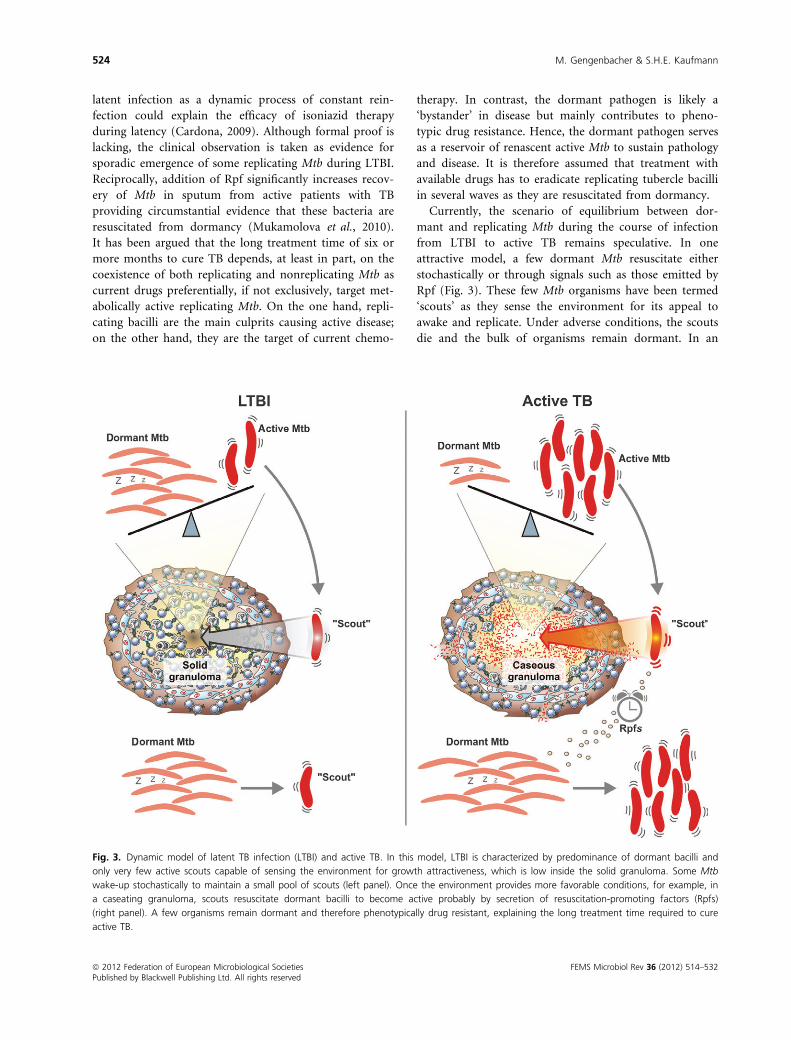
Currently, the scenario of equilibrium between dor-
mant and replicating Mtb during the course of infection
from LTBI to active TB remains speculative. In one
attractive model, a few dormant Mtb resuscitate either
stochastically or through signals such as those emitted by
Rpf (Fig. 3). These few Mtb organisms have been termed
‘scouts’ as they sense the environment for its appeal to
awake and replicate. Under adverse conditions, the scouts
die and the bulk of organisms remain dormant. In an
Fig. 3. Dynamic model of latent TB infection (LTBI) and active TB. In this model, LTBI is characterized by predominance of dormant bacilli and
only very few active scouts capable of sensing the environment for growth attractiveness, which is low inside the solid granuloma. Some Mtb
wake-up stochastically to maintain a small pool of scouts (left panel). Once the environment provides more favorable conditions, for example, in
a caseating granuloma, scouts resuscitate dormant bacilli to become active probably by secretion of resuscitation-promoting factors (Rpfs)
(right panel). A few organisms remain dormant and therefore phenotypically drug resistant, explaining the long treatment time required to cure
active TB.
ª 2012 Federation of European Microbiological Societies FEMS Microbiol Rev 36 (2012) 514–532Published by Blackwell Publishing Ltd. All rights reserved
524 M. Gengenbacher & S.H.E. Kaufmann
appealing environment, scouts send activation signals to
the dormant majority of bacteria which then resuscitate
(Epstein, 2009; Chao & Rubin, 2010). The biochemical
nature of these signals could involve Rpfs but has to be
further investigated. In an environment attractive for
growth, the bacterial population starts replicating, causes
pathology, and leads to reactivation of TB. A small popu-
lation will stay in or fall into dormancy thus showing tol-
erance to drug treatment (Lewis, 2010). Active disease
caused by reinfection could involve similar factors as Mtb
newly entering the host is actively growing and hence can
provide wake-up signals to dormant bacilli in an LTBI
host.
Deletion of Rv2623, a gene of unknown function under
the control of the dosR regulon, for instance, confers
hypervirulence to the tubercle bacillus in mice and guinea
pigs, while its overexpression leads to growth delay in vitro
(Drumm et al., 2009). Thus, Rv2623 could be involved in
restricting replication. Reciprocally, regrowth of dormant
bacilli could be promoted by the downregulation of
Rv2623. The dosR regulon provides an instructive example
of how environmental changes can trigger a complex adap-
tation program in Mtb. It is well understood how DosR
responds to decreasing oxygen tension (Park et al., 2003).
But, how a change in oxygen availability is sensed on a
molecular level and ultimately translated into an impulse
for the dos regulatory system is not yet understood. Simply
turning off the ‘dos program’ will most likely not be suffi-
cient to allow for resuscitation. Although the molecular
mechanisms of resuscitation are largely unknown, evidence
for several aspects is unfolding.
Concluding remarks
The last decade has witnessed a remarkable increase in
our knowledge about Mtb, not only on the bacillus itself
but also on the communication with its host. We have
learned that Mtb has invented complex mechanisms to
survive in the intracellular environment, that it counter-
acts or evades the numerous defense mechanisms of mac-
rophages, and that the cross-talk between pathogen and
host immune system is focused on granulomas, which
serve as both, habitat and containment for Mtb. However,
many interesting observations remain fragmented in our
current view of Mtb. Remarkable efforts are ongoing to
integrate the relatively new science of systems biology
into mycobacterial research (http://www.systemtb.org/
and http://www.broadinstitute.org/annotation/tbsysbio/
home.html). Ideally, this will tie up loose ends and
improve our understanding, allowing for a broad net-
work-oriented view of Mtb and its host.
After decades of dormancy, TB drug discovery has
reawakened. Although the boost that was expected from
whole genome sequencing of Mtb and application of
high-throughput screening to target-based approaches
has not paid off so far, nine TB drug candidates are
currently undergoing clinical investigation – more than
ever before (Cole et al., 1998; Payne et al., 2007; Ma
et al., 2010). Most of these compounds belong to exist-
ing classes of antibiotics and thus are derivates of the
respective parental molecule: oxazolidinones target pro-
tein synthesis; fluoroquinolones are well-known DNA
gyrase inhibitors; 1,2-ethylene diamines interfere with
cell wall biosynthesis pathways; nitroimidazoles cause
multiple damage to Mtb by RNS. The target of sudoterb
has yet to be identified. The ATP synthase inhibitor
TMC207 is a diarylquinoline and represents a relatively
new antimycobacterial class (current TB drug discovery
reviewed by Koul et al., 2011). Which new entity will
complete the stony road to drug application is yet to be
seen. Applying our increasing knowledge of host–patho-gen interactions to TB drug discovery will most likely
result in more new candidate compounds (Nathan et al.,
2008).
Most impressive are the dynamics and the plasticity of
the infection process, which unfolds as a continuum in
which different populations of Mtb, as well as different
pathologic forms of granulomas, coexist. As a corollary,
different stages of infection are characterized by different
ratios in the abundance of pathogen populations rather
than by distinct periods of life cycle. Accordingly, analysis
of single cells ‘frozen’ in a distinct stage, rather than of
populations will be required in the future.
Development of next-generation vaccines will benefit
from our deeper insights into the adaption of Mtb to dif-
ferent host environments, as well. Currently, over a dozen
vaccine candidates are at different stages of clinical trial
development (Kaufmann et al., 2010; Kaufmann, 2011a).
All these candidates are preexposure vaccines, which do
not prevent or eradicate infection with Mtb but rather
aim at precluding emergence of active TB. Accordingly,
these vaccine candidates stimulate an immune response
that targets the pathogen promptly after infection, pre-
sumably by means of antigens expressed by the metaboli-
cally active, replicating pathogen. However, during LTBI,
Mtb changes its genetic program and antigens expressed
by the dormant pathogen prevail. To sustain efficacious
control of dormant Mtb, the vaccine-induced immune
response needs to target antigens expressed by dormant
bacilli, the so-called latency antigens (Kaufmann, 2010).
This stratagem becomes even more valid for postexposure
vaccines that are administered during LTBI – more than
2 billion individuals with elevated risk of developing TB.
A first example of a postexposure vaccine comprising an
antigen selectively expressed during nutrient starvation in
addition to canonical preexposure vaccine antigens has
FEMS Microbiol Rev 36 (2012) 514–532 ª 2012 Federation of European Microbiological SocietiesPublished by Blackwell Publishing Ltd. All rights reserved
Mycobacterium tuberculosis: success through dormancy 525
been described recently (Aagaard et al., 2011; Kaufmann,
2011b). This vaccine was highly successful in containing
Mtb in the mouse model. Hence, better understanding of
the tubercle bacillus’ life cycle will facilitate rational
design of next-generation vaccine candidates.
Definition of terms
Active TB: characterized by the presence of clinical symp-
toms caused by a high bacterial burden in the lung and
sometimes in other organs; patients spread the disease.
Latent TB infection (LTBI): asymptomatic infection with
a low number of Mtb in the absence of clinical signs;
infected individuals do not spread the disease.
Reactivation of TB: transition from latency to full-blown
active disease.
Relapse of TB: redevelopment of active disease after
incomplete or false treatment with antibiotics; single- or
multiple-drug resistance is often observed.
Active Mtb: metabolically active replicating bacilli; suscep-
tible to drug-inhibiting processes essential for growth (i.e.
DNA replication, RNA synthesis, cell wall biogenesis).
Dormant Mtb: nonreplicating bacilli maintaining full via-
bility at a very low metabolic rate; organisms show minor
susceptibility or phenotypic drug resistance to antibiotics
targeting functions required for growth.
Resuscitation of Mtb: transition of the pathogen from
dormancy to growth.
Drug resistance: inheritable resistance to a drug conferred
by genetic mutation.
Phenotypic drug resistance: noninheritable resistance to a
drug conferred by a specific metabolic state (usually dor-
mancy).
Viable but not culturable (VBNC): refers to a state of via-
ble Mtb that is not capable of colonizing on nutrient-rich
solid media without being resuscitated.
Acknowledgements
We would like to thank Olivier Neyrolles and colleagues
for providing most recent experimental data prior to
publication. We are grateful for outstanding editorial sup-
port of M.L. Grossman and excellent graphic design of
D. Schad. This work received financial support from
the European 7th Framework Program SYSTEMTB
(HEALTH-2009-2.1.2-1-241587) and the National Insti-
tutes of Health (SysBio, 745090 HHSN272200800059C).
References
Aagaard C, Hoang T, Dietrich J, Cardona PJ, Izzo A, Dolganov
G, Schoolnik GK, Cassidy JP, Billeskov R & Andersen P
(2011) A multistage tuberculosis vaccine that confers
efficient protection pre- and post-exposure. Nat Med 17:
189–194.Abramovitch RB, Rohde KH, Hsu FF & Russell DG (2011)
aprABC: a Mycobacterium tuberculosis complex-specific locus
that modulates pH-driven adaptation to the macrophage
phagosome. Mol Microbiol 80: 678–694.Aderem A & Underhill DM (1999) Mechanisms of
phagocytosis in macrophages. Annu Rev Immunol 17: 593–623.
Aldridge BB, Fernandez-Suarez M, Heller D, Ambravaneswaran
V, Irimia D, Toner M & Fortune SM (2011) Asymmetry
and aging of mycobacterial cells lead to variable growth and
antibiotic susceptibility. Science 335: 100–104.Alonso S, Pethe K, Russell DG & Purdy GE (2007) Lysosomal
killing of Mycobacterium mediated by ubiquitin-derived
peptides is enhanced by autophagy. P Natl Acad Sci USA
104: 6031–6036.Amigorena S, Drake JR, Webster P & Mellman I (1994)
Transient accumulation of new class II MHC molecules in a
novel endocytic compartment in B lymphocytes. Nature
369: 113–120.Armstrong JA & Hart PD (1975) Phagosome-lysosome
interactions in cultured macrophages infected with virulent
tubercle bacilli. Reversal of the usual nonfusion pattern and
observations on bacterial survival. J Exp Med 142: 1–16.Axelrod S, Oschkinat H, Enders J, Schlegel B, Brinkmann V,
Kaufmann SH, Haas A & Schaible UE (2008) Delay of
phagosome maturation by a mycobacterial lipid is reversed
by nitric oxide. Cell Microbiol 10: 1530–1545.Barry CE III, Boshoff HI, Dartois V, Dick T, Ehrt S, Flynn J,
Schnappinger D, Wilkinson RJ & Young D (2009) The
spectrum of latent tuberculosis: rethinking the biology and
intervention strategies. Nat Rev Microbiol 7: 845–855.Beckman JS, Beckman TW, Chen J, Marshall PA & Freeman
BA (1990) Apparent hydroxyl radical production by
peroxynitrite: implications for endothelial injury from
nitric oxide and superoxide. P Natl Acad Sci USA 87: 1620–1624.
Bedard K & Krause KH (2007) The NOX family of ROS-
generating NADPH oxidases: physiology and
pathophysiology. Physiol Rev 87: 245–313.Behar SM, Divangahi M & Remold HG (2010) Evasion of
innate immunity by Mycobacterium tuberculosis: is death an
exit strategy? Nat Rev Microbiol 8: 668–674.Beste DJ, Bonde B, Hawkins N, Ward JL, Beale MH, Noack S,
Noh K, Kruger NJ, Ratcliffe RG & McFadden J (2011) (1)
(3)C metabolic flux analysis identifies an unusual route for
pyruvate dissimilation in mycobacteria which requires
isocitrate lyase and carbon dioxide fixation. PLoS Pathog 7:
e1002091.
ª 2012 Federation of European Microbiological Societies FEMS Microbiol Rev 36 (2012) 514–532Published by Blackwell Publishing Ltd. All rights reserved
526 M. Gengenbacher & S.H.E. Kaufmann
Betts JC, Lukey PT, Robb LC, McAdam RA & Duncan K
(2002) Evaluation of a nutrient starvation model of
Mycobacterium tuberculosis persistence by gene and protein
expression profiling. Mol Microbiol 43: 717–731.Bogdan C (2001) Nitric oxide and the immune response. Nat
Immunol 2: 907–916.Boon C & Dick T (2002) Mycobacterium bovis BCG response
regulator essential for hypoxic dormancy. J Bacteriol 184:
6760–6767.Botella H, Peyron P, Levillain F et al. (2011) Mycobacterial p
(1)-type ATPases mediate resistance to zinc poisoning in
human macrophages. Cell Host Microbe 10: 248–259.Braunstein M, Espinosa BJ, Chan J, Belisle JT & Jacobs WR Jr
(2003) SecA2 functions in the secretion of superoxide
dismutase A and in the virulence of Mycobacterium
tuberculosis. Mol Microbiol 48: 453–464.Brennan PJ & Nikaido H (1995) The envelope of
mycobacteria. Annu Rev Biochem 64: 29–63.Bryk R, Gold B, Venugopal A et al. (2008) Selective killing
of nonreplicating mycobacteria. Cell Host Microbe 3: 137–145.
Brzostek A, Dziadek B, Rumijowska-Galewicz A, Pawelczyk J &
Dziadek J (2007) Cholesterol oxidase is required for
virulence of Mycobacterium tuberculosis. FEMS Microbiol Lett
275: 106–112.Buchmeier N, Blanc-Potard A, Ehrt S, Piddington D, Riley L
& Groisman EA (2000) A parallel intraphagosomal survival
strategy shared by Mycobacterium tuberculosis and
Salmonella enterica. Mol Microbiol 35: 1375–1382.Cambi A, Koopman M & Figdor CG (2005) How C-type
lectins detect pathogens. Cell Microbiol 7: 481–488.Cardona PJ (2009) A dynamic reinfection hypothesis of latent
tuberculosis infection. Infection 37: 80–86.Chang JC, Miner MD, Pandey AK, Gill WP, Harik NS, Sassetti
CM & Sherman DR (2009) igr Genes and Mycobacterium
tuberculosis cholesterol metabolism. J Bacteriol 191: 5232–5239.
Chao MC & Rubin EJ (2010) Letting sleeping dos lie: does
dormancy play a role in tuberculosis? Annu Rev Microbiol
64: 293–311.Cohen-Gonsaud M, Barthe P, Bagneris C, Henderson B, Ward
J, Roumestand C & Keep NH (2005) The structure of a
resuscitation-promoting factor domain from Mycobacterium
tuberculosis shows homology to lysozymes. Nat Struct Mol
Biol 12: 270–273.Cole ST, Brosch R, Parkhill J et al. (1998) Deciphering the
biology of Mycobacterium tuberculosis from the complete
genome sequence (vol 393, pg 537, 1998). Nature 396: 190–198.
Converse PJ, Karakousis PC, Klinkenberg LG et al. (2009) Role
of the dosR-dosS two-component regulatory system in
Mycobacterium tuberculosis virulence in three animal models.
Infect Immun 77: 1230–1237.Cooper AM, Dalton DK, Stewart TA, Griffin JP, Russell DG &
Orme IM (1993) Disseminated tuberculosis in interferon
gamma gene-disrupted mice. J Exp Med 178: 2243–2247.
Daniel J, Maamar H, Deb C, Sirakova TD & Kolattukudy PE
(2011) Mycobacterium tuberculosis uses host triacylglycerol
to accumulate lipid droplets and acquires a dormancy-like
phenotype in lipid-loaded macrophages. PLoS Pathog 7:
e1002093.
Darwin KH (2009) Prokaryotic ubiquitin-like protein (Pup),
proteasomes and pathogenesis. Nat Rev Microbiol 7: 485–491.
Deretic V (2010) Autophagy in infection. Curr Opin Cell Biol
22: 252–262.Deretic V, Delgado M, Vergne I, Master S, De HS, Ponpuak M
& Singh S (2009) Autophagy in immunity against
Mycobacterium tuberculosis: a model system to dissect
immunological roles of autophagy. Curr Top Microbiol
Immunol 335: 169–188.Desjardins M (2009) The good fat: a link between lipid
bodies and antigen cross-presentation. Immunity 31: 176–178.
Dittrich D, Keller C, Ehlers S, Schultz JE & Sander P (2006)
Characterization of a Mycobacterium tuberculosis mutant
deficient in pH-sensing adenylate cyclase Rv1264. Int J Med
Microbiol 296: 563–566.Drumm JE, Mi K, Bilder P et al. (2009) Mycobacterium
tuberculosis universal stress protein Rv2623 regulates
bacillary growth by ATP-Binding: requirement for
establishing chronic persistent infection. PLoS Pathog 5:
e1000460.
Dussurget O, Stewart G, Neyrolles O, Pescher P, Young D &
Marchal G (2001) Role of Mycobacterium tuberculosis
copper-zinc superoxide dismutase. Infect Immun 69: 529–533.
Dye C & Williams BG (2010) The population dynamics and
control of tuberculosis. Science 328: 856–861.Dye C, Scheele S, Dolin P, Pathania V & Raviglione MC
(1999) Consensus statement. Global burden of tuberculosis:
estimated incidence, prevalence, and mortality by country.
WHO Global Surveillance and Monitoring Project. JAMA
282: 677–686.Edwards KM, Cynamon MH, Voladri RK, Hager CC,
DeStefano MS, Tham KT, Lakey DL, Bochan MR &
Kernodle DS (2001) Iron-cofactored superoxide dismutase
inhibits host responses to Mycobacterium tuberculosis. Am J
Respir Crit Care Med 164: 2213–2219.Ehrt S & Schnappinger D (2009) Mycobacterial survival
strategies in the phagosome: defence against host stresses.
Cell Microbiol 11: 1170–1178.Epstein SS (2009) Microbial awakenings. Nature 457: 1083.
Ernst JD (1998) Macrophage receptors for Mycobacterium
tuberculosis. Infect Immun 66: 1277–1281.Etlinger JD & Goldberg AL (1977) A soluble ATP-dependent
proteolytic system responsible for the degradation of
abnormal proteins in reticulocytes. P Natl Acad Sci USA 74:
54–58.Fang FC (2004) Antimicrobial reactive oxygen and nitrogen
species: concepts and controversies. Nat Rev Microbiol 2:
820–832.
FEMS Microbiol Rev 36 (2012) 514–532 ª 2012 Federation of European Microbiological SocietiesPublished by Blackwell Publishing Ltd. All rights reserved
Mycobacterium tuberculosis: success through dormancy 527
Fisher MA, Plikaytis BB & Shinnick TM (2002) Microarray
analysis of the Mycobacterium tuberculosis transcriptional
response to the acidic conditions found in phagosomes.
J Bacteriol 184: 4025–4032.Fox W, Ellard GA & Mitchison DA (1999) Studies on the
treatment of tuberculosis undertaken by the British Medical
Research Council tuberculosis units, 1946–1986, withrelevant subsequent publications. Int J Tuberc Lung Dis 3:
S231–S279.Gandotra S, Schnappinger D, Monteleone M, Hillen W & Ehrt
S (2007) In vivo gene silencing identifies the Mycobacterium
tuberculosis proteasome as essential for the bacteria to
persist in mice. Nat Med 13: 1515–1520.Gengenbacher M, Rao SP, Pethe K & Dick T (2010) Nutrient-
starved, non-replicating Mycobacterium tuberculosis requires
respiration, ATP synthase and isocitrate lyase for
maintenance of ATP homeostasis and viability. Microbiology
156: 81–87.Giebel JD, Carr KA, Anderson EC & Hanna PC (2009) The
germination-specific lytic enzymes SleB, CwlJ1, and CwlJ2
each contribute to Bacillus anthracis spore germination and
virulence. J Bacteriol 191: 5569–5576.Gill WP, Harik NS, Whiddon MR, Liao RP, Mittler JE &
Sherman DR (2009) A replication clock for Mycobacterium
tuberculosis. Nat Med 15: 211–214.Glickman MS, Cox JS & Jacobs WR (2000) A novel mycolic
acid cyclopropane synthetase is required for cording,
persistence, and virulence of Mycobacterium tuberculosis. Mol
Cell 5: 717–727.Greenberg S (1999) Fc receptor-mediated phagocytosis.
Phagocytosis and the Host (Gordon S, ed), pp. 149–191. JAIPress, Stamford, CT.
Grode L, Seiler P, Baumann S et al. (2005) Increased vaccine
efficacy against tuberculosis of recombinant Mycobacterium
bovis bacille Calmette-Guerin mutants that secrete
listeriolysin. J Clin Invest 115: 2472–2479.Gruenberg J & Stenmark H (2004) The biogenesis of
multivesicular endosomes. Nat Rev Mol Cell Biol 5: 317–323.Haase H & Rink L (2009) Functional significance of zinc-
related signaling pathways in immune cells. Annu Rev Nutr
29: 133–152.Harper J, Skerry C, Davis SL, Tasneen R, Weir M, Kramnik I,
Bishai WR, Pomper MG, Nuermberger EL & Jain SK (2012)
Mouse model of necrotic tuberculosis granulomas develops
hypoxic lesions. J Infect Dis 205: 595–602.Huynh KK & Grinstein S (2007) Regulation of vacuolar pH
and its modulation by some microbial species. Microbiol
Mol Biol Rev 71: 452–462.Jackson M, Stadthagen G & Gicquel B (2007) Long-chain
multiple methyl-branched fatty acid-containing lipids of
Mycobacterium tuberculosis: biosynthesis, transport,
regulation and biological activities. Tuberculosis (Edinb) 87:
78–86.Kana BD, Gordhan BG, Downing KJ et al. (2008) The
resuscitation-promoting factors of Mycobacterium
tuberculosis are required for virulence and resuscitation from
dormancy but are collectively dispensable for growth
in vitro. Mol Microbiol 67: 672–684.Katti MK, Dai G, Armitige LY, Rivera MC, Daniel S, Singh
CR, Lindsey DR, Dhandayuthapani S, Hunter RL &
Jagannath C (2008) The Delta fbpA mutant derived from
Mycobacterium tuberculosis H37Rv has an enhanced
susceptibility to intracellular antimicrobial oxidative
mechanisms, undergoes limited phagosome maturation and
activates macrophages and dendritic cells. Cell Microbiol 10:
1286–1303.Kaufmann SHE (2001) How can immunology contribute to
the control of tuberculosis? Nat Rev Immunol 1: 20–30.Kaufmann SH (2010) Future vaccination strategies against
tuberculosis: thinking outside the box. Immunity 33: 567–577.
Kaufmann SH (2011a) Fact and fiction in tuberculosis vaccine
research: 10 years later. Lancet Infect Dis 11: 633–640.Kaufmann SH (2011b) Tuberculosis vaccines – a new kid on
the block. Nat Med 17: 159–160.Kaufmann SHE & Winau F (2005) From bacteriology to
immunology: the dualism of specificity. Nat Immunol 6:
1063–1066.Kaufmann SHE, Hussey G & Lambert PH (2010) New
vaccines for tuberculosis. Lancet 375: 85–94.Keren I, Shah D, Spoering A, Kaldalu N & Lewis K (2004)
Specialized persister cells and the mechanism of
multidrug tolerance in Escherichia coli. J Bacteriol 186:
8172–8180.Keren I, Minami S, Rubin E & Lewis K (2011)
Characterization and transcriptome analysis of
Mycobacterium tuberculosis persisters. MBio 2: 1–10.Koch R (1882) Die Aetiologie der Tuberculose (Nach einem
in der physiologischen Gesellschaft zu Berlin am 24.Marz
gehaltenem Vortrage). Berliner klin Wochenschr 19: 221–230.
Koch R (1932) The Aetiology of Tuberculosis (Transl: Pinner B
& Pinner M). National Tuberculosis Association, New York.
Koul A, Arnoult E, Lounis N, Guillemont J & Andries K
(2011) The challenge of new drug discovery for tuberculosis.
Nature 469: 483–490.Kumar A, Toledo JC, Patel RP, Lancaster JR Jr & Steyn AJ
(2007) Mycobacterium tuberculosis DosS is a redox sensor
and DosT is a hypoxia sensor. P Natl Acad Sci USA 104:
11568–11573.Kurtz S, McKinnon KP, Runge MS, Ting JP & Braunstein M
(2006) The SecA2 secretion factor of Mycobacterium
tuberculosis promotes growth in macrophages and inhibits
the host immune response. Infect Immun 74: 6855–6864.Leistikow RL, Morton RA, Bartek IL, Frimpong I, Wagner K &
Voskuil MI (2010) The Mycobacterium tuberculosis DosR
regulon assists in metabolic homeostasis and enables rapid
recovery from nonrespiring dormancy. J Bacteriol 192:
1662–1670.Leong FJ, Dartois V & Dick T, eds (2011) A Color Atlas of
Comparative Pathology of Pulmonary Tuberculosis. CRC
Press, Boca Raton, FL.
ª 2012 Federation of European Microbiological Societies FEMS Microbiol Rev 36 (2012) 514–532Published by Blackwell Publishing Ltd. All rights reserved
528 M. Gengenbacher & S.H.E. Kaufmann
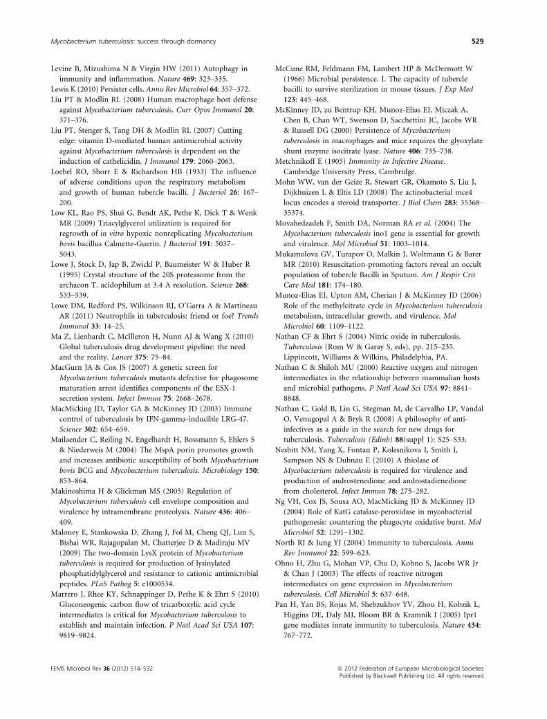
Levine B, Mizushima N & Virgin HW (2011) Autophagy in
immunity and inflammation. Nature 469: 323–335.Lewis K (2010) Persister cells. Annu RevMicrobiol 64: 357–372.Liu PT & Modlin RL (2008) Human macrophage host defense
against Mycobacterium tuberculosis. Curr Opin Immunol 20:
371–376.Liu PT, Stenger S, Tang DH & Modlin RL (2007) Cutting
edge: vitamin D-mediated human antimicrobial activity
against Mycobacterium tuberculosis is dependent on the
induction of cathelicidin. J Immunol 179: 2060–2063.Loebel RO, Shorr E & Richardson HB (1933) The influence
of adverse conditions upon the respiratory metabolism
and growth of human tubercle bacilli. J Bacteriol 26: 167–200.
Low KL, Rao PS, Shui G, Bendt AK, Pethe K, Dick T & Wenk
MR (2009) Triacylglycerol utilization is required for
regrowth of in vitro hypoxic nonreplicating Mycobacterium
bovis bacillus Calmette-Guerin. J Bacteriol 191: 5037–5043.
Lowe J, Stock D, Jap B, Zwickl P, Baumeister W & Huber R
(1995) Crystal structure of the 20S proteasome from the
archaeon T. acidophilum at 3.4 A resolution. Science 268:
533–539.Lowe DM, Redford PS, Wilkinson RJ, O’Garra A & Martineau
AR (2011) Neutrophils in tuberculosis: friend or foe? Trends
Immunol 33: 14–25.Ma Z, Lienhardt C, McIlleron H, Nunn AJ & Wang X (2010)
Global tuberculosis drug development pipeline: the need
and the reality. Lancet 375: 75–84.MacGurn JA & Cox JS (2007) A genetic screen for
Mycobacterium tuberculosis mutants defective for phagosome
maturation arrest identifies components of the ESX-1
secretion system. Infect Immun 75: 2668–2678.MacMicking JD, Taylor GA & McKinney JD (2003) Immune
control of tuberculosis by IFN-gamma-inducible LRG-47.
Science 302: 654–659.Mailaender C, Reiling N, Engelhardt H, Bossmann S, Ehlers S
& Niederweis M (2004) The MspA porin promotes growth
and increases antibiotic susceptibility of both Mycobacterium
bovis BCG and Mycobacterium tuberculosis. Microbiology 150:
853–864.Makinoshima H & Glickman MS (2005) Regulation of
Mycobacterium tuberculosis cell envelope composition and
virulence by intramembrane proteolysis. Nature 436: 406–409.
Maloney E, Stankowska D, Zhang J, Fol M, Cheng QJ, Lun S,
Bishai WR, Rajagopalan M, Chatterjee D & Madiraju MV
(2009) The two-domain LysX protein of Mycobacterium
tuberculosis is required for production of lysinylated
phosphatidylglycerol and resistance to cationic antimicrobial
peptides. PLoS Pathog 5: e1000534.
Marrero J, Rhee KY, Schnappinger D, Pethe K & Ehrt S (2010)
Gluconeogenic carbon flow of tricarboxylic acid cycle
intermediates is critical for Mycobacterium tuberculosis to
establish and maintain infection. P Natl Acad Sci USA 107:
9819–9824.
McCune RM, Feldmann FM, Lambert HP & McDermott W
(1966) Microbial persistence. I. The capacity of tubercle
bacilli to survive sterilization in mouse tissues. J Exp Med
123: 445–468.McKinney JD, zu Bentrup KH, Munoz-Elias EJ, Miczak A,
Chen B, Chan WT, Swenson D, Sacchettini JC, Jacobs WR
& Russell DG (2000) Persistence of Mycobacterium
tuberculosis in macrophages and mice requires the glyoxylate
shunt enzyme isocitrate lyase. Nature 406: 735–738.Metchnikoff E (1905) Immunity in Infective Disease.
Cambridge University Press, Cambridge.
Mohn WW, van der Geize R, Stewart GR, Okamoto S, Liu J,
Dijkhuizen L & Eltis LD (2008) The actinobacterial mce4
locus encodes a steroid transporter. J Biol Chem 283: 35368–35374.
Movahedzadeh F, Smith DA, Norman RA et al. (2004) The
Mycobacterium tuberculosis ino1 gene is essential for growth
and virulence. Mol Microbiol 51: 1003–1014.Mukamolova GV, Turapov O, Malkin J, Woltmann G & Barer
MR (2010) Resuscitation-promoting factors reveal an occult
population of tubercle Bacilli in Sputum. Am J Respir Crit
Care Med 181: 174–180.Munoz-Elias EJ, Upton AM, Cherian J & McKinney JD (2006)
Role of the methylcitrate cycle in Mycobacterium tuberculosis
metabolism, intracellular growth, and virulence. Mol
Microbiol 60: 1109–1122.Nathan CF & Ehrt S (2004) Nitric oxide in tuberculosis.
Tuberculosis (Rom W & Garay S, eds), pp. 215–235.Lippincott, Williams & Wilkins, Philadelphia, PA.
Nathan C & Shiloh MU (2000) Reactive oxygen and nitrogen
intermediates in the relationship between mammalian hosts
and microbial pathogens. P Natl Acad Sci USA 97: 8841–8848.
Nathan C, Gold B, Lin G, Stegman M, de Carvalho LP, Vandal
O, Venugopal A & Bryk R (2008) A philosophy of anti-
infectives as a guide in the search for new drugs for
tuberculosis. Tuberculosis (Edinb) 88(suppl 1): S25–S33.Nesbitt NM, Yang X, Fontan P, Kolesnikova I, Smith I,
Sampson NS & Dubnau E (2010) A thiolase of
Mycobacterium tuberculosis is required for virulence and
production of androstenedione and androstadienedione
from cholesterol. Infect Immun 78: 275–282.Ng VH, Cox JS, Sousa AO, MacMicking JD & McKinney JD
(2004) Role of KatG catalase-peroxidase in mycobacterial
pathogenesis: countering the phagocyte oxidative burst. Mol
Microbiol 52: 1291–1302.North RJ & Jung YJ (2004) Immunity to tuberculosis. Annu
Rev Immunol 22: 599–623.Ohno H, Zhu G, Mohan VP, Chu D, Kohno S, Jacobs WR Jr
& Chan J (2003) The effects of reactive nitrogen
intermediates on gene expression in Mycobacterium
tuberculosis. Cell Microbiol 5: 637–648.Pan H, Yan BS, Rojas M, Shebzukhov YV, Zhou H, Kobzik L,
Higgins DE, Daly MJ, Bloom BR & Kramnik I (2005) Ipr1
gene mediates innate immunity to tuberculosis. Nature 434:
767–772.
FEMS Microbiol Rev 36 (2012) 514–532 ª 2012 Federation of European Microbiological SocietiesPublished by Blackwell Publishing Ltd. All rights reserved
Mycobacterium tuberculosis: success through dormancy 529
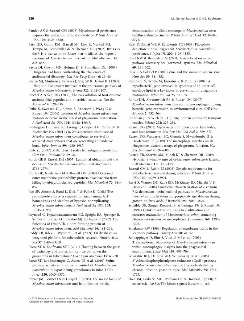
Pandey AK & Sassetti CM (2008) Mycobacterial persistence
requires the utilization of host cholesterol. P Natl Acad Sci
USA 105: 4376–4380.Park HD, Guinn KM, Harrell MI, Liao R, Voskuil MI,
Tompa M, Schoolnik GK & Sherman DR (2003) Rv3133c/
dosR is a transcription factor that mediates the hypoxic
response of Mycobacterium tuberculosis. Mol Microbiol 48:
833–843.Payne DJ, Gwynn MN, Holmes DJ & Pompliano DL (2007)
Drugs for bad bugs: confronting the challenges of
antibacterial discovery. Nat Rev Drug Discov 6: 29–40.Pearce MJ, Mintseris J, Ferreyra J, Gygi SP & Darwin KH (2008)
Ubiquitin-like protein involved in the proteasome pathway of
Mycobacterium tuberculosis. Science 322: 1104–1107.Peschel A & Sahl HG (2006) The co-evolution of host cationic
antimicrobial peptides and microbial resistance. Nat Rev
Microbiol 4: 529–536.Pethe K, Swenson DL, Alonso S, Anderson J, Wang C &
Russell DG (2004) Isolation of Mycobacterium tuberculosis
mutants defective in the arrest of phagosome maturation.
P Natl Acad Sci USA 101: 13642–13647.Piddington DL, Fang FC, Laessig T, Cooper AM, Orme IM &
Buchmeier NA (2001) Cu, Zn superoxide dismutase of
Mycobacterium tuberculosis contributes to survival in
activated macrophages that are generating an oxidative
burst. Infect Immun 69: 4980–4987.Pieters J (1997) MHC class II restricted antigen presentation.
Curr Opin Immunol 9: 89–96.Purdy GE & Russell DG (2007) Lysosomal ubiquitin and the
demise of Mycobacterium tuberculosis. Cell Microbiol 9:
2768–2774.Purdy GE, Niederweis M & Russell DG (2009) Decreased
outer membrane permeability protects mycobacteria from
killing by ubiquitin-derived peptides. Mol Microbiol 73: 844–857.
Rao SP, Alonso S, Rand L, Dick T & Pethe K (2008) The
protonmotive force is required for maintaining ATP
homeostasis and viability of hypoxic, nonreplicating
Mycobacterium tuberculosis. P Natl Acad Sci USA 105:
11945–11950.Raynaud C, Papavinasasundaram KG, Speight RA, Springer B,
Sander P, Bottger EC, Colston MJ & Draper P (2002) The
functions of OmpATb, a pore-forming protein of
Mycobacterium tuberculosis. Mol Microbiol 46: 191–201.Reddy TB, Riley R, Wymore F et al. (2009) TB database: an
integrated platform for tuberculosis research. Nucleic Acids
Res 37: D499–D508.Reece ST & Kaufmann SHE (2011) Floating between the poles
of pathology and protection: can we pin down the
granuloma in tuberculosis? Curr Opin Microbiol 15: 63–70.Reece ST, Loddenkemper C, Askew DJ et al. (2010) Serine
protease activity contributes to control of Mycobacterium
tuberculosis in hypoxic lung granulomas in mice. J Clin
Invest 120: 3365–3376.Reyrat JM, Berthet FX & Gicquel B (1995) The urease locus of
Mycobacterium tuberculosis and its utilization for the
demonstration of allelic exchange in Mycobacterium bovis
bacillus Calmette-Guerin. P Natl Acad Sci USA 92: 8768–8772.
Rifat D, Bishai WR & Karakousis PC (2009) Phosphate
depletion: a novel trigger for Mycobacterium tuberculosis
persistence. J Infect Dis 200: 1126–1135.Rigel NW & Braunstein M (2008) A new twist on an old
pathway–accessory Sec [corrected] systems. Mol Microbiol
69: 291–302.Rink L & Gabriel P (2000) Zinc and the immune system. Proc
Nutr Soc 59: 541–552.Robinson N, Wolke M, Ernestus K & Plum G (2007) A
mycobacterial gene involved in synthesis of an outer cell
envelope lipid is a key factor in prevention of phagosome
maturation. Infect Immun 75: 581–591.Rohde KH, Abramovitch RB & Russell DG (2007)
Mycobacterium tuberculosis invasion of macrophages: linking
bacterial gene expression to environmental cues. Cell Host
Microbe 2: 352–364.Rothman JE & Wieland FT (1996) Protein sorting by transport
vesicles. Science 272: 227–234.Russell DG (2001) Mycobacterium tuberculosis: here today,
and here tomorrow. Nat Rev Mol Cell Biol 2: 569–577.Russell DG, Vanderven BC, Glennie S, Mwandumba H &
Heyderman RS (2009) The macrophage marches on its
phagosome: dynamic assays of phagosome function. Nat
Rev Immunol 9: 594–600.Rustad TR, Sherrid AM, Minch KJ & Sherman DR (2009)
Hypoxia: a window into Mycobacterium tuberculosis latency.
Cell Microbiol 11: 1151–1159.Sassetti CM & Rubin EJ (2003) Genetic requirements for
mycobacterial survival during infection. P Natl Acad Sci
USA 100: 12989–12994.Savvi S, Warner DF, Kana BD, McKinney JD, Mizrahi V &
Dawes SS (2008) Functional characterization of a vitamin
B12-dependent methylmalonyl pathway in Mycobacterium
tuberculosis: implications for propionate metabolism during
growth on fatty acids. J Bacteriol 190: 3886–3895.Schaible UE, Sturgill-Koszycki S, Schlesinger PH & Russell DG
(1998) Cytokine activation leads to acidification and
increases maturation of Mycobacterium avium-containing
phagosomes in murine macrophages. J Immunol 160: 1290–1296.
Schekman RW (1994) Regulation of membrane traffic in the
secretory pathway. Harvey Lect 90: 41–57.Schnappinger D, Ehrt S, Voskuil MI et al. (2003)
Transcriptional adaptation of Mycobacterium tuberculosis
within macrophages: insights into the phagosomal
environment. J Exp Med 198: 693–704.Senaratne RH, De Silva AD, Williams SJ et al. (2006)
5′-Adenosinephosphosulphate reductase (CysH) protects
Mycobacterium tuberculosis against free radicals during
chronic infection phase in mice. Mol Microbiol 59: 1744–1753.
Shah IM, Laaberki MH, Popham DL & Dworkin J (2008) A
eukaryotic-like Ser/Thr kinase signals bacteria to exit
ª 2012 Federation of European Microbiological Societies FEMS Microbiol Rev 36 (2012) 514–532Published by Blackwell Publishing Ltd. All rights reserved
530 M. Gengenbacher & S.H.E. Kaufmann
dormancy in response to peptidoglycan fragments. Cell 135:
486–496.Shi S & Ehrt S (2006) Dihydrolipoamide acyltransferase is
critical for Mycobacterium tuberculosis pathogenesis. Infect
Immun 74: 56–63.Shleeva MO, Bagramyan K, Telkov MV, Mukamolova GV,
Young M, Kell DB & Kaprelyants AS (2002) Formation and
resuscitation of “non-culturable” cells of Rhodococcus
rhodochrous and Mycobacterium tuberculosis in prolonged
stationary phase. Microbiology 148: 1581–1591.Smith NH, Hewinson RG, Kremer K, Brosch R & Gordon SV
(2009) Myths and misconceptions: the origin and evolution
of Mycobacterium tuberculosis. Nat Rev Microbiol 7: 537–544.Sow FB, Florence WC, Satoskar AR, Schlesinger LS, Zwilling
BS & Lafuse WP (2007) Expression and localization of
hepcidin in macrophages: a role in host defense against
tuberculosis. J Leukoc Biol 82: 934–945.Striebel F, Imkamp F, Sutter M, Steiner M, Mamedov A &
Weber-Ban E (2009) Bacterial ubiquitin-like modifier Pup is
deamidated and conjugated to substrates by distinct but
homologous enzymes. Nat Struct Mol Biol 16: 647–651.Sturgill-Koszycki S, Schlesinger PH, Chakraborty P, Haddix
PL, Collins HL, Fok AK, Allen RD, Gluck SL, Heuser J &
Russell DG (1994) Lack of acidification in Mycobacterium
phagosomes produced by exclusion of the vesicular proton-
ATPase. Science 263: 678–681.Sturgill-Koszycki S, Schaible UE & Russell DG (1996)
Mycobacterium-containing phagosomes are accessible to
early endosomes and reflect a transitional state in normal
phagosome biogenesis. EMBO J 15: 6960–6968.Sutter M, Striebel F, Damberger FF, Allain FH & Weber-Ban E
(2009) A distinct structural region of the prokaryotic
ubiquitin-like protein (Pup) is recognized by the N-terminal
domain of the proteasomal ATPase Mpa. FEBS Lett 583:
3151–3157.Tailleux L, Waddell SJ, Pelizzola M et al. (2008) Probing host
pathogen cross-talk by transcriptional profiling of both
Mycobacterium tuberculosis and infected human dendritic
cells and macrophages. PLoS ONE 3: e1403.
Teh JS, Yano T & Rubin H (2007) Type II NADH:
menaquinone oxidoreductase of Mycobacterium tuberculosis.
Infect Disord Drug Targets 7: 169–181.Tews I, Findeisen F, Sinning I, Schultz A, Schultz JE & Linder
JU (2005) The structure of a pH-sensing mycobacterial
adenylyl cyclase holoenzyme. Science 308: 1020–1023.Tulp A, Verwoerd D, Dobberstein B, Ploegh HL & Pieters J
(1994) Isolation and characterization of the intracellular
MHC class II compartment. Nature 369: 120–126.van der Wel N, Hava D, Houben D, Fluitsma D, van Zon M,
Pierson J, Brenner M & Peters PJ (2007) M. tuberculosis and
M. leprae translocate from the phagolysosome to the cytosol
in myeloid cells. Cell 129: 1287–1298.Vandal OH, Pierini LM, Schnappinger D, Nathan CF & Ehrt S
(2008) A membrane protein preserves intrabacterial pH in
intraphagosomal Mycobacterium tuberculosis. Nat Med 14:
849–854.
Vandal OH, Roberts JA, Odaira T, Schnappinger D, Nathan
CF & Ehrt S (2009) Acid-susceptible mutants of
Mycobacterium tuberculosis share hypersusceptibility to cell
wall and oxidative stress and to the host environment.
J Bacteriol 191: 625–631.Vazquez-Laslop N, Lee H & Neyfakh AA (2006) Increased
persistence in Escherichia coli caused by controlled
expression of toxins or other unrelated proteins. J Bacteriol
188: 3494–3497.Velayati AA, Masjedi MR, Farnia P, Tabarsi P, Ghanavi J,
Ziazarifi AH & Hoffner SE (2009) Emergence of new forms
of totally drug-resistant tuberculosis bacilli: super
extensively drug-resistant tuberculosis or totally drug-
resistant strains in Iran. Chest 136: 420–425.Vergne I, Chua J & Deretic V (2003) Tuberculosis toxin
blocking phagosome maturation inhibits a novel Ca2+/Calmodulin-PI3K hVPS34 cascade. J Exp Med 198: 653–659.
Vergne I, Chua J, Lee HH, Lucas M, Belisle J & Deretic V
(2005) Mechanism of phagolysosome biogenesis block by
viable Mycobacterium tuberculosis. P Natl Acad Sci USA 102:
4033–4038.Via LE, Fratti RA, McFalone M, Pagan-Ramos E, Deretic D &
Deretic V (1998) Effects of cytokines on mycobacterial
phagosome maturation. J Cell Sci 111(Pt 7): 897–905.Via LE, Lin PL, Ray SM et al. (2008) Tuberculous granulomas
are hypoxic in guinea pigs, rabbits, and nonhuman
primates. Infect Immun 76: 2333–2340.Vilcheze C, Av-Gay Y, Attarian R et al. (2008) Mycothiol
biosynthesis is essential for ethionamide susceptibility
in Mycobacterium tuberculosis. Mol Microbiol 69: 1316–1329.
Walburger A, Koul A, Ferrari G, Nguyen L, Prescianotto-
Baschong C, Huygen K, Klebl B, Thompson C, Bacher G &
Pieters J (2004) Protein kinase G from pathogenic
mycobacteria promotes survival within macrophages. Science
304: 1800–1804.Wang T, Li H, Lin G, Tang C, Li D, Nathan C, Darwin KH &
Li H (2009) Structural insights on the Mycobacterium
tuberculosis proteasomal ATPase Mpa. Structure 17: 1377–1385.
Wayne LG & Hayes LG (1996) An in vitro model for
sequential study of shiftdown of Mycobacterium tuberculosis
through two stages of nonreplicating persistence. Infect
Immun 64: 2062–2069.West MA, Lucocq JM & Watts C (1994) Antigen processing
and class II MHC peptide-loading compartments in human
B-lymphoblastoid cells. Nature 369: 147–151.Wolf PR & Ploegh HL (1995) How MHC class II molecules
acquire peptide cargo: biosynthesis and trafficking through
the endocytic pathway. Annu Rev Cell Dev Biol 11: 267–306.
Wu X, Yang Y, Han Y, Zhang J, Liang Y, Li H, Li B & Wang
L (2008) Effect of recombinant Rv1009 protein on
promoting the growth of Mycobacterium tuberculosis. J Appl
Microbiol 105: 1121–1127.
FEMS Microbiol Rev 36 (2012) 514–532 ª 2012 Federation of European Microbiological SocietiesPublished by Blackwell Publishing Ltd. All rights reserved
Mycobacterium tuberculosis: success through dormancy 531
Xie Z, Siddiqi N & Rubin EJ (2005) Differential antibiotic
susceptibilities of starved Mycobacterium tuberculosis isolates.
Antimicrob Agents Chemother 49: 4778–4780.Yang X, Nesbitt NM, Dubnau E, Smith I & Sampson NS
(2009) Cholesterol metabolism increases the metabolic pool
of propionate in Mycobacterium tuberculosis. Biochemistry
48: 3819–3821.Yano T, Li LS, Weinstein E, Teh JS & Rubin H (2006) Steady-
state kinetics and inhibitory action of antitubercular
phenothiazines on mycobacterium tuberculosis type-II
NADH-menaquinone oxidoreductase (NDH-2). J Biol Chem
281: 11456–11463.Yates RM, Hermetter A & Russell DG (2005) The kinetics of
phagosome maturation as a function of phagosome/
lysosome fusion and acquisition of hydrolytic activity.
Traffic 6: 413–420.Yew WW & Leung CC (2008) Update in tuberculosis 2007.
Am J Respir Crit Care Med 177: 479–485.Zhang Y & Mitchison D (2003) The curious characteristics of
pyrazinamide: a review. Int J Tuberc Lung Dis 7: 6–21.
ª 2012 Federation of European Microbiological Societies FEMS Microbiol Rev 36 (2012) 514–532Published by Blackwell Publishing Ltd. All rights reserved
532 M. Gengenbacher & S.H.E. Kaufmann


![[Micro] mycobacterium tuberculosis](https://img.pdfslide.us/doc/110x75/55d6fc67bb61ebfa2a8b47ea/micro-mycobacterium-tuberculosis.jpg)