Embed Size (px)
Citation preview
RESEARCH ARTICLE Open Access
MYC, FBXW7 and TP53 copy number variation andexpression in Gastric CancerDanielle Queiroz Calcagno1,2*, Vanessa Morais Freitas3, Mariana Ferreira Leal2, Carolina Rosal Teixeira de Souza1,Samia Demachki4, Raquel Montenegro1, Paulo Pimentel Assumpção5, André Salim Khayat1,Marília de Arruda Cardoso Smith2, Andrea Kely Campos Ribeiro dos Santos6 and Rommel Rodriguez Burbano1
Abstract
Background: MYC deregulation is a common event in gastric carcinogenesis, usually as a consequence of geneamplification, chromosomal translocations, or posttranslational mechanisms. FBXW7 is a p53-controlled tumor-suppressorthat plays a role in the regulation of cell cycle exit and reentry via MYC degradation.
Methods: We evaluated MYC, FBXW7, and TP53 copy number, mRNA levels, and protein expression in gastric cancer andpaired non-neoplastic specimens from 33 patients and also in gastric adenocarcinoma cell lines. We also determined theinvasion potential of the gastric cancer cell lines.
Results: MYC amplification was observed in 51.5% of gastric tumor samples. Deletion of one copy of FBXW7 and TP53was observed in 45.5% and 21.2% of gastric tumors, respectively. MYC mRNA expression was significantly higher intumors than in non-neoplastic samples. FBXW7 and TP53 mRNA expression was markedly lower in tumors than in pairednon-neoplastic specimens. Moreover, deregulated MYC and FBXW7 mRNA expression was associated with the presenceof lymph node metastasis and tumor stage III-IV. Additionally, MYC immunostaining was more frequently observed inintestinal-type than diffuse-type gastric cancers and was associated with MYC mRNA expression. In vitro studies showedthat increased MYC and reduced FBXW7 expression is associated with a more invasive phenotype in gastric cancer celllines. This result encouraged us to investigate the activity of the gelatinases MMP-2 and MMP-9 in both cell lines. Bothgelatinases are synthesized predominantly by stromal cells rather than cancer cells, and it has been proposed that bothcontribute to cancer progression. We observed a significant increase in MMP-9 activity in ACP02 compared with ACP03cells. These results confirmed that ACP02 cells have greater invasion capability than ACP03 cells.
Conclusion: In conclusion, FBXW7 and MYC mRNA may play a role in aggressive biologic behavior of gastric cancercells and may be a useful indicator of poor prognosis. Furthermore, MYC is a candidate target for new therapies againstgastric cancer.
Keywords: Gastric cancer, MYC, FBXW7, TP53
BackgroundGastric cancer (GC) is the fourth most common can-cer and the second leading cause of cancer deathworldwide [1]. GC is considered a major public healthconcern, especially in developing countries, includingBrazil [2].
A fundamental aspect of carcinogenesis is uncon-trolled cell proliferation resulting from the accumulationof changes that promote the expression or repression ofcell cycle-control genes [3]. MYC is a transcriptionalfactor involved in cell cycle regulation and cell growtharrest that is commonly deregulated in cancers and hasbeen described as a key element of gastric carcinogenesis[4,5]. Several different types of posttranslational modifi-cations of MYC have been described, including phos-phorylation, acetylation, and ubiquitination [6]. Theubiquitin-proteasome system is the major protein degrad-ation regulatory pathway involved in cell differentiation
* Correspondence: [email protected]ório de Citogenética Humana, Instituto de Ciências Biológicas,Universidade Federal do Pará, Belém, PA, Brasil2Disciplina de Genética, Departamento de Morfologia e Genética, EscolaPaulista de Medicina, Universidade Federal de São Paulo, Rua Botucatu 740,CEP 04023-900, São Paulo, SP, BrazilFull list of author information is available at the end of the article
© 2013 Calcagno et al.; licensee BioMed Central Ltd. This is an Open Access article distributed under the terms of the CreativeCommons Attribution License (http://creativecommons.org/licenses/by/2.0), which permits unrestricted use, distribution, andreproduction in any medium, provided the original work is properly cited.
Calcagno et al. BMC Gastroenterology 2013, 13:141http://www.biomedcentral.com/1471-230X/13/141
and growth control [7]. FBXW7 encodes an F-box proteinsubunit of the Skp1/Cul1/F-box complex (SCF) ubiquitinligase complex. SCFFBXW7 induces degradation of theproducts of positive cell cycle regulator genes, such as cyclinE, MYC, NOTCH, and JUN, through phosphorylation-dependent ubiquitination [8]. Among SCFFBXW7 substrates,MYC is of particular importance in cell cycle exit becauseit is thought to play a role in determining whether mam-malian cells divide or not [9].Deregulated FBXW7 expression is a major cause of
carcinogenesis [10-12]. Loss of FBXW7 expression canlead to MYC overexpression and has been associatedwith poor prognosis in GC patients [13]. However, MYCactivation by FBXW7 loss triggers activation of p53,which plays a key role in the regulation of cellularresponses to DNA damage and abnormal expression ofoncogenes. Induction of cell cycle arrest by p53 allowsfor DNA repair or apoptosis induction [14]. Thus,concomitant loss of FBXW7 and TP53 is necessary toinduce genetic instability and tumorigenesis [11].In the present study, we investigated MYC, FBXW7,
and TP53 gene copy number variation and mRNA andprotein expression in GC samples and gastric adenocar-cinoma cell lines. Possible associations between ourfindings and the clinicopathological features and/orinvasion and migration capability of the cell lines werealso evaluated.
MethodsClinical samplesSamples were obtained from 33 GC patients who under-went surgical treatment at the João de Barros BarretoUniversity Hospital in Pará State, Brazil. Dissectedtumor and paired non-neoplastic tissue specimens wereimmediately cut from the stomach and frozen in liquidnitrogen until RNA extraction.The clinicopathological features of the patient samples
are shown in Table 1. GC samples were classifiedaccording to Lauren [15]. All GC samples showed thepresence of Helicobacter pylori, and the cagA virulencefactor was determined by PCR analysis of ureA and cagAas described by Clayton et al. [16] and Covacci et al.[17], respectively. All patients had negative histories ofexposure to either chemotherapy or radiotherapy beforesurgery, and there were no other co-occurrences of diag-nosed cancers. Informed consent with approval of theethics committee of the Federal University of Pará wasobtained.
Cells linesGastric adenocarcinoma cell lines ACP02 and ACP03[18] were cultured in complete RPMI medium (InvitrogenCorp., Carlsbad, CA, USA) supplemented with 10% fetal
bovine serum (FBS), 1% penicillin/streptomycin, and 1%kanamycin.
Copy number variation (CNV)DNA was extracted using a DNAQiamp mini kit (Qiagen,Hilden, Germany) according to the manufacturer’sinstructions. Duplex quantitative real-time PCR (real-timeqPCR) was performed using the FAM/MGB-labeledTaqMan probes for MYC (Hs01764918_cn), FBXW7(Hs01362464_cn), or TP53 (Hs06423639_cn), and VIC/TAMRA-labeled TaqMan CNV RNAse P (#4403326) wasused for the internal control. All real-time qPCR reactionswere performed in quadruplicate with gDNA according tothe manufacturer’s protocol using a 7500 Fast Real-TimePCR system (Life Technologies, Foster City, CA, USA).The copy number of each sample was estimated by CNVanalysis using Copy Caller Software V1.0 (Life Technologies,Foster City, CA, USA). Known Human Genomic DNA(Promega, Madison, USA) was used for calibration.
Quantitative real-time reverse transcriptase PCRTotal RNA was extracted with TRI Reagent® Solution(Life Technologies, Carlbad, CA, USA) following themanufacturer’s instructions. RNA concentration andquality were determined using a NanoDrop spectropho-tometer (Thermo Scientific, Wilmington, DE, USA) and1% agarose gels. Complementary DNA (cDNA) wassynthesized using a High-Capacity cDNA Archive kitaccording to the manufacturer’s recommendations(Life Technologies, Foster City, CA, USA). Real-timeqPCR primers and TaqMan probes targeting MYC(Hs00153408_m1), FBXW7 (Hs00217794_m1), and TP53(Hs01034249_m1) were purchased as Assays-on-DemandProducts for Gene Expression ((Life Technologies, FosterCity, CA, USA). Real time qPCR was performed using anABI Prism 7500 system (Life Technologies, Foster City,CA, USA) according to the manufacturer’s instructions.GAPDH (NM_002046.3; Life Technology, USA) wasselected as an internal control for monitoring RNA inputand reverse transcription efficiency. All real-time qPCRreactions for target genes and internal controls wereperformed in triplicate on the same plate. The relativequantification (RQ) of gene expression was calculatedusing the ΔΔCt method [19], in which the non-neoplasticsample was designated as a calibrator for each pairedtumor sample.
ImmunohistochemistryImmunohistochemical analyses for MYC and p53 wereperformed on formalin-fixed, paraffin-embedded surgicalsections. Serial 3-μm sections were used. Heat-inducedantigen retrieval was employed (microprocessor-controlledpressure Pascal® DakoCytomation, Carpinteria, CA, USA).A universal peroxidase-conjugated secondary antibody kit
Calcagno et al. BMC Gastroenterology 2013, 13:141 Page 2 of 10http://www.biomedcentral.com/1471-230X/13/141
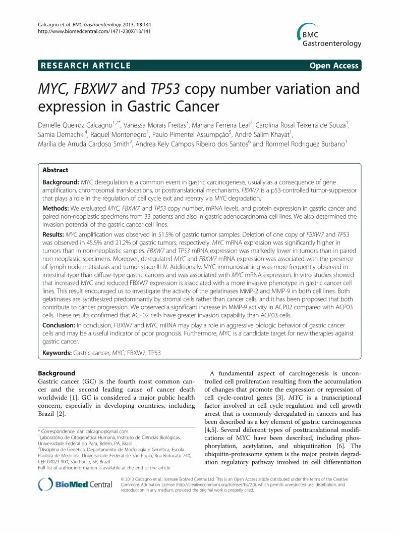
Table 1 MYC, FBXW7 and TP53 gene copy number variation, MYC and p53 protein expression and clinicopathological features of 33 GC patients
CNV MYC CNV FBXW7 CNV TP53 IHC MYC IHC p53
2 copies(n = 16)
≥ 3 copies(n = 17)
p- value 2 copies(n = 18)
1 copy(n = 15)
p- value 2 copies(n = 25)
1 copy(n = 7)
p- value P (n = 19) N (n = 14) p- value P (n = 6) N (n = 25) p- value
Age (y) (mean ± SD)
>50 (65.3 ± 9.1) 7 12 0.166 12 7 0.304 15 3 0.393 10 4 0.310 5 9 0.094
≤50 (42.1 ± 8.2) 9 5 6 8 10 4 10 9 2 17
Gender
Male 8 7 0.437 7 8 1.000 13 1 0.104 12 3 0.072 2 13 0.413
Female 8 10 8 10 12 6 8 10 5 13
Histopathology
Intestinal 12 10 0.465 12 10 1.000 16 6 0.387 17 5 0.009* 6 16 0.378
Diffuse 4 7 6 5 9 1 3 8 1 10
Depth of tumor invasion
T1 3 3 1.000 5 1 0.186 5 1 1.000 2 4 0.182 1 5 1.000
T2-T4 13 14 13 14 20 6 18 9 6 21
Lymph node metastasis
Absent 5 8 0.481 8 5 0.722 10 3 1.000 7 6 0.717 2 11 0.676
Present 11 9 10 10 15 4 13 7 5 15
Stage
I-II 8 10 0.732 12 6 0.170 14 3 0.678 12 6 0.493 3 15 0.674
III-IV 8 7 6 9 11 4 8 7 4 11
MYC IHC
Negative 5 8 0.481 7 6 1.000 11 2 0.671
Positive 11 9 11 9 14 5
p53 IHC
Negative 14 12 0.398 14 12 1.000 21 4 0.157
Positive 2 5 4 3 4 3
*p < 0.05; P: positive; N: negative.
Calcagno
etal.BM
CGastroenterology
2013,13:141Page
3of
10http://w
ww.biom
edcentral.com/1471-230X/13/141
(LSAB System, DakoCytomation, Carpinteria, CA, USA)was used for detection with diaminobenzidine (DAB) asthe chromogen. The following primary antibodies wereused: mouse monoclonal antibodies directed against MYC(dilution 1:150; sc-40, Santa Cruz Biotechnology, SantaCruz, CA, USA and clone 9E10, Zymed®, San Francisco,CA, USA), FBXW7 (dilution 1:50, Abnova Corp., TaipeiCity, Taiwan), and p53 (dilution 1:50; DakoCytomation,Carpinteria, CA, USA). Positive protein expression wasdefined as clear nuclear staining in more than 10% of thecells.
Migration and invasion assayMigration and invasion assays were carried out in amodified Boyden chamber with filter inserts (8-μmpores) for 12-well plates (BD Biosciences, San Jose, CA,USA). To assess invasion, filters were coated with 10 μlof Matrigel (10–13 mg/ml) (BD Biosciences, San Jose,CA, USA) while on ice. Cells (2 × 10⁵) were plated intothe upper chamber in 1 ml of RPMI without FBS. Thelower chamber was filled with 1.5 ml of RPMI with FBS.After 48 h in culture, cells were fixed with 4% parafor-maldehyde and post-fixed with 0.2% crystal violet in 20%methanol. Cells on the upper side of the filter, includingthose in the Matrigel, were removed with a cotton swab.Invading cells (on the lower side of the filter) werephotographed and counted. Experiments were performedin triplicate.
ImmunofluorescenceCells grown on glass coverslips were fixed with 1% para-formaldehyde in phosphate-buffered saline (PBS) for10 min, then permeabilized with 0.5% Triton X-100(Sigma-Aldrich, St. Louis, MO, USA) in PBS for 15 minand blocked with 1% bovine serum albumin (BSA) inPBS. The cells were stained with mouse antibodiesagainst MYC (diluted 1:50; Zymed®, USA), p53 (diluted1:50; DakoCytomation, Carpinteria, CA, USA), andFBXW7 (diluted 1:50; Abnova Corp., Taipei City,Taiwan). Primary antibodies were revealed using an anti-mouse Alexa-568-conjugated secondary antibody(Invitrogen). All incubations were carried out for 60 minat room temperature. Nuclei were stained with DAPI inProlong anti-fade mounting medium (Invitrogen).Negative control samples were processed as describedabove except that primary antibodies were omitted andreplaced with PBS alone.
Western blottingProtein extraction from cells was performed accordingto standard procedures. Briefly, total protein was extractedfrom ACP02 and ACP03 cells using 50 mM Tris–HClbuffer containing 100 mmol/L NaCl, 50 mM NaF, 1 mMNaVO4, 0.5% NP-40, and complete protease inhibitor
cocktail (Roche, Germany). Protein concentration wasestimated using a Bradford assay (Sigma-Aldrich). About30 μg of total protein extract was loaded onto a 12%sodium dodecyl sulfate-polyacrylamide gel electrophoresis(SDS-PAGE) gel and electrophoresed. Resolved proteinswere then transferred from the gel onto a nitrocellulosemembrane. The membrane was blocked with 5% nonfatmilk in Tris-buffered saline containing 5% Tween (Sigma-Aldrich, Sant Louis, MO, USA) and then incubated withmouse monoclonal anti-MYC (Santa Cruz Biotechnology),anti-FBXW7 (Abnova, Taipei City, Taiwan), anti-p53(DakoCytomation, Carpinteria, CA, USA), and anti-β-actin (Sigma-Aldrich, Sant Louis, MO, USA) antibodiesdiluted 1:200, 1:100, 1:100, and 1:2,000, respectively.Subsequently, membranes were incubated with a 1:5,000dilution of horseradish peroxidase (HRP)-conjugatedsheep anti-mouse antibody (Amersham Biosciences,Piscataway, NJ, USA) for 1 h at room temperature.Proteins were visualized by enhanced chemiluminescence.
ZymographyACP02 and ACP03 cells (5 × 10⁴ of each) were platedand allowed to adhere and spread for at least 8 h. Adher-ent cells were washed three times with PBS, and theculture medium was replaced with serum-free mediumfor 24 h. The activity of MMP2 and MMP9 in the condi-tioned medium was assessed by zymography. Condi-tioned medium was collected, concentrated (Microcon30 K, Merck Millipore, Darmstadt, Germany) andresuspended in SDS-PAGE sample buffer (without β-mercaptoethanol). The remaining cells were lysed andthe protein concentration was estimated using a BCAassay (Thermo Scientific Pierce, Rockford, IL, USA). Atotal of 1 μg of protein from each conditioned mediumwas separated on 10% polyacrylamide gels containing0.2% gelatin. After electrophoresis, the gels were washedin 2.5% Triton X-100 for 30 min, then equilibrated in10 mM Tris (pH 8.0) and incubated at 37°C for 16–24 hin a development buffer containing 50 mM Tris(pH 8.0), 5 mM CaCl2, and 0.02% NaN3. The gels werestained with 0.2% Coomassie blue R250 (GE Amersham,Piscataway, NJ, USA) and destained with 1:1 acetic acid/methanol solution. Experiments were performed in trip-licate. Zymographic bands, which are indicative of MMPactivity, were quantified by scanning densitometry.
Statistical analysesThe normality of variable distributions was determinedusing the Shapiro-Wilk test. Associations between MYC,FBXW7, and TP53 copy number variation, mRNA levels,protein expression, clinicopathological features, and cellinvasion and migration capability were analyzed usingthe chi-square (χ2) and Mann–Whitney tests. Correl-ation between expression of the different target mRNAs
Calcagno et al. BMC Gastroenterology 2013, 13:141 Page 4 of 10http://www.biomedcentral.com/1471-230X/13/141
was determined using Spearman’s test, in which a valuebelow 0.3 indicated a weak correlation, 0.3-0.7 indicateda medium correlation, and values above 0.7 indicated astrong correlation. Data are shown as the median andinterquartile range; p values less than 0.05 were consid-ered significant.
ResultsGastric tumor specimens showed amplification of MYCand deletion of FBXW7 and TP53Three or more copies of MYC were found in 51.5% (17/33) of gastric tumor cells. In contrast, 45.5% (15/33) and21.2% (7/33) of gastric tumor cells contained only onecopy of FBXW7 and TP53, respectively.The association between clinicopathological features
and MYC, FBXW7, and TP53 copy number is summa-rized in Table 1. One gastric tumor that contained threecopies of TP53 was excluded from the chi-squareanalysis. No association was found between copy num-ber variation of the genes studied and clinicopathologi-cal features.
MYC mRNA expression was higher in tumors than innon-neoplastic specimens, whereas FBXW7 and TP53mRNA expression was lower in tumor specimensThe expression level of MYC mRNA (2.01 ± 1.72 foldchange) in tumor tissue samples was significantly higherthan in non-neoplastic tissue (p = 0.0002), whereas theexpression level of FBXW7 mRNA (0.53 ± 0.40 foldchange) and TP53 mRNA (0.84 ± 0.55 fold change) intumor tissue specimens was significantly lower than innon-neoplastic tissue (p < 0.0001 and p = 0.0011, respect-ively). We did not find a significant correlation betweenMYC, FBXW7, and TP53 mRNA expression (MYC/FBXW7 mRNA r = −0.3464, p = 0.0562; MYC/TP53mRNA r = 0.0950, p = 0.6113; FBXW7/TP53 mRNAr = −0.0745, p = 0.4747). Thus, only a tendency towardcorrelation between an increase in MYC mRNA ex-pression and a decrease in FBXW7 mRNA expressionwas detected.Table 2 summarizes the associations between various
clinicopathological features and the RQ of MYC, FBXW7,and TP53 mRNA expression in tumor and paired non-neoplastic specimens. An increase in MYC mRNA levelwas associated with the presence of lymph node metasta-sis (p = 0.016) and GC tumor stage III-IV (p = 0.036). Asignificant reduction in FBXW7 mRNA level was alsoassociated with the presence lymph node metastasis(p = 0.015) and tumor stage III-IV (p = 0.008).
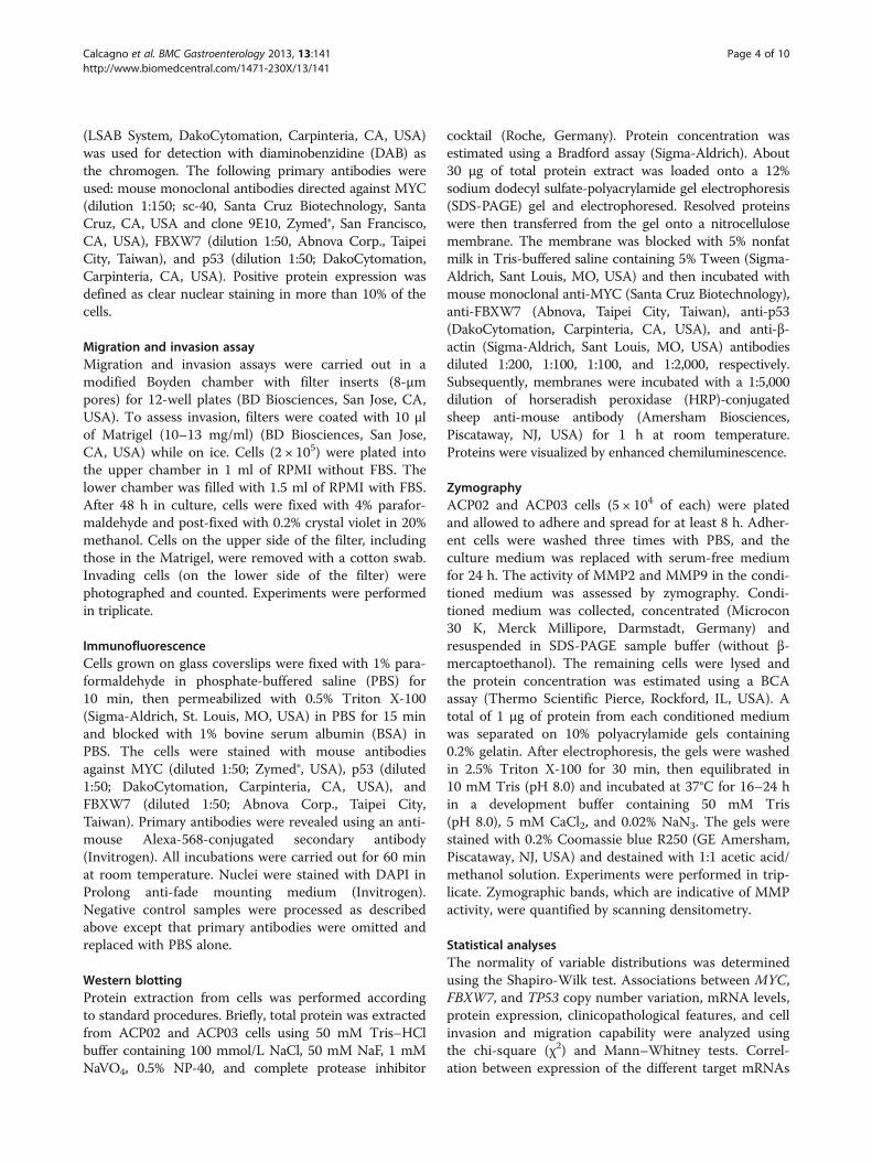
Nuclear MYC protein staining is associated withintestinal-type GCPositive staining for nuclear MYC and p53 was found in64.5% (20/31) and 19.4% (6/31) of GC samples,
respectively (Figure 1). No positivity was found forFBXW7. Table 1 summarizes the clinicopathologicalfeatures and MYC and p53 immunostaining results.Expression of MYC was more frequent in intestinal-typethan diffuse-type GC (p = 0.007). Furthermore, MYCimmunostaining was associated with increased MYCmRNA level (p = 0.0022). No association was foundbetween p53 immunostaining and clinicopathologicalcharacteristics, TP53 copy number, or TP53 mRNAexpression.
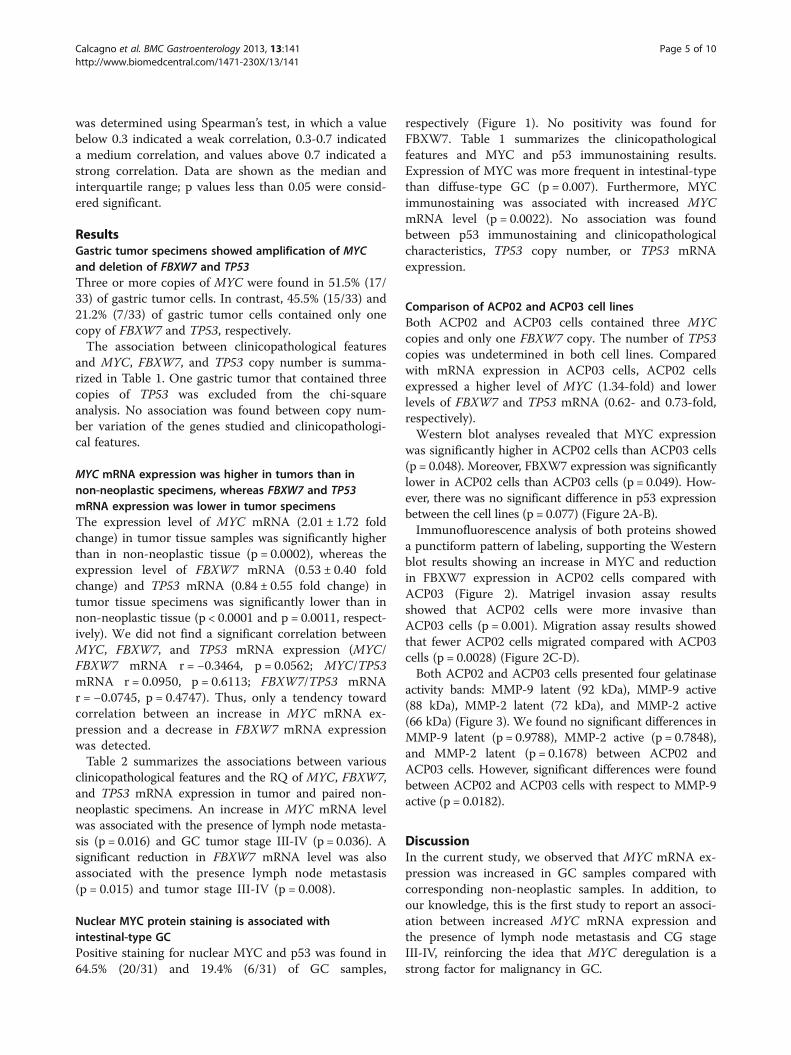
Comparison of ACP02 and ACP03 cell linesBoth ACP02 and ACP03 cells contained three MYCcopies and only one FBXW7 copy. The number of TP53copies was undetermined in both cell lines. Comparedwith mRNA expression in ACP03 cells, ACP02 cellsexpressed a higher level of MYC (1.34-fold) and lowerlevels of FBXW7 and TP53 mRNA (0.62- and 0.73-fold,respectively).Western blot analyses revealed that MYC expression
was significantly higher in ACP02 cells than ACP03 cells(p = 0.048). Moreover, FBXW7 expression was significantlylower in ACP02 cells than ACP03 cells (p = 0.049). How-ever, there was no significant difference in p53 expressionbetween the cell lines (p = 0.077) (Figure 2A-B).Immunofluorescence analysis of both proteins showed
a punctiform pattern of labeling, supporting the Westernblot results showing an increase in MYC and reductionin FBXW7 expression in ACP02 cells compared withACP03 (Figure 2). Matrigel invasion assay resultsshowed that ACP02 cells were more invasive thanACP03 cells (p = 0.001). Migration assay results showedthat fewer ACP02 cells migrated compared with ACP03cells (p = 0.0028) (Figure 2C-D).Both ACP02 and ACP03 cells presented four gelatinase
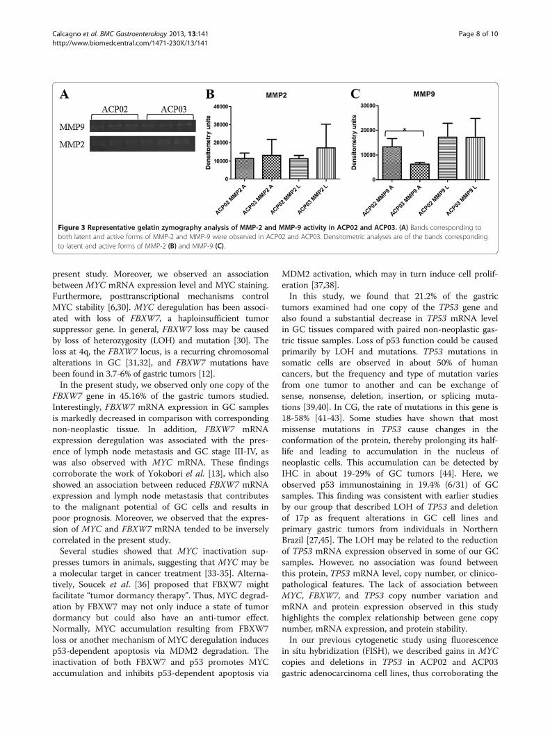
activity bands: MMP-9 latent (92 kDa), MMP-9 active(88 kDa), MMP-2 latent (72 kDa), and MMP-2 active(66 kDa) (Figure 3). We found no significant differences inMMP-9 latent (p = 0.9788), MMP-2 active (p = 0.7848),and MMP-2 latent (p = 0.1678) between ACP02 andACP03 cells. However, significant differences were foundbetween ACP02 and ACP03 cells with respect to MMP-9active (p = 0.0182).
DiscussionIn the current study, we observed that MYC mRNA ex-pression was increased in GC samples compared withcorresponding non-neoplastic samples. In addition, toour knowledge, this is the first study to report an associ-ation between increased MYC mRNA expression andthe presence of lymph node metastasis and CG stageIII-IV, reinforcing the idea that MYC deregulation is astrong factor for malignancy in GC.
Calcagno et al. BMC Gastroenterology 2013, 13:141 Page 5 of 10http://www.biomedcentral.com/1471-230X/13/141
Adams et al. [20] and Leder et al. [21] demonstratedthat MYC mRNA expression deregulation can promotethe development of cancer in transgenic mouse models.The increase in MYC mRNA level in human cancersmay result from both direct and indirect mechanisms,which could have several explanations. First, MYCamplification is the most common mechanism of MYCderegulation in GC [5]. This mechanism leads toincreased production of oncogenic products in quan-tities that exceed the transcriptional capacity of a normaldouble copy gene. Here, we observed three or moreMYC gene copies in 51.5% of gastric tumors specimens.Previous studies from our group also showed that MYCamplification or trisomy of chromosome 8, on whichMYC is located, was present in all GC samples examinedfrom individuals in Northern Brazil, as well as in GC celllines established by our group from tumors of Brazilianpatients [18,22-27]. The presence of MYC amplificationhas also been reported in plasma samples from individ-uals with GC [28]. However, no direct association
between MYC copy number variation and mRNA expres-sion was detected in the present study.Second, the increase in MYC mRNA expression may
result from consistent recombination between theimmunoglobulin (Ig) locus and the MYC oncogene. Thisphenomenon is frequently described in Burkitt’s lymph-oma and is associated with a longer half-life of MYCmRNA in affected cells [29]. Previously, our researchgroup observed MYC insertions in diffuse-type GCmainly into chromosomes that are mapped to genes ofimmunoglobulins (chromosomes 2, 14, and 22) [26].Thus, chromosomal translocations involving the MYClocus (8q24) in diffuse-type CG in individuals fromNorthern Brazil might also reflect an increase in MYCmRNA level.Immunohistochemistry (IHC) analysis revealed that
MYC expression is more frequently found in intestinal-type GC than diffuse-type GC specimens. These alter-ations could lead to an abnormal MYC protein that isnot recognized by either of the antibodies used in the
Table 2 MYC, FBXW7 and TP53 mRNA expression levels and clinicopathological factors of 33 gastric cancer patients
n (%) MYC p- value FBXW7 p- value TP53 p- value
Median ± IQR Median ± IQR Median ± IQR
Age (y) (mean ± SD)
>50 (65.3 ± 9.1) 19 (57.6%) 2.04 ± 1.35 0.8873 0.55 ± 0.37 0.9247 0.86 ± 0.62 0.7409
≤50 (42.1 ± 8.2) 14 (42.4%) 1.44 ± 4.88 0.53 ± 0.50 0.94 ± 1.65
Gender
Male 15 (45.5%) 2.01 ± 1.01 0.4065 0.53 ± 0.22 0.6353 0.89 ± 0.58 0.8125
Female 18 (54.5%) 1.67 ± 2.03 0.56 ± 0.56 0.87 ± 0.69
Histopathology
Intestinal 22 (66.7%) 2.06 ± 0.99 0.3525 0.53 ± 0.16 0.1391 0.81 ±0.78 0.3311
Diffuse 11 (33.3%) 1.40 ± 2.32 0.77 ±0.74 0.94 ± 0.38
Depth of tumor invasion
T1 6 (18.2%) 0.89 ± 0.47 0.0857 0.88 ± 0.58 0.0678 0.85 ± 0.15 0.7069
T2-T4 27 (81.8%) 2.08 ± 1.42 0.53 ± 0.43 0.91 ± 0.79
Lymph node metastasis
Absent 13 (39.4%) 0.98 ± 1.09 0.0225* 0.68 ± 0.36 0.0238* 0.84 ± 0.44 0.6121
Present 20 (60.6%) 2.10 ± 2.20 0.46 ± 0.38 0.94 ± 0.79
Stage
I-II 18 (54.5%) 1.39 ± 1.23 0.0362* 0.57 ± 0.38 0.0380* 0.83 ± 0.55 0.0892†
III-IV 15 (45.5%) 2.41 ± 2.79 0.34 ± 0.45 0.96 ± 1.19
MYC IHC
Positive 20 (60.6%) 2.18 ± 1.59 0.0022* 0.53 ± 0.32 0.4090 0.81 ± 0.57 0.1372
Negative 13 (39.4%) 0.89 ± 0.85 0.58 ± 0.53 0.99 ± 0.90
p53 IHC
Positive 7 (21.2%) 4.25 ± 6.53 0.0891 0.64 ± 0.68 0.9203 1.06 ± 0.83 0.2937
Negative 26 (78.8%) 2.00 ± 1.44 0.55 ± 0.35 0.87 ± 0.60
*p < 0.05; IQR: interquartile range.
Calcagno et al. BMC Gastroenterology 2013, 13:141 Page 6 of 10http://www.biomedcentral.com/1471-230X/13/141
Figure 1 Immunohistochemical analysis of MYC and p53 protein expression in GC. (A) Negative MYC immunostaining in diffuse-type GC;(B) MYC immune positivity in intestinal-type GC; (C) Positive p53 immunostaining in diffuse-type GC; (D) p53 immune positivity in intestinal-typeGC (magnification × 40).
Figure 2 MYC, FBXW7 and p53 expression, migration and invasion ability in ACP02 and ACP03. (A) Graph show mean ± SD of MYC,FBXW7 and p53 protein expression in ACP02 and ACP03. These proteins were normalized to the level of beta actin; (B) Representative data ofMYC, FBXW7 and p53 protein expression; (C-D) Graphs show mean ± SD of migration and invasive cells triplicates assay; (E) Representativeresults of MYC, FBXW7 and p53 immunofluorescence.
Calcagno et al. BMC Gastroenterology 2013, 13:141 Page 7 of 10http://www.biomedcentral.com/1471-230X/13/141
present study. Moreover, we observed an associationbetween MYC mRNA expression level and MYC staining.Furthermore, posttranscriptional mechanisms controlMYC stability [6,30]. MYC deregulation has been associ-ated with loss of FBXW7, a haploinsufficient tumorsuppressor gene. In general, FBXW7 loss may be causedby loss of heterozygosity (LOH) and mutation [30]. Theloss at 4q, the FBXW7 locus, is a recurring chromosomalalterations in GC [31,32], and FBXW7 mutations havebeen found in 3.7-6% of gastric tumors [12].In the present study, we observed only one copy of the
FBXW7 gene in 45.16% of the gastric tumors studied.Interestingly, FBXW7 mRNA expression in GC samplesis markedly decreased in comparison with correspondingnon-neoplastic tissue. In addition, FBXW7 mRNAexpression deregulation was associated with the pres-ence of lymph node metastasis and GC stage III-IV, aswas also observed with MYC mRNA. These findingscorroborate the work of Yokobori el al. [13], which alsoshowed an association between reduced FBXW7 mRNAexpression and lymph node metastasis that contributesto the malignant potential of GC cells and results inpoor prognosis. Moreover, we observed that the expres-sion of MYC and FBXW7 mRNA tended to be inverselycorrelated in the present study.Several studies showed that MYC inactivation sup-
presses tumors in animals, suggesting that MYC may bea molecular target in cancer treatment [33-35]. Alterna-tively, Soucek et al. [36] proposed that FBXW7 mightfacilitate “tumor dormancy therapy”. Thus, MYC degrad-ation by FBXW7 may not only induce a state of tumordormancy but could also have an anti-tumor effect.Normally, MYC accumulation resulting from FBXW7loss or another mechanism of MYC deregulation inducesp53-dependent apoptosis via MDM2 degradation. Theinactivation of both FBXW7 and p53 promotes MYCaccumulation and inhibits p53-dependent apoptosis via
MDM2 activation, which may in turn induce cell prolif-eration [37,38].In this study, we found that 21.2% of the gastric
tumors examined had one copy of the TP53 gene andalso found a substantial decrease in TP53 mRNA levelin GC tissues compared with paired non-neoplastic gas-tric tissue samples. Loss of p53 function could be causedprimarily by LOH and mutations. TP53 mutations insomatic cells are observed in about 50% of humancancers, but the frequency and type of mutation variesfrom one tumor to another and can be exchange ofsense, nonsense, deletion, insertion, or splicing muta-tions [39,40]. In CG, the rate of mutations in this gene is18-58% [41-43]. Some studies have shown that mostmissense mutations in TP53 cause changes in theconformation of the protein, thereby prolonging its half-life and leading to accumulation in the nucleus ofneoplastic cells. This accumulation can be detected byIHC in about 19-29% of GC tumors [44]. Here, weobserved p53 immunostaining in 19.4% (6/31) of GCsamples. This finding was consistent with earlier studiesby our group that described LOH of TP53 and deletionof 17p as frequent alterations in GC cell lines andprimary gastric tumors from individuals in NorthernBrazil [27,45]. The LOH may be related to the reductionof TP53 mRNA expression observed in some of our GCsamples. However, no association was found betweenthis protein, TP53 mRNA level, copy number, or clinico-pathological features. The lack of association betweenMYC, FBXW7, and TP53 copy number variation andmRNA and protein expression observed in this studyhighlights the complex relationship between gene copynumber, mRNA expression, and protein stability.In our previous cytogenetic study using fluorescence
in situ hybridization (FISH), we described gains in MYCcopies and deletions in TP53 in ACP02 and ACP03gastric adenocarcinoma cell lines, thus corroborating the
Figure 3 Representative gelatin zymography analysis of MMP-2 and MMP-9 activity in ACP02 and ACP03. (A) Bands corresponding toboth latent and active forms of MMP-2 and MMP-9 were observed in ACP02 and ACP03. Densitometric analyses are of the bands correspondingto latent and active forms of MMP-2 (B) and MMP-9 (C).
Calcagno et al. BMC Gastroenterology 2013, 13:141 Page 8 of 10http://www.biomedcentral.com/1471-230X/13/141
present results obtained using real-time qPCR [27]. Bothalterations were observed in the primary tumors fromwhich these cell lines were established. Since ACP02 andACP03 cells present alterations similar to those ofgastric tumors, these cell lines may be useful as tools forexperimental modeling of gastric carcinogenesis andmay enhance understanding of the genetic basis under-lying GC behavior and treatment and perhaps maychange the landscape of GC.In the present study, we also observed increased MYC
and reduced FBXW7 mRNA and protein expression inACP02 cells compared with ACP03 cells. Furthermore,ACP02 cells were more invasive than ACP03 cells. Onthe other hand, ACP03 cells had a higher migrationcapability than ACP02 cells. Thus, despite the ability tomigrate, ACP03 cells probably do not have efficient inva-sive machinery such as active proteases necessary todegrade the substrate. These findings are in agreementwith observations in gastric tumors and reinforce thehypothesis that deregulation of MYC and FBXW7 iscrucial for the invasive ability of GC cells. This resultencouraged us to investigate the MMP-2 and MMP-9activities of cells using zymography. The MMPs aresynthesized as latent enzymes and later activated viaproteolytic cleavage by themselves or other proteins inthe intracellular space. Both proteases are synthesizedpredominantly by stromal cells rather than cancer cellsand both contribute to cancer progression [46]. Ourzymography analysis revealed no significant differencesin the activity of MMP2 between ACP02 and ACP03cells. Additionally, MMP-9 was more active in ACP02than ACP03 cells. Studies have shown that high levels ofMMP-2 and/or MMP-9 are significantly correlated withGC invasion and are associated with poor prognosis[47,48]. Sampieri et al. [49] showed that MMP-9 expres-sion is enhanced in GC mucosa compared to non-neoplastic mucosa and that gelatinase activity differssignificantly between cancerous and normal tissue.
ConclusionsIn conclusion, our findings show that FBXW7 and MYCmRNA levels reflect the potential for aggressive biologicbehavior of gastric tumors and may be used as indicatorsof poor prognosis in GC patients. Furthermore, MYCcan be a potential biomarker for use in development ofnew targets for GC therapy.
Competing interestsThe authors declare that they have no competing interests.
Authors’ contributionsDQC, VMF, ASK, MACS, AKRS and RRB participated in conception and designof study. DQC, VMF, SD participated in acquisition and performed theanalysis of data. DQC, VMF, MFL, SD, CRTS performed interpretation of data.DQC, VMF, RM, PPA and RRB involved in drafting the manuscript. All authorsread and approved the final manuscript.
AcknowledgmentsThis study was supported by Conselho Nacional de DesenvolvimentoCientífico e Tecnológico (CNPq) as grants and fellowship awards. RRB, MACS,RM, ASK and AKRS have research grants from CNPq. DQC, MFL and CRTS hasa fellowship granted by CNPq and Fundação de Amparo a Pesquisa doEstado de São Paulo (FAPESP).
Author details1Laboratório de Citogenética Humana, Instituto de Ciências Biológicas,Universidade Federal do Pará, Belém, PA, Brasil. 2Disciplina de Genética,Departamento de Morfologia e Genética, Escola Paulista de Medicina,Universidade Federal de São Paulo, Rua Botucatu 740, CEP 04023-900, SãoPaulo, SP, Brazil. 3Departamento de Biologia Celular e do Desenvolvimento,Instituto de Ciências Biomédicas, Universidade de São Paulo, São Paulo, SP,Brasil. 4Laboratorio de Imunoistoquímica, Serviço de Anatomia Patológica,Faculdade de Medicina, Hospital Universitário João de Barros Barreto,Universidade Federal do Pará, Belém, PA, Brasil. 5Serviço de Cirurgia HospitalUniversitário João de Barros Barreto, Universidade Federal do Pará, Belém, PA,Brasil. 6Laboratório de Genética Humana, Instituto de Ciências Biológicas,Universidade Federal do Pará, Belém, PA, Brasil.
Received: 17 January 2013 Accepted: 10 September 2013Published: 23 September 2013
References1. Brenner H, Rothenbacher D, Arndt V: Epidemiology of stomach cancer.
Methods in molecular biology (Clifton, NJ 2009, 472:467–477.2. Dicken BJ, Bigam DL, Cass C, Mackey JR, Joy AA, Hamilton SM: Gastric
adenocarcinoma: review and considerations for future directions.Ann Surg 2005, 241(1):27–39.
3. Smith MG, Hold GL, Tahara E, El-Omar EM: Cellular and molecular aspectsof gastric cancer. World J Gastroenterol 2006, 12(19):2979–2990.
4. Dang CV, O’Donnell KA, Zeller KI, Nguyen T, Osthus RC, Li F: The c-Myctarget gene network. Semin Cancer Biol 2006, 16(4):253–264.
5. Calcagno DQ, Leal MF, Assumpcao PP, Smith MA, Burbano RR: MYC andgastric adenocarcinoma carcinogenesis. World J Gastroenterol 2008,14(39):5962–5968.
6. Vervoorts J, Luscher-Firzlaff J, Luscher B: The ins and outs of MYCregulation by posttranslational mechanisms. J Biol Chem 2006,281(46):34725–34729.
7. Bashir T, Pagano M: Aberrant ubiquitin-mediated proteolysis of cell cycleregulatory proteins and oncogenesis. Adv Cancer Res 2003, 88:101–144.
8. Nakayama KI, Nakayama K: Regulation of the cell cycle by SCF-typeubiquitin ligases. Seminars in cell & developmental biology 2005,16(3):323–333.
9. Onoyama I, Nakayama KI: Fbxw7 in cell cycle exit and stem cellmaintenance: insight from gene-targeted mice. Cell cycle (Georgetown, Tex2008, 7(21):3307–3313.
10. Mao JH, Perez-Losada J, Wu D, Delrosario R, Tsunematsu R, Nakayama KI,Brown K, Bryson S, Balmain A: Fbxw7/Cdc4 is a p53-dependent,haploinsufficient tumour suppressor gene. Nature 2004,432(7018):775–779.
11. Welcker M, Clurman BE: Fbw7/hCDC4 dimerization regulates its substrateinteractions. Cell division 2007, 2:7.
12. Akhoondi S, Sun D, von der Lehr N, Apostolidou S, Klotz K, Maljukova A,Cepeda D, Fiegl H, Dafou D, Marth C, et al: FBXW7/hCDC4 is a generaltumor suppressor in human cancer. Cancer Res 2007, 67(19):9006–9012.
13. Yokobori T, Mimori K, Iwatsuki M, Ishii H, Onoyama I, Fukagawa T, KuwanoH, Nakayama KI, Mori M: p53-Altered FBXW7 expression determines poorprognosis in gastric cancer cases. Cancer Res 2009, 69(9):3788–3794.
14. Kountouras J, Zavos C, Chatzopoulos D: New concepts of molecularbiology on gastric carcinogenesis. Hepatogastroenterology 2005,52(64):1305–1312.
15. Lauren P: The Two Histological Main Types of Gastric Carcinoma: Diffuseand So- Called Intestinal-Type Carcinoma. an Attempt at a Histo-ClinicalClassification. Acta pathologica et microbiologica Scandinavica 1965,64:31–49.
16. Clayton CL, Kleanthous H, Morgan DD, Puckey L, Tabaqchali S: Rapidfingerprinting of Helicobacter pylori by polymerase chain reaction andrestriction fragment length polymorphism analysis. J Clin Microbiol 1993,31:1420–1425.
Calcagno et al. BMC Gastroenterology 2013, 13:141 Page 9 of 10http://www.biomedcentral.com/1471-230X/13/141
17. Covacci A, Falkow S, Berg DE, Rappuoli R: Did the inheritance of apathogenicity island modify the virulence of Helicobacter pylori? TrendsMicrobiol 1997, 5:205–208.
18. Leal MF, Martins do Nascimento JL, da Silva CE, Vita Lamarao MF, CalcagnoDQ, Khayat AS, Assumpcao PP, Cabral IR, de Arruda Cardoso Smith M,Burbano RR: Establishment and conventional cytogeneticcharacterization of three gastric cancer cell lines. Cancer Genet Cytogenet2009, 195(1):85–91.
19. Livak KJ, Schmittgen TD: Analysis of relative gene expression data usingreal-timequantitative PCR and the 2(−Delta Delta C(T)) Method. Methods2001, 25:402–408.
20. Adams JM, Harris AW, Pinkert CA, Corcoran LM, Alexander WS, Cory S,Palmiter RD, Brinster RL: The c-myc oncogene driven by immunoglobulinenhancers induces lymphoid malignancy in transgenic mice. Nature1985, 318:533–538.
21. Leder A, Pattengale PK, Kuo A, Stewart TA, Leder P: Consequences ofwidespread deregulation of the c-myc gene in transgenic mice: multipleneoplasms and normal development. Cell 1986, 45:485–495.
22. Calcagno DQ, Leal MF, Taken SS, Assumpcao PP, Demachki S, Smith Mde A,Burbano RR: Aneuploidy of chromosome 8 and C-MYC amplification inindividuals from northern Brazil with gastric adenocarcinoma. AnticancerRes 2005, 25(6B):4069–4074.
23. Calcagno DQ, Leal MF, Seabra AD, Khayat AS, Chen ES, Demachki S,Assumpcao PP, Faria MH, Rabenhorst SH, Ferreira MV, et al:Interrelationship between chromosome 8 aneuploidy, C-MYCamplification and increased expression in individuals from northernBrazil with gastric adenocarcinoma. World J Gastroenterol 2006,12(38):6207–6211.
24. Burbano RR, Assumpcao PP, Leal MF, Calcagno DQ, Guimaraes AC, KhayatAS, Takeno SS, Chen ES, De Arruda Cardoso Smith M: C-MYC locusamplification as metastasis predictor in intestinal-type gastricadenocarcinomas: CGH study in Brazil. Anticancer Res 2006,26(4B):2909–2914.
25. Costa Raiol LC, Figueira Silva EC, Mendes da Fonseca D, Leal MF, GuimaraesAC, Calcagno DQ, Khayat AS, Assumpcao PP, de Arruda Cardoso Smith M,Burbano RR: Interrelationship between MYC gene numerical aberrationsand protein expression in individuals from northern Brazil with earlygastric adenocarcinoma. Cancer Genet Cytogenet 2008, 181(1):31–35.
26. Calcagno DQ, Guimaraes AC, Leal MF, Seabra AD, Khayat AS, Pontes TB,Assumpcao PP, De Arruda Cardoso Smith M, Burbano RR: MYC insertions indiffuse-type gastric adenocarcinoma. Anticancer Res 2009,29(7):2479–2483.
27. Leal MF, Calcagno DQ, Borges da Costa Jde F, Silva TC, Khayat AS, Chen ES,Assumpção PP, de Arruda Cardoso Smith M, Burbano RR: MYC, TP53, andchromosome 17 copy-number alterations in multiple gastric cancer celllines and in their parental primary tumors. J Biomed Biotechnol 2011,2011:631268.
28. Park KU, Lee HE, Park do J, Jung EJ, Song J, Kim HH, Choe G, Kim WH, LeeHS: MYC quantitation in cell-free plasma DNA by real-time PCR forgastric cancer diagnosis. Clin Chem Lab Med 2009, 47(5):530–536.
29. Eick D, Piechaczyk M, Henglein B, Blanchard JM, Traub B, Kofler E, Wiest S,Lenoir GM, Bornkamm GW: Aberrant c-myc RNAs of Burkitt’s lymphomacells have longer half-lives. EMBO J 1985, 4(13B):3717–3725.
30. Payne SR, Kemp CJ: Tumor suppressor genetics. Carcinogenesis 2005,26(12):2031–2045.
31. Takada H, Imoto I, Tsuda H, Sonoda I, Ichikura T, Mochizuki H, Okanoue T,Inazawa J: Screening of DNA copy-number aberrations in gastric cancercell lines by array-based comparative genomic hybridization. Cancerscience 2005, 96(2):100–110.
32. Panani AD: Cytogenetic and molecular aspects of gastric cancer: clinicalimplications. Cancer letters 2008, 266(2):99–115.
33. Shachaf CM, Kopelman AM, Arvanitis C, Karlsson A, Beer S, Mandl S,Bachmann MH, Borowsky AD, Ruebner B, Cardiff RD, et al: MYC inactivationuncovers pluripotent differentiation and tumour dormancy inhepatocellular cancer. Nature 2004, 431(7012):1112–1117.
34. Yu D, Dews M, Park A, Tobias JW, Thomas-Tikhonenko A: Inactivation ofMyc in murine two-hit B lymphomas causes dormancy with elevatedlevels of interleukin 10 receptor and CD20: implications for adjuvanttherapies. Cancer Res 2005, 65(12):5454–5461.
35. Shachaf CM, Felsher DW: Tumor dormancy and MYC inactivation: pushingcancer to the brink of normalcy. Cancer Res 2005, 65(11):4471–4474.
36. Soucek L, Whitfield J, Martins CP, Finch AJ, Murphy DJ, Sodir NM, KarnezisAN, Swigart LB, Nasi S, Evan GI: Modelling Myc inhibition as a cancertherapy. Nature 2008, 455:679–683.
37. Zindy F, Eischen CM, Randle DH, Kamijo T, Cleveland JL, Sherr CJ, RousselMF: Myc signaling via the ARF tumor suppressor regulates p53-dependent apoptosis and immortalization. Genes & development 1998,12:2424–2433.
38. Pelengaris S, Khan M: The many faces of c-MYC. Arch Biochem Biophys2003, 416:129–136.
39. Harris CC, Hollstein M: Clinical implications of the p53 tumor-suppressorgene. N Engl J Med 1993, 329(18):1318–1327.
40. Chang F, Syrjanen S, Syrjanen K: Implications of the p53 tumor-suppressorgene in clinical oncology. J Clin Oncol 1995, 13(4):1009–1022.
41. Renault B, van den Broek M, Fodde R, Wijnen J, Pellegata NS, Amadori D,Khan PM, Ranzani GN: Base transitions are the most frequent geneticchanges at P53 in gastric cancer. Cancer Res 1993, 53(11):2614–2617.
42. Seruca R, David L, Castedo S, Veiga I, Borresen AL, Sobrinho-Simoes M: p53alterations in gastric carcinoma: a study of 56 primary tumors and 204nodal metastases. Cancer Genet Cytogenet 1994, 75(1):45–50.
43. Poremba C, Yandell DW, Mellin W, Schmid KW, Reers B, Bocker W,Dockhorn-Dworniczak B: Adenocarcinoma of the cardia in a young man:detection of somatic p53 mutation by immunohistochemistry andautomated direct sequencing. Pathol Res Pract 1995, 191(10):1004–1009.
44. Hollstein M, Sidransky D, Vogelstein B, Harris CC: p53 mutations in humancancers. Science 1991, 253:49–53.
45. Khayat AS, Guimaraes AC, Calcagno DQ, Seabra AD, Lima EM, Leal MF, FariaMH, Rabenhorst SH, Assumpcao PP, Demachki S, et al: Interrelationshipbetween TP53 gene deletion, protein expression and chromosome 17aneusomy in gastric adenocarcinoma. BMC gastroenterology 2009, 9:55.
46. Kessenbrock K, Plaks V, Werb Z: Matrix metalloproteinases: regulators ofthe tumor microenvironment. Cell 2010, 14(1):52–67.
47. Kong L, Sun JW, Zhang CH: The expression of integrinalphanubeta6 andMMP-9 in gastric cancer and the correlation with clinicopathologiccharacteristic. Xi Bao Yu Fen Zi Mian Yi Xue Za Zhi 2011, 27(1):92–94.
48. Kubben FJ, et al: Matrix metalloproteinase-2 is a consistent prognosticfactor in gastric cancer. Br J Cancer 2006, 94(7):1035–1040.
49. Sampieri CL, et al: Expression of matrix metalloproteinases 2 and 9 inhuman gastric cancer and superficial gastritis. World J Gastroenterol 2010,16(12):1500–1505.
doi:10.1186/1471-230X-13-141Cite this article as: Calcagno et al.: MYC, FBXW7 and TP53 copy numbervariation and expression in Gastric Cancer. BMC Gastroenterology2013 13:141.
Submit your next manuscript to BioMed Centraland take full advantage of:
• Convenient online submission
• Thorough peer review
• No space constraints or color figure charges
• Immediate publication on acceptance
• Inclusion in PubMed, CAS, Scopus and Google Scholar
• Research which is freely available for redistribution
Submit your manuscript at www.biomedcentral.com/submit
Calcagno et al. BMC Gastroenterology 2013, 13:141 Page 10 of 10http://www.biomedcentral.com/1471-230X/13/141

![Research Paper A Propensity Score-adjusted Analysis of the ...c-Myc during the G1 to S phase to regulate cell [23] cycle transition. FBXW7 targets several key regulatory proteins involved](https://img.pdfslide.us/doc/110x75/60a67b84bf583c63835d6c5f/research-paper-a-propensity-score-adjusted-analysis-of-the-c-myc-during-the.jpg)


![MYC 2012-2013 Application Packet - Wichita, Kansas€¦ · Web viewIn the subject line, please type, “[First Name] [Last Name] – MYC Application.” Example: John Doe – MYC](https://img.pdfslide.us/doc/110x75/5f09a1057e708231d427bfd9/myc-2012-2013-application-packet-wichita-kansas-web-view-in-the-subject-line.jpg)