Embed Size (px)
Citation preview
1
My, what big eyes you have: The evolution of the trilobite eye.
Andrea Freshwater
Geology 250: Invertebrate Paleontology
7 December 2006
2
Abstract
Trilobites, which appeared during the Lower Cambrian period, are believed to have been among
the oldest arthropods and are the oldest oculate animals in the fossil record. Trilobite eyes have been
remarkably well-preserved due to their calcified chitinous exoskeleton and the calcite lenses of their
compound eyes. Three categories of trilobite eyes are observed in the fossil record: holochroal,
abathochroal, and schizochroal. Trilobite eye structure studies comparing the three eye types with each
other and with modern arthropods has led paleontologists to propose a trilobite eye evolutionary model
that suggests abathochroal and schizochroal eye types were neotenically derived from holochroal eyes.
Empirical studies of trilobite lenses indicate that schizochroal eyes may not have been true compound
eyes, but rather a collection of simple eyes that are homeomorphic to compound eyes. Several unusual
trilobite eyes are also observed in fossilized trilobite specimens; they are believed to be the result of
paedomorphic heterochrony or ecophenotypy.
Introduction
What are trilobites?
Class Trilobita, extinct marine arthropods commonly known as trilobites, were the earliest
animals to develop true visual systems, and even the oldest fossil specimens have sophisticated compound
eyes. The trilobites emerged in the early Cambrian period, some 542 million years ago and are among the
most frequently found and well-preserved arthropods in the fossil record. Trilobite phylogenic diversity
peaked in the late Cambrian and early Ordovician periods. At the pinnacle of the trilobites’ reign in the
Late Cambrian, at least 63 taxonomic families containing a plethora of genera and species formed the
most diverse and numerous invertebrate group in history to leave a fossil record. Trilobites flourished for
the next several million years, but following the Ordovician Extinction, the population dropped
significantly. The trilobites then began a steady decline in abundance until their extinction at the end of
the Permian period 251 million years ago. Despite their waning numbers, the trilobites advanced in
3
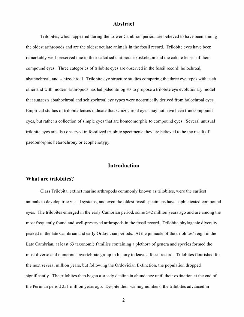
Figure 1. Fossilized Trilobite photograph (left) and artist’s interpretation of trilobite while alive (right). <http://www.trilobites.info/trilobite.htm >
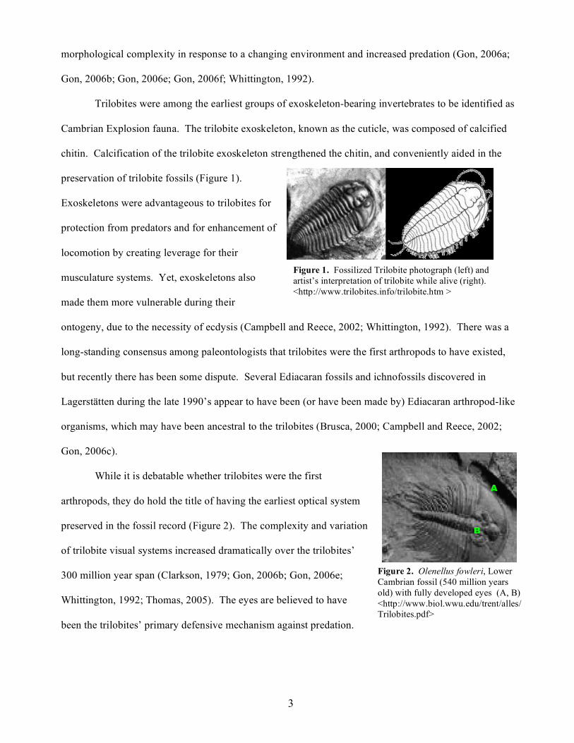
Figure 2. Olenellus fowleri, Lower Cambrian fossil (540 million years old) with fully developed eyes (A, B) <http://www.biol.wwu.edu/trent/alles/Trilobites.pdf>
A
B
morphological complexity in response to a changing environment and increased predation (Gon, 2006a;
Gon, 2006b; Gon, 2006e; Gon, 2006f; Whittington, 1992).
Trilobites were among the earliest groups of exoskeleton-bearing invertebrates to be identified as
Cambrian Explosion fauna. The trilobite exoskeleton, known as the cuticle, was composed of calcified
chitin. Calcification of the trilobite exoskeleton strengthened the chitin, and conveniently aided in the
preservation of trilobite fossils (Figure 1).
Exoskeletons were advantageous to trilobites for
protection from predators and for enhancement of
locomotion by creating leverage for their
musculature systems. Yet, exoskeletons also
made them more vulnerable during their
ontogeny, due to the necessity of ecdysis (Campbell and Reece, 2002; Whittington, 1992). There was a
long-standing consensus among paleontologists that trilobites were the first arthropods to have existed,
but recently there has been some dispute. Several Ediacaran fossils and ichnofossils discovered in
Lagerstätten during the late 1990’s appear to have been (or have been made by) Ediacaran arthropod-like
organisms, which may have been ancestral to the trilobites (Brusca, 2000; Campbell and Reece, 2002;
Gon, 2006c).
While it is debatable whether trilobites were the first
arthropods, they do hold the title of having the earliest optical system
preserved in the fossil record (Figure 2). The complexity and variation
of trilobite visual systems increased dramatically over the trilobites’
300 million year span (Clarkson, 1979; Gon, 2006b; Gon, 2006e;
Whittington, 1992; Thomas, 2005). The eyes are believed to have
been the trilobites’ primary defensive mechanism against predation.

4
Figure 3. Compound eye with multiple lenses and individual ommatidia (B) underlying each lens (A). <http://www.mbl.edu/animals/Limulus/vision/compound.eye2.jpg>
A
B
Figure 4. Simple eye with single lens (A) and underlying single ocellus (B). <http://biodidac.bio.uottawa.ca/Thumbnails/filedet.htm?File_name=INSE109B&File_type=GIF>
A
B
There are correlations among the evolution of trilobite eye complexity and other defensive mechanisms,
such as enrollment/burrowing behaviors and the development of protective spines (Clarkson, 1979;
Horváth et al., 1997).
General Eye Anatomy
Two basic types of eyes exist in animals: compound and simple. Compound eyes (Figure 3) are
typically found in arthropods, and consist of a visual surface covered in many individual “facet-like”
cylindrical units, known as ommatidia. Each ommatidium is fixed in position
on the eye and has its own lens and rhabdomeres (photoreceptors). Since the
ommatidia are completely separate units, the animal sees either an overall
composite image of all the visual inputs from each ommatidium, or multiple
images from each of the many ommatidia. Image resolution is generally poor
in compound eyes; however, they do provide a wide viewing field and have
the ability to easily detect minor movements (Campbell and Reece, 2002;
Clarkson, 1979; Thomas, 2005).
Simple eyes (Figure 4) have a single ocellus, or ocellar unit, which is
similar to the ommatidium, though the unit is not fixed in position. The ocellus has a single lens
underlain by a single retina containing photoreceptors. Retinal eyes have better
overall image resolution than compound eyes due to lens movement that allows
image focusing (Figure 5). Despite the animal’s ability to move the eyes, the
single lens causes the field of vision to be limited, particularly in the periphery,
unlike in compound eyes, which afford a view of the entire visual field
(Campbell and Reece, 2002; Clarkson, 1979; Thomas, 2005).

5
Figure 6. Trilobite ecdysis process. <http://www.trilobites.info/sutures.htm>
Trilobite Eye Anatomy and Models of Trilobite Eyes
Trilobites had complex, multi-lensed eyes. The visual surfaces and lenses were made of calcite,
and as with the rest of their chitinous/calcitic cuticle, re-grew following every molt (Figure 6). Ecdysis
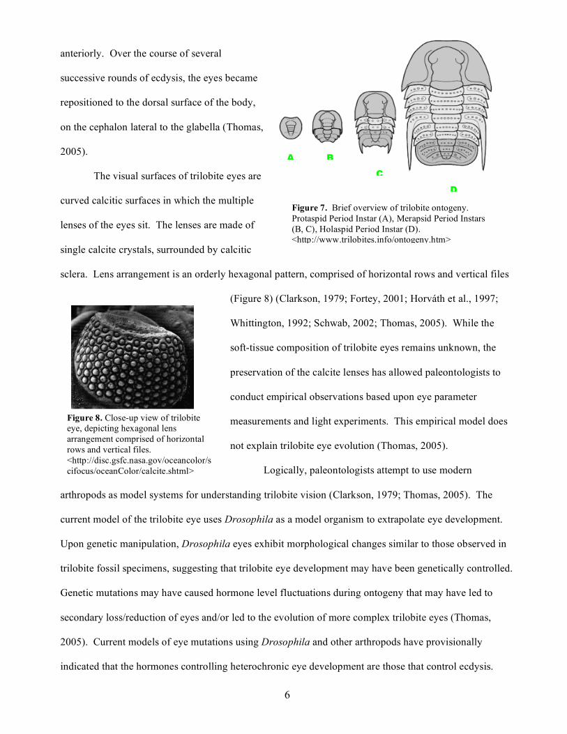
occurred throughout trilobite ontogeny (Figure 7), which consisted of three basic phases: the protaspid
period (larval stages without segmentation), the merapsid
period (larval stages marked by thoracic segmentation), and
the holaspid period (stages in which the trilobites had gained
their adult segmentation, but continued to molt as they grew
in overall body size) (Gon, 2006d;(Whittington, 1992). In
fossilized protaspid stage trilobites, the eyes were located
Compound Eye
Simple Eye
Figure 5. Comparison of compound eye and simple eye image resolution. <http://www.kcl.ac.uk/ip/christerhogstrand/courses/hb0223/images/E-R_7-42.jpg>
Compound Eye
Simple Eye
6
Figure 7. Brief overview of trilobite ontogeny. Protaspid Period Instar (A), Merapsid Period Instars (B, C), Holaspid Period Instar (D). <http://www.trilobites.info/ontogeny.htm>
A B
C
D
anteriorly. Over the course of several
successive rounds of ecdysis, the eyes became
repositioned to the dorsal surface of the body,
on the cephalon lateral to the glabella (Thomas,
2005).
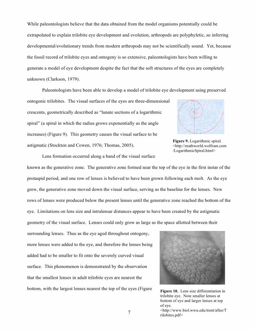
The visual surfaces of trilobite eyes are
curved calcitic surfaces in which the multiple
lenses of the eyes sit. The lenses are made of
single calcite crystals, surrounded by calcitic
sclera. Lens arrangement is an orderly hexagonal pattern, comprised of horizontal rows and vertical files
(Figure 8) (Clarkson, 1979; Fortey, 2001; Horváth et al., 1997;
Whittington, 1992; Schwab, 2002; Thomas, 2005). While the
soft-tissue composition of trilobite eyes remains unknown, the
preservation of the calcite lenses has allowed paleontologists to
conduct empirical observations based upon eye parameter
measurements and light experiments. This empirical model does
not explain trilobite eye evolution (Thomas, 2005).
Logically, paleontologists attempt to use modern
arthropods as model systems for understanding trilobite vision (Clarkson, 1979; Thomas, 2005). The
current model of the trilobite eye uses Drosophila as a model organism to extrapolate eye development.
Upon genetic manipulation, Drosophila eyes exhibit morphological changes similar to those observed in
trilobite fossil specimens, suggesting that trilobite eye development may have been genetically controlled.
Genetic mutations may have caused hormone level fluctuations during ontogeny that may have led to
secondary loss/reduction of eyes and/or led to the evolution of more complex trilobite eyes (Thomas,
2005). Current models of eye mutations using Drosophila and other arthropods have provisionally
indicated that the hormones controlling heterochronic eye development are those that control ecdysis.
Figure 8. Close-up view of trilobite eye, depicting hexagonal lens arrangement comprised of horizontal rows and vertical files. <http://disc.gsfc.nasa.gov/oceancolor/scifocus/oceanColor/calcite.shtml>
7
Figure 9. Logarithmic spiral. <http://mathworld.wolfram.com/LogarithmicSpiral.html>
While paleontologists believe that the data obtained from the model organisms potentially could be
extrapolated to explain trilobite eye development and evolution, arthropods are polyphyletic, so inferring
developmental/evolutionary trends from modern arthropods may not be scientifically sound. Yet, because
the fossil record of trilobite eyes and ontogeny is so extensive, paleontologists have been willing to
generate a model of eye development despite the fact that the soft structures of the eyes are completely
unknown (Clarkson, 1979).
Paleontologists have been able to develop a model of trilobite eye development using preserved
ontogenic trilobites. The visual surfaces of the eyes are three-dimensional
crescents, geometrically described as “lunate sections of a logarithmic
spiral” (a spiral in which the radius grows exponentially as the angle
increases) (Figure 9). This geometry causes the visual surface to be
astigmatic (Stockton and Cowen, 1976; Thomas, 2005).
Lens formation occurred along a band of the visual surface
known as the generative zone. The generative zone formed near the top of the eye in the first instar of the
protaspid period, and one row of lenses is believed to have been grown following each molt. As the eye
grew, the generative zone moved down the visual surface, serving as the baseline for the lenses. New
rows of lenses were produced below the present lenses until the generative zone reached the bottom of the
eye. Limitations on lens size and intralensar distances appear to have been created by the astigmatic
geometry of the visual surface. Lenses could only grow as large as the space allotted between their
surrounding lenses. Thus as the eye aged throughout ontogeny,
more lenses were added to the eye, and therefore the lenses being
added had to be smaller to fit onto the severely curved visual
surface. This phenomenon is demonstrated by the observation
that the smallest lenses in adult trilobite eyes are nearest the
bottom, with the largest lenses nearest the top of the eyes (Figure Figure 10. Lens size differentiation in trilobite eye. Note smaller lenses at bottom of eye and larger lenses at top of eye. <http://www.biol.wwu.edu/trent/alles/Trilobites.pdf>
8
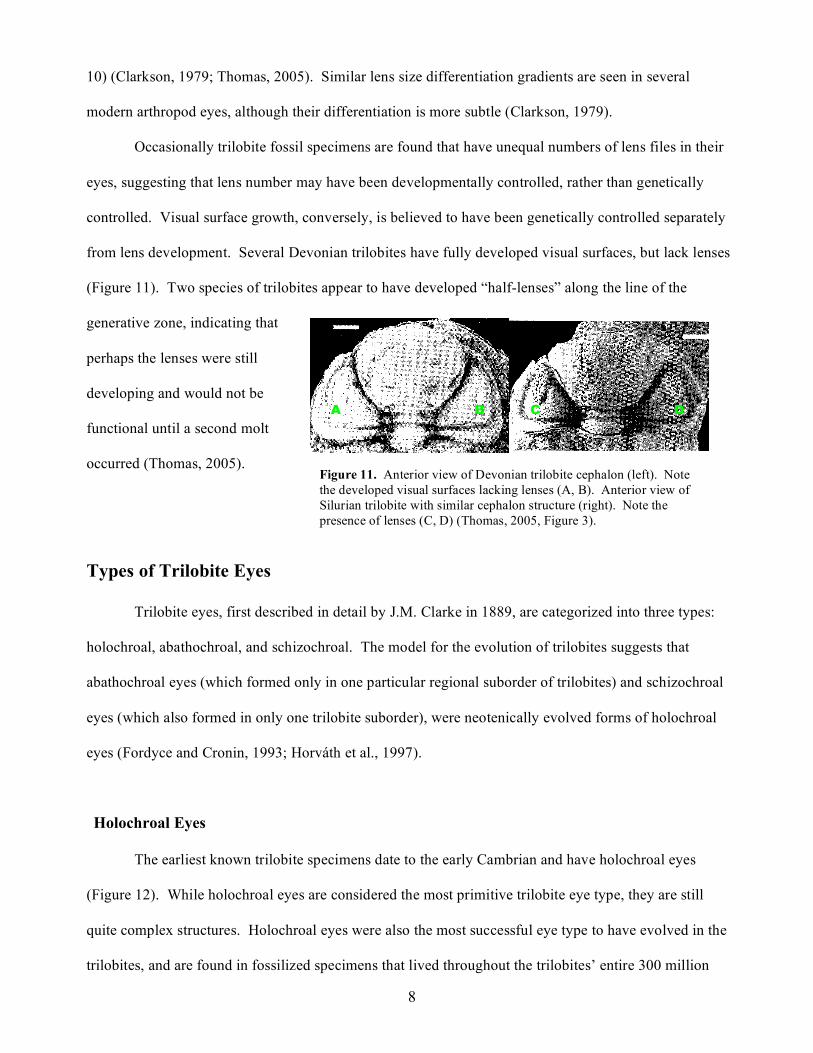
Figure 11. Anterior view of Devonian trilobite cephalon (left). Note the developed visual surfaces lacking lenses (A, B). Anterior view of Silurian trilobite with similar cephalon structure (right). Note the presence of lenses (C, D) (Thomas, 2005, Figure 3).
B A D C
10) (Clarkson, 1979; Thomas, 2005). Similar lens size differentiation gradients are seen in several
modern arthropod eyes, although their differentiation is more subtle (Clarkson, 1979).
Occasionally trilobite fossil specimens are found that have unequal numbers of lens files in their
eyes, suggesting that lens number may have been developmentally controlled, rather than genetically
controlled. Visual surface growth, conversely, is believed to have been genetically controlled separately
from lens development. Several Devonian trilobites have fully developed visual surfaces, but lack lenses
(Figure 11). Two species of trilobites appear to have developed “half-lenses” along the line of the
generative zone, indicating that
perhaps the lenses were still
developing and would not be
functional until a second molt
occurred (Thomas, 2005).
Types of Trilobite Eyes
Trilobite eyes, first described in detail by J.M. Clarke in 1889, are categorized into three types:
holochroal, abathochroal, and schizochroal. The model for the evolution of trilobites suggests that
abathochroal eyes (which formed only in one particular regional suborder of trilobites) and schizochroal
eyes (which also formed in only one trilobite suborder), were neotenically evolved forms of holochroal
eyes (Fordyce and Cronin, 1993; Horváth et al., 1997).
Holochroal Eyes
The earliest known trilobite specimens date to the early Cambrian and have holochroal eyes
(Figure 12). While holochroal eyes are considered the most primitive trilobite eye type, they are still
quite complex structures. Holochroal eyes were also the most successful eye type to have evolved in the
trilobites, and are found in fossilized specimens that lived throughout the trilobites’ entire 300 million
9
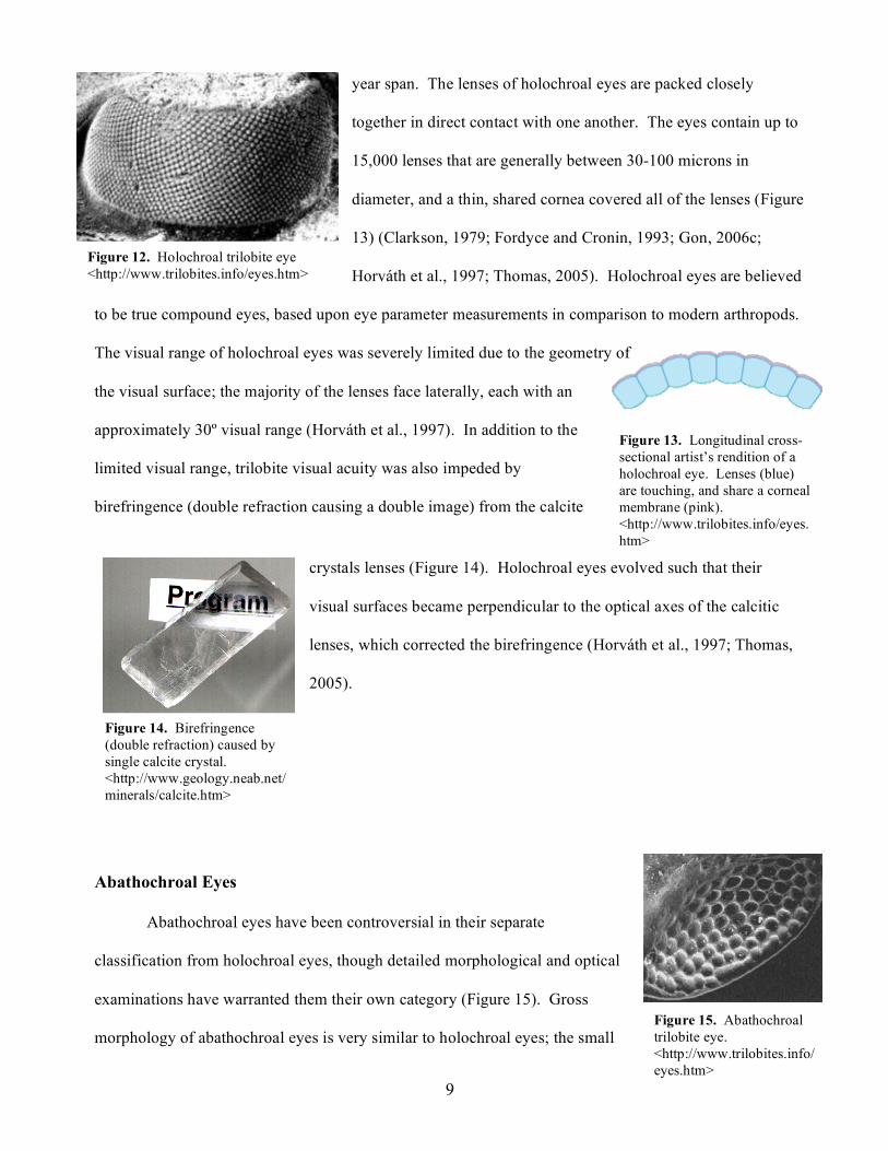
Figure 12. Holochroal trilobite eye <http://www.trilobites.info/eyes.htm>
Figure 13. Longitudinal cross-sectional artist’s rendition of a holochroal eye. Lenses (blue) are touching, and share a corneal membrane (pink). <http://www.trilobites.info/eyes.htm>
Figure 14. Birefringence (double refraction) caused by single calcite crystal. <http://www.geology.neab.net/minerals/calcite.htm>
Figure 15. Abathochroal trilobite eye. <http://www.trilobites.info/eyes.htm>
year span. The lenses of holochroal eyes are packed closely
together in direct contact with one another. The eyes contain up to
15,000 lenses that are generally between 30-100 microns in
diameter, and a thin, shared cornea covered all of the lenses (Figure
13) (Clarkson, 1979; Fordyce and Cronin, 1993; Gon, 2006c;
Horváth et al., 1997; Thomas, 2005). Holochroal eyes are believed
to be true compound eyes, based upon eye parameter measurements in comparison to modern arthropods.
The visual range of holochroal eyes was severely limited due to the geometry of
the visual surface; the majority of the lenses face laterally, each with an
approximately 30º visual range (Horváth et al., 1997). In addition to the
limited visual range, trilobite visual acuity was also impeded by
birefringence (double refraction causing a double image) from the calcite
crystals lenses (Figure 14). Holochroal eyes evolved such that their
visual surfaces became perpendicular to the optical axes of the calcitic
lenses, which corrected the birefringence (Horváth et al., 1997; Thomas,
2005).
Abathochroal Eyes
Abathochroal eyes have been controversial in their separate
classification from holochroal eyes, though detailed morphological and optical
examinations have warranted them their own category (Figure 15). Gross
morphology of abathochroal eyes is very similar to holochroal eyes; the small
Figure 14. Birefringence (double refraction) caused by single calcite crystal. <http://www.geology.neab.net/minerals/calcite.htm>
Figure 14. Birefringence (double refraction) caused by single calcite crystal. <http://www.geology.neab.net/minerals/calcite.htm>
10
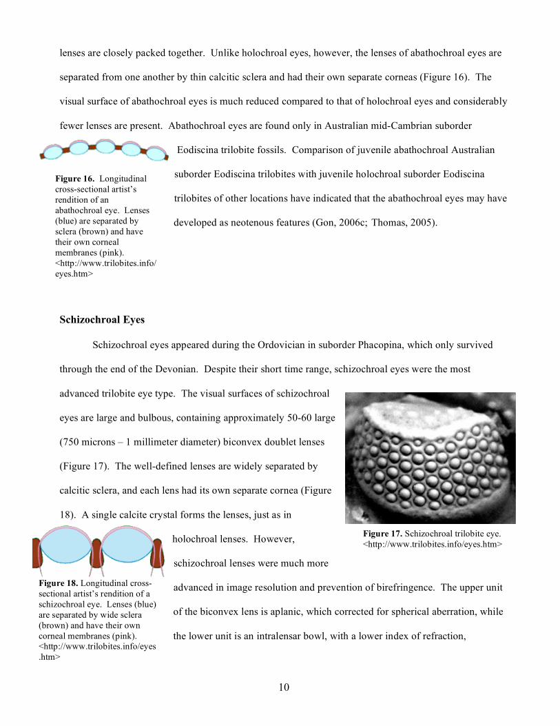
Figure 16. Longitudinal cross-sectional artist’s rendition of an abathochroal eye. Lenses (blue) are separated by sclera (brown) and have their own corneal membranes (pink). <http://www.trilobites.info/eyes.htm>
Figure 17. Schizochroal trilobite eye. <http://www.trilobites.info/eyes.htm>
Figure 18. Longitudinal cross-sectional artist’s rendition of a schizochroal eye. Lenses (blue) are separated by wide sclera (brown) and have their own corneal membranes (pink). <http://www.trilobites.info/eyes.htm>
lenses are closely packed together. Unlike holochroal eyes, however, the lenses of abathochroal eyes are
separated from one another by thin calcitic sclera and had their own separate corneas (Figure 16). The
visual surface of abathochroal eyes is much reduced compared to that of holochroal eyes and considerably
fewer lenses are present. Abathochroal eyes are found only in Australian mid-Cambrian suborder
Eodiscina trilobite fossils. Comparison of juvenile abathochroal Australian
suborder Eodiscina trilobites with juvenile holochroal suborder Eodiscina
trilobites of other locations have indicated that the abathochroal eyes may have
developed as neotenous features (Gon, 2006c;(Thomas, 2005).
Schizochroal Eyes
Schizochroal eyes appeared during the Ordovician in suborder Phacopina, which only survived
through the end of the Devonian. Despite their short time range, schizochroal eyes were the most
advanced trilobite eye type. The visual surfaces of schizochroal
eyes are large and bulbous, containing approximately 50-60 large
(750 microns – 1 millimeter diameter) biconvex doublet lenses
(Figure 17). The well-defined lenses are widely separated by
calcitic sclera, and each lens had its own separate cornea (Figure
18). A single calcite crystal forms the lenses, just as in
holochroal lenses. However,
schizochroal lenses were much more
advanced in image resolution and prevention of birefringence. The upper unit
of the biconvex lens is aplanic, which corrected for spherical aberration, while
the lower unit is an intralensar bowl, with a lower index of refraction,
11
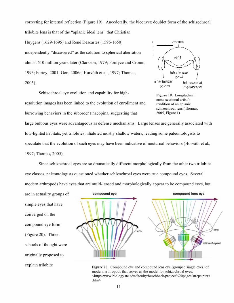
Figure 20. Compound eye and compound lens eye (grouped single eyes) of modern arthropods that serves as the model for schizochroal eyes. <http://www.biology.uc.edu/faculty/buschbeck/project%20pages/strepsiptera.htm>
correcting for internal reflection (Figure 19). Anecdotally, the biconvex doublet form of the schizochroal
trilobite lens is that of the “aplanic ideal lens” that Christian
Huygens (1629-1695) and René Descartes (1596-1650)
independently “discovered” as the solution to spherical aberration
almost 510 million years later (Clarkson, 1979; Fordyce and Cronin,
1993; Fortey, 2001; Gon, 2006c; Horváth et al., 1997; Thomas,
2005).
Schizochroal eye evolution and capability for high-
resolution images has been linked to the evolution of enrollment and
burrowing behaviors in the suborder Phacopina, suggesting that
large bulbous eyes were advantageous as defense mechanisms. Large lenses are generally associated with
low-lighted habitats, yet trilobites inhabited mostly shallow waters, leading some paleontologists to
speculate that the evolution of such eyes may have been indicative of nocturnal behaviors (Horváth et al.,
1997; Thomas, 2005).
Since schizochroal eyes are so dramatically different morphologically from the other two trilobite
eye classes, paleontologists questioned whether schizochroal eyes were true compound eyes. Several
modern arthropods have eyes that are multi-lensed and morphologically appear to be compound eyes, but
are in actuality groups of
simple eyes that have
converged on the
compound eye form
(Figure 20). Three
schools of thought were
originally proposed to
explain trilobite
Figure 19. Longitudinal cross-sectional artist’s rendition of an aplanic schizochroal lens (Thomas, 2005, Figure 1)
12
schizochroal eyes: true compound eyes with ommatidia, agglomerations of simple eyes with retinas, or
something else entirely different from any visual system known in modern arthropods. Eye parameter
measurements of schizochroal eyes suggest that they are not true compound eyes, but are instead
agglomerations of simple eyes. Based upon the angles of refraction created by the doublet lens, it seems
unlikely that schizochroal eyes would have been able to focus on images if they had ommatidia. A
comparison of schizochroal eyes to modern arthropods also supports the schizochroal simple eye model;
modern arthropods with biconvex lenses generally have ocelli rather than ommatidia (Clarkson, 1979;
Fortey, 2001). A recent paper disputes the biconvex doublet character of schizochroal lenses; however,
the study has not been replicated and is considered provisional (Lane et al., 2003; Thomas, 2005).
Assuming the model of the schizochroal eye as an agglomerate of simple retinal eyes is correct, there are
no modern analogs (no modern arthropod has this eye type), therefore making the trilobite schizochroal
eye one of a kind. There are some recent and modern arthropods which possess some aspects of
schizochroal eyes, but never in the same combination observed in trilobites (large separated lenses, ocelli,
and aplanic biconvex doublet character) (Horváth et al., 1997).
All trilobites had stereoscopic vision due to the overlapping visual field created by each eye,
however, schizochroal eyes may have had the capability to be stereoscopic within each eye. Conditions
for stereoscopic vision include lenses with high resolution power, lenses positioned to create overlapping
images, and a neural network sophisticated enough to interpret such overlapping images (Clarkson, 1979;
Stockton and Cowen, 1976; Thomas, 2005). If schizochroal eyes were in fact collections of simple eyes,
the potential for high-resolution power was likely (Clarkson, 1979). Schizochroal eyes are believed to
have had resolving power of an order of magnitude better than modern compound eyes. This has been
equated to the resolution power of frog eyes (Thomas, 2005). The arrangement of lenses on the visual
surface, and the highly convex nature of the lenses themselves are within the parameters needed to create
overlapping images (Fordyce and Cronin, 1993; Thomas, 2005). Despite the empirical evidence
supporting the possibility of stereoscopic vision within the eye, the absence of living trilobite specimens
means that the sophistication of the nervous system remains unknown. Thus, the ability of stereoscopic

13
vision within an individual schizochroal eye will never be determined (Fordyce and Cronin, 1993; Fortey,
2001; Horváth et al., 1997; Stockton and Cowen, 1976; Thomas, 2005).
Trilobite Eye Evolutionary Theories and Models
Paedomorphic heterochrony is the primary theory behind trilobite eye evolution. The chief
evidence for this theory is the observation that larval trilobites with holochroal eyes have large, separated
lenses during the protaspid period. As the holochroal trilobites progressed through their ontogeny, they
generated smaller lenses that were packed more closely in order to fit lenses onto their severely curved
visual surfaces. Thus, paleontologists have introduced the idea that the large and/or separated lenses
observed in abathochroal and schizochroal eyes may be neotenous features (Horváth et al., 1997; Thomas,
2005).
The general model for trilobite eye evolution suggests the following:
1. Holochroal eyes developed; birefringence was corrected by orienting the optical axis
perpendicular to the visual surface and incoming photons.
2. Abathochroal eyes developed; lenses were slightly larger and set further apart, perhaps due to
constraints on habitat lighting. The lenses were still small enough that spherical aberration was not an
overwhelming problem. Birefringence was corrected by orientation of the optical axis as in holochroal
eyes. Many of the suborder Eodiscina trilobites with abathochroal eyes secondarily lost their lenses or
visual surfaces, and this group was a dead-end clade.
3. Schizochroal eyes developed; lenses were biconvex doublets, much larger, much fewer in
number, and were separated by wide sclera. The aplanic lens corrected for the spherical aberrations and
internal reflections caused by a larger calcite lens. The suborder Phacopina was also a dead-end clade
(Horváth et al., 1997).
Both abathochroal and schizochroal eyes may have evolved as the result of a delay in action of
ecdysone (the primary ecdysis hormone that is believed to have controlled lens differentiation). In the
case of the suborder Phacopina, the lenses may have had a longer time to develop between molts, leading
14
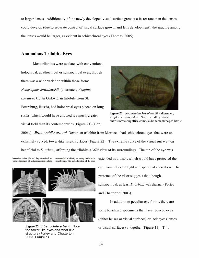
Figure 21. Neoasaphus kowalewskii, (alternately Asaphus kowalewskii). Note the tall eyestalks. <http://www.angelfire.com/ks2/boneman8/page8.html>
to larger lenses. Additionally, if the newly developed visual surface grew at a faster rate than the lenses
could develop (due to separate control of visual surface growth and lens development), the spacing among
the lenses would be larger, as evident in schizochroal eyes (Thomas, 2005).
Anomalous Trilobite Eyes
Most trilobites were oculate, with conventional
holochroal, abathochroal or schizochroal eyes, though
there was a wide variation within those forms.
Neoasaphus kowalewskii, (alternately Asaphus
kowalewskii) an Ordovician trilobite from St.
Petersburg, Russia, had holochroal eyes placed on long
stalks, which would have allowed it a much greater
visual field than its contemporaries (Figure 21) (Gon,
2006c). Erbenochile erbeni, Devonian trilobite from Morocco, had schizochroal eyes that were on
extremely curved, tower-like visual surfaces (Figure 22). The extreme curve of the visual surface was
beneficial to E. erbeni, affording the trilobite a 360º view of its surroundings. The top of the eye was
extended as a visor, which would have protected the
eye from deflected light and spherical aberration. The
presence of the visor suggests that though
schizochroal, at least E. erbeni was diurnal (Fortey
and Chatterton, 2003).
In addition to peculiar eye forms, there are
some fossilized specimens that have reduced eyes
(either lenses or visual surfaces) or lack eyes (lenses
or visual surfaces) altogether (Figure 11). This Figure 22. Erbenochile erbeni. Note the tower-like eyes and visor-like structure (Fortey and Chatterton, 2003, Figure 1).
15
secondary blindness appears to have been ecophenotypic. Unlike Drosophila in which secondary
blindness can be genetically created, secondarily blind trilobites appear in successive generations of
multiple clades, suggesting that the reduction of the eyes was due to environmental constraints rather than
a particular genetic mutation. The order Agnostida (excluding the Cambrian Suborder Eodiscina) in
particular is primarily eyeless (lacking visual surfaces). Such secondary loss of eyes in order Agnostida is
thought to have occurred as an adaptation to living infaunally below the photic zone (Clarkson, 1979;
Horváth et al., 1997; Thomas, 2005).
Conclusion
Trilobites are among the most recognizable fossils in the United States and have a tremendous
evolutionary importance as the first animals with true eyes. Particularly remarkable is the complexity of
their eyes, despite the fact that they lived more than 540 million years ago. Paleontological studies of
trilobite eyes may never be completed, and due to the lack of soft tissue preservation, the visual system
will never be totally understood. Yet, using the trilobites’ preserved calcitic lenses. paleontologists have
been able to uncover many of the evolutionary and ontogenic trends of the oldest visual system in history.

16
References Cited
Brusca, R. C., 2000, Unraveling the history of arthropod biodiversification: Ann. Missouri Bot. Gard., v. 87, p. 13-25. Campbell, N. A., and Reece, J. B., 2002, Biology: San Francisco, Benjamin Cummings, 1247 p. Clarkson, E. N. K., 1979, The visual system of trilobites: Palaeontology, v. 22, p. 1-22. Fordyce, D., and Cronin, T. W., 1993, Trilobite vision: A comparison of schizochroal and holochroal eyes with the compound eyes of modern arthropods: Paleobiology, v. 19, no. 3, p. 288-303. Fortey, R. A., 2001, Trilobite systematics: the last 75 years: Journal of Paleontology, v. 75, no. 6, p. 1141-1151. Fortey, R. A., and Chatterton, B., 2003, A Devonian trilobite with an eyeshade: Science, v. 301, p. 1689. Gon, S.M. III, 2006a, A Chart of Geological Time (from a trilobite's point of view) <http://www.trilobites.info/geotime.htm>. Accessed 11 November 2006. Gon, S.M. III, 2006b, Origins of Trilobites < http://www.trilobites.info/origins.htm>. Accessed 11 November 2006. Gon, S.M. III, 2006c, The Trilobite Eye <http://www.trilobites.info/eyes.htm>. Accessed 11 November 2006. Gon, S.M. III, 2006d, Trilobite Reproduction and Development <http://www.trilobites.info/ontogeny.htm>. Accessed 11 November 2006. Gon, S.M. III, 2006e, Trilobite Systematic Relationships <http://www.trilobites.info/triloclass.htm#orders>. Accessed 11 November 2006. Gon, S.M. III, 2006f, What are Trilobites? <http://www.trilobites.info/trilobite.htm>. Accessed 11 November 2006. Horváth, G., Clarkson, E. N. K., and Pix, W., 1997, Survey of modern counterparts of schizochroal trilobite eyes: structural and functional similarities and differences: Historical Biology, v. 12, p. 229-263. Lane, P. D., Siveter, D. J., and Fortey, R. A., 2003, Special papers in Palaeontology: trilobites and their relatives: London, The Palaeontological Association, p. 397. Schwab, I. R., 2002, The eyes have it: Br. J. Opthalmol, v. 86, p. 372. Stockton, W. L., and Cowen, R., 1976, Stereoscopic vision in one eye: paleophysiology of the schizochroal eye of trilobites: Paleobiology, v. 2, p. 304-315. Thomas, A. T., 2005, Developmental palaeobiology of trilobite eyes and its evolutionary significance: Earth-Science Reviews, v. 71, p. 77-93. Whittington, H.B. 1992, Trilobites: Rochester, NY, The Boydell Press, 160 p.