Embed Size (px)
Citation preview

My Experience in the final part exam of FRCS Glasgow,
Ophthalmology
Eslam ElkohlyFRCS, 2017

I am Eslam Elkohly, Ophthalmology senior registrar. Thanks to Allah, I have passed the final part exam of FRCS Glasgow (FRCS 3) in Amman, Jordan October 30 – November 2, 2017.
I’m so grateful to my family and teachers. Thanks very much my father/mother for blessings, my wife/sons (Mohamed/Ahmed) for patience and my teachers/colleagues for support.
Thanks my colleagues and previous candidates who greatly affected me and made a big difference in my way to FRCS 3 exam specially Dr. Ahmed Ali Megahed, Dr. Emad Alaaedin, Dr. Mohamed Badie, Dr. Ahmed Alsherbeny, Dr. Mohamed Hatab Dr. Mohamed Magdy, Dr. Ahmed Adly and Dr. Osama Rass.

Thanks my Mentors
Prof/ Gehad Elnahri Prof/ Tarek Shaarawy
Prof/ Ahmed Samir Dr/ Mohamed Raslan
Prof/ M El Kafrawy
Prof/ Hussein Swelem

My preparation: Practice Practice Practice
The principle oneGood Clinical approach
Was the principle on in the second part exam and needed here for some tables
Thanks Dr. Abdelsattar Farrag, Dr. Mahmoud Rabie, Dr. Sameh Galal, DrOmar Abdelghany and Dr Ashraf Shaat, the members of e_learnersacademy for the critical point they added to me and to my colleagues which is the very valuable course organized by the academy.
Chua web page: very important specially past candidate experience.
Eyerounds: For me it was very important and was really helpful as it is a clinical exam so you will find real cases with full approach, atlas, videos and differential diagnosis of each and this is so important in the final exam.

• Special thanks also to Dr. Muthusamy virtual university.
• This connection was the first stone in my FRCS building as there was a great patience regarding correction of the mistakes of solving the scenarios and sending emails with interesting cases and motivating sayings and continued till the end.
• Very important book • I took some points in general medicine
from this book• VIVA scenarios in the beginning of
each section are very important.

The exam center King Hussein medical city, Amman, Jordan.
• Well equipped/organized (I took 2 pen torches, fixation target, 2 rulers, occluder and white/red targets)
• Try to reach there early (3 days before the exam)
• Group discussion is very important and is more important in the off day between VIVA and clinical exams during which you have to practice all possible relevant tests with your colleagues. I consider it a corner stone in the success.
• Try to be in the exam center 2 hours or more before the exam, keep calm and get ready.

My advises:
• During preparation for the exam, don’t depend on one source of photos like Kanski book but google and check varieties of case presentations, that’s why I used variable sources as eyerounds.org
• During the exam: take time looking at the photo then start to comment, describe all what you see in a systematic manner as you trained yourself before, don’t panic if you don’t know the case and just describe, the examiner will mostly help.
• Some photos are very unclear, Guess & Describe

Posterior Segment Session

• Describe! Part of fundus photo, hazy view, line between attached and slightly elevated retina with reddish dot (she said it is tear! and patient is complaining of floaters and photopsia!!)
• Diagnosis? Rhegmatogenous RD
• Management? History, examination, ….
• Treatment options? I cannot take decision from this photo as it is part of the fundus. She said: tell me all options you know. I gave all conditions with appropriate option for each. She said what are advantages of PPV, answered.
First Examiner

• Describe! Exactly same like this photo with subretinalexudates along upper and lower temporal arcades with some telangiectatic vessels.
• Your impression? Coats disease
• Management? History, examination, investigations
• Treatment options? Counsel, Laser, ..
• Where in this case will you fire your laser?

• Describe! Peripapillaryatrophy, tilted disc, I cannot see cup/rim, situs inversus.
• Diagnosis? Tilted disc. He said what else?? I said, there is some yellowish material, could be OD drusen.
• What are the early sings of POAG?! I said “is that OD cupping? Very unclear” > I told him early signs of POAG.
• How to treat it surgically?
Second Examiner

• Describe!• Impression? Rubeosis iridis
for DD.• Causes?• Management? History,
examination (IOP, gonio, fundus), investigations (US if associated vitreous hge)
• Treatment? Counsel, Topical: antiglaucoma, cyclo, steroids + urgent PRP if view is OK +adjuvant anti-VEGF
• If no view? If vitreous hge, there is visual potential > PPV + endolaser.
• If IOP not controlled by medications? Laser cycloablation

• Describe!
• Impression? RP
• Management? History, examination, investigations
• Ocular associations?
• Systemic associations?
• Medical treatment?Phytanic aicd free diet in Refsum, Vitamin A/E in abeta lipoprotienemia, acetazolamide in CME.
• Surgical? Cataract Sx, if associated keratoconus or glaucoma.


General medicine: English examiner
45 year old with limited abduction followed by limited adduction 2 days later.
• DD? Paralytic (CN plasies, supranuclear palsies, …), myogenic (as MG, myopathies, …), restrictive (as TED, …)
• OK, He is MG. The patient presented to you later with dyspnoea, distress. Dx? Myasthenic crisis. Mechanism? Respiratory failure ….
• Asked about details of diagnosis of MG like ice pack test, Tensilon test, EMG, ….

5 year old child presented in your clinic with respiratory distress after swallowing a pea nut.
• Dx? Could be air way obstruction or anaphylactic shock.
• Management? Call for help, ABC, clinical signs of anaphylaxis in the form of … (tachycardia/hypotension), ….
• Asked about treatment of anaphylactic shock with doses
• If just air way obstruction? heimlich manoeuvre
• What the advise you will give to the mother? Not to give the child Pea nut again

55 year-old patient presented with palpitation in your clinic.
• Manifestations suggesting AF? Irregularly irregular pulse, apical pulse > radial pulse, ..
• Causes of AF? Idiopathic, cardiac, non-cardiac.
• Treatment? Stepwise (Digoxin > beta blockers > Amiodarone/DC shock)
• What else important? Anticoagulants.

Neuro-ophthalmology, 2nd Examiner
36 year old patient presented after motor car accident with unequal pupil size.
• DD? Could be: dilated pupil is the abnormal (traumatic mydriasis, surgical 3rd CN palsy) or the small pupil is the abnormal one (horner syndrome)
• Detailed discussion about horner syndrome:causes, manifestations, investigations, pharmacological tests, … all possible details.

• The examiner gave me Humphrey visual field: he put the field of the left eye in my Rt hand. I rearranged it and started interpreting: which eye, personal data of the patient, strategy, reliability indices, grey scale, MD, PSD.
• Dx? Bitemporal superior quadrantanopia.• Causes? Chiasmal lesion: neoplastic (pituitary adenoma,
…), vascular (aneurysm, ..), trauma, inflammatory (MS, sarcoidosis, ..)
• Asked about pituitary adenoma in details: history (++ ICP, hromonal manifestations, ..), hormones you will investigate for, investigations, …

• Describe! Burred disc margin, yellowish material on the surface of Optic disc.
• Dx? Pseudopapilledema, OD drusen.
• Why do you say pseudo? Blood vessels are not obscured, no hge/CWSs, …
• Causes of visual loss? Vascular occlusions, CNV, angioid streaks with choroidal rupture.
• Do you see angioid streaks? NO
• Where do you look for it? Lines radiating from the optic disc area
• Systemic associations of angioidstreaks? PEPSI


• Again the same photo!!!• Describe!• Impression? Rubeosis iridis for DD.• Causes?• He is diabetic patient. What if the
other eye normal? Asymmetric diabetic retinopathy although will not be asymmetric to that extent, CRVO, …
• Management? History, examination (IOP, gonio, fundus), investigations (US if associated vitreous hge)
• Treatment? Counsel, Topical: antiglaucoma, cyclo, steroids + urgent PRP if view is OK + adjuvant anti-VEGF
• If IOP not controlled by medications? Laser cycloablation
First Examiner

• Describe!
• DD? HSV keratitis, HZV, acanthamoeba, healing corneal abrasion.
• Treatment of herpetic keratitis? Aciclovir 3 % oint, 5x/day/14 days.
• Alternatives to aciclovir?Trifluridine, Ganciclovir.
• Treatment of Disciformkeratitis?

• Describe!• DD? Infectious keratitis for DD. Could be
bacterial, fungal or acanthamoeba, ..• If bacterial, causative agents?• Management? History (risk factors as trauma, CL,
…), examination/documentation of all signs with size and other criteria, admission, scraping, culture/sensitivity, fortified antibiotics hourly in the form of … + cyclo, systemic antibiotics (do not mention steroids under any condition even if in the healing phase)
• If 48 hrs later, still the same? Stop treatment, re-scraping, widen the staining and culture media, …
• If perforation? Corneal glue if small (< 1 mm), tectonic graft is larger.
• If it is fungal keratitis: what are staining/culture media, treatment options … (counsel patient about 6-8 weeks of treatment).
• If it is acanthamoeba: culture media, treatment options … (counsel the patient about long duration of treatment - months).

The second examiner
1 year old boy, mother complaining of watery eye.
• Management? My 1st priority will be toward excluding congenital glaucoma. then it could be obstruction at any level of lacrimal excretory system.
• Asked about details of congenital glaucoma:diagnosis, follow ups, treatment options
• How to suspect NLD obstruction?
• Treatment options? Massage > probing +intubation

• Describe!• Dx? Giant papillary conjunctivitis.• Causes? Chronic irritation (chronic CL use,
protruding corneal suture, buckle, prosthesis), Allergy (VKC, AKC), floppy eyelid.
• Possible associations? KC, …• Treatment? Frequent lubricants, stop CL use,
treat the cause, topical steroids if associated inflammation/corneal vascularization.
• If the patient high myope and cannot live without CL? I will counsel her about using glasses.
• If she does not like glasses? I will counsel her about glasses and limited CL use during the work time.
• Other options? I will refer to specialist of refractive Sx to discuss with her the available options if possible.

• Describe!
• Dx? Peripheral ulcerative keratitis for DD. Could be PUK with systemic cause as RA, SLE, .. Or ocular as Mooren ulcer or infectious keratitis.
• What is Mooren ulcer? Diagnosis of exclusion, ..
• How to treat mooren ulcer?

• Thanks to Allah. I was very stressed before the exam but relieved after finishing the day and was satisfied.
• My performance in the viva exam kept me calm to some extent and was a very strong pushing power and motivated me to prepare very well to the clinical exam.
• Me and my colleagues spent the 1 ½ day practicing all relevant tests on each other in a proper way and many many times till we were doing them by the subconscious and this is so important not to let the examiner think you are doing the test for the first time. That helped me a lot beside my way of thinking as if I’m inside the exam in my clinic for the last 6 months before the exam “Live the FRCS”

My advises:
• Practice as much as you can in your clinic, deal with every case in your clinic as an exam case.
• Do all tests in your clinic as many as you can and explain what you see and process all to get the case.
• Keep calm during the exam and try to have an eye on the examiner as he mostly will help you.
• Any problem with any case, forget about it and concentrate in the next not to lose the whole exam.


Old man sitting on the chair of the slit lamp. • Examiner switched off the light and said: Examine
the pupil! I said I want to observe first in light. He witched on the light. I said there is anisocoria in light. Please switch off the light to observe in dark. I said it is nearly the same difference as the smaller pupil was having sluggish reaction. OK, did you finish pupil examination? I said no I will test for direct/consensual pupil reaction. I said the larger one does not react at all. He said well, your DD? I said my priority is to exclude 3rd CN palsy (the patient had bilateral severe involutional ptosis). He said OK do. I said there is severe ptosis but bilateral. Do you want me to measure, he said no. I tested ocular motility and found it full. He said: then?? I said now it could be Adies pupil and I want to test near reflex, he said normal, I said must now pharmacologically dilated pupil, he said yes it is. (my opinion was to exclude dangerous causes first)
• OK look at the optic disc in the dilated eye by 90 D lens! I said optic disc swelling, splinter hge.
• Your DD? Optic neuritis, AION, compressive lesion, ….
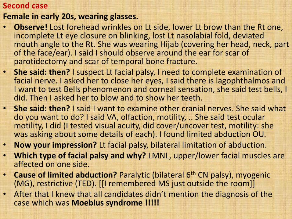
Second caseFemale in early 20s, wearing glasses.• Observe! Lost forehead wrinkles on Lt side, lower Lt brow than the Rt one,
incomplete Lt eye closure on blinking, lost Lt nasolabial fold, deviated mouth angle to the Rt. She was wearing Hijab (covering her head, neck, part of the face/ear). I said I should observe around the ear for scar of parotidectomy and scar of temporal bone fracture.
• She said: then? I suspect Lt facial palsy, I need to complete examination of facial nerve. I asked her to close her eyes, I said there is lagophthalmos and I want to test Bells phenomenon and corneal sensation, she said test bells, I did. Then I asked her to blow and to show her teeth.
• She said: then? I said I want to examine other cranial nerves. She said what do you want to do? I said VA, olfaction, motility, .. She said test ocular motility, I did (I tested visual acuity, did cover/uncover test, motility: she was asking about some details of each). I found limited abduction OU.
• Now your impression? Lt facial palsy, bilateral limitation of abduction.• Which type of facial palsy and why? LMNL, upper/lower facial muscles are
affected on one side.• Cause of limited abduction? Paralytic (bilateral 6th CN palsy), myogenic
(MG), restrictive (TED). [[I remembered MS just outside the room]]• After that I knew that all candidates didn’t mention the diagnosis of the
case which was Moebius syndrome !!!!!


Old man in 80s • Observe! The patient is taking chin up position, bilateral complete
ptosis, bandage around his head and plaster on the forehead may be due to Sx or trauma. I went closer and said the Lt eyelid is swollen, hyperemic, yellowish discharge. I said I need to elevate this lid to see what is behind! She said do. I said there is proptosis, congested eye, discharge, very hazy cornea, dilated pupil. She said what is your impression? My priority will be to exclude orbital cellulitis, however it could be ….., So I need to test motility, she said no (after sometime agreed), I found the Lt eye frozen, no movement.
• Ok, your impression now? Still I need to exclude orbital cellulitis, but it also could be IOID, tolosa hunt syndrome, cavernous sinus thrombosis, orbital apex syndrome, orbital tumour, …..
• Could you explain ptosis in the Rt eye? I said it could be traumatic or involutional.
• She did not look satisfied by my DD and wanted a final diagnosis but it was a complicated case and after that I knew that no one gave a diagnosis as this man underwent many neurosurgeries. After my session they removed him from room although some cases continued for more than a session !!!!

About 20 year old female• Observe! Apparent protrusion of both eyes with inferior scleral show. I need
to confirm proptosis. I looked from sides and from behind and said no decompression scars. I need to measure for proptosis, the patient replied:please do : I measured, found one eye about 24 mm, the other 22 mm
• Do you want me to measure for dystopia? He said do. I measured and said no horizontal/vertical dystopia.
• Then? I said ocular motility, he said do. I tested the vision, did cover/uncover and said no tropia or phoria, asked about diplopia in primary position, did motility test and said there is limited abduction in both eyes, and demonstrated signs of TED and found lid lag and said +ve Von Graefesign and tested convergence and said –ve mobius sign.
• Then? I said I need to test protective measures as bells phenomenon, corneal sensation and to test pupil to exclude compressive optic neuropathy and see her on slit lamp and test the cornea to exclude exposure keratopathy. Hes said no all are OK
• She is concerned about her look? I will refer to endocrinologist to control thyroid. He said it is OK. I said I didn’t find any tropia or phoria and she is not complaining of diplopia, so no place here for prisms.
• She need cosmetic solution? I said so I will refer her to oculoplastic surgeon to counsel her about decompression.
[I was stressed from the first case but Thanks to Allah I did well in this case and forgot the first case temporarily]. الحمد هلل


Old man on slit lamp.
• Observe before slit lamp exam! Both eyes are congested. He said anything in the lashes? I said the room is dark and I want to see on SL.
• Punctal stenosis, upper lid notching with cut lashes rubbing the cornea, inferior symblepharon, shortened fornices, conjunctival congestion, corneal opacification/vascularization, patient is pseudophakic. Same findings in the other eye.
• Cause of lid notching? Electrolysis, cryo or Sx.
• DD? Cicatrizing conjunctivitis: …..
• Well this patient had chemical injury, how to deal with the chemical burn? I will not take history from the start but immediately will do irrigation. Then asked about some details and possibilities

Male, about 18 year old.
• Observe before slit lamp exam! I said he is wearing high myopic glasses. Then asked the patient to remove his glasses and said what else? I said the cornea is large and this patient might have congenital glaucoma.
• Ok look on slit lamp! OD: Large cornea, Haabs striae, 2 tubes in the AC, I double-everted upper/lower lids and found both shunts working. OS: Haabs striae, PI, iris pigments on the lens surface, bleb which is partially encysted.
• How to counsel a patient with congenital glaucoma?Blinding disease, Treatment options, compliance, follow ups, Sx, …
• Inheritance, parameters you look for during the examination, ….
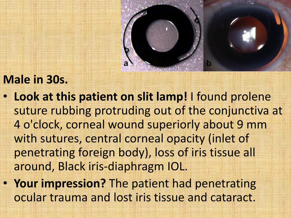
Male in 30s.
• Look at this patient on slit lamp! I found prolenesuture rubbing protruding out of the conjunctiva at 4 o'clock, corneal wound superiorly about 9 mm with sutures, central corneal opacity (inlet of penetrating foreign body), loss of iris tissue all around, Black iris-diaphragm IOL.
• Your impression? The patient had penetrating ocular trauma and lost iris tissue and cataract.


Middle aged man • Examine the fundus with 90 D lens!
Blackish irregular lines radiating from the optic disc giving the appearance of angioidstreaks and there is macular scar taking most of the macula. Rt eye the same but the scar is taking ½ of the fovea.
• Impression? Angioid streaks with macular scar OS > OD, small yellowish dot inferotemporal to the fovea in the Rt eye.
• What do you expect the vision in the Lt eye? Poor.
• Causes of macular scar? Idiopathic, trauma with choroidal rupture, CNV.
• What will you do for him? After Hx, examination, I will not do anything for the Lt eye. Rt eye: I will record the vision, give glasses, do FFA to see if this yellowish dot represents any activity of CNV. The examiner did not like doing FFA as there is no strong clinical evidence to suspect activity and I agreed with him.

17 year-old female• Examine the macula in both eyes with 90 D
lens!• Impression? Stargardt disease.• Details about stargardt: inheritance (AR),
stages in fundus appearance, investigations, management (very important to tell genetic counseling, glasses, low vision aids, support groups, …)
• Then again in details: how will you counsel your patient, will you tell him you are going to be blind, is it a blinding disease, any good news you will tell the patient about, ….
• We had long time and there were many cases in the room and I liked to have a 3rd
case but he seemed he didn’t want and for the whole day they did not give a 3rd case.

Results • Thanks to Allah, the results
released on Thursday November 30, 2017 and found my number the first in the list. Was a great moment when I found my dream came true.
• I hope all of that will be for the benefit of my patients as the real exam is managing your patient properly.


![Doctors Academydoctorsacademy.org/Course/TCATLevel2/downloads/... · 1610 - 1810 Individual viva [along with refreshments and feedback] ... - FRCS (General Surgery) Exit Exam - Cadaveric](https://img.pdfslide.us/doc/110x75/5ed541aaa8496c11e9719109/doctors-a-1610-1810-individual-viva-along-with-refreshments-and-feedback-.jpg)