Embed Size (px)
Citation preview

Validation of the Microbial-Vac System for Use in Food
Safety Monitoring Activities within Beef Abattoirs
J. G. Maughan*, K. J. Church, K. M. Tesar, A. S. Vos, and W. D. Carlsen
Microbial-Vac Systems®, Inc., 14621 S 800 W #100, Bluffdale, UT 84065
2
Table of Contents Abstract ...............................................................................................................3
Introduction.........................................................................................................5
Objective..............................................................................................................7
Procedures ..........................................................................................................8
Environmental Sampling................................................................................8 Carcass Sampling........................................................................................10 Trim Sampling .............................................................................................11 Statistical Analysis .......................................................................................15
Results...............................................................................................................17
Environmental Sampling..............................................................................17 Carcass Sampling........................................................................................17 Trim Sampling .............................................................................................17
Conclusions ......................................................................................................19
Summary ...........................................................................................................21
Appendix ...........................................................................................................22
Table 1.........................................................................................................22 Table 2.........................................................................................................23 Table 3.........................................................................................................24 Table 4.........................................................................................................25 Table 5.........................................................................................................26 Figures.........................................................................................................27 Graphs.........................................................................................................27

3
Abstract
The Microbial-Vac System (M-Vac) is a novel wet vacuum surface sampling
device with numerous applications within food processing facilities. A broad spectrum
of validations, including environmental, carcass/primal, and beef trim sampling, have
been conducted to determine whether the M-Vac is capable of producing bacterial
recoveries equivalent, or better, than currently utilized sampling techniques. Pre-
operational environmental sampling testing showed the M-Vac was able to recover
significantly higher (P < 0.05) levels of Listeria and Salmonella from environmental
surfaces (simulated) than cellulose sponge and cotton swab. Surfaces tested include
stainless steel, ceramic tile, ultra high molecular weight polyethelene (UHMWP) cutting
board, and conveyor belt.
Carcass/primal surfaces were simulated using brisket and flank meat from freshly
slaughtered carcasses, with inoculated pieces being held for 18 hours at 4oC prior to
sampling. Under these conditions the M-Vac was able to more frequently recover
detectable levels of E. coli O157:H7, inoculated at low levels (1 – 5 CFU), than cellulose
sponge. The difference between the M-Vac and sponge in this testing was significant at
the 5% probability level.
Trim testing was conducted in two phases with the first phase being three separate
plant staged field trials. The M-Vac and excision techniques were used to sample sites on
the same pieces of trim with samples being tested for aerobic plate count (APC), total
coliforms (TC), and generic E. coli. In all head to head sampling, no significant
differences existed between M-Vac and excision, excepting TC results in plant C where
M-Vac recovered significantly more than excision (P < 0.05).

4
Phase 2 of the trim testing was conducted in the laboratory and involved low level
(1 – 5 CFU) E. coli O157:H7 inoculated onto pre and post rigor mortis brisket and flank
meat. Following inoculation, pieces of meat were held at 4oC for 18 hours prior to
sampling. Following sampling, all samples were pre-enriched and processed via real-
time PCR for presence/absence of the target organism. The M-Vac showed recoveries
that were not significantly different than excision regardless of the condition of the meat
at the time of inoculation.
Throughout all three validations (environmental monitoring, carcass/primal
sampling, and trim sampling) the M-Vac was able to produce bacterial recoveries that
were either significantly better than or not different than the evaluated conventional
methods. These results suggest the M-Vac is a suitable alternative to current sampling
techniques for food safety monitoring activities.

5
Introduction
The Microbial-Vac System (M-Vac) is a novel pathogen sampling device which
utilizes wet-vacuum principles to aggressively sample surfaces of interest. The system
consists of a durable Support Equipment Case (SEC) which allows for the generation of
vacuum and a means for delivering pressurized (15 PSI) sterile solution required for
collecting samples. The SEC (Figure 1) is factory calibrated to provide over 2,000 hours
of repeatable, service free operation when basic maintenance recommendations are
followed. The SEC is available in multiple formats to accommodate the electrical and
other needs of users. In addition to the SEC an M-Vac Kit (Figure 2) is also required to
collect samples. The M-Vac Kit consists of a handheld sampling head connected, via co-
extruded tubing, to an air/liquid separator with an attached plastic bottle in which a liquid
sample is collected/retained for analyses in the laboratory. The sampling head consists of
a solution control (On/Off) switch and a flexible surface contact ring. The solution
control switch gives the operator the ability to start and stop the spray of solution onto the
surface being sampled. The flexible ring allows the sampling head to interface with
surfaces of interest. Within this ring is a nozzle, which produces a consistent spray for
application of liquid to a surface, and a vacuum port around the circumference of the
ring. With most uses of the M-Vac, solution is sprayed while vacuum is simultaneously
used to aspirate the dispensed solution. The co-extruded tubing connecting the sampling
head to the air/liquid separator consists of two attached tubes. One of the tubes serves as
a supply line for delivering sterile liquid to the sampling head while the other tube is used
to carry vacuumed air/liquid/particles from the surface being sampled to the sample
collection bottle. When a sample has been collected in the sample collection bottle, the

6
bottle is unthreaded from the air/liquid separator. The bottle is sealed with a cap and is
delivered to a laboratory for further analyses of the sample. The M-Vac is capable of
collecting samples from a diverse array of surfaces including hard/nonporous surface
such as stainless steel and UHMWP to pliable/irregular surfaces like carcasses/primals
(carcass), and beef trimmings or porous surfaces like brick and carpet.
A sampling screen tool (Figure 3) has been developed to allow sampling
personnel to easily sample pliable surfaces such as beef carcasses and trimmings. This
tool may also include a hook, as shown in Figure 3, for the user’s convenience. The hook
portion of this tool is similar to the meat hooks which are ubiquitous in abattoirs and
allow for ease in moving meat around as needed. The stainless steel screen and handle
are configured to allow the user to easily place the screen on the surface of the meat to be
sampled. A screen between the meat and the sampling head prevents the vacuum from
pulling meat into the sampling head and blocking air flow. The spacing of the sampling
screen grid used in the validations was approximately 1 cm x 1 cm. Using this tool also
provides a template for users to ensure the same surface area is covered at every site. The
sampling screen tool is easily decontaminated by rinsing in 180oF water as is commonly
done with other stainless steel devices (knives, meat hooks, etc.) currently used within
beef processing facilities.

7
Objective
Several studies were done to compare the effectiveness of the M-Vac sampling
technique to the techniques currently used in environmental monitoring, carcass/primal
sampling, and beef trim sampling. Each of these studies represents potential applications
for the M-Vac sampling system within food safety monitoring activities for food
processors.
For environmental monitoring and carcass sampling studies, the M-Vac was
compared to cellulose sponges. The objective in the environmental monitoring study was
to show how the M-Vac compared to sponging in quantitative recovery of bacteria from
simulated pre-operational environmental surfaces. A simulated carcass sampling study
was performed to determine qualitatively if the M-Vac provided comparable results to
sponging in recovering detectable levels of E. coli O157:H7. For beef trim sampling, two
studies were conducted which compared the M-Vac to the excision technique, as
currently utilized by several facilities within the beef industry. The first of these two
studies consisted of multiple plant staged field trials which evaluated both sampling
techniques for recovery of total aerobic bacteria, total coliforms, and generic E. coli. The
second trim related study was conducted in the laboratory and evaluated the two
sampling techniques for recovery of low level (1 – 5 CFU) E. coli O157:H7
contamination. This low level testing was conducted both with freshly slaughtered (pre-
rigor) meat and meat that had been chilled at least 18 hours at 4oC (post-rigor) at the time
of inoculation. These two variables (pre-rigor/post-rigor) were evaluated since both
modes of trim contamination are possibilities in the beef fabrication process. Results for
this study were qualitative (+/–).

8
Procedures
Environmental Sampling
Two strains were examined for recovery in this study: Salmonella enterica (ATCC#:
700720) and Listeria inocua (ATCC#: 49595). Continuous cultures of these bacterial
strains were kept by transferring a loop full of a 48 hour culture into a fresh test tube of
sterile broth. Salmonella was cultured in 10 mL Tryptic Soy Broth (TSB) and Listeria
was cultured in 10 mL Demi-Fraser Broth (DFB). Both cultures were cultured at 37° C
for 24 hours prior to use in inoculation.
Four surfaces were examined in this study: stainless steel, ceramic tile, cutting
board, and conveyor belt. 100 cm2 (10cm x 10cm) squares were outlined on each of the
surfaces to be the designated area to inoculate and sample. This surface area was
sufficient for accomplishing the desired objective while also being convenient to
accommodate in the lab. One square represented one sample in the study. Stainless steel
coupons, ceramic tiles, and the conveyor belt were autoclaved at 121° C for 20 minutes
and allowed to cool to room temperature prior to inoculation. The cutting boards were
cleaned using 70% EtOH and allowed to dry prior to inoculation.
Three sampling techniques were examined in this study: M-Vac, Sponge, and
Swab (Salmonella only). Sterile surfaces were inoculated with 0.2 mL of the stock
culture in broth (~109 CFU/mL). Inoculum was spread evenly over the surface using an
L-spreader. Surfaces were allowed to dry completely before sampling (~30 minutes).
Upon drying, the inoculated Salmonella squares were divided equally and
sampled with each of the three techniques. Dried Listeria squares were divided evenly
and sampled with M-Vac and sponge.

9
M-Vac samples were collected using Butterfield’s Buffer with 0.1% Tween 80
and 0.05% antifoam (mBB) as the sterile rinse solution. Sampling was performed by
passing the M-Vac sampling head with the vacuum and solution turned ON over the
entire surface slowly using 5 strokes, and then again in the other direction. Solution was
then turned off and the M-Vac sampling head was run quickly back over the surface with
only the vacuum turned ON to collect residual solution, if any, remaining on the surface.
The sample collection bottle was removed, capped, and hand shaken for 5 seconds.
Sponge samples were obtained using a sterile, dehydrated cellulose sponge (3M;
St. Paul, MN) that was pre-moistened with 10 mL Butterfield’s Buffer (BB). Each
square designated to be sampled using a sponge was sampled by passing the sponge over
the surface 10 times in one direction and 10 times in a perpendicular direction. The
sponge was then placed back into its pouch. 15 mL sterile BB was added to each sponge
and the pouch was stomached for 2 minutes.
Swab samples were obtained using a sterile, cotton tipped swab (Puritan Medical
Products; Guilford, ME) that was dipped into BB for pre-moistening. Each square
designated to be sampled using a swab was sampled by passing the swab over the entire
surface in one direction and then again in a perpendicular direction. The swab was then
placed in a test tube containing 10 mL BB. Each test tube containing a swab was
vortexed 12 times for 10 seconds each time.
For all samples, an appropriate dilution was prepared in BB. This dilution was
thoroughly mixed and plated in duplicate onto XLD plates (Becton Dickinson; Sparks,
MD) for Salmonella samples and OLA plates (Becton Dickinson) for Listeria samples.

10
Plates were incubated at 37°C overnight followed by enumeration via direct plate count
with results converted to log10 values.
Carcass Sampling
The recovery efficiency of the M-Vac and Sponge was compared at low levels of
simulated carcass contamination with E. coli O157:H7. Pre-rigor flank and brisket pieces
of beef were obtained from a local slaughter facility and transported to the laboratory
within 2 hours from slaughter for this study. Collected pieces had been dehided,
eviscerated, and exposed to a 2% lactic acid wash by the manufacturer prior to
transportation to the lab. Sampling sites for this study were established by using a
template to outline 100 cm2 squares. Each square represented one sample. Two squares
were outlined on each piece of meat – one designated to be sampled using the M-Vac and
one designated to be sampled using a sponge. The samples were inoculated with 1-5
CFU E. coli. Each square was labeled with the type of sampling technique that was to be
exercised.
A 10-8 dilution of a 24 hour E. coli O157:H7 (ATCC#: 700728) culture was
prepared and 0.2 mL of the dilution was used to inoculate the squares. Inoculated meat
was placed in a plastic tote and a wet paper towel was added to the tote to provide a
source of humidity to prevent desiccation of the organisms on the meat. A lid was then
placed on each tote and chilled at 4°C for 24 hours.
After refrigeration, sponge samples were obtained using a sterile, cellulose sponge
pre-moistened with 10 mL of Butterfield’s Buffer. Each square designated to be sampled
using a sponge was sampled by passing the sponge over the surface of the meat 10 times
in one direction and 10 times in a perpendicular direction. The sponge was then placed

11
back into its pouch. 25 mL 1X mEHEC broth was added to the pouch followed by
stomaching (Weber Scientific; Hamilton, NJ), at 240 RPMs for 2 minutes. An additional
75 mL 1X mEHEC broth was added to the pouch and the sample was incubated at 42° C
for 18 hours.
M-Vac samples were collected by first placing a sampling screen template on the
surface of the inoculated square. The sampling head was then moved back and forth over
the template twice with vacuum ON but no buffer spraying. Solution spray, Butterfield’s
Buffer (BB), was then turned ON and the head was again moved back and forth twice for
a total of 2 seconds. After 2 seconds of sampling, the spray of BB was stopped, and an
identical procedure was performed for a strip immediately adjacent to the first strip. A
third strip was also sampled the same way so the entire square had been covered. This
sample was weighed and 5X mEHEC broth was added to each sample at a ratio of 1:5 of
5X mEHEC to the final weight of sample and mEHEC. Samples were incubated at 42°C
for 18 hours.
Following incubation, samples were prepared for PCR analysis in the BioControl
Genetic Detection System (GDS) using E. coli O157:H7 test kits (BioControl Part #:
61007-100) according to manufacturers written instructions by appropriately trained
laboratory personnel. Results were reported as +/– for the target organism.
Trim Sampling
Plant Staged Field Trials
Three identical trials were conducted in separate beef processing facilities.
Within each facility combo bins were pulled from the fabrication process and moved to a
separate area where samples could be collected using both M-Vac and excision sampling

12
techniques. From each bin, 12 individual pieces of trim were selected for sampling. For
the excision technique, an excision tool designed for surface N-60 testing (G-R
Manufacturing; Manhattan, KS) with a diameter of 3.8 cm was used to cut circular sites
in each of the 12 selected pieces of trim. A scalpel was then used to cut the circular sites
of meat from the pieces of trim at a thickness of 1/8”. The 12 excised sites were then
combined in one sterile Whirl-pak pouch (Weber Scientific), labeled, and placed in a
cooler for transportation to a laboratory. In actual use in industry, this excision tool has
been shown to produce 60 excision disks with a combined weight of 375 ± 37 g and is
part of standard trim sampling protocols within several beef processing establishments in
the United States. Since this tool allows users to accurately and reproducibly obtain a
consistent surface area, it was employed in this study as a suitable technique for excision
sampling.
The M-Vac was used to sample a site, of equivalent surface area, in close
proximity to the excised site on each of the 12 pieces of trim. M-Vac sites were sampled
by first placing a sampling screen on the surface of the trim. The sampling head was then
moved back and forth over the template with vacuum and BB spray ON for 2 seconds.
After 2 seconds of sampling, the spray of buffer was stopped, the screen was removed
and the sampling head quickly dabbed over the sampled site to remove any remaining
moisture from the surface of the meat. The same procedure was then used to sample the
11 remaining pieces of trim. For each sample of n=12 sites, a sterile M-Vac Kit was
used.
Following transportation to the laboratory, excision samples had 100 mL of
Butterfield’s phosphate diluent added. Samples were then hand massaged for 2 minutes

13
to promote release of organisms into the surrounding solution. M-Vac samples were
hand shaken for five seconds to mix the contents of each sample. A 1.0 mL aliquot of
each sample was then deposited on different Petrifilm (3M) for total aerobic plate count
(APC), total coliforms (TC), and presence/absence of generic E. coli (EC). Petrifilm
were then processed according to manufacturer’s written instructions and incubated at
37oC for 24 hours. Following incubation, Petrifilm were enumerated, again following
manufacturer’s written instructions, by direct counting and visual observation.
Low Level E. coli O157:H7 Trim Contamination Testing
Pre-rigor mortis contamination testing:
Several pieces of brisket and flank meat from freshly slaughtered, dehided,
eviscerated, and lactic acid washed carcasses were obtained from a local slaughter
facility. Collected pieces were transported to the laboratory within 2 hours from
slaughter. In the laboratory, each piece of meat was laid in a foil lined plastic tote with
the carcass external side of the meat facing up. Meat branding ink was used to outline
specific sites on each piece of meat for inoculation. Five sites were outlined for both
excision and M-Vac sampling techniques per sample. For excision sites, 1.5” diameter
circles were outlined using a disposable template dipped in the ink and then “stamped” on
the meat. M-Vac sites were rectangular in shape with dimensions of 1 ⅜” x 4”.
A strain of E. coli O157:H7 (ATCC#: 700728) was cultured in Brain Heart
Infusion (BHI) broth (Becton Dickinson) for 48 hours at 37oC. The culture was
transferred a minimum of 3 times prior to use in testing. On the day of inoculation, a 48
hour culture was diluted to 10-8 and labeled as Working Inoculation Solution (WIS). For

14
each set of 5 outlined sites a 0.2 mL aliquot of WIS was deposited drop-wise over the 5
sites (all 5 sites contained a total of 0.2 mL of WIS). In between every two inoculations
of WIS onto the meat, two test tubes were also inoculated with 0.2 mL of WIS. One of
the test tubes contained BHI while the other contained Buffered Peptone Water (BPW).
Following inoculation all of the meat and test tubes were transferred to cold storage at
4oC for 24 hours. A water saturated paper towel was placed inside each tote containing
inoculated meat during refrigeration to provide a source of humidity and prevent
desiccation of the inoculated cells.
Following refrigeration, inoculated test tubes were filtered through individual
0.45 µ membranes (VWR; West Chester, PA). Membranes were then transferred to
Tryptic Soy Agar (TSA) plates (Becton Dickinson) and incubated at 37oC for 24 hours.
Plates were then enumerated by direct count of the membranes to establish an average
number of cells inoculated on the sites for each sample on the day of inoculation.
Totes were removed one at a time from refrigeration and sampled via excision or
M-Vac. For excision samples, a flamed scalpel was used to cut around the outlined sites
on the meat and each site was removed at a thickness of ⅛”. All five excised sites were
combined in a sterile Whirl-pak pouch. Each pouch was then weighed and mEHEC broth
(BioControl), pre-warmed to 42oC, was added at a 1:5 dilution of sample weight to total
weight of sample plus media followed by two minutes of hand massaging.
For M-Vac sampling, a 1 cm x 1 cm sampling screen was placed on an inoculated
site. The sampling head was placed on the screen and moved back and forth over the site
two times with vacuum ON but without solution spraying. Then with Butterfield’s buffer
solution spraying, the sampling head was again moved back and forth two times over the

15
site for a total of 2 seconds at which time the spray was stopped. The screen was
removed from the site and the sampling head was dabbed on the surface of the meat to
collect any residual buffer spray visually remaining on the site of meat. This procedure
was repeated four more times until all five inoculated sites were sampled. The sample
collection bottle was removed, weighed, and 5X concentrated mEHEC media, pre-
warmed to 42oC, was added to the bottle at a dilution of 1:5 of the weight of the 5X
mEHEC media to the weight of the sample plus media.
Excision and M-Vac samples were then incubated for 18 hours at 42oC.
Following incubation, samples were prepared for PCR analysis in the BioControl Genetic
Detection System (BioControl International; Bellevue, WA) using E. coli O157:H7 test
kits (BioControl) according to manufacturers written instructions by appropriately trained
laboratory personnel with results reported as +/– for the target organism.
Post-rigor mortis contamination testing:
For post-rigor contamination testing, the exact same procedures as detailed in the “Pre-
rigor mortis contamination testing” section above were followed with one deviation.
Collected pieces of flank and brisket were held at least overnight at 4oC prior to
inoculation with WIS.
Statistical Analysis
All data from quantitative studies, including environmental sampling along with
APC and TC from the plant staged field trials, was imported into ProStat (version 3.0)
software (Poly Software International; Pearl River, NY). The data was analyzed using an
un-paired T-test to determine if any significant differences exist between the different

16
sampling techniques utilized. EC data from plant staged field trials was analyzed using
the Fisher Exact test with results interpreted at the 5% probability level. The qualitative
data, including carcass sampling and both pre/post-rigor trim contamination sampling,
were analyzed using a Chi Square Goodness of Fit test with significance determined at
the 5% probability level.

17
Results
Environmental Sampling
The M-Vac recovered significantly higher levels of Listeria and Salmonella from
each simulated environmental surface type (Table 1). The M-Vac samples were
calculated to have an average of over 7 log CFU recovered for both Listeria and
Salmonella testing across all surfaces tested. Considerably more variability was observed
from sponge samples when comparing recovery for Listeria and Salmonella on identical
surfaces. Given the performance of swabs in the Salmonella testing, they were not
included in the Listeria trial.
Carcass Sampling
For recovery of low level E. coli O157:H7 contamination, the sponge technique,
currently recommended for use by beef processors, recovered significantly less positive
samples than the M-Vac (Table 2).
Trim Sampling
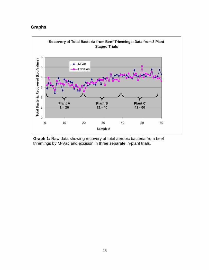
Overall results from the plant staged field trials showed the M-Vac achieved
recoveries of APC and TC that were, for the most part, not significantly different from
excision (Table 3). The one exception was in plant C where the M-Vac recovered
significantly more (P < 0.05) coliforms than excision. Analysis of the EC data within
each plant showed no statistically significant differences, at the 5% probability level,
between the two sampling techniques. Given the variability of the EC data from plant to
plant, a statistical analysis was not performed on the combined data. Raw data from the

18
in-plant trials (Graphs 1 – 2) also showed a definite correlation between M-Vac and
excision recoveries. Additional testing in the lab demonstrated the M-Vac was able to
recover low level E. coli O157:H7 contamination without any significant differences to
excision. This was shown to be true for both pre-rigor contamination (Table 4) and post-
rigor contamination (Table 5).

19
Conclusions Compared to sampling techniques currently utilized in food safety monitoring
procedures the M-Vac showed comparable recoveries of bacteria and in some cases
statistically outperformed these techniques. Results from the environmental monitoring
study showed the M-Vac produces bacterial recoveries which are superior to those
produced by the sponge/swab technique. Utilizing the M-Vac for such testing would give
quality control personnel a more accurate estimation of the cleanliness of surfaces
throughout their facilities which are involved in the production process. More efficient
bacterial recovery would also allow quality personnel to detect the presence of food
pathogens when such organisms are still at relatively low levels in the processing
environment. Earlier detection of pathogens in the environment would make it less likely
for manufacturers to produce pathogen containing product and could make it easier for
manufacturers to implement steps to eliminate the pathogens while at lower levels.
Simulated carcass/primal sampling showed the M-Vac was capable of recovering
low level O157:H7 contamination more frequently than sponging. These results suggest
that processing facilities which screen carcasses for O157:H7 contamination with
sponges are more likely to have false negative results leading to increased introduction of
contaminated meat into the fabrication process. Utilizing the M-Vac sampling technique
in the carcass screening process could have an immediate impact on lowering the
incidence of contaminated meat entering downstream processing.
The M-Vac demonstrated results that were not significantly different than
excision on beef surfaces; both in the field trials and in extensive laboratory testing for
O157:H7. The lack of a significant difference between the two sampling techniques

20
suggests that, regardless of the technique utilized to sample beef surfaces, O157:H7
contamination will be collected with the same efficiency. Therefore, abattoirs that
determine utilization of the M-Vac would be a better fit in their processes would still be
using a sampling technique as robust as excision to validate the safety of their product.
21
Summary
The M-Vac is a non-destructive wet-vacuum based sampling system which is
capable of aggressively collecting bacteria from a diverse array of surfaces. Many
applications within food processing establishments exist for the M-Vac. In the beef
industry these applications include environmental monitoring, carcass/primal sampling,
and beef trimmings sampling. Validations of each of these applications have shown the
M-Vac is capable of recovering bacteria as well as, or better than, techniques currently
utilized for these applications within the beef industry. The M-Vac could provide
significant advantages for food manufacturers by recovering bacteria more efficiently
than currently utilized surface sponges/swabs. Carcass screening for O157:H7 with
sponges is more likely to introduce contaminated meat into the fabrication process than
the M-Vac due to the high frequency of false negative results produced by sponging. For
beef processors who wish to utilize the M-Vac for trim sampling, the M-Vac has been
shown to be a robust sampling technique not statistically different than excision. This
was demonstrated both in plant staged field trials and in the laboratory.

22
Appendix
Table 1
Environmental Sampling
Organism DeviceStainless
SteelCutting Board
Ceramic Tile
Conveyor Belt
M-Vac 7.49 a 7.07 a 7.31 a 7.51 aSponge 5.65 b 5.77 b 5.52 b 5.55 bM-Vac 7.29 a 7.37 a 7.05 a 7.30 a
Sponge 6.06 b 6.57 b 5.92 b 6.03 bSwab 6.02 b 5.48 c 5.62 c 5.75 b
Recovery of Common Food Pathogen Simulants from Representative Food Processing Environmental Surfaces
Listeria
Salmonella
Table 1: Recovery of Listeria and Salmonella from common food processing environmental surfaces, reported as Log10 values, using three sampling techniques. In all treatments M-Vac recoveries were statistically higher than those for sponge and swab. Sponge and swab results for Salmonella with the same letters were not significantly different (P < 0.05). n = 15 for each treatment.

23
Table 2: Recovery of E. coli O157:H7 from simulated carcass surfaces using two sampling techniques. Differences between the two techniques were statistically significant (χ2 > 3.84).
Table 2 Carcass/Primal Sampling
Positive NegativeM-Vac 15 9 24Sponge 9 15 24
6.4
Sample ResultSampling Technique Total
Recovery of Low Level (1 - 5 CFU) E. coli O157:H7 Contamination from Simulated Beef Carcass Surfaces
χχχχ2 Value

24
Table 3 Plant Staged Field Trials
Plant TechniqueAverage Aerobic
Bacteria Recovered
Average Total Coliform
RecoveredSamples E. coli +
0.0%(0/20)
0.0%(0/20)
35.0%(7/20)
35.0%(7/20)
25.0%(5/20)
0.0%(0/20)
20.0%(12/60)
11.7%(7/60)
Multiparameter Plant Staged Field Trials for Recovery of Bacteria from Beef Trimmings
TotalM-Vac 3.76 a 2.21 a
Excision 3.70 a 1.78 a
CM-Vac 4.24 a 3.41 a
Excision 4.16 a 2.93 b
BM-Vac 3.80 a 2.33 a
Excision 3.69 a 1.86 a
AM-Vac 3.23 a 0.87 a
Excision 3.25 a 0.53 a
Table 3: Recovery of APC, TC, and generic E. coli from beef trimmings. APC and TC results reported as Log10 values. Results with the same letter for APC and TC are not significantly different (P < 0.05). Using the Fisher Exact test at the 5% probability level, no significant differences were observed between techniques for recovery of E. coli.

25
Table 4 Pre-rigor Contamination Trim Testing
Positives NegativesM-Vac 9 7Excision 6 10M-Vac 9 11Excision 12 8M-Vac 9 7Excision 10 6M-Vac 27 25Excision 28 24
Result χχχχ2 Value
0.08
0.27
1.88
2.41
DeviceTrial
Recovery of Low Level (1 - 5 CFU) E. coli O157:H7 Contamination from Beef Trimmings: Inoculated Pre-rigor
Total Observations
16
Average Inoculum
2.9
20
16
52Combined
3
2
3.4
3.0
4.0
Table 4: Recovery of E. coli O157:H7 inoculated on pre-rigor mortis meat surfaces and sampled after 24 hour refrigeration. No significant differences were observed between the two techniques in individual trials or in the combined data at the 5% probability level (χ2 value < 3.84).

26
Table 5 Post-rigor Contamination Trim Testing
Positives NegativesM-Vac 12 6 18Excision 7 8 15M-Vac 10 2 12Excision 10 1 11M-Vac 22 8 30Excision 17 9 26
χχχχ2 Value
0.6
Not Calculated
2.89
2.7
2.7
Trial DeviceTotal
Observations
Recovery of Low Level (1 - 5 CFU) E. coli O157:H7 Contamination from Beef Trimmings: Inoculated Post-rigor
1
2
Results
Combined
Average Inoculum
2.6
Table 5: Recovery of E. coli O157:H7 inoculated on post-rigor mortis meat surfaces and sampled after 24 hour refrigeration. No significant differences were observed between the two techniques in the combined data at the 5% probability level (χ2 value < 3.84).

27
Figure 2: M-Vac Kit
Figure 3: Sampling Hook and Screen Tool
Figures
Figure 1: Support Equipment Case (SEC)
28
Graphs
Recovery of Total Bacteria from Beef Trimmings: Data from 3 Plant Staged Trials
0
1
2
3
4
5
6
0 10 20 30 40 50 60
Sample #
Tota
l Bac
teri
a R
ecov
ered
(Log
Val
ues)
M-Vac
Excision
Plant A 1 – 20
Plant B 21 - 40
Plant C 41 - 60
Graph 1: Raw data showing recovery of total aerobic bacteria from beef trimmings by M-Vac and excision in three separate in-plant trials.

29
Recovery of Total Coliforms from Beef Trimmings: Data from 3 Plant Staged Trials
0
1
2
3
4
5
0 10 20 30 40 50 60
Sample #
To
tal
Co
lifo
rms
Rec
ove
red
(L
og
V
alu
es)
M-Vac
Excision
Plant A 1 – 20
Plant B 21 - 40
Plant C 41 - 60
Graph 2: Raw data showing recovery of total coliforms from beef trimmings by M-Vac and excision in three separate in-plant trials. Data from samples showing no recovery of coliforms was omitted to enable visualization of the overall trends.