Embed Size (px)
Citation preview

IntroductionCongenital myasthenic syndromes (CMSs) are hetero-geneous disorders caused by presynaptic, synaptic, orpostsynaptic defects (1). Postsynaptic CMSs can bebroadly classified according to whether the response toACh is increased or decreased relative to controlresponses. An increased response is observed in slow-channel syndromes due to prolonged activationepisodes of AChR. A decreased response is observedwith AChR deficiency due to mutations in AChR sub-unit genes (2) or in rapsyn (3), and in the fast-channelsyndromes. Fast-channel syndromes are characterizedby attenuated and abnormally rapidly decaying end-plate (EP) currents, abnormally brief single-channel cur-rents, and decreased probability of channel opening.
Several molecular mechanisms underlying fast-channel syndromes have been identified. At the lig-and-binding site formed by α and ε subunits, themutation εP121L reduces ACh affinity for the openchannel state and slows the rate of channel opening,resulting in a moderately severe myasthenic pheno-type (4). Also at the α-ε site, the mutation εD175Nreduces ACh affinity of the resting closed state andimpairs gating efficiency, while the nearby mutationεN182Y increases ACh affinity for the resting closedstate and also impairs gating (5). The phenotypic con-sequences of these combined mutations are also mod-erately severe. At the ligand-binding site formed bythe α and δ subunits, the mutation δE59K likelyreduces ACh affinity (6), but the altered steps in theactivation process have not been identified. Thismutation was shown to result in hypomotility inutero, multiple congenital joint contractures, andneonatal respiratory distress, but the patient subse-quently improved and could walk short distances. Inthe third transmembrane domain of the α subunit,the mutation αV285I impairs gating efficiency (7) butresults in a mild phenotype. Finally, in the amphi-pathic helix of the long cytoplasmic loop of the ε sub-unit, either a duplication of codons 413–418 (8) or anαA411P missense mutation (9) result in heteroge-neous channel gating kinetics; the phenotypic conse-quences are mild to moderately severe.
The Journal of Clinical Investigation | February 2003 | Volume 111 | Number 4 497
Mutation causing severe myastheniareveals functional asymmetry of AChR signature cystine loops in agonist binding and gating
Xin-Ming Shen,1 Kinji Ohno,1 Akira Tsujino,1 Joan M. Brengman,1 Monique Gingold,2
Steven M. Sine,3 and Andrew G. Engel1
1Department of Neurology and Neuromuscular Research Laboratory, Mayo Clinic, Rochester, Minnesota, USA2Childrens Neurology Services, Morgantown, West Virginia, USA3Receptor Biology Laboratory, Department of Physiology and Biophysics, Mayo Clinic, Rochester, Minnesota, USA
We describe a highly disabling congenital myasthenic syndrome (CMS) associated with rapidly decay-ing, low-amplitude synaptic currents, and trace its cause to a valine to leucine mutation in the signa-ture cystine loop (cys-loop) of the AChR α subunit. The recently solved crystal structure of an ACh-bind-ing protein places the cys-loop at the junction between the extracellular ligand-binding andtransmembrane domains where it may couple agonist binding to channel gating. We therefore analyzedthe kinetics of ACh-induced single-channel currents to identify elementary steps in the receptor acti-vation mechanism altered by the αV132L mutation. The analysis reveals that αV132L markedly impairsACh binding to receptors in the resting closed state, decreasing binding affinity for the second bindingstep 30-fold, but attenuates gating efficiency only about twofold. By contrast, mutation of the equiva-lent valine residue in the δ subunit impairs channel gating approximately fourfold with little effect onACh binding, while corresponding mutations in the β and ε subunits are without effect. The uniquefunctional contribution of the α subunit cys-loop likely owes to its direct connection via a β strand toαW149 at the center of the ligand-binding domain. The overall findings reveal functional asymmetrybetween cys-loops of the different AChR subunits in contributing to ACh binding and channel gating.
J. Clin. Invest. 111:497–505 (2003). doi:10.1172/JCI200316997.
Received for publication September 26, 2002, and accepted in revisedform November 26, 2002.
Address correspondence to: Andrew G. Engel, Department of Neurology, Mayo Clinic, Rochester, Minnesota 55905, USA.Phone: (507) 284-5102; Fax: (507) 284-5831; E-mail: [email protected] of interest: The authors have declared that no conflict ofinterest exists.Nonstandard abbreviations used: cystine loop (cys-loop);congenital myasthenic syndrome (CMS); endplate (EP);acetylcholinesterase (AChE); α-bungarotoxin (α-bgt); endplatepotential (EPP); endplate current (EPC); miniature endplatepotential (MEPP); miniature endplate current (MEPC); gatingequilibrium constant (θ); open probability (Popen).
See the related Commentary beginning on page 436.

Here we describe a fast-channel CMS caused by anαV132L mutation in the signature cystine loop (cys-loop) of the AChR α subunit that is more severely dis-abling than any previously reported fast-channel CMS.The cys-loop, formed by a disulfide bond between cys-teines 128 and 142 of the α subunit and equivalent cys-teines in non-α subunits, is one of the most highly con-served structural domains among members of theAChR superfamily (10). Previous studies suggested thatthe cys-loop is required for assembly of AChR subunitsand formation of the ligand-binding sites (11–13), butα or β subunits lacking the cys-loop disulfide bond canstill form stable complexes with other subunits (14, 15).Other reports implicate the cys-loop in α-neurotoxinbinding (11, 15, 16), but the cys-loop is not fullyexposed on the surface of the native receptor (17), andrecent atomic structural evidence places it remote fromthe toxin-binding site (18). The present study uses sin-gle-channel kinetic analysis to identify elementaryfunctional steps altered by αV132L, and show that themutation profoundly impairs ACh binding to the rest-ing closed state but only slightly attenuates channelgating. Mutations of equivalent residues in β and ε sub-units are without effect, but mutation of the δ subunitmarkedly impairs channel gating. The overall findingsreveal distinct contributions of cys-loops of the differ-ent AChR subunits to ACh binding and channel gating.
MethodsMuscle specimens. Intercostal muscle specimens wereobtained intact from origin to insertion from thepatient and from control subjects without muscle dis-ease undergoing thoracic surgery. All human studieswere in accord with the guidelines of the InstitutionalReview Board of the Mayo Clinic.
AChR and acetylcholinesterase (AChE) were detect-ed in cryostat sections by two-color fluorescence (19).EPs were localized for electron microscopy and ana-lyzed by established methods (20, 21). Peroxidase-labeled α-bungarotoxin (α-bgt) was used for the ultra-structural localization of AChR (22). The number ofAChR’s per EP was measured with [125I]α-bgt (23).
Electrophysiology of muscle specimens. Recordings of EPpotentials (EPPs) and EP currents (EPCs) and esti-mates of the number of transmitter quanta releasedby nerve impulse were performed as described else-where (23, 24), except that the amplitude of theminiature EPPs (MEPPs) and currents (MEPCs) wereestimated from the quantal components of the EPPand EPC, respectively (25). Single-channel patch-clamp recordings from the EP were performed in thecell-attached mode as described (26).
Mutation analysis. We directly sequenced the AChR αand ε subunit genes using genomic DNA (27).αV132L results in gain of an MnlI site, and α381delCresults in loss of a Bst4CI site. Therefore we screenedfor these mutations in relatives by restriction analysis.We also screened for αV132L in 200 normal allelesusing allele-specific PCR.
Construction and expression of wild-type and mutantAChR’s. Sources of human α, β, δ, and ε subunitcDNAs were as previously described (28, 29). All fourcDNAs were subcloned into the CMV-based expres-sion vector pRBG4 (4) for expression in humanembryonic kidney fibroblast (HEK 293) cells. TheαV132L, α381delC, αV132I, βV132L, δV134L, andεV132L mutations were engineered into the wild-typeα, β, δ, and ε subunit cDNA in pRBG4 using theQuikChange Site-Directed Mutagenesis Kit (Strata-gene, La Jolla, California, USA) (30). Presence of thedesired mutation and absence of unwanted muta-tions were confirmed by sequencing the entireinserts. HEK cells were transfected with mutant andcomplementary wild-type AChR subunit cDNAsusing calcium phosphate precipitation (31).
498 The Journal of Clinical Investigation | February 2003 | Volume 111 | Number 4
Figure 1(a) Patient at age 4. Note eyelid ptosis, facial diplegia, open mouth,gothic palate, and gastrostomy. (b) Ultrastructural localization ofEP AChR with peroxidase-labeled α-bgt. The density and distribu-tion of AChR on the junctional folds is normal. ×9,500.

Measurements of α-bgt binding. The total number of[125I]α-bgt sites on the surface of transfected HEKcells and ACh competition against the initial rate of[125I]α-bgt binding were determined as previouslydescribed (4). ACh competition measurements wereanalyzed according to the Hill equation: 1 – Y = 1/(1 +([ACh]/Kov)n), where Y is fractional occupancy by ACh,n is the Hill coefficient, and Kov is an overall dissocia-tion constant.
Patch-clamp recordings from AChR’s expressed in HEKcells. Recordings were obtained in the cell-attachedconfiguration at a membrane potential of –80 mV at22°C, with bath and pipette solutions containing142 mM KCl, 5.4 mM NaCl, 1.8 mM CaCl2, 1.7 mMMgCl2, and 10 mM HEPES, pH 7.4 (4, 31). Single-channel currents were recorded using an Axopatch200A amplifier (Axon Instruments Inc., Union City,California, USA) at a bandwidth of 50 kHz, digitizedat 5-µs intervals using a Digidata 1200A (AxonInstruments Inc.), and recorded to hard disk usingthe program pCLAMP (Axon Instruments Inc.).Records were analyzed at a uniform bandwidth of11.7 kHz with TAC × 4.0.9 software (Bruxton Corpo-ration, Seattle, Washington, USA).
To estimate rate constants underlying AChR activa-tion (see Results), we used desensitizing concentra-tions of ACh that cause events from a single channelto cluster into identifiable activation episodes (32).Clusters were identified as series of closely spacedopenings preceded and followed by closed intervalsgreater than a specified critical time. The latter wasobtained from the intersection of the predominantclosed time component with the succeeding closed
time component in the histogram. Clusters with fewerthan five openings were excluded from analysis. Indi-vidual clusters were examined for homogeneity bydetermining the mean open probability and openduration for each cluster, and clusters within two stan-dard deviations of the mean were accepted for furtheranalysis (33, 34). The resulting open and closed inter-vals for a given set of clusters were analyzed accordingto kinetic schemes of receptor activation using theprogram MIL, which uses an interval-based maximumlikelihood method that also corrects for missed events(33). A dead time of 23 µs was superimposed on allrecordings. For each type of AChR, single-channeldwell times obtained at several concentrations of AChwere fitted simultaneously. Data for different AChR’swere obtained at the following concentrations of ACh(in µM): wild type, αV132I, δV134L, βV132L, andεV132L at 10, 20, 30, 50, 70, 100, 200, and 300; andαV132L at 50, 70, 100, 200, 300, and 1,000. Individualrecordings contained 4,913–20,651 events in 57–311clusters. To check the final set of fitted rate constants,probability density curves were calculated from fittedrate constants and superimposed on the experimentaldwell-time histograms.
ResultsCharacteristics of CMS patient. A 4-year-old girl has hadlife-threatening myasthenic symptoms since birth,requiring frequent ventilator support. She cannot
The Journal of Clinical Investigation | February 2003 | Volume 111 | Number 4 499
Table 1EP studies
Controls Patient
[125I]α-bgt 12.82 ± 0.79 E6 (13) 10.7 E6binding sites/EPAChR index 3.3 ± 0.08 (155) 3.20 ± 0.19 (50)mA 31 ± 1 (190)B 26 ± 2.10 (26)MEPP amplitude (mV)B 1.00 ± 0.025 (165) 0.096 ± 0.011 (26)MEPC amplitude (nA)C 3.95 ± 0.10 (79) 0.32 ± 0.044 (17)BurstsD
τ1 (ms) 0.12 ± 0.012 (32) 0.44 ± 0.057 (3)Area 0.16 ± 0.014 0.86 ± 0.059 τ2 (ms) 3.04 ± 0.175 (34) 1.35 ± 0.32 (2)Area 0.85 ± 0.015 0.14 ± 0.059
Values indicate mean ± SEM. Measurements were taken at 29°C ± 0.5°C forEPPs and at 22°C ± 0.5°C for EPCs and single-channel recordings. Numbersin parentheses indicate number of patients studied for [125I]α-bgt bindingsites/EP, number of EP regions used for AChR index, and number of EPsincluded in other measurements. AQuantal content of EPP at 1 Hz stimulationcorrected for a resting membrane potential of –80 mV, nonlinear summation,and non-Poisson release. BCorrected for resting membrane potential of –80mV; MEPP amplitude in patient was determined from quantal component ofEPP and corrected for a fiber diameter of 50 µm. CMEPC amplitude in patientwas estimated from quantal component of EPC. DACh, 1 µM; pipette poten-tial, 80 mV; bandwidth, 5.8 kHz. The τ1 component of bursts was absent attwo control EPs and the τ2 component was absent at one patient EP. Valuesin bold indicate dominant components of bursts.
Figure 2Channel events elicited by 50 nM ACh from control (a) andpatient EPs (b), and from HEK cells expressing wild-type (c) andmutant AChR’s (d). Channel openings are upward deflections.Logarithmically binned burst duration histograms are fitted to thesum of exponentials.

hold her head erect, stand, or walk. She has markedeyelid ptosis and facial diplegia, is unable to close hermouth, and cannot speak or swallow (Figure 1a). Shehas a 25–50% decrement of the fifth compared withthe first evoked compound muscle fiber actionpotential, has no anti-AChR antibodies, andresponds partially to anticholinesterase drugs and3,4-diaminopyridine.
EP studies. EP configuration was normal, as judgedfrom the cytochemical reaction product for AChE onlongitudinally oriented teased single muscle fibers.Two-color fluorescence localization of AChR andAChE at the EPs revealed no abnormality. On electronmicroscopy, the structural integrity of the nerve ter-minals and junctional folds was preserved. Ultra-structural localization of AChR with peroxidase-labeled α-bgt revealed normal density anddistribution of AChR on the crests of the junctionalfolds (Figure 1b). The AChR index, defined as theratio of the length of the postsynaptic membranereacting for AChR to the length of the primary synap-tic cleft (21), was normal (Table 1). The number of EPspecific α-bgt binding sites was close to the meanadult control value (Table 1).
Although AChR was normally abundant at the EPsby multiple criteria, the MEPP and MEPC amplitudeswere so low that most were lost in baseline noise.Normally, d-tubocurarine is required to preventtwitching during recording of EPPs, but in thepatient these could be recorded in its absence. The
quantal content of the EPP (m) fell in the range ofvalues observed in adult controls. The MEPP andMEPC amplitudes, calculated from the respectivequantal component of the EPP and EPC, werereduced to about 10% of normal (Table 1).
Single-channel patch-clamp recordings from EPsrevealed AChR channels opening to a normal con-ductance of approximately 59 picosiemens. Analysisof bursts of channel openings elicited by low concen-trations of ACh yielded dwell-time histograms thatwere best fitted by two exponential components: amajor component whose duration was only 16% thatof wild-type, and a longer minor component (Table 1and Figure 2, a and b). Thus the burst open durationspredict an abnormally fast decay of the response toquantal release of ACh. The overall EP studiesrevealed no AChR deficiency, abnormally brief chan-nel activation episodes, and a markedly decreasedsynaptic response to ACh.
Mutation analysis. To look for a genetic basis for theobserved kinetic abnormality, we directly sequencedthe AChR ε and α subunits and detected two heterozy-gous mutations in the α subunit. The first mutation,αV132L, is a G-to-C transversion of nucleotide 394 thatconverts a valine to a leucine at codon 132 in the cys-loop of the α subunit. Valine at codon 132 is conservedin all human AChR subunits (Figure 3a), and in AChRα subunits of all species. The second mutation is a Cnucleotide deletion at position 381 (α381delC) in theextracellular domain of the AChR α subunit that pre-dicts 16 missense codons followed by a stop codon.Restriction analysis indicates that the mutations arerecessive and heteroallelic (Figure 3b). The αV132Lmutation was not detected in 200 normal alleles.
Expression studies in HEK cells. The total binding of[125I]α-bgt to mutant receptors on the cell surface wasnormalized to that measured for wild-type AChR. Therewas robust expression of αV132L-AChR compared withwild type (mean ± SD: 135% ± 12%, n = 3), but essentiallyno expression of α381delC-AChR (0.21% ± 0.22%, n = 3;P < 0.0001 vs. wild type). Robust expression of αV132L-AChR indicates that all three non-α subunits incorporateinto surface receptors because omission of one or moresubunits would abolish or markedly reduce expression(35, 36). Thus α381delC is a null mutation and αV132Ldetermines the phenotype.
500 The Journal of Clinical Investigation | February 2003 | Volume 111 | Number 4
Table 2Bursts of wild-type AChR and αV132L-AChR expressed in HEK cells
τ1 τ2 τ3
Wild type 0.036 ± 0.0017A 0.47 ± 0.059 3.31 ± 0.12n = 21 (0.24 ± 0.021) (0.21 ± 0.027) (0.58 ± 0.038)αV132L-AChR 0.080 ± 0.0058 0.50 ± 0.024 1.18 ± 0.084n = 7 (0.15 ± 0.019) (0.66 ± 0.021) (0.19 ± 0.019)
Values indicate mean ± SEM. n, number of patches. Values in parentheses rep-resent fractional histogram areas. Values in bold indicate dominant burstcomponent. ACh concentration was 50 nM, temperature was 22°C ± 0.5°C,and bandwidth was 11.7 kHz. ANot detected at three patches.
Figure 3(a) Multiple alignment of cys-loop and its flanking regions. Valine132 is conserved in all human AChR subunits and in α subunits ofall species. (b) MnlI and Bst4CI restriction analysis of PCR productsamplified from genomic DNA of patient’s and relatives’ blood.Closed and open arrows point, respectively, to mutant and wild-typefragments. Unaffected grandmother and mother are heterozygousfor α381delC, and a half sister carries no mutation. Father was notavailable for genetic analysis. Arrow and closed circle indicatepatient; half-shaded circles represent asymptomatic carriers.
To compare the affinity of ACh for αV132L and wild-type receptors, we measured ACh binding at steady state by competition against the initial rate of [125I]α-bgt binding to intact cells. The apparent dissociation constant of αV132L-AChR (6.18 × 10⁻⁷)increased by fivefold relative to wild-type (1.26 × 10⁻⁷),but the Hill coefficients were similar (1.2 and 1.4,respectively). The increased dissociation constant sug-gests that αV132L alters ACh binding, channel gating,desensitization, or a combination of these (4, 5).
Kinetics of activation of the αV132L mutant. To deter-mine whether αV132L causes the fast-channel CMS,we expressed the αV132L mutant with complementa-ry wild-type subunits in HEK 293 cells, recorded sin-gle-channel currents activated by a low concentrationof ACh (50 nM), and compared these with single-channel currents recorded from control and patientEPs (Figure 2). As observed at patient EPs, single-channel currents through the engineered mutantAChR activate in bursts of much shorter duration
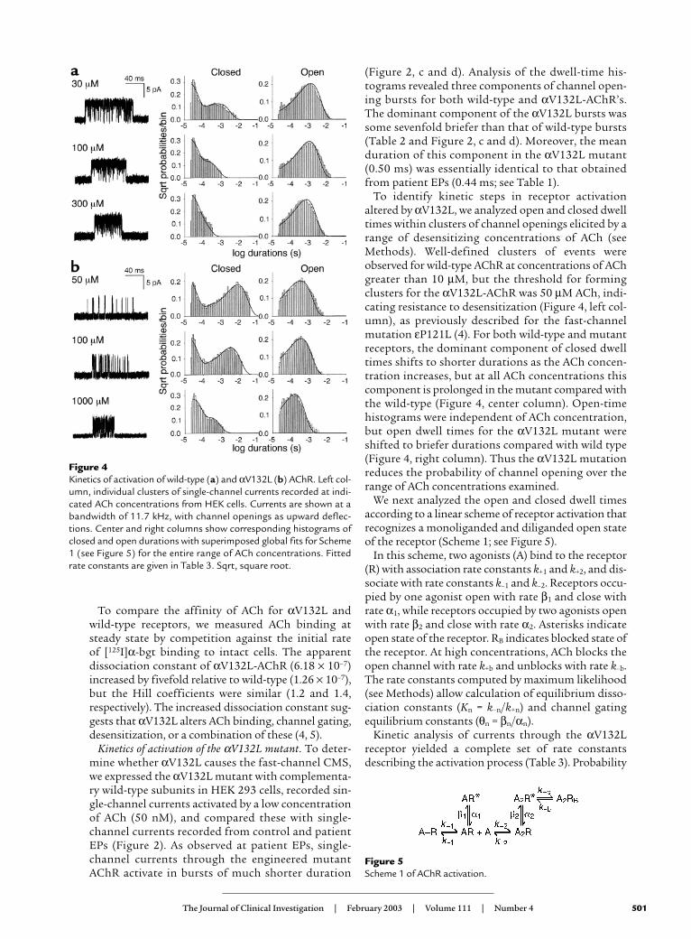
(Figure 2, c and d). Analysis of the dwell-time his-tograms revealed three components of channel open-ing bursts for both wild-type and αV132L-AChR’s.The dominant component of the αV132L bursts wassome sevenfold briefer than that of wild-type bursts(Table 2 and Figure 2, c and d). Moreover, the meanduration of this component in the αV132L mutant(0.50 ms) was essentially identical to that obtainedfrom patient EPs (0.44 ms; see Table 1).
To identify kinetic steps in receptor activationaltered by αV132L, we analyzed open and closed dwelltimes within clusters of channel openings elicited by arange of desensitizing concentrations of ACh (seeMethods). Well-defined clusters of events wereobserved for wild-type AChR at concentrations of AChgreater than 10 µM, but the threshold for formingclusters for the αV132L-AChR was 50 µM ACh, indi-cating resistance to desensitization (Figure 4, left col-umn), as previously described for the fast-channelmutation εP121L (4). For both wild-type and mutantreceptors, the dominant component of closed dwelltimes shifts to shorter durations as the ACh concen-tration increases, but at all ACh concentrations thiscomponent is prolonged in the mutant compared withthe wild-type (Figure 4, center column). Open-timehistograms were independent of ACh concentration,but open dwell times for the αV132L mutant wereshifted to briefer durations compared with wild type(Figure 4, right column). Thus the αV132L mutationreduces the probability of channel opening over therange of ACh concentrations examined.
We next analyzed the open and closed dwell timesaccording to a linear scheme of receptor activation thatrecognizes a monoliganded and diliganded open stateof the receptor (Scheme 1; see Figure 5).
In this scheme, two agonists (A) bind to the receptor(R) with association rate constants k+1 and k+2, and dis-sociate with rate constants k–1 and k–2. Receptors occu-pied by one agonist open with rate β1 and close withrate α1, while receptors occupied by two agonists openwith rate β2 and close with rate α2. Asterisks indicateopen state of the receptor. RB indicates blocked state ofthe receptor. At high concentrations, ACh blocks theopen channel with rate k+b and unblocks with rate k–b.The rate constants computed by maximum likelihood(see Methods) allow calculation of equilibrium disso-ciation constants (Kn = k–n/k+n) and channel gatingequilibrium constants (θn = βn/αn).
Kinetic analysis of currents through the αV132Lreceptor yielded a complete set of rate constantsdescribing the activation process (Table 3). Probability
The Journal of Clinical Investigation | February 2003 | Volume 111 | Number 4 501
Figure 4Kinetics of activation of wild-type (a) and αV132L (b) AChR. Left col-umn, individual clusters of single-channel currents recorded at indi-cated ACh concentrations from HEK cells. Currents are shown at abandwidth of 11.7 kHz, with channel openings as upward deflec-tions. Center and right columns show corresponding histograms ofclosed and open durations with superimposed global fits for Scheme1 (see Figure 5) for the entire range of ACh concentrations. Fittedrate constants are given in Table 3. Sqrt, square root.
Figure 5Scheme 1 of AChR activation.
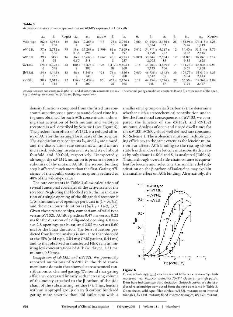
density functions computed from the fitted rate con-stants superimpose upon open and closed time his-tograms obtained for each ACh concentration, show-ing that activation of both mutant and wild-typereceptors is well described by Scheme 1 (see Figure 5).The predominant effect of αV132L is a reduced affin-ity of ACh for the resting, closed state of the receptor.The association rate constants k+1 and k+2 are slowed,and the dissociation rate constants k–1 and k–2 areincreased, yielding increases in K1 and K2 of aboutfourfold and 30-fold, respectively. Unexpectedly,although the αV132L mutation is present in both αsubunits of the mutant AChR, the second bindingstep is affected much more than the first. Gating effi-ciency of the doubly occupied receptor is reduced to48% of the wild-type value.
The rate constants in Table 3 allow calculation ofseveral functional correlates of the active state of thereceptor. Neglecting the blocked state, the mean dura-tion of a single opening of the diliganded receptor is1/α2; the number of openings per burst is (1 + β2/k–2);and the mean burst duration is (β2/k–2 + 1)/α2 (37).Given these relationships, comparison of wild-typeversus αV132L AChR’s predicts 0.47 ms versus 0.22ms for the duration of a diliganded opening, 6.0 ver-sus 2.8 openings per burst, and 2.83 ms versus 0.60ms for the burst duration. The burst duration pre-dicted from kinetic analysis is similar to that observedat the EPs (wild type, 3.04 ms; CMS patient, 0.44 ms)and to that observed in transfected HEK cells at lim-iting low concentrations of ACh (wild-type, 3.31 ms;mutant, 0.50 ms).
Comparison of αV132L and αV132I. We previouslyreported mutations of αV285 in the third trans-membrane domain that showed stereochemical con-tributions to channel gating. We found that gatingefficiency decreased linearly with increasing volumeof the moiety attached to the β carbon of the sidechain of the substituting residue (7). Thus, leucinewith an isopropyl group on its β carbon hinderedgating more severely than did isoleucine with a
smaller ethyl group on its β carbon (7). To determinewhether such a stereochemical contribution under-lies the functional consequences of αV132, we com-pared the kinetics of the αV132L and αV132Imutants. Analysis of open and closed dwell times forthe αV132I-AChR yielded well-defined rate constantsfor Scheme 1. The isoleucine mutation reduces gat-ing efficiency to the same extent as the leucine muta-tion but affects ACh binding to the resting closedstate less than does the leucine mutation; K2 decreas-es by only about 14-fold and K1 is unaltered (Table 3).Thus, although overall side-chain volume is equiva-lent for leucine and isoleucine, the smaller ethyl sub-stitution on the β carbon of isoleucine may explainthe smaller effect on ACh binding. Alternatively, the
502 The Journal of Clinical Investigation | February 2003 | Volume 111 | Number 4
Figure 6Open probability (Popen) as a function of ACh concentration. Symbolsrepresent mean Popen computed for 75–311 clusters in a single patch.Error bars indicate standard deviation. Smooth curves are the pre-dicted relationships computed from the rate constants in Table 3.Open circles, wild type; filled circles, αV132L mutant; open invertedtriangles, δV134L mutant; filled inverted triangles, αV132I mutant.
Table 3Activation kinetics of wild-type and mutant AChR’s expressed in HEK cells
k+1 k–1 K1/µM k+2 k–2 K2/µM β1 α1 θ1 β2 α2 θ2 k+b k–b KB/mM
Wild-type 102 ± 1,951 ± 19 88 ± 10,363 ± 117 196 ± 3,066 ± 0.064 54,240 ± 2,136 ± 25 133.96 ± 171,415 ± 1.288 200 2 169 15 250 1,094 52 5.26 1,819
αV132L 37 ± 2,712 ± 73 8 ± 31,269 ± 3,909 92 ± 7,869 ± 0.012 54,911 ± 4,587 ± 12 14.40 ± 53,214 ± 3.706 485 0.36 894 8 397 4,190 277 0.72 2,816
αV132I 39 ± 643 ± 16 16 ± 26,668 ± 1,667 45 ± 4,925 ± 0.0091 30,044 ± 2,554 ± 12 34.07 ± 107,063 ± 3.145 92 0.50 518 3 257 2,095 85 9.35 1,628
δV134L 174 ± 8,323 ± 48 180 ± 18,475 ± 103 1,417 ± 9,403 ± 0.15 33,083 ± 4,489 ± 7 181.78 ± 165,034 ± 0.916 548 8 382 99 268 1,133 106 6.61 1,908
βV132L 84 ± 1,143 ± 13 68 ± 8,245 ± 121 76 ± 1,526 ± 0.050 46,735 ± 1,542 ± 30 104.77 ± 135,010 ± 1.297 137 2 149 12 200 1,542 35 5.04 2,143
εV132L 90 ± 2,013 ± 22 116 ± 10,454 ± 90 417 ± 2,176 ± 0.19 44,334 ± 1,596 ± 28 56.30 ± 114,968 ± 2.045 185 3 207 35 195 948 37 3.24 2,467
Association rate constants are in µM–1s–1, and all other rate constants are in s–1. The channel gating equilibrium constants θ1 and θ2 are the ratios of the open-ing to closing rate constants, β1/α1 and β2/α2, respectively.

different branching patterns of the leucine andisoleucine side chains may be responsible for the dif-ferent functional consequences.
Mutations of equivalent residues in non-α subunits. Todetermine whether cys-loops of the non-α subunitscontribute similarly to the kinetics of receptor acti-vation, we substituted leucine for valine at positionsequivalent to α132 in AChR non-α subunits and ana-lyzed single-channel dwell times from the resultingmutant receptors. Single-channel dwell times fromthe βV132L, εV132L, and δV134L mutants also yield-ed well-defined rate constants in Scheme 1. Theequivalent mutation in the δ subunit, δV134L, hasonly a minor effect on ACh affinity, but it impairsgating efficiency of the doubly liganded receptor: θ2
is reduced to 28% of the wild-type counterpart, andthe predicted burst duration falls from about 3 msfor wild type to 0.62 ms for the mutant. By contrastto mutations in α and δ subunits, the correspondingvaline to leucine mutations in the β and ε subunitsdo not affect the kinetics of receptor activation(Table 3). Thus, the overall findings demonstratefunctional asymmetry in the contributions of cys-loops from different AChR subunits.
Effects of the cys-loop mutants on channel open probabili-ty. Next we compared fractional activation of wild-type and mutant receptors as a function of ACh con-centration. Fractional activation is taken as theprobability that a channel is open (Popen) within adefined cluster of channel events; for each concen-tration of ACh, the mean Popen computed for all clus-ters is plotted (Figure 6). Each of the mutations,αV132L, αV132I, and δV134L, shifts the Popen curveto higher concentrations of ACh, with αV132L iden-tified as having the most severe effect. Correspond-ing Popen curves for the βV132L and εV132L muta-tions did not differ significantly from wild type (datanot shown). Theoretical Popen curves computed fromthe fitted rate constants superimpose upon the Popen
measurements, further supporting the validity of theestimated rate constants in Table 3.
DiscussionWe trace the clinical phenotype of a highly disablingfast-channel CMS to two heteroallelic mutations in theAChR α subunit: a missense mutation, αV132L, in thesignature cys-loop, and a frame-shifting null mutation,381delC, that unmasks the functional consequences ofαV132L. The robust expression of the αV132L-AChRat EPs and in HEK cells indicates that the pathologicconsequences stem entirely from abnormal activationkinetics of the mutant channels.
Neuromuscular transmission is compromised by thevery small and rapidly decaying synaptic currentsresulting from abnormally brief single-channel activa-tion episodes that occur with low probability. Single-channel kinetic analysis reveals that, compared withwild-type, αV132L impairs ACh binding to receptors inthe resting closed state by about 30-fold, but attenuateschannel gating by only about twofold. Mutation of theequivalent valine residue in the δ subunit has littleeffect on ACh binding but reduces channel gating effi-ciency about 3.6-fold, while corresponding mutationsin the β and ε subunits are without effect. The overallfindings reveal functional asymmetry between cys-loops of the different AChR subunits in contributingto ACh binding and channel gating.
We can assess the consequences of the mutation onneuromuscular transmission using our estimatedreceptor activation rate constants (Table 3). Both theamplitude and the decay rate of the EPC determine themagnitude of the postsynaptic response. The usualapproximation to assess changes in EPC amplitude isthe probability that a receptor will open following fulloccupancy of the binding sites by the impulse of ACh,which is given by β2/(β2 + k–2). However, for the mutantreceptor, the assumption of full occupancy is likely notvalid because the rate of ACh association is slowed byan order of magnitude at the second occupancy step ofScheme 1. We therefore used our estimated rate con-stants to compute the fraction of active receptors fol-lowing an impulse of ACh according to a subset ofScheme 1 (Scheme 2; see Figure 7).
Scheme 2 neglects the return transition from theopen state because it is too slow to contribute to thepeak of the MEPC. We also make the further assump-tion that the first ACh binding step, which is onlyslightly altered in the mutant, is complete at theinstant of the pulse of ACh. Given these simplifyingassumptions, the fraction of active receptors as a
The Journal of Clinical Investigation | February 2003 | Volume 111 | Number 4 503
Figure 7Scheme 2 of AChR activation.
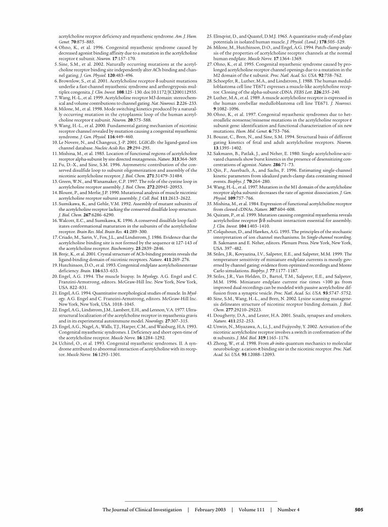
Figure 8Location of V132 in the AChR α subunit. Stereo view of a structuralmodel of the human α subunit (42) is shown in magenta with the cys-loop and contiguous β strand 7 highlighted in green. Ball and stickrepresentations indicate valine (Val) 132 within the cys-loop, cysteines(Cys) 128 and 142 which form the loop, and tryptophan (Trp) 149 atthe center of the α subunit portion of the binding pocket.

function of time can be predicted by writing equa-tions for the time dependence of each receptor stateand solving the set of equations using the Laplacetransform to yield
Equation 1
where λ1 = sqrt{(k+2[ACh] + k–2 + β2)2 – 4k+2[ACh]β2} andλ2 = (k+2[ACh] + k–2 + β2).
Given that the synaptic ACh concentration is a squarewave pulse with a concentration amplitude of 1 mMand duration of 50–80 µs (38, 39), our fitted rate con-stants predict activation of 78% of wild-type receptorsand 14% of mutant receptors following an impulse ofACh, corresponding to an 18% reduction of the MEPCamplitude relative to wild type. This predicted reduc-tion of the MEPC approaches our observed reductionof 8%, but the difference between predicted and exper-imental amplitudes would vanish with only a twofoldreduction of the peak ACh concentration, the durationof the ACh transient, or a combination of these. Ele-mentary functional steps responsible for this reductionof the MEPC amplitude are the tenfold reduced rate ofACh association in the second binding step of Scheme1 and the threefold increased rate of ACh dissociationfrom the diliganded receptor.
Neuromuscular transmission is further compromisedby the rapid decay of MEPCs caused by the mutation.The MEPC decay time constant is approximated by themean duration of bursts of single-channel openingselicited by a low concentration of ACh, and is given by(1 + β2/k–2)/α2. In the mutant receptor, the threefoldincrease of k–2 and twofold increase of α2 reduce meanburst duration from 2.8 ms for wild type to 0.6 ms forthe αV132 mutant. The shortened burst duration alonepredicts reduction of the total current flow during anEPC to 21% of that of the wild-type receptor.
To summarize, we attribute the extremely severe phe-notypic consequences of αV132L to the approximate-ly 30-fold decrease of ACh binding affinity for the sec-ond of two closed-state binding sites, so that theprevailing ACh concentration in the synaptic spacesaturates only about 10% of these sites in the mutantreceptor, and to a further decrease of about fivefold inthe total ACh-induced current due to an accelerateddecay of the synaptic response.
Our mechanistic findings can be interpreted in lightof the crystal structure of an ACh binding protein froma fresh water mollusk (18), together with a structuralmodel of the receptor counterpart based on ACh bind-ing protein and lysine scanning mutagenesis (40). AChbinding protein is a homopentamer of α-like subunitsthat are 24% homologous to the neuronal α7 subunit.Moreover, ACh binding protein contains many of thestructural cornerstones that give nicotinic receptors
their unique signature, including ligand-binding sitesat interfaces between subunits, key aromatic residuesat the ligand-binding sites, and the signature cys-loopformed between cysteines 128 and 142 of the musclereceptor α subunit. In the structural model of thehuman AChR ligand-binding domain, the cys-loop islocated at the bottom of each subunit, bridging βstrands 6 and 7, and is directly connected to αW149,the center of the binding site through β strand 7 (40)(see Figure 8). Moreover, the cys-loop is located at thejunction between ligand-binding and transmembranedomains (41), and recent cryoelectron microscopystudies suggest that it may be in close apposition withthe second and third transmembrane domains (42),where it may couple structural changes at the bindingsite to opening and closing of the channel gate. Ourobservation that the cys-loop in the α subunit con-tributes primarily to ACh binding is explained by itslocation on the same β strand that harbors αW149 at the center of the binding site (Figure 8), which is the most likely residue to stabilize the quaternaryammonium group of ACh (43). Insertion of the largerleucine side chain in the cys-loop likely displaces the βstrand, and consequently αW149, thus causing lowaffinity for ACh. Consistent with this notion is that theisoleucine mutation, which has a smaller ethyl ratherthan isopropyl substitution on its β carbon, has asmaller effect on ACh binding.
Unexpectedly, the mutation in the α subunit cys-loop reduces gating efficiency only about twofold,compared with an approximately 30-fold effect onACh binding. A strong gating effect might be expect-ed because of the proximity of the cys-loop to thetransmembrane domains. However, the equivalentmutation in the δ subunit cys-loop attenuates channelgating nearly fourfold, with little effect on ACh bind-ing. The δV134 mutation and the contiguous β strand7 are far away from the ACh binding site, and wouldnot be expected to affect ACh binding. The functionalconsequences of the mutation in the δ subunit cys-loop suggest similar effects following mutation ofequivalent residues in β and ε subunits, but these showno effect. Therefore, although the four types of AChRsubunits have similar folded structures, our findingsdemonstrate an unexpected functional asymmetry ofthe cys-loops in the different subunits.
AcknowledgmentsThis work was supported by grants from the NIH toA.G. Engel (NS-6277) and to S.M. Sine (NS-31744)and by a Muscular Dystrophy Association grant toA.G. Engel. We thank Zeljko Bajzer for providingEquation 1.
1. Engel, A.G., Ohno, K., and Sine, S.M. 1999. Congenital myasthenic syn-dromes. In Myasthenia gravis and myasthenic disorders. A.G. Engel, editor.Oxford University Press. New York, New York, USA. 251–297.
2. Ohno, K., and Engel, A.G. 2002. Congenital myasthenic syndromes:genetic defects at the neuromuscular junction. Curr. Neurol. Neurosci. Rep.2:78–88.
3. Ohno, K., et al. 2002. Rapsyn mutations in humans cause endplate
504 The Journal of Clinical Investigation | February 2003 | Volume 111 | Number 4
acetylcholine receptor deficiency and myasthenic syndrome. Am. J. Hum.Genet. 70:875–885.
4. Ohno, K., et al. 1996. Congenital myasthenic syndrome caused bydecreased agonist binding affinity due to a mutation in the acetylcholinereceptor ε subunit. Neuron. 17:157–170.
5. Sine, S.M., et al. 2002. Naturally occurring mutations at the acetyl-choline receptor binding site independently alter ACh binding and chan-nel gating. J. Gen. Physiol. 120:483–496.
6. Brownlow, S., et al. 2001. Acetylcholine receptor δ subunit mutationsunderlie a fast-channel myasthenic syndrome and arthrogryposis mul-tiplex congenita. J. Clin. Invest. 108:125–130. doi:10.1172/JCI200112935.
7. Wang, H.-L., et al. 1999. Acetylcholine receptor M3 domain: stereochem-ical and volume contributions to channel gating. Nat. Neurosci. 2:226–233.
8. Milone, M., et al. 1998. Mode switching kinetics produced by a natural-ly occurring mutation in the cytoplasmic loop of the human acetyl-choline receptor ε subunit. Neuron. 20:575–588.
9. Wang, H.-L., et al. 2000. Fundamental gating mechanism of nicotinicreceptor channel revealed by mutation causing a congenital myasthenicsyndrome. J. Gen. Physiol. 116:449–460.
10. Le Novere, N., and Changeux, J.-P. 2001. LGICdb: the ligand-gated ionchannel database. Nucleic Acids Res. 29:294–295.
11. Mishina, M., et al. 1985. Location of functional regions of acetylcholinereceptor alpha-subunit by site directed mutagenesis. Nature. 313:364–369.
12. Fu, D.-X., and Sine, S.M. 1996. Asymmetric contribution of the con-served disulfide loop to subunit oligomerization and assembly of thenicotinic acetylcholine receptor. J. Biol. Chem. 271:31479–31484.
13. Green, W.N., and Wanamaker, C.P. 1997. The role of the cystine loop inacetylcholine receptor assembly. J. Biol. Chem. 272:20945–20953.
14. Blount, P., and Merlie, J.P. 1990. Mutational analysis of muscle nicotinicacetylcholine receptor subunit assembly. J. Cell. Biol. 111:2613–2622.
15. Sumikawa, K., and Gehle, V.M. 1992. Assembly of mutant subunits ofthe acetylcholine receptor lacking the conserved disulfide loop structure.J. Biol. Chem. 267:6286–6290.
16. Walcott, E.C., and Sumikawa, K. 1996. A conserved disulfide loop facil-itates conformational maturation in the subunits of the acetylcholinereceptor. Brain Res. Mol. Brain Res. 41:289–300.
17. Criado, M., Sarin, V., Fox, J.L., and Lindstrom, J. 1986. Evidence that theacetylcholine binding site is not formed by the sequence α 127-143 ofthe acetylcholine receptor. Biochemistry. 25:2839–2846.
18. Brejc, K., et al. 2001. Crystal structure of ACh-binding protein reveals theligand-binding domain of nicotinic receptors. Nature. 411:269–276.
19. Hutchinson, D.O., et al. 1993. Congenital endplate acetylcholinesterasedeficiency. Brain. 116:633–653.
20. Engel, A.G. 1994. The muscle biopsy. In Myology. A.G. Engel and C.Franzini-Armstrong, editors. McGraw-Hill Inc. New York, New York,USA. 822–831.
21. Engel, A.G. 1994. Quantitative morphological studies of muscle. In Myol-ogy. A.G. Engel and C. Franzini-Armstrong, editors. McGraw-Hill Inc.New York, New York, USA. 1018–1045.
22. Engel, A.G., Lindstrom, J.M., Lambert, E.H., and Lennon, V.A. 1977. Ultra-structural localization of the acetylcholine receptor in myasthenia gravisand in its experimental autoimmune model. Neurology. 27:307–315.
23. Engel, A.G., Nagel, A., Walls, T.J., Harper, C.M., and Waisburg, H.A. 1993.Congenital myasthenic syndromes. I. Deficiency and short open-time ofthe acetylcholine receptor. Muscle Nerve. 16:1284–1292.
24. Uchitel, O., et al. 1993. Congenital myasthenic syndromes. II. A syn-drome attributed to abnormal interaction of acetylcholine with its recep-tor. Muscle Nerve. 16:1293–1301.
25. Elmqvist, D., and Quastel, D.M.J. 1965. A quantitative study of end-platepotentials in isolated human muscle. J. Physiol. (Lond.) 178:505–529.
26. Milone, M., Hutchinson, D.O., and Engel, A.G. 1994. Patch-clamp analy-sis of the properties of acetylcholine receptor channels at the normalhuman endplate. Muscle Nerve. 17:1364–1369.
27. Ohno, K., et al. 1995. Congenital myasthenic syndrome caused by pro-longed acetylcholine receptor channel openings due to a mutation in theM2 domain of the ε subunit. Proc. Natl. Acad. Sci. USA. 92:758–762.
28. Schoepfer, R., Luther, M.A., and Lindstrom, J. 1988. The human medul-loblastoma cell line TE671 expresses a muscle-like acetylcholine recep-tor. Cloning of the alpha-subunit cDNA. FEBS Lett. 226:235–240.
29. Luther, M.A., et al. 1989. A muscle acetylcholine receptor is expressed inthe human cerebellar medulloblastoma cell line TE671. J. Neurosci.9:1082–1096.
30. Ohno, K., et al. 1997. Congenital myasthenic syndromes due to het-eroallelic nonsense/missense mutations in the acetylcholine receptor εsubunit gene: identification and functional characterization of six newmutations. Hum. Mol. Genet. 6:753–766.
31. Bouzat, C., Bren, N., and Sine, S.M. 1994. Structural basis of differentgating kinetics of fetal and adult acetylcholine receptors. Neuron.13:1395–1402.
32. Sakmann, B., Patlak, J., and Neher, E. 1980. Single acetylcholine-acti-vated channels show burst kinetics in the presence of desensitizing con-centrations of agonist. Nature. 286:71–73.
33. Qin, F., Auerbach, A., and Sachs, F. 1996. Estimating single-channelkinetic parameters from idealized patch-clamp data containing missedevents. Biophys. J. 70:264–280.
34. Wang, H.-L., et al. 1997. Mutation in the M1 domain of the acetylcholinereceptor alpha subunit decreases the rate of agonist dissociation. J. Gen.Physiol. 109:757–766.
35. Mishina, M., et al. 1984. Expression of functional acetylcholine receptorfrom cloned cDNAs. Nature. 307:604–608.
36. Quiram, P., et al. 1999. Mutation causing congenital myasthenia revealsacetylcholine receptor β/δ subunit interaction essential for assembly. J. Clin. Invest. 104:1403–1410.
37. Colquhoun, D., and Hawkes, A.G. 1995. The principles of the stochasticinterpretation of ion channel mechanisms. In Single-channel recording.B. Sakmann and E. Neher, editors. Plenum Press. New York, New York,USA. 397–482.
38. Stiles, J.R., Kovyazina, I.V., Salpeter, E.E., and Salpeter, M.M. 1999. Thetemperature sensitivity of miniature endplate currents is mostly gov-erned by channel gating: evidence from optimized recordings and MonteCarlo simulations. Biophys. J. 77:1177–1187.
39. Stiles, J.R., Van Helden, D., Bartol, T.M., Salpeter, E.E., and Salpeter,M.M. 1996. Miniature endplate current rise times <100 µs fromimproved dual recordings can be modeled with passive acetylcholine dif-fusion from a synaptic vesicle. Proc. Natl. Acad. Sci. USA. 93:5747–5752.
40. Sine, S.M., Wang, H.-L., and Bren, N. 2002. Lysine scanning mutagene-sis delineates structure of nicotinic receptor binding domain. J. Biol.Chem. 277:29210–29223.
41. Dougherty, D.A., and Lester, H.A. 2001. Snails, synapses and smokers.Nature. 411:252–253.
42. Unwin, N., Miyazawa, A., Li, J., and Fujiyoshy, Y. 2002. Activation of thenicotinic acetylcholine receptor involves a switch in conformation of theα subunits. J. Mol. Biol. 319:1165–1176.
43. Zhong, W., et al. 1998. From ab initio quantum mechanics to molecularneurobiology: a cation-π binding site in the nicotinic receptor. Proc. Natl.Acad. Sci. USA. 95:12088–12093.
The Journal of Clinical Investigation | February 2003 | Volume 111 | Number 4 505