Embed Size (px)
Citation preview
MutaPLATE HLA DQ 2+8(TM)
Version 2.3 / November 2018
KF190532
KF190596
32
96
Nur für in-vitro Diagnostik
Real-Time PCR Kit
Real-Time PCR Kit für den Nachweis der HLA-Allele DQA1*05,DQB1*02 und DQB1*03:02/*03:05 auf Basis der TaqMan
Technologie für den LightCycler 1.5 und 2.0
®
Immundiagnostik AG, Stubenwald-Allee 8a, 64625 Bensheim, Deutschlandwww.immundiagnostik.com Tel.: +49 (0)6251/ [email protected] Fax: +49 (0)6251/ 849430
2
© 2018 Immundiagnostik AG / Version 2.3
MutaPLATE® HLA DQ 2+8 (TM)
Inhaltsverzeichnis
1 Verwendungszweck 3
2 Einleitung 3
3 Testprinzip 3
4 Kitbestandteile 4
5 Erforderliche Materialien 4
6 Lagerung und Haltbarkeit 4
7 Hinweise und Vorsichtsmaßnahmen 4
8 Testdurchführung 5
8.1 PCR Ansatz 5
8.2 PCR Protokoll 6
9 Auswertung 7
10 Troubleshooting 10
11 Grenzen des Tests 11
3
© 2018 Immundiagnostik AG / Version 2.3
MutaPLATE® HLA DQ 2+8 (TM)
1 Verwendungszweck
Das HLA DQ 2+8 (TaqMan) Real-Time PCR Kit ist ein auf der TaqMan-Technologiebasierter Test für den Nachweis der HLA-Allele DQA1*05, DQB1*021 und DQB1*03:02/*03:052,3.
1 Die Kopienanzahl von DQB1*02 wird mit dieser Methode nicht bestimmt.2 Auf Grund der Sequenzhomologie kann mit dieser Methode DQB1*03:05 nicht von DQB1*03:02
unterschieden werden. Die Häufigkeit von DQB1*03:05 ist jedoch sehr gering, ca. 0,4 % in derKaukasischen Bevölkerung, während DQB1*03:02 eine Häufigkeit von ca. 15 % aufweist (http://www.allelefrequencies.net, Nov. 2018). Weltweit beträgt die Häufigkeit von DQB1*03:05 0,07 % und dieHäufigkeit von DQB1*03:02 11,5 % (Solberg et al., Hum Immunol, 2008).
3 Die Detektion anderer sehr seltener Allele kann nicht vollständig ausgeschlossen werden.
2 Einleitung
Zöliakie / Glutenunverträglichkeit (GU) stellt eine der häufigsten chronisch gastro-intestinalen Erkrankungen dar. Es handelt sich um eine genetisch bedingte Erkrankung,bei der der Körper das in vielen Getreidesorten enthaltene Gluten nicht verarbeiten kann.Nahezu alle Zöliakie-Patienten sind Träger von HLA-DQ2 (HLA-DQA1*05 und HLA-DQB1*02) oder HLA-DQ8 (HLA-DQB1*03:02, häufig in Kombination mit DQA1*03). Wennkein DQ2 und DQ8 nachgewiesen werden kann, kann mit einer Wahrscheinlichkeit vonüber 95 % eine Glutenunverträglichkeit ausgeschlossen werden.
3 Testprinzip
Der Assay beinhaltet zwei spezifische Primer, die die Zielsequenz flankieren und zweiHydrolysesonden (TaqMan Sonden), die spezifisch in der Region der Mutation binden. Diebeiden Hydrolysesonden sind am 5' Ende mit unterschiedlichen Fluorophoren (ReporterFarbstoffen) markiert, welche für die Unterscheidung der Allele genutzt werden. Am 3'Ende sind die Sonden mit einem nicht-fluoreszierenden Quencher markiert. Die Nähe desReporter Farbstoffes zu dem Quencher inhibiert die Fluoreszenz des Reportermoleküls.Während der Amplifikation binden die Sonden spezifisch an die DNA Fragmente. Die 5'Nukleaseaktivität der Polymerase spaltet die hybridisierten Sonden, wodurch der Reportervom Quencher getrennt und ein Fluoreszenzsignal generiert wird.
4
© 2018 Immundiagnostik AG / Version 2.3
MutaPLATE® HLA DQ 2+8 (TM)
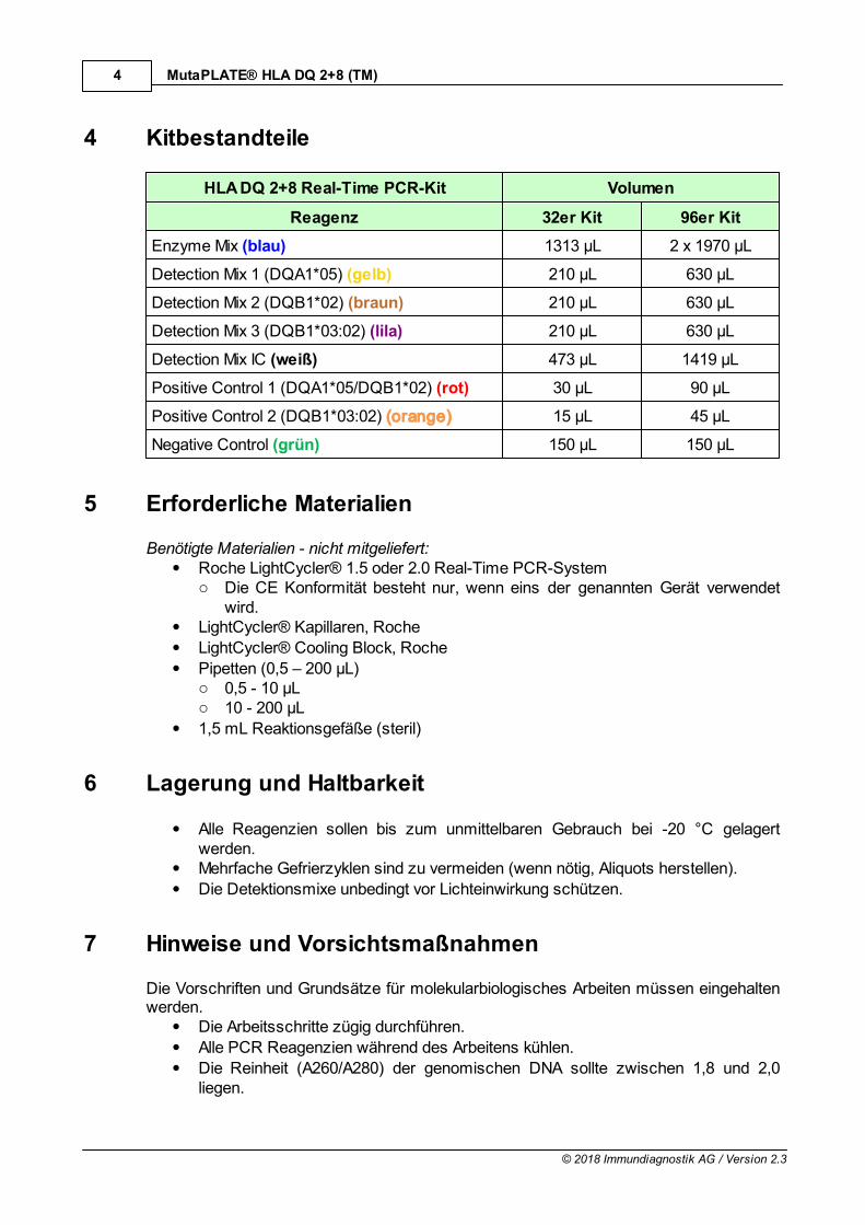
4 Kitbestandteile
HLA DQ 2+8 Real-Time PCR-Kit Volumen
Reagenz 32er Kit 96er Kit
Enzyme Mix (blau) 1313 µL 2 x 1970 µL
Detection Mix 1 (DQA1*05) (gelb) 210 µL 630 µL
Detection Mix 2 (DQB1*02) (braun) 210 µL 630 µL
Detection Mix 3 (DQB1*03:02) (lila) 210 µL 630 µL
Detection Mix IC (weiß) 473 µL 1419 µL
Positive Control 1 (DQA1*05/DQB1*02) (rot) 30 µL 90 µL
Positive Control 2 (DQB1*03:02) (orange) 15 µL 45 µL
Negative Control (grün) 150 µL 150 µL
5 Erforderliche Materialien
Benötigte Materialien - nicht mitgeliefert:Roche LightCycler® 1.5 oder 2.0 Real-Time PCR-Systemo Die CE Konformität besteht nur, wenn eins der genannten Gerät verwendet
wird.LightCycler® Kapillaren, Roche
LightCycler® Cooling Block, Roche
Pipetten (0,5 – 200 µL)o 0,5 - 10 µLo 10 - 200 µL
1,5 mL Reaktionsgefäße (steril)
6 Lagerung und Haltbarkeit
Alle Reagenzien sollen bis zum unmittelbaren Gebrauch bei -20 °C gelagertwerden.Mehrfache Gefrierzyklen sind zu vermeiden (wenn nötig, Aliquots herstellen).
Die Detektionsmixe unbedingt vor Lichteinwirkung schützen.
7 Hinweise und Vorsichtsmaßnahmen
Die Vorschriften und Grundsätze für molekularbiologisches Arbeiten müssen eingehaltenwerden.
Die Arbeitsschritte zügig durchführen.
Alle PCR Reagenzien während des Arbeitens kühlen.
Die Reinheit (A260/A280) der genomischen DNA sollte zwischen 1,8 und 2,0liegen.
5
© 2018 Immundiagnostik AG / Version 2.3
MutaPLATE® HLA DQ 2+8 (TM)
8 Testdurchführung
8.1 PCR Ansatz
Alle Kitbestandteile schonend auftauen lassen, vorsichtig vor dem Benutzendurchmischen (nicht vortexen) und kurz anzentrifugieren. Den Detektionsmix vorLichteinwirkung schützen. Während der Arbeiten alle PCR Reagenzien und den PCRAnsatz kühlen.
Für die Amplifikation werden drei Reaktionsgefäße (LightCycler® Kapillaren) pro Probeund zwei zusätzliche Reaktionsgefäße pro Mastermix für die negative und die positiveKontrolle benötigt. Die folgende Tabelle zeigt die zu pipettiernden Volumen pro Probe. Fürdie Analyse wird empfohlen ein Mastermix für die Anzahl an Proben (inkl. negativer undpositiver Kontrolle) (N) plus 10 % herzustellen, um Ungenauigkeiten auszugleichen. DerMastermix wird wie im Folgenden beschrieben pipettiert:
Mastermix 1 (DQA1*05):
Reagenz Volumen pro 25 µL-Reaktionsansatz
Master Mix Volumen
Detection Mix 1 (gelb) 6,0 µL 6,0 µL * (N + (N * 0,1))
Detection Mix IC (weiß) 4,5 µL 4,5 µL * (N + (N * 0,1))
Enzyme Mix (blau) 12,5 µL 12,5 µL * (N + (N * 0,1))
Den Mastermix vorsichtig durch auf- und abpipettierten oder durch Invertierendurchmischen und kurz anzentrifugieren. In jede Kapillare/Well 23 µL desMastermix vorlegen.Für die negative Kontrolle 2 µL von der mitgelieferten negativen Kontrolle (grün)dazugeben.Für die positive Kontrolle 2 µL von der mitgelieferten positiven Kontrolle 1 (rot)dazugeben.Für die zu analysierenden Proben jeweils 2 µL der Proben-DNA in dasentsprechende Reaktionsgefäß dazugeben.
Mastermix 2 (DQB1*02):
Reagenz Volumen pro 25 µL-Reaktionsansatz
Master Mix Volumen
Detection Mix 2 (braun) 6,0 µL 6,0 µL * (N + (N * 0,1))
Detection Mix IC (weiß) 4,5 µL 4,5 µL * (N + (N * 0,1))
Enzyme Mix (blau) 12,5 µL 12,5 µL * (N + (N * 0,1))
Den Mastermix vorsichtig durch auf- und abpipettierten oder durch Invertierendurchmischen und kurz anzentrifugieren. In jede Kapillare/Well 23 µL desMastermix vorlegen.Für die negative Kontrolle 2 µL von der mitgelieferten negativen Kontrolle (grün)dazugeben.
6
© 2018 Immundiagnostik AG / Version 2.3
MutaPLATE® HLA DQ 2+8 (TM)
Für die positive Kontrolle 2 µL von der mitgelieferten positiven Kontrolle1 (rot)dazugeben.Für die zu analysierenden Proben jeweils 2 µL der Proben-DNA in dasentsprechende Reaktionsgefäß dazugeben.
Mastermix 3 (DQB1*03:02):
Reagenz Volumen pro 25 µL-Reaktionsansatz
Master Mix Volumen
Detection Mix 3 (lila) 6,0 µL 6,0 µL * (N + (N * 0,1))
Detection Mix IC (weiß) 4,5 µL 4,5 µL * (N + (N * 0,1))
Enzyme Mix (blau) 12,5 µL 12,5 µL * (N + (N * 0,1))
Den Mastermix vorsichtig durch auf- und abpipettierten oder durch Invertierendurchmischen und kurz anzentrifugieren. In jede Kapillare/Well 23 µL desMastermix vorlegen.
Für die negative Kontrolle 2 µL von der mitgelieferten negativen Kontrolle (grün)dazugeben.Für die positive Kontrolle 2 µL von der mitgelieferten positiven Kontrolle 2 (orange)dazugeben.Für die zu analysierenden Proben jeweils 2 µL der Proben-DNA in dasentsprechende Reaktionsgefäß dazugeben.
Die Kapillaren mit den Deckeln verschließen, in das LightCycler® Karussell überführenund in der LightCycler® Zentrifuge abzentrifugieren (sollte eine Tischzentrifuge verwendetwerden die Kapillaren in den Einsätzen des Cooling Blocks bei 3000 rpm für 15 szentrifugieren). Anschließend das Karussell in den LightCycler® überführen und das unter8.2 beschriebene PCR Programm starten.
8.2 PCR Protokoll
Schritt Temperatur [°C] Zeit [s] Heizrate [°C/s] Zyklen Acquisition
InitialeDenaturierung
94 120 max. 1 x keine
Denaturierung 94 10 max.
45 x
keine
PrimerAnlagerung
und Elongation60 50 max. Single
Kühlen 40 30 max. 1 x ---
7
© 2018 Immundiagnostik AG / Version 2.3
MutaPLATE® HLA DQ 2+8 (TM)
9 Auswertung
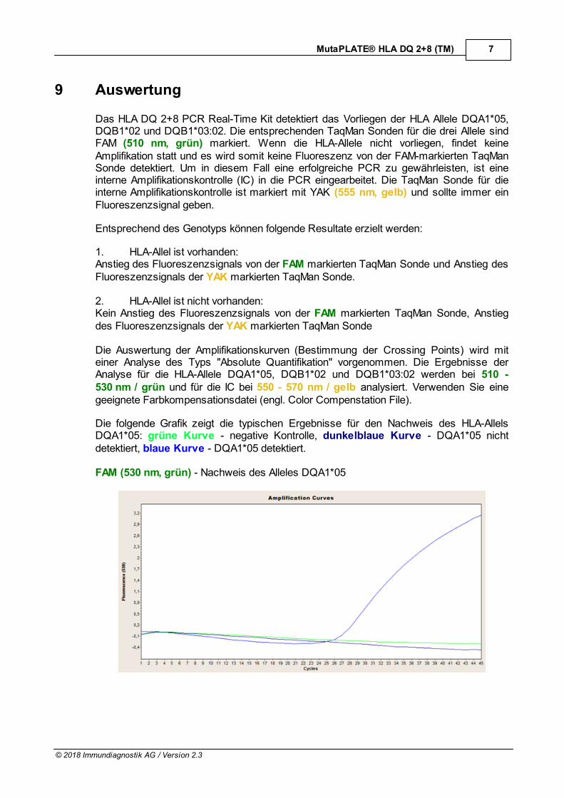
Das HLA DQ 2+8 PCR Real-Time Kit detektiert das Vorliegen der HLA Allele DQA1*05,DQB1*02 und DQB1*03:02. Die entsprechenden TaqMan Sonden für die drei Allele sindFAM (510 nm, grün) markiert. Wenn die HLA-Allele nicht vorliegen, findet keineAmplifikation statt und es wird somit keine Fluoreszenz von der FAM-markierten TaqManSonde detektiert. Um in diesem Fall eine erfolgreiche PCR zu gewährleisten, ist eineinterne Amplifikationskontrolle (IC) in die PCR eingearbeitet. Die TaqMan Sonde für dieinterne Amplifikationskontrolle ist markiert mit YAK (555 nm, gelb) und sollte immer einFluoreszenzsignal geben.
Entsprechend des Genotyps können folgende Resultate erzielt werden:
1. HLA-Allel ist vorhanden:Anstieg des Fluoreszenzsignals von der FAM markierten TaqMan Sonde und Anstieg desFluoreszenzsignals der YAK markierten TaqMan Sonde.
2. HLA-Allel ist nicht vorhanden:Kein Anstieg des Fluoreszenzsignals von der FAM markierten TaqMan Sonde, Anstiegdes Fluoreszenzsignals der YAK markierten TaqMan Sonde
Die Auswertung der Amplifikationskurven (Bestimmung der Crossing Points) wird miteiner Analyse des Typs "Absolute Quantifikation" vorgenommen. Die Ergebnisse derAnalyse für die HLA-Allele DQA1*05, DQB1*02 und DQB1*03:02 werden bei 510 -530 nm / grün und für die IC bei 550 - 570 nm / gelb analysiert. Verwenden Sie einegeeignete Farbkompensationsdatei (engl. Color Compenstation File).
Die folgende Grafik zeigt die typischen Ergebnisse für den Nachweis des HLA-AllelsDQA1*05: grüne Kurve - negative Kontrolle, dunkelblaue Kurve - DQA1*05 nichtdetektiert, blaue Kurve - DQA1*05 detektiert.
FAM (530 nm, grün) - Nachweis des Alleles DQA1*05
8
© 2018 Immundiagnostik AG / Version 2.3
MutaPLATE® HLA DQ 2+8 (TM)
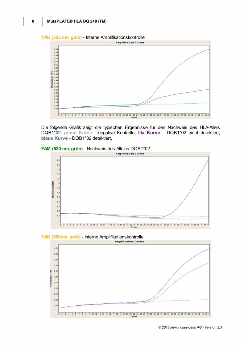
YAK (555 nm, gelb) - Interne Amplifikationskontrolle
Die folgende Grafik zeigt die typischen Ergebnisse für den Nachweis des HLA-AllelsDQB1*02: graue Kurve - negative Kontrolle, lila Kurve - DQB1*02 nicht detektiert,blaue Kurve - DQB1*02 detektiert.
FAM (530 nm, grün) - Nachweis des Alleles DQB1*02
YAK (560nm, gelb) - Interne Amplifikationskontrolle
9
© 2018 Immundiagnostik AG / Version 2.3
MutaPLATE® HLA DQ 2+8 (TM)
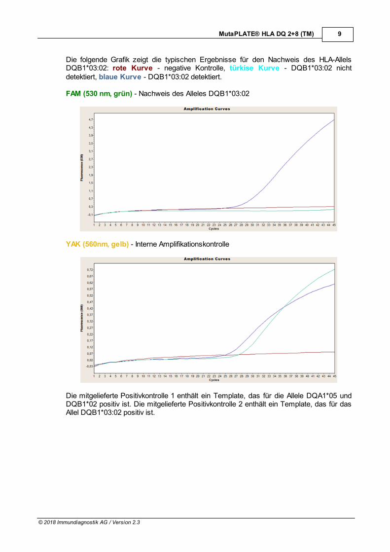
Die folgende Grafik zeigt die typischen Ergebnisse für den Nachweis des HLA-AllelsDQB1*03:02: rote Kurve - negative Kontrolle, türkise Kurve - DQB1*03:02 nichtdetektiert, blaue Kurve - DQB1*03:02 detektiert.
FAM (530 nm, grün) - Nachweis des Alleles DQB1*03:02
YAK (560nm, gelb) - Interne Amplifikationskontrolle
Die mitgelieferte Positivkontrolle 1 enthält ein Template, das für die Allele DQA1*05 undDQB1*02 positiv ist. Die mitgelieferte Positivkontrolle 2 enthält ein Template, das für dasAllel DQB1*03:02 positiv ist.
10
© 2018 Immundiagnostik AG / Version 2.3
MutaPLATE® HLA DQ 2+8 (TM)
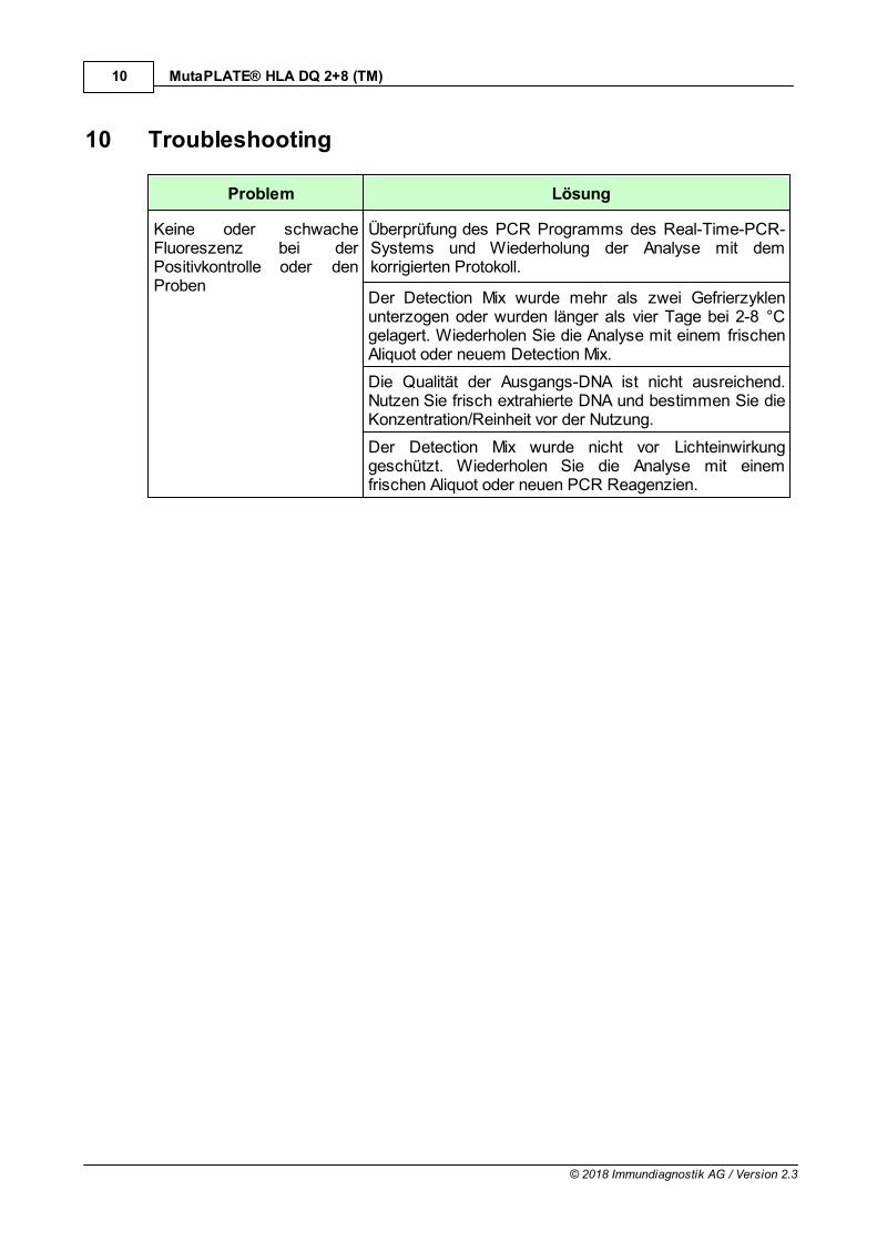
10 Troubleshooting
Problem Lösung
Keine oder schwacheFluoreszenz bei derPositivkontrolle oder denProben
Überprüfung des PCR Programms des Real-Time-PCR-Systems und Wiederholung der Analyse mit demkorrigierten Protokoll.
Der Detection Mix wurde mehr als zwei Gefrierzyklenunterzogen oder wurden länger als vier Tage bei 2-8 °Cgelagert. Wiederholen Sie die Analyse mit einem frischenAliquot oder neuem Detection Mix.
Die Qualität der Ausgangs-DNA ist nicht ausreichend.Nutzen Sie frisch extrahierte DNA und bestimmen Sie dieKonzentration/Reinheit vor der Nutzung.
Der Detection Mix wurde nicht vor Lichteinwirkunggeschützt. Wiederholen Sie die Analyse mit einemfrischen Aliquot oder neuen PCR Reagenzien.
11
© 2018 Immundiagnostik AG / Version 2.3
MutaPLATE® HLA DQ 2+8 (TM)
11 Grenzen des Tests
Das Ergebnis wird dem behandelnden Arzt als unterstützendes Material zur Verfügunggestellt und sollte niemals ausschließlich zur Diagnostik oder zuBehandlungsempfehlungen herangezogen werden. Die Diagnose sowie dieeinzuleitenden Behandlungsentscheidungen bleiben in der vollen Verantwortung desArztes.
Die Genauigkeit von genetischen Tests beträgt nicht 100 %. Es wurde jedoch eineGenauigkeit von über 98 % basierend auf den Validierungsdaten für diesen Testfestgestellt. Weiterhin müssen Ergebnisse von genetischen Tests im Kontext derklinischen Repräsentation des Patienten sowie bekannten familiären Risiken im Umfelddes Patienten betrachtet werden.
Der Test analysiert nur eine Auswahl an Markern. Daher schließt ein negativesTestergebnis des Patienten ein Risiko jedweder Art nicht vollständig aus.
MutaPLATE HLA DQ 2+8(TM)
Version 2.3 / November 2018
KF190532
KF190596
32
96
Only for in vitro diagnostics
Real-Time PCR Kit
Real-Time PCR Kit for the Detection of the HLA-alleles DQA1*05,DQB1*02 und DQB1*03:02/*03:05 on Basis of the TaqMan
Technology for the LightCycler 1.5 and 2.0
®
Immundiagnostik AG, Stubenwald-Allee 8a, 64625 Bensheim, Germany www.immundiagnostik.com Tel.: +49 (0)6251/ [email protected] Fax: +49 (0)6251/ 849430
2
© 2018 Immundiagnostik AG / Version 2.3
MutaPLATE® HLA DQ 2+8 (TM)
Table of Contents
1 Intended Use 3
2 Introduction 3
3 Concept of the Assay 3
4 Kit Components 4
5 Required Materials 4
6 Storage and Handling 4
7 Considerations and Precautions 4
8 Test Procedure 5
8.1 PCR Preparation 5
8.2 PCR Protocol 6
9 Evaluation 7
10 Troubleshooting 10
11 Test Limitations 11
3
© 2018 Immundiagnostik AG / Version 2.3
MutaPLATE® HLA DQ 2+8 (TM)
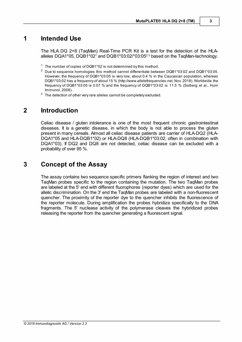
1 Intended Use
The HLA DQ 2+8 (TaqMan) Real-Time PCR Kit is a test for the detection of the HLA-alleles DQA1*05, DQB1*021 and DQB1*03:02/*03:052,3 based on the TaqMan-technology.
1 The number of copies of DQB1*02 is not determined by this method.2 Due to sequence homologies this method cannot differentiate between DQB1*03:02 and DQB1*03:05.
However, the frequency of DQB1*03:05 is very low, about 0.4 % in the Caucasian population, whereasDQB1*03:02 has a frequency of about 15 % (http://www.allelefrequencies.net, Nov. 2018). Worldwide thefrequency of DQB1*03:05 is 0.07 % and the frequency of DQB1*03:02 is 11.5 % (Solberg et al., HumImmunol, 2008).
3 The detection of other very rare alleles cannot be completely excluded.
2 Introduction
Celiac disease / gluten intolerance is one of the most frequent chronic gastrointestinaldiseases. It is a genetic disease, in which the body is not able to process the glutenpresent in many cereals. Almost all celiac disease patients are carrier of HLA-DQ2 (HLA-DQA1*05 and HLA-DQB1*02) or HLA-DQ8 (HLA-DQB1*03:02, often in combination withDQA1*03). If DQ2 and DQ8 are not detected, celiac disease can be excluded with aprobability of over 95 %.
3 Concept of the Assay
The assay contains two sequence specific primers flanking the region of interest and twoTaqMan probes specific to the region containing the mutation. The two TaqMan probesare labeled at the 5' end with different fluorophores (reporter dyes) which are used for theallelic discrimination. On the 3' end the TaqMan probes are labeled with a non-fluorescentquencher. The proximity of the reporter dye to the quencher inhibits the fluorescence ofthe reporter molecule. During amplification the probes hybridize specifically to the DNAfragments. The 5' nuclease activity of the polymerase cleaves the hybridized probesreleasing the reporter from the quencher generating a fluorescent signal.
4
© 2018 Immundiagnostik AG / Version 2.3
MutaPLATE® HLA DQ 2+8 (TM)
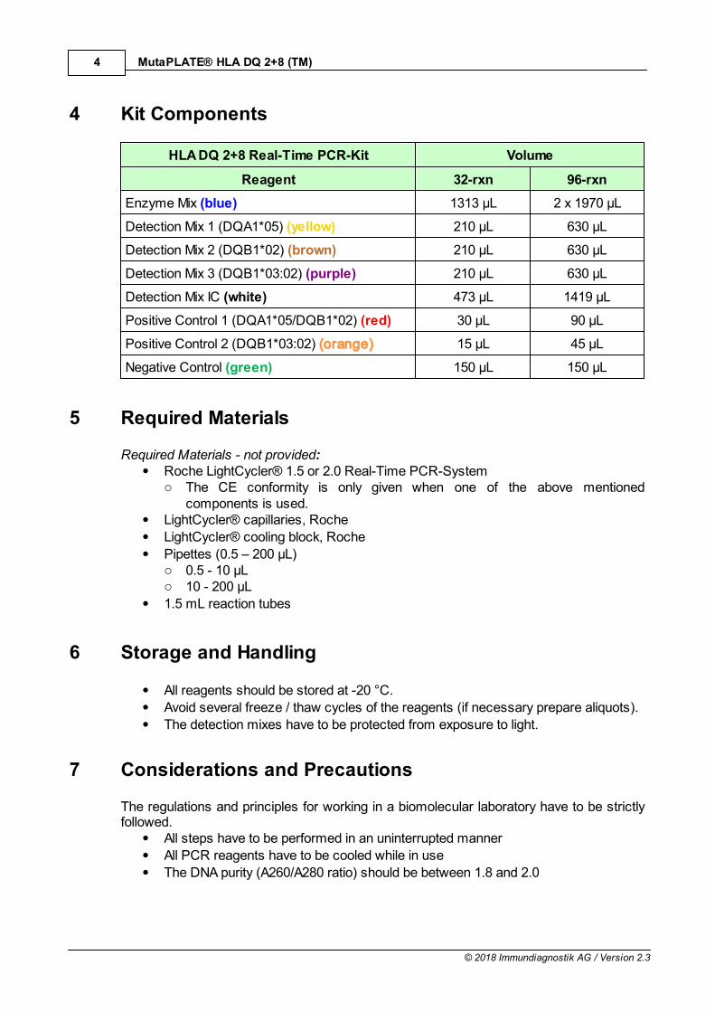
4 Kit Components
HLA DQ 2+8 Real-Time PCR-Kit Volume
Reagent 32-rxn 96-rxn
Enzyme Mix (blue) 1313 µL 2 x 1970 µL
Detection Mix 1 (DQA1*05) (yellow) 210 µL 630 µL
Detection Mix 2 (DQB1*02) (brown) 210 µL 630 µL
Detection Mix 3 (DQB1*03:02) (purple) 210 µL 630 µL
Detection Mix IC (white) 473 µL 1419 µL
Positive Control 1 (DQA1*05/DQB1*02) (red) 30 µL 90 µL
Positive Control 2 (DQB1*03:02) (orange) 15 µL 45 µL
Negative Control (green) 150 µL 150 µL
5 Required Materials
Required Materials - not provided:Roche LightCycler® 1.5 or 2.0 Real-Time PCR-Systemo The CE conformity is only given when one of the above mentioned
components is used.LightCycler® capillaries, Roche
LightCycler® cooling block, Roche
Pipettes (0.5 – 200 µL)o 0.5 - 10 µLo 10 - 200 µL
1.5 mL reaction tubes
6 Storage and Handling
All reagents should be stored at -20 °C.
Avoid several freeze / thaw cycles of the reagents (if necessary prepare aliquots).
The detection mixes have to be protected from exposure to light.
7 Considerations and Precautions
The regulations and principles for working in a biomolecular laboratory have to be strictlyfollowed.
All steps have to be performed in an uninterrupted manner
All PCR reagents have to be cooled while in use
The DNA purity (A260/A280 ratio) should be between 1.8 and 2.0
5
© 2018 Immundiagnostik AG / Version 2.3
MutaPLATE® HLA DQ 2+8 (TM)
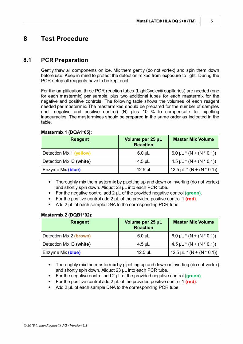
8 Test Procedure
8.1 PCR Preparation
Gently thaw all components on ice. Mix them gently (do not vortex) and spin them downbefore use. Keep in mind to protect the detection mixes from exposure to light. During thePCR setup all reagents have to be kept cool.
For the amplification, three PCR reaction tubes (LightCycler® capillaries) are needed (onefor each mastermix) per sample, plus two additional tubes for each mastermix for thenegative and positive controls. The following table shows the volumes of each reagentneeded per mastermix. The mastermixes should be prepared for the number of samples(incl. negative and positive control) (N) plus 10 % to compensate for pipettinginaccuracies. The mastermixes should be prepared in the same order as indicated in thetable.
Mastermix 1 (DQA1*05):
Reagent Volume per 25 µLReaction
Master Mix Volume
Detection Mix 1 (yellow) 6.0 µL 6.0 µL * (N + (N * 0,1))
Detection Mix IC (white) 4.5 µL 4.5 µL * (N + (N * 0,1))
Enzyme Mix (blue) 12.5 µL 12.5 µL * (N + (N * 0,1))
Thoroughly mix the mastermix by pipetting up and down or inverting (do not vortex)and shortly spin down. Aliquot 23 µL into each PCR tube. For the negative control add 2 µL of the provided negative control (green).
For the positive control add 2 µL of the provided positive control 1 (red).
Add 2 µL of each sample DNA to the corresponding PCR tube.
Mastermix 2 (DQB1*02):
Reagent Volume per 25 µLReaction
Master Mix Volume
Detection Mix 2 (brown) 6.0 µL 6.0 µL * (N + (N * 0,1))
Detection Mix IC (white) 4.5 µL 4.5 µL * (N + (N * 0,1))
Enzyme Mix (blue) 12.5 µL 12.5 µL * (N + (N * 0,1))
Thoroughly mix the mastermix by pipetting up and down or inverting (do not vortex)and shortly spin down. Aliquot 23 µL into each PCR tube. For the negative control add 2 µL of the provided negative control (green).
For the positive control add 2 µL of the provided positive control 1 (red).
Add 2 µL of each sample DNA to the corresponding PCR tube.
6
© 2018 Immundiagnostik AG / Version 2.3
MutaPLATE® HLA DQ 2+8 (TM)
Mastermix 3 (DQB1*03:02):
Reagent Volume per 25 µLReaction
Master Mix Volume
Detection Mix 3 (purple) 6.0 µL 6.0 µL * (N + (N * 0,1))
Detection Mix IC (white) 4.5 µL 4.5 µL * (N + (N * 0,1))
Enzyme Mix (blue) 12.5 µL 12.5 µL * (N + (N * 0,1))
Thoroughly mix the mastermix by pipetting up and down or inverting (do not vortex)and shortly spin down. Aliquot 23 µL into each PCR tube. For the negative control add 2 µL of the provided negative control (green).
For the positive control add 2 µL of the provided positive control 2 (orange).
Add 2 µL of each sample DNA to the corresponding PCR tube.
Close the capillaries with the corresponding lids, transfer them into the LightCycler®carousel and spin them down in the LightCycler® centrifuge (if a table top centrifuge isused, spin the capillaries in the adapters of the cooling block at 3000 rpm for 15 s).Subsequently place the carousel in the LightCycler® and use the PCR protocol describedin 8.2.
8.2 PCR Protocol
Step Temperature [°C] Time [s] Ramp rate [°C/s] Cycles Acquisition
InitialDenaturation
94 120 20 1 x none
Denaturation 94 10 20
45 x
none
PrimerAnnealing
andElongation
60 50 20 single
Cooling 40 30 20 1 x ---
7
© 2018 Immundiagnostik AG / Version 2.3
MutaPLATE® HLA DQ 2+8 (TM)
9 Evaluation
The HLA DQ 2+8 PCR Real-Time kit detects the presence of the HLA alleles DQA1*05,DQB1*02 and DQB1*03:02. The corresponding TaqMan probe for the three alleles islabeled with FAM (510 nm, green). If no HLA allele is present, no amplification occurs andtherefore no fluorescence of the FAM-labeled TaqMan probe is detected. To ensure in thiscase that the PCR was correctly carried out and an internal amplification control (IC) isincluded into the PCR. The TaqMan probe for the internal amplification control is labeledwith YAK (555 nm, yellow) and should always give a signal.
Corresponding to the genotype the following results can be achieved:
1. HLA allele present:Increase of the fluorescence signal of the FAM-labeled TaqMan probe and increase of thefluorescence signal of the YAK-labeled TaqMan probe.
2. HLA allele is not present:No increase of the fluorescence signal of the FAM-labeled TaqMan probe and increase ofthe fluorescence signal of the YAK-labeled TaqMan probe.
The evaluation of the amplification curves (determination of the crossing points) is doneby adding an analysis of the type "absolute quantification". The results of the analysis forthe HLA alleles DQA1*05, DQB1*02 and DQB1*03:02 are analysed at 510 - 530 nm /green and for the IC at 550 - 570 nm / yellow. Please use a corresponding colorcompensation file.
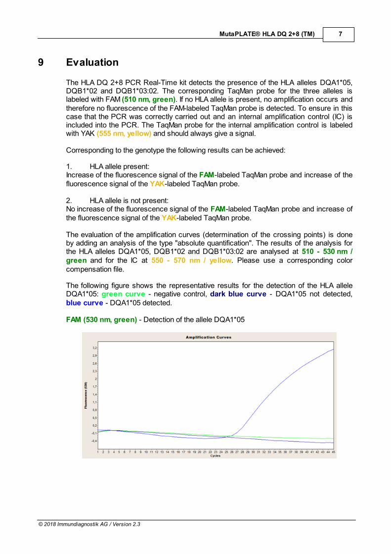
The following figure shows the representative results for the detection of the HLA alleleDQA1*05: green curve - negative control, dark blue curve - DQA1*05 not detected,blue curve - DQA1*05 detected.
FAM (530 nm, green) - Detection of the allele DQA1*05
8
© 2018 Immundiagnostik AG / Version 2.3
MutaPLATE® HLA DQ 2+8 (TM)
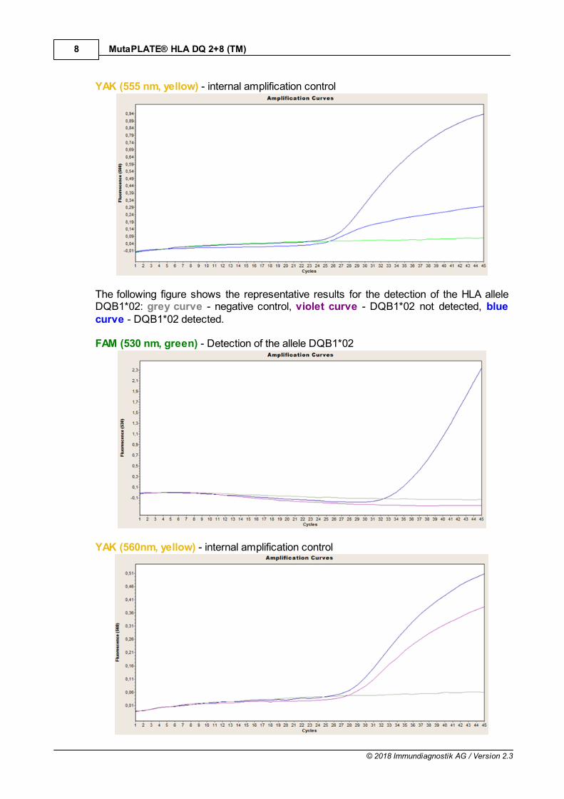
YAK (555 nm, yellow) - internal amplification control
The following figure shows the representative results for the detection of the HLA alleleDQB1*02: grey curve - negative control, violet curve - DQB1*02 not detected, bluecurve - DQB1*02 detected.
FAM (530 nm, green) - Detection of the allele DQB1*02
YAK (560nm, yellow) - internal amplification control
9
© 2018 Immundiagnostik AG / Version 2.3
MutaPLATE® HLA DQ 2+8 (TM)
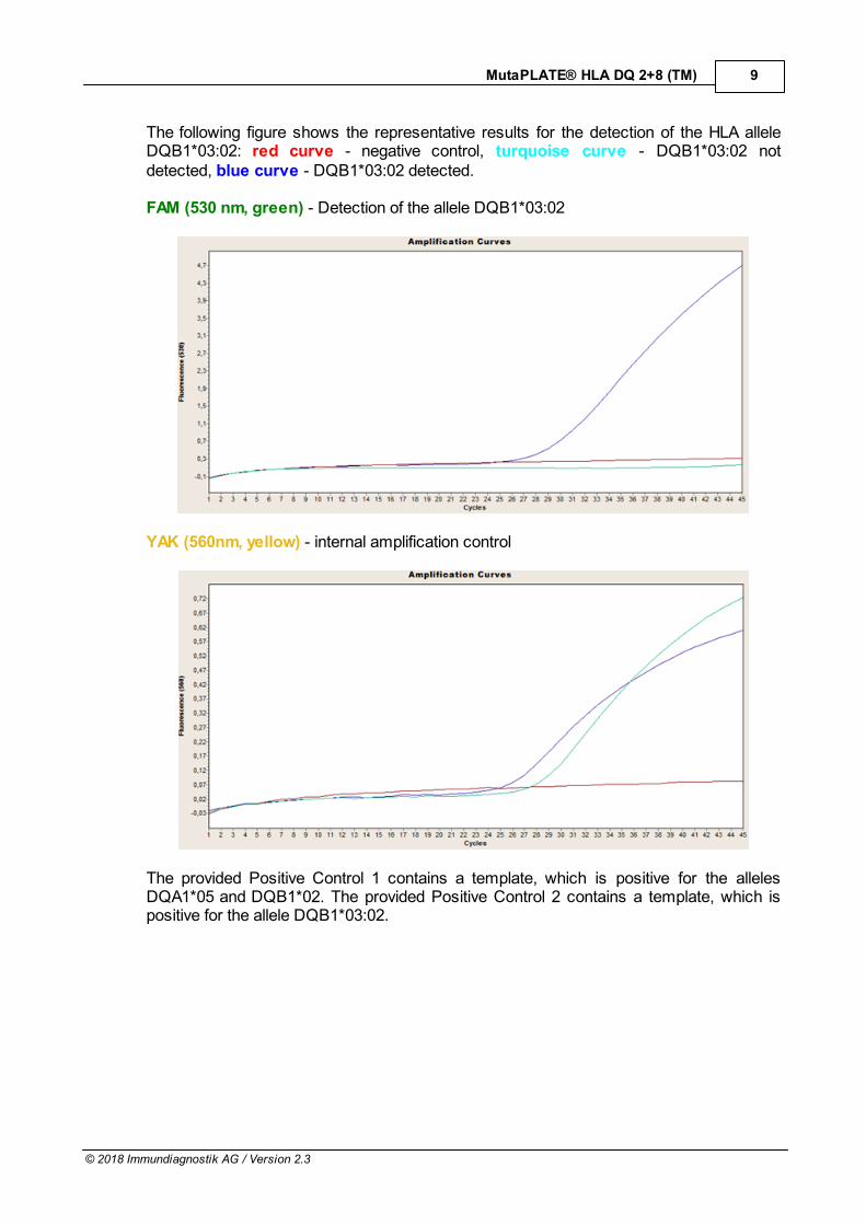
The following figure shows the representative results for the detection of the HLA alleleDQB1*03:02: red curve - negative control, turquoise curve - DQB1*03:02 notdetected, blue curve - DQB1*03:02 detected.
FAM (530 nm, green) - Detection of the allele DQB1*03:02
YAK (560nm, yellow) - internal amplification control
The provided Positive Control 1 contains a template, which is positive for the allelesDQA1*05 and DQB1*02. The provided Positive Control 2 contains a template, which ispositive for the allele DQB1*03:02.
10
© 2018 Immundiagnostik AG / Version 2.3
MutaPLATE® HLA DQ 2+8 (TM)
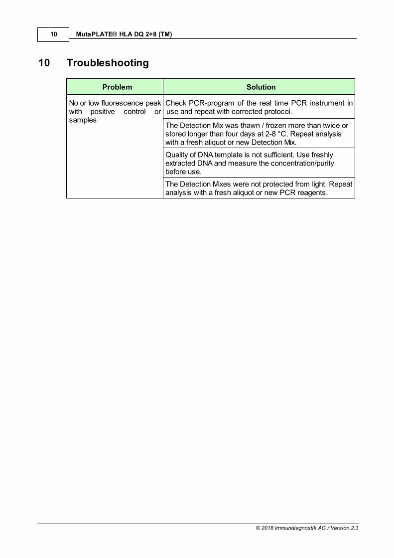
10 Troubleshooting
Problem Solution
No or low fluorescence peakwith positive control orsamples
Check PCR-program of the real time PCR instrument inuse and repeat with corrected protocol.
The Detection Mix was thawn / frozen more than twice orstored longer than four days at 2-8 °C. Repeat analysiswith a fresh aliquot or new Detection Mix.
Quality of DNA template is not sufficient. Use freshlyextracted DNA and measure the concentration/puritybefore use.
The Detection Mixes were not protected from light. Repeatanalysis with a fresh aliquot or new PCR reagents.
11
© 2018 Immundiagnostik AG / Version 2.3
MutaPLATE® HLA DQ 2+8 (TM)
11 Test Limitations
The accuracy of genetic testing is never 100%. However, an accuracy of more than 98%was determined based on the validation data. The attending physician is free to use thetest results as a guidance in the decision making process in terms of diagnosis andtherapy. However, these recommendations are based on genetic test outcomes and needto be interpreted in the context of medical history and known familiar risks of eachindividual patient. The attending physician is fully responsible for the final diagnosis andtreatment.