Embed Size (px)
Citation preview

ANTIMICROBIAL AGENTS AND CHEMOTHERAPY, Aug. 1974, p. 156-165Copyright 0 1974 American Society for Microbiology
Vol. 6, No. 2Printed in U.S.A.
Mutanolysin, Bacteriolytic Agent for Cariogenic Streptococci:Partial Purification and Properties
KANAE YOKOGAWA, SHIGEO KAWATA, SHINZO NISHIMURA, YASUHIKO IKEDA,AND YOSHIO YOSHIMURA
Research and Development Division, Dainippon Pharmaceutical Co. Ltd., Osaka, Japan
Received for publication 29 March 1974
Mutanolysin partially purified from the culture filtrate of Streptomycesglobisporus 1829 consists of two main lytic enzymes with an isoelectric point nearpH 8.5 and 10, respectively, and proteolytic enzyme is associated with the latterlytic enzyme. Mutanolysin exhibited maximal lytic activity at 60 C in the pHrange 6.5 to 7.0 and was stable at 50 C in the acid range. N-bromosuccinimidecaused complete inhibition of lytic activity at 1 mM, whereas calcium andmagnesium ions at the same concentration caused activation. Mutanolysin hadlytic or bactericidal activity against the living cells of Streptococcus mutans,Streptococcus salivarius, Streptococcus sanguis, Lactobacillus acidophilus, andActinomyces viscosus, which are considered to be etiologic agents of dentalcaries, but had no activity against S. aureus and all gram-negative strains tested.The lytic activity was well retained in human saliva. Digestion of the cell walls ofS. mutans BHT by mutanolysin was accompanied by the liberation of free aminogroups and reducing sugars. Mutanolysin may be expected to be a useful agentfor dental caries control.
Egg white lysozyme has been used frequentlyfor investigation of the intracellular compo-nents of cells. Because the lytic spectrum of thisenzyme was limited, other bacteriolytic en-zymes from microbial sources have been usedinstead by numerous investigators to obtaininformation about the structural and immuno-chemical characteristics of bacterial cell wallsand plasma membranes (1, 13, 17). Lyso-staphin, an enzyme that rapidly lyses the cellwalls of Staphylococcus aureus, has been foundto be effective in the treatment of establishedstaphylococci infections in experimental ani-mals (3, 8) and to reduce the incidence of S.aureus in nasal carriers (9, 14). We have re-ported that the culture filtrate of Streptomycesglobisporus 1829, a strain isolated from soil, iscapable of rapidly lysing cells of cariogenicsteptococci isolated from carious lesions inboth rodents and humans (24, 25). Streptococ-cus mutans, a type of cariogenic bacteria, hasbeen shown to form dental plaques composed ofextracellular polysaccharides of a dextran typeand to induce dental caries when inoculatedinto the oral cavities of experimental animalsmaintained on a high sucrose diet (5, 16, 27).Fitzgerald (4) found that in some hamstersreceiving dextranase in their drinking watersignificantly less plaque formation occurred,and fewer dental caries developed than in
animals not receiving the enzyme; an attempthas been made to control dental caries byapplying dextranase (15, 18, 21). The lytic andbactericidal actions of a culture broth from S.globisporus 1829 against S. mutans also sug-gested its possible application for dental cariescontrol.
In the present report, we describe some prop-erties of the partially purified lytic enzyme(termed "mutanolysin") from S. globisporus1829 and its lytic action against strains of S.mutans.
MATERIALS AND METHODSOrganisms. Strains of S. mutans used in this study
were maintained in brain heart infusion agar medium(Difco). They were cultivated by stab culture at 37 Cfor 3 days in 1 atm of 95% nitrogen and 5% carbondioxide. For the experiments on enzymatic lysis, thestock cultures were transferred into brain heart infu-sion (Difco) broth or into a medium containing 1%polypeptone (Difco), 1% meat extract (Difco), 0.5%yeast extract (Difco), 2% glucose, 1% sodium acetate,0.5% NaCl, and 0.1 mM MnSO4 (pH 7.5), and thenwere incubated at 37 C for 24 h in 1 atm of 95%nitrogen and 5% carbon dioxide. The culture brothswere chilled in ice, and the cells were harvested bycentrifugation at 10,000 rpm under cooling, washedthree times with chilled deionized water, and usedimmediately or as lyophilized cells.
All bacteria, including Bacillus subtilis, Brucella156

MUTANOLYSIN PROPERTIES 157
abortus, Diplococcus pneumoniae, Escherichia coli,Klebsiella species, Lactobacillus acidophilus, Listeriamonocytogenes, Proteus vulgaris, Pseudomonasaeruginosa, Salmonella typhimurium, Shigella flex-neri, Shigella sonnei, and S. aureus (all our collec-tion), were grown on nutrient agar medium, exceptthat brain heart infusion medium was employed forActinomyces viscosus, Streptococcus salivarius, andStreptococcus sanguis, and Sabouraud medium wasused for Candida utilis. The freshly cultured cellswere harvested and then subjected to lysis to deter-mine the lytic spectrum of mutanolysin.
Preparation of mutanolysin. S. globisporus 1829was grown in a 500-ml shaking flask containing 50 mlof a medium consisting of 2% dextrin, 0.5% soybeanmeal (Ajinomoto), 0.2% polypeptone (Wako), 0.1%NaCl, 0.5% Na2HPO4, 12H20, 0.1% MgSO4 7H20,and 0.02% CaCl2, pH 7.5. After 3 days of growth at30 C, 1 liter of culture broth was filtered through filterpaper, mixed with 40 g of Amberlite CG-50 weakcationic exchange resins (Rohm and Haas, Philadel-phia) in the H+ form, and stirred for 1 h. The resinswere removed through filter paper in a Buchnerfunnel and washed with deionized water. The lyticcomponents adsorbed on the resins were eluted outwith 130 ml of 0.2 M Na2HPO4 solution (pH 7.5). Theeluate was brought to 60% saturation with solidammonium sulfate. The precipitate was allowed tosettle overnight at 4 C and collected by passingthrough a pad of Radiolite no. 700 (Showa Chemicals)in a Buchner funnel. The lytic components in the padwere extracted with the minimal amount of deionizedwater, and the colored materials in the extracts wereremoved by treating with Duolite A-2 resins (Dia-mond Shamrock Chemicals) in the Cl- form. Thedecolorized solution was first adjusted to pH 2.0 withhydrochloric acid and then immediately to pH 6.0with sodium hydroxide solution. The insolubilizedproteolytic components were removed by centrifuga-tion. The clarified solution was desalted, concen-trated by ultrafiltration (ULVAC, Nihonshinku), andlyophilized. At this stage, 61 mg of mutanolysin wasobtained.Assay procedures. Lytic activity was determined
as follows: a mixture of 1.9 ml of cell suspension and2.0 ml of 10 mM tris(hydroxymethyl)aminomethane(Tris)-malate NaOH buffer (pH 7.0) was made to givean optical density of 0.6 at 600 nm in a Bausch andLomb Spectronic 20 colorimeter and preincubated ina water bath at 37 C for 1 min. A 0.1-ml amount ofadequately diluted mutanolysin solution was added tothis suspension and reacted at 37 C. The decrease inoptical density at 600 nm was read against a waterblank at appropriate time intervals. One unit of lyticactivity was defined as the amount of lytic enzymecausing a decrease in optical density of 0.01/min. Forviable cell counts, the reaction mixture containing 80,gg of mutanolysin and S. mutans BHT cells in 4 ml of10 mM Tris-malate NaOH buffer (pH 7.0) wasincubated at 37 C for 90 min. A 0.5-ml amount of thereaction mixture was pipetted out at appropriate timeintervals and added to 9.5 ml of 0.05% yeast extractsolution sterilized and chilled to stop enzymaticaction. A sample of the solution was admixed in mitis
salivarius agar medium, poured into a petri dish, andincubated at 37 C for 3 days in 1 atm of 95% nitrogenand 5% carbon dioxide, followed by counting thecolonies of viable streptococcal cells.
Proteolytic activity was determined as follows: to 2ml of 0.6% casein (Merck) solution in 0.05 M Tris-hydrochloride buffer (pH 8.0) was added 1 ml ofappropriately diluted enzyme solution. After incuba-tion at 37 C for 10 min, reaction was stopped byaddition of 3 ml of 0.01 M trichloroacetic acid, 0.02 Msodium acetate, and 0.03 M acetic acid. The precipi-tate was filtered off, and the absorbancy of the filtratewas determined at 275 nm in a spectrophotometer.
Protein was determined by the method of Lowry etal. (19) by using bovine serum albumin (NutritionalBiochemicals Corp.) as a standard.
Isoelectric focusing of mutanolysin was carried outby using an electrofocusing column (LKB 8100) of 110ml capacity with a density gradient of sucrose and pHgradient of carrier ampholites ranging from 3 to 10. A20-mg amount of mutanolysin was applied on thecolumn and subjected to electrofocusing at 0.5 mA at4 C for 46 h as described by Vesterberg and Svensson(26).
Free amino groups were determined according tothe method of Ghuysen (7) and free reducing sugarswere measured by the procedure of Park and Johnson(22).
Microscopy observation. Intact whole cells of S.mutans BHT were used in this investigation. The cellswere statically grown at 30 C for 2 days in a mediumcontaining 2% glucose, 1% polypeptone, 1% meatextract, 0.5% NaCl, 0.2% yeast extract, 1% sodiumacetate, and 0.1 mM MnSO4 (pH 7.5). The cells wereharvested by centrifugation and washed twice withdistilled water. To the mixture of 1.9 ml of cellsuspension and 2 ml of 0.02 M Tris-hydrochloridebuffer (containing 2 mM MgCl2) was added 0.1 ml ofmutanolysin solution (120 U/ml), and the reactionwas carried out at 37 C for 60 min. The initial opticaldensity at 600 nm gave 0.6. Samples of the solutionwere taken out at 0-, 5-, 10-, 15-, 20-, 30-, and 60-minintervals and diluted with a large volume of coldwater to stop the enzyme reaction, followed by imme-diate centrifugation at 10,000 rpm at 4 C for 10 min.The precipitates were resuspended in distilled water,and a droplet of the suspension was placed on electronmicroscope grids. The specimens were shadowed withgold-palladium alloy at an angle of 130 and exam-ined in an Akashi-type TRC 50 electron microscope.
Preparation of saliva. Hamster saliva was col-lected by stimulating the secretory ducts of salivaryglands by applying 2% pilocarpine in eyes of animalsanesthetized with pentobarbital. Human and hamstersaliva was centrifuged at 10,000 x g at 5 C for 15 minand sterilized by exposure to ultraviolet irradiationfor 15 min from a distance of 15 cm with a 2537-Alamp. The sterilized saliva was kept at -20 C andthawed just before use.
RESULTSPurification. The lytic enzymes in the cul-
ture filtrate of S. globisporus 1829 were found to
VOL. 6, 1974

158 YOKOGAWA ET AL.
be basic proteins and, therefore, they wereadsorbed on a weak cationic exchange resin,Amberlite CG-50, directly from culture filtrate,and easily eluted out with 0.2 M Na2HPO,solution. On the other hand, proteolytic activityremained in the filtrate unadsorbed on theresins, and nearly 75% of proteolytic activitywas removed. The precipitate obtained bybringing the eluate to 60% saturation with solidammonium sulfate was dissolved in distilledwater. The solution, with considerable brownishcolor, was treated successfully with a weak basicresin, Duolite A-2, to remove the color. Prelimi-nary experiments by using isoelectric focusingresulted in the eluate from Duolite A-2 resinscontaining, in addition to lytic enzymes, twodifferent proteolytic enzymes with isoelectricpoints near pH 5 and 10, respectively. Theactivity of the former proteolytic enzyme was
destroyed immediately when pH was brought to2.0 and precipitated when the pH of the solutionwas readjusted to its isoelectric point of 5.0. Onthe other hand, the lytic activity did not changeduring that treatment. After this treatment,96% of the original proteolytic activity was
removed as shown in Table 1. Ultrafiltrationwas conducted to separate enzyme proteinsfrom the acid-treated solution containing salts.The recovery of the lytic activity by ultrafiltra-tion was compared with the three membranes ofdifferent pore sizes: HF-35, HFA-180, andG-20T. The HF-35 membrane (Eastman Or-ganic Chemicals) is reported to retain moleculeswith a molecular weight of 5,000 or above. TheHFA-180 (Abcor) and G-20T (Nihonshinku)membranes are reported to have pore sizeswhich cut off molecules with molecular weightsof 15,000 and 20,000, rspectively. The recoveryof lytic or proteolytic activities after treatmentwith the HF-35, HFA-180, and G-20T mem-
branes were 100, 86, and 47%, or 100, 85, and43%, respectively. Although an accurate recov-
ery could not be obtained because of a slightinactivation of enzyme during ultrafiltration,leakages of proteolytic enzyme from the mem-
TABLE 1. Summary of partial purification data for t
branes were entirely parallel to that of the lyticenzyme(s). Then the HF-35 membrane was
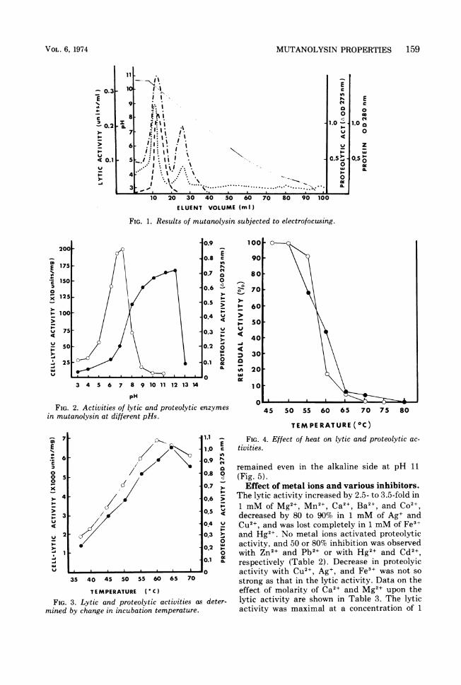
used to remove the excess salts from the decol-orized solution and to concentrate the solution,and 93% of the initial activity was recoveredfrom the concentrate obtained by ultrafiltra-tion. At this purification stage, about 32% oflytic activity was recovered with an increaseonly twice in specific lytic activity in compari-son with that of the starting material, whereasthe specific proteolytic activity decreased toabout 0.25 (Table 1). Mutanolysin was sub-jected to electrofocusing (Fig. 1). When theoptical density of the eluted fractions weremeasured at 280 nm, two main protein peakswere observed. The one with an isoelectric pointnear pH 8.5 had lytic activity, but the otherpeak with an isoelectric point near pH 10 hadproteolytic activity in addition to the lyticactivity.Enzymatic properties. Figure 2 shows the
activities of the lytic and proteolytic enzymes inmutanolysin at different pHs. The lytic activitywas affected greatly by pH. The optimal pH forthe lytic activity ranged from 6.5 to 7.0 inphosphate buffer. The curve of proteolytic ac-
tivity clearly differed from that of lytic activity,and the optimal pH was 12. The proteolyticactivity decreased remarkably as pH was low-ered below 7. The lytic and proteolytic activitiesdetermined by changing the incubation temper-ature from 37 to 70 C are shown in Fig. 3. Thelytic activity was highest at 60 C, whereas themaximal proteolytic activity was obtained at65 C. Upon heating for 10 min, a slight decreasein lytic activity at 55 C, a rapid decrease at 55to 65 C, and inactivation at over 70 C were
observed (Fig. 4). The stability curve of proteo-lytic enzyme was similar to that of lytic en-
zymes. When mutanolysin was incubated at37 C for 24 h in the various buffers withdifferent pH, the lytic activity was found to beretained in the acidic pH side, but the activitydecreased with increase in pH over 6.0. Approx-imately 50% of the proteolytic activity still
'he lytic enzyme from Streptomyces globisporus 1829
Step Vol |Total _ _Lytic activity Proteolytic activity
(mS)o protein U X 102 Recovery Sp actU x 102 RecO)(ml) ~(mg) (% (U/mg) M
0 cvr
Culture filtrate 1,000 750 1,140 (100) 152 924 (100)Amberlite CG-50 eluate 171 319 730 64 229 240 27(NH4)2S04 precipitate 22 257 547 48 212 223 24Duolite A-2 eluate 26 192 516 45 269 153 17pH treatment 26 122 394 35 323 36 4Ultrafiltration 3 122 368 32 302 38 4Lyophilized material 61mg 110 367 32 335 33 3
ANTIMICROB. AG. CHEMOTHER.
MUTANOLYSIN PROPERTIES 159
I II'I.FlU.:I.Igl:). .;l
. II"
I .I. i,,, ..
I .I '
.....
E
cmC
1.o
u
a.5
0I-0c-0L
10 20 30 40 50 60 70 80 90 100ELUENT VOLUME (ml)
FIG. 1. Results of mutanolysin subjected to electrofocusing.
EC0
041.0 o
0
0.50.lxXL
200E0.8 c
E175 I0.7 0a'A10
0.~~~ ~ ~ ~~~~~~~..2 125 I-
0.5
-10004
1 75 0.3
50 -0.2 o
0.~25 0.1 I%-a 0
3 4 5 6 7 8 9 10 11 12 13 14
pH
FIG. 2. Activities of lytic and proteolytic enzymesin mutanolysin at different pHs.
/XC//,
1.1 _E
1.0 cin
0.9 N
0.8 0
0.7 >.I-1
0.6 >
0.5 li
04 V0.3 >
00.2 o
0.1 X
35 40 45 50 55 60 65 70
TEMPERATURE (C)
FIG. 3. Lytic and proteolytic activities as deter-mined by change in incubation temperature.
470
f60
( 50v
40
30
~"20
10
45 50 55 60 65 70 75 80
TEMPERATURE (Cc)FIG. 4. Effect of heat on lytic and proteolytic ac-
tivities.
remained even in the alkaline side at pH 11(Fig. 5).Effect of metal ions and various inhibitors.
The lytic activity increased by 2.5- to 3.5-fold in1 mM of Mg2+, Mn2+, Ca2+, Ba2+, and Co2+,decreased by 80 to 90% in 1 mM of Ag+ andCu2+, and was lost completely in 1 mM of Fe3+and Hg2+. No metal ions activated proteolyticactivity, and 50 or 80% inhibition was observedwith Zn2+ and Pb2+ or with Hg2+ and Cd2+,respectively (Table 2). Decrease in proteolyicactivity with Cu2+, Ag+, and Fe3+ was not sostrong as that in the lytic activity. Data on theeffect of molarity of Ca2+ and Mg2+ upon thelytic activity are shown in Table 3. The lyticactivity was maximal at a concentration of 1
11
10
9~
VOL. 6, 1974
- 0.3E
.
0.2
"-
I-
4 0.1
-
z8
7
6
5
4
-.7E
±-6C2
00 50
x
"-
; 3U
v 2
U-A
v I- %F
1 c

160 YOKOGAWA ET AL.
100 0-0-0-090
80
070
I-60
50
40
.430
-20
10
2 3 4 5 6 7 8 9 1 11 12
pH
FIG. 5. Effect of pH on the stability of lytic andproteolytic enzymes.
TABLE 2. Effect of various metal ions on lytic andproteolytic activities in mutanolysin
Relative activity (%)Metal ion (nM)
Cell lysis Proteolysis
None 100 100Li+ 92 97Mg2+ 350 95Ca2+ 345 82Mn2+ 335 65CU2+ 21 90Zn2+ 54 59Fe2+ 53 104Fe3+ 0 74CO2+ 244 67Ni2+ 89 63Ag+ 13 88Cd2+ 53 23Ba2+ 358 91Hg2+ 0 16Pb2+ 71 43Ethylenediaminetetraacetic 76 87
acid
mM and decreased as ionic strengths were
increased.Table 4 shows the effect of some inhibitors on
the lytic and proteolytic activities. Mutanolysinwas preincubated with various enzyme inhibi-tors at 37 C for 5 min, and the residual activitieswere assayed. Both lytic and proteolytic activi-ties were not affected by chelating agents,sulfhydryl inhibitors, carbonyl reagents, sulfhy-dryl compounds, soybean trypsin inhibitor(Sigma, type 1-S), and diisopropyl phospho-fluoridate, but were inhibited nearly completely
at a concentration of 0.1 mM N-bromosuccini-mide. The lytic activity decreased to 13%, evenat the low concentration of 0.01 mM of theinhibitor.Lytic and bactericidal action of mutanoly-
sin. Figure 6 represents the typical time coursecurves of lysis and viable cell counts whenmutanolysin is allowed to act on S. mutansAHT and BHT. The optical density at 600 nm,due to whole cell suspensions of S. mutans AHTor BHT, decreased to 40 or 20% in 90 min in thepresence of 20,ug of mutanolysin per ml. How-ever, the rate of decrease in viable cell countswith S. mutans AHT was, on the contrary,about 1,000-fold greater than that with S.mutans BHT. For example, the viable cellcounts with S. mutans AHT decreased from 2 x10⁷ to 2 x 10² cells/ml after 90 min of incubation(equivalent to 99.999% killing of the initialcells), whereas the decrease in those with S.mutans BHT was from 5 x 10⁶ to 3 x 10⁴ cells(99.0% were killed). Mutanolysin causes lysis ofS. mutans BHT in human and hamster saliva(Fig. 7). Although the lytic activity in muta-nolysin was suppressed in the golden hamster'snatural saliva with pH 9.5, the activity wasrecovered when the pH of the saliva was ad-justed to neutral. In human natural saliva,streptococcal cells were well lysed, even thougha complete decrease in optical density did notoccur because of turbidity attributed to theopaque saliva.Spectrum of lytic activity. Mutanolysin was
tested against cell suspensions of various livingmicroorganisms to obtain its lytic spectrum.The results (Table 5) show the relative lysis ofmicroorganisms tested by mutanolysin whenlysis with S. mutans BHT was expressed as100%. All gram-negative organisms were resist-ant to lysis. The genus Streptococcus was foundto have a specific susceptibility to lysis by
TABLE 3. Effect of concentration of calcium andmagnesium ions on lytic activity in mutanolysin
Concn Relative activity (%)(mM)
Ca2+ Mg2+
None 100 1000.05 174 1860.10 200 1930.25 208 1980.50 213 2081.00 219 2112.50 195 1985.00 178 14810.00 87 8125.00 22 37
ANTIMICROB. AG. CHEMOTHER.

MUTANOLYSIN PROPERTIES 161
TABLE 4. Effect of various inhibitors on lytic andproteolytic activities in mutanolysin
Relative activity (%)Inhibitor
Cell lysis Proteolysis
NoneChelating agenta-a Dipyridyl8-HydroxyquinolinePotassium oxalateSuccinic acidO-phenanthrolineSodium diethyldithiocarba-mate
ThioureaSodium pyrophosphate
Sulfhydryl inhibitorIodoacetic acidSodium arseniteSodium arsenateSodium monofluoroacetate
Carbonyl reagentHydroxylamine hydrochlorideSemicarbazid hydrochloridePhenylhydrazine hydrochlo-
rideThiosemicarbazid hydrochlo-
rideHydrazine sulfate
Sulfhydryl comoundL-Ascorbic acid2-MercaptoethanolGlutathioneL-Cysteine hydrochlorideDimercaprol
Other inhibitorNaFSodium azideSoybean trypsin inhibitorDiisopropyl fluorophosphate1.0mM0.1mM
N-Bromosuccinimide1.0 mM0.1mM0.01 mM0.001 mM
100
101105979810194
10285
96908790
97112100
109
107
1069410510486
919075b
6980
081361
100
116a838390
93
9077
93119104 E105 o
0~o98 I-88110 >
VW
98 za
85 -i
99 a.0
1109810274
11010285b
7682
00
7396
a The agent was used at a concentration of 0.1 mM.,'To 10 ug of mutanolysin, 400 and 300 ,ug of
inhibitor for lysis and proteolysis were added, respec-tively.
mutanolysin. A. viscosus which causes perio-dontal disease and fissure caries in hamsterswas found to be highly susceptible to lysis justas was S. mutans BHT. Among the gram-posi-tive strains tested, only S. aureus was resistantto mutanolysin. Candida albicans, a species ofyeast, was not lysed.
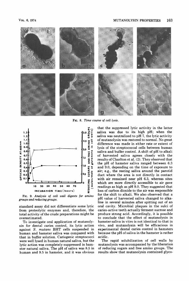
Electron microscopy observation on lysis.The course of lysis of S. mutans BHT was
followed by electron microscopy. Concentrationof mutanolysin was adjusted so that lysis withthe whole-cell suspension initiated by an opticaldensity of 0.6 at 600 nm would be completed in 1h. Figure 8 shows the time course of the celllysis. The oval masses of uniform thickness inFig. 8a are untreated streptococcal cells. Whenmutanolysin was added to a cell suspension, itwas observed that a cell was spurting theprotoplasm out of one side of the central cell bybursting in 5 min as shown in Fig. 8b. The edgesof the cells lost some thickness. The cell seen inFig. 8b left only a semicircular shell. Figure 8c
EU,
I.-z00v
.4v
-8
10 20 30 40 50 60 70 80 90
INCUBATION TIME (minutes)FIG. 6. Time course curves of Iysis and viable cell
counts by mutanolysis on S. mutans.
0.5
E 0.4 |o
0.3
z 0O.2
0.1IL~~ ~
0.
0 5 10 15 20
INCUBATION TIME (minutes)FIG. 7. Lysis of S. mutans by mutanolysin in human
and hamster saliva.
VOL. 6, 1974

162 YOKOGAWA ET AL.
TABLE 5. Susceptibility of various bacteria tomutanolysin
Organism
Actinomyces (Odontomyces) viscosus15987
Bacillus subtilis PCI 219Brucella abortus KusayanagiDiplococcus pneumoniae IEscherichia coli K-12 p-512Klebsiella sp. no. 13Lactobacillus acidophilus IFO 1110Listeria monocytogenes LI-2402Proteus vulgaris OX1,Pseudomonas aeruginosa TsuchijimaSalmonella typhimurium S-9Shigella flexneri 2a EW 10Shigella sonneiEW 33Staphylococcus aureus TerajimaStreptococcus mutansAHTS. mutans BHTS. mutans FA-1S. mutans HS-1S. mutans HS-6S. mutans IngbrittS. mutans K1-RStreptococcus salivarius IFO 3350S. salivarius HHTStreptococcus sanguis OMZ 9S. sanguis M5S. sanguis ATCC 10556Streptococcus CHT (unidentified)Candida albicans ATCC 10257
Lyticactivityrelativevalue
112
280
380
0
82320
0
0
0
0
0
113
100
73
117
101
53
114
42
33
21
96
8
116
0
'aRelative ratio of lysis for the tested organismswhen the lysis on S. mutans BHT was expressed as
100%.
shows some loss in thickness of the cells at theterminal positions and a large, flattened shellwhich occurred by rupturing at the equatorialring of the cell. This effect became more markedby digesting to 10 min, and many cells changedto circular or semicircular shells (Fig. 8d). Apair of semicircular shells split into two partswas observed after 20 min of incubation (Fig.8e), and the lysis was most drastic on the crosswall at the equator. In the semicircular shells, avisible inner line which appears to be a crosswall at next cell division, appeared in parallelwith a diametrical outer edge. A cell in Fig. 8f,which loses thickness by leaking intracellularmaterials, shows three faded lines in it. Two ofthem are situated symmetrically about a dia-metrical line. After 60 min, a number of cellsdisappeared against background, and somethin, indefinite sheets remained (Fig. 8g). How-ever, the enlarged photograph (Fig. 8f) shows
that the sheets consist of semicircular frag-ments or secondarily torn off fragments.Products of cell walls degraded by mutan-
olysin. A reaction mixture containing 250 mg ofcell walls of S. mutans BHT, 400 mg of NaN,,1,100 U of mutanolysin, and 1 mM MgSO4 in 50ml of 0.02 M phosphate buffer (pH 6.5) wasincubated at 37 C for 72 h. The samples werecollected at different time intervals and heatedat 100 C for 5 min. After centrifugation, thesupematant was analyzed for amino groups andreducing sugars. The results are shown in Fig. 9.Maximal lysis of cell walls was obtained in 7 hwith the liberation of 1 mol of reducing sugarsand 2 mol of free amino groups per 1 mol of totalglutamic acid in the digests.
DISCUSSIONMutanolysin from S. globisporus 1829 con-
tains mainly one proteolytic and two lyticenzymes. The lytic enzymes found were basicproteins with an isoelectric point near pH 8.5and 10 by electrofocusing. The latter lytic en-zyme possessed the same isoelectric point as theproteolytic enzyme, and they seemed to have asimilar molecular size from the fact that bothenzymatic activities leaked out in nearly equalratios into the filtrates through three kinds ofmembranes. Furthermore, they seemed to havesome specific tryptophan residues on the activesites, as postulated from the fact that they wereinactivated by oxidation with N-bromosuccini-mide. Final proof of the identity of both en-zymes must await complete purification data.However, it seems more probable that both ofthe lytic and proteolytic enzymes are differentupon further purification by diethylaminoethyl-cellulose and O-(carboxymethyl)-Sephadex col-umn chromatography.
In the purification process, removal of cationssuch as calcium and magnesium derived fromthe culture filtrate results in apparent loss oflytic activity. Hence, the assay of lytic activityin each of the purification processes was madein the presence of 1 mM MgCI, to obtain a trueyield. Nevertheless, the specific lytic activitydid not increase as we expected in the purifica-tion procedure. The main reason for a littleincrease in specific lytic activity probably lies inthe removal of proteolytic enzymes, which en-hances an apparent lytic activity by its clearingaction against cell debris during the purificationsteps. Sudo and Dworkin (23) have made asimilar interpretation in studies on lytic enzymefrom Myxococcus xanthus that the degree ofpurification could not be calculated in such astandard assay to measure decrease in turbidityof a suspension of whole cells, because the
ANTIMICROB. AG. CHEMOTHER.
MUTANOLYSIN PROPERTIES 163
FIG. 8. Time course of cell lysis.
1.3 3.0 01.2
2.5 07
c1.0~~ ~ ~ ~ ~~~~15
lo. 203 0so 07
0~~~~~~~~~~~~~~~.
0.8~~ ~ ~ ~ ~~~ Su 0.
FIG. 9.Aayiso elwaldgssEomn
0.1
0.6~ ~ ~ ~ ~ ~
z0.5~ ~ ~ ~ ~
groups and reducing groups.
standard assay did not differentiate some lytic
from proteolytic enzymes and, therefore, the
total activity of the crude preparations might be
overestimated.
To investigate oral application of mutanoly-
sin for dental caries control, its lytic action
against S. mutans BHT cells suspended in
human and hamster saliva was compared with
that in buffer solution. Cariogenic streptococci
were well lysed in human natural saliva, but the
lytic action was completely suppressed in ham-
ster natural saliva. The pH of saliva was 8.5 in
human and 9.5 in hamster, and it was obvious
that the suppressed lytic activity in the lattersaliva was due to its high pH; when thesaliva was neutralized to pH 7, the lytic activityof mutanolysin was restored to normal. No greatdifference was made in either rate or extent oflysis of the streptococcal cells between humansaliva and buffer control. A shift of pH to alkaliof harvested saliva agrees closely with theresults of Charlton et al. (2). They observed thatthe pH of hamster saliva ranged between 6.3and 9.0, depending on the time of exposure toair; e.g., the resting saliva around the parotidduct where the area is not directly in contactwith air remained near pH 6.3, whereas siteswhich are more directly accessible to air gavereadings as high as pH 9.0. They suggested thatloss of carbon dioxide to the air was responsiblefor the shift to alkali. We also observed that apH value of harvested saliva changed to alka-line in several minutes after spitting out of anoral cavity. Microbial plaques in the sulci ofcaries-active teeth actually ferment sucrose andproduce strong acid. Accordingly, it is possibleto conclude that the effect of mutanolysin inhamster saliva in vitro is not identical to that invivo, and mutanolysin will be efficacious inexperimental dental caries control in hamstersbecause the pH of saliva in the hamster is ratheracidic.The rapid solubilization of cell walls by
mutanolysin was accompanied by the liberationof reducing sugars and free amino groups. Theresults show that mutanolysin contained glyco-
VOL. 6, 1974

164 YOKOGAWA ET AL.
sidase and peptidase or amidase. We haveobtained some evidence that the two main lyticenzymes seemed to be N-acetyl muramidase.However, the lytic mechanisms must be eluci-dated on pure cell walls by using perfectlypurified enzymes to explain whether peptidaseor amidase in mutanolysin is identical with theproteolytic enzyme or not.Mutanolysin was found to have certain prop-
erties similar to egg white lysozyme; e.g., muta-nolysin is inert to S. aureus and all gram-nega-tive bacteria tested, exhibits some glucosidaseactivity, consists of basic proteins with alkalineisoelectric points and is inactivated by N-bromosuccinimide-like egg white lysozyme.However, there is a big difference betweenmutanolysin and lysozyme; that is, the former iscapable of lysing the living cells and cell walls ofS. mutans, whereas the latter is inactive againstthese strains.
Fitzgerald et al. (4) have reported that theactive Lactobacillus developed in dental cariesin weanling rats appeared to be a variety ofLactobacillus acidophilus. Recently, Ikeda etal. (11) have observed that S. mutans andLactobacillus in plaque related to the initiationof human dental caries, and the latter bacteriabecame a sizable plaque microflora only afterthe appearance of caries. Odontomyces viscosus(renamed Actinomyces viscosus) isolated fromsubgingival plaque in hamsters has been dem-onstrated to have an ability to produce experi-mental periodontal disease in hamsters. Muta-nolysin was easily capable of lysing cariogenicstreptococci with four morphological categoriesconveniently classified by Jablon and Zinner(12). In addition, mutanolysin was also capableof lysing the living cells of A. viscosus and L.acidophilus. Mutanolysin may be expected to bea potentially useful agent for dental cariescontrol because it (i) has lytic action against S.mutans, S. sanguis L. acidophilus, and A.viscosus, (ii) has optimal pH with acidity, (iii)has lytic activity in human saliva, and (v) isactivated by calcium ions at concentrationsgenerally found in human saliva (20).
In a subsequent paper, we will describe muta-nolysin efficacy in vitro on the elimination ofmicrobial plaques.
ACKNOWLEDGMENTSWe express our sincere thanks to K. Ogata, Department of
Agricultural Chemistry, Kyoto University, T. Morioka, De-partment of Preventive Dentistry, School of Dentistry, Kyu-shu University, Japan, and S. Ose, T. Mizuma, and S.Takamatsu of this laboratory for their suggestions during theinvestigation.
LITERATURE CITED
1. Barkulis, S. S., C. Smith, J. J. Boltralik, and H.Heymann. 1964. Structure of streptococcal cell walls.IV. Purification and properties of streptococcal phagemuralysin. J. Biol. Chem. 239:4027-4043.
2. Charlton, G., R. J. Fitzgerald, and P. H. Keyes. 1971.Determination of saliva and dental plaque pH inhamsters with glass micro-electrodes. Arch. Oral Biol.16:649-654.
3. Dixon, R. E., J. S. Godman, and M. G. Koenig. 1968.Lysostaphin: an enzymatic approach to staphylococcaldisease. III. Combined lysostaphin-methicillin therapyof established staphylococcal abscesses in mice. Yale J.Biol. Med. 41:62-68.
4. Fitzgerald R. J., H. V. Jordan, and H. 0. Archard. 1966.Dental caries in gnotobiotic rats infected with a varietyof Lactobacillus acidophilus. Arch. Oral Biol.11:473-476.
5. Fitzgerald R. J., and P. H. Keyes. 1963. Ecologic factorsin dental caries. The fate of antibiotic-resistant organicstreptococci in humans. Amer. J. Pathol. 42:759-772.
6. Ghuysen, J. M., D. J. Tipper, and J. L. Strominger.1966. Enzymes that degrade bacterial cell walls, p.685-699. In E. F. Newfeld and V. Ginsburg (ed.),Methods in enzymology, vol. 8. Academic Press Inc.,New York.
7. Goldberg L. M., J. M. De Franco, C. Watanakunakorn,and M. Hamburger. 1968. Studies in experimentalstaphylococcal endocarditis in dogs. VI. Treatmentwith lysostaphin, p. 45-53. Antimicrob. Ag. Chemo-ther. 1967.
8. Guggenheim, B., K. G. Konig, H. R. Miihlemann, and B.Regolati. 1969. Effect of dextranase on caries in ratsharbouring an indigenous cariogenic bacterial flora.Arch. Oral Biol. 14:555-558.
9. Harris, R. L., A. W. Nunnery, and H. D. Riley, Jr. 1968.Effect of lysostaphin on staphylococcal carriage ininfants and children, p. 110-112. Antimicrob. Ag.Chemother. 1967.
10. Hayashi, K., T. Imoto, and M. Funatsu. 1965. Theposition of the active tryptophan residue in lysozyme.J. Biochem. 58:227-235.
11. Ikeda, T., H. J. Sand Lam, and E. L. Bradley, Jr. 1973.Changes in Streptococcus mutans and Lactobacilli inplaque in relation to the initiation of dental caries innegro children. Arch. Oral Biol. 18:555-556.
12. Jablon, J. M., and D. D. Zinner. 1966. Differentiation ofcariogenic streptococci by fluorescent antibody. J.Bacteriol. 92:1590-1596.
13. Kato, K., T. Hirota, Y. Murayama, H. Suginaka, and S.Kotani. 1968. Studies on the mode of action of Flavo-bacterium L-11 enzyme on the cell walls of Staphylo-coccus aureus strain Copenhagen. Identification ofisolated cell wall peptides. Biken J. 11:1-12.
14. Kenneth, E. Q. Jr., S. Richard, R. C. Jacques, F. N.Nedo, and S. William. 1971. Efficacy and safety oftopical lysostaphin treatment of persistent nasal car-riage of Staphylococcus aureus. Appl. Microbiol.22:446-450.
15. Konig, K. G., and B. Guggenheim. 1968. In vivo effects ofdextranase on plaque and caries. Helv. Odontol. Acta12:48-55.
16. Krasse, B., and J. Carlsson. 1970. Various type ofstreptococci and experimental caries in hamsters.Arch. Oral Biol. 15:25-32.
17. Krause, R. M. 1963. Antigenic and biochemical composi-tion of hemolytic streptococci cell walls. Bacteriol. Rev.27:369-380.
18. Lobene, R. R. 1971. A clinical study of the effect ofdextranase on human dental plaque. J. Amer. Dent.
ANTIMICROB. AG. CHEMOTHER.

MUTANOLYSIN PROPERTIES 165
Ass. 82:132-135.19. Lowry, 0. H., N. J. Rosebrough, A. L. Farr, and R. J.
Randall. 1951. Protein measurement with the Folinphenol reagent. J. Biol. Chem. 193:265-275.
20. McCann, H. G. 1968. Inorganic components of salivarysecretion, p. 56-57. In R. S. Harris (ed.), Art andscience of dental caries research. Academic Press Inc.,New York.
21. Minah, G. E., W. J. Loesche, and D. D. Dziewiavkowski.1972. The in vitro effect of fungal dextranase on humandental plaque. Arch. Oral Biol. 17:35-42.
22. Park, J. T., and M. J. Johnson. 1949. A submicrodetermi-nation of glucose. J. Biol. Chem. 181:149-151.
23. Sudo, S., and M. Dworkin. 1972. Bacteriolytic enzymesproduced by Myxococcus xanthus. J. Bacteriol.110:236-245.
24. Yokogawa, K., S. Kawata, and Y. Yoshimura. 1972.Studies on lytic enzyme against cariogenic strepto-cocci. I. Bacteriolytic activity of enzymes derived fromStreptomyces species. Agr. Biol. Chem. 36:2055-2065.
25. Yokogawa K., S. Kawata, and Y. Yoshimura. 1973.Studies on lytic enzyme against cariogenic strepto-cocci. II. Lytic enzyme from Streptomyces globisporus1829 strain. Agr. Biol. Chem. 37:799-808.
26. Vesterberg, O., and H. Svensson. 1966. Isoelectric frac-tionation. Analysis and characterization of ampholitein natural pH gradients. Acta Chem. Scand.20:820-834.
27. Zinner, D. D., J. M. Jablon, A. P. Aran, and M. S.Saslaw. 1965. Experimental caries induced in animalsby streptococci of human origin. Proc. Soc. Exp. Biol.Med. 118:766-770.
VOL. 6, 1974