Embed Size (px)
Citation preview

J. Anat.
(2006)
208
, pp153–167
© 2006 The Authors Journal compilation © 2006 Anatomical Society of Great Britain and Ireland
Blackwell Publishing Ltd
Muscles of facial expression in the chimpanzee (
Pan troglodytes
): descriptive, comparative and phylogenetic contexts
Anne M. Burrows,
1,2
Bridget M. Waller,
3
Lisa A. Parr
4,5
and Christopher J. Bonar
6
1
Department of Physical Therapy, Duquesne University, Pittsburgh, USA
2
Department of Anthropology, University of Pittsburgh, Pittsburgh, USA
3
Department of Psychology, University of Portsmouth, UK
4
Yerkes National Primate Research Center, Atlanta, USA
5
Department of Psychiatry and Behavioral Sciences, Emory University, Atlanta, USA
6
Cleveland Metroparks Zoo, USA
Abstract
Facial expressions are a critical mode of non-vocal communication for many mammals, particularly non-human pri-
mates. Although chimpanzees (
Pan troglodytes
) have an elaborate repertoire of facial signals, little is known about
the facial expression (i.e. mimetic) musculature underlying these movements, especially when compared with some
other catarrhines. Here we present a detailed description of the facial muscles of the chimpanzee, framed in com-
parative and phylogenetic contexts, through the dissection of preserved faces using a novel approach. The arrange-
ment and appearance of muscles were noted and compared with previous studies of chimpanzees and with
prosimians, cercopithecoids and humans. The results showed 23 mimetic muscles in
P. troglodytes
, including a thin
sphincter colli muscle, reported previously only in adult prosimians, a bi-layered zygomaticus major muscle and a
distinct risorius muscle. The presence of these muscles in such definition supports previous studies that describe an
elaborate and highly graded facial communication system in this species that remains qualitatively different from
that reported for other non-human primate species. In addition, there are minimal anatomical differences
between chimpanzees and humans, contrary to conclusions from previous studies. These results amplify the impor-
tance of understanding facial musculature in primate taxa, which may hold great taxonomic value.
Key words
chimpanzee; hominines; mimetic, phylogeny.
Introduction
The study of facial expressions and communication has
a rich history from Duchenne (1862) to Darwin (1872)
and through to the present (e.g. Ekman, 1973; Ekman
& Oster, 1979; Kaiser, 2002). Darwin (1872), in particu-
lar, stressed the importance of comparing facial expres-
sions among humans and other mammals in order to
understand the evolution of non-vocal communication
systems. Additionally, he posited that the means for
expressing emotion through movements of the face
may be similar among humans and other mammals,
particularly among the primates. However, there is
very little research that has actually attempted to com-
pare the facial expression musculature (mimetic muscu-
lature) among primates. This is a particularly important
endeavour, as the majority of the literature, although
old, adopts a hierarchical ascending phylogenetic model
that proposes that as species get more closely related
to humans, their communicative facial repertoire and
underlying facial musculature become more elaborate
(Gregory, 1929; Huber, 1931).
The muscles of facial expression are branchiomeric in
origin and are innervated by the seventh cranial nerve
(Young, 1957, 1962; Walker & Liem, 1994). These
muscles generally serve to move the vibrissae, change
the sizes of the oral, orbital and nasal openings, aid in
Correspondence
Dr Anne M. Burrows, Department of Physical Therapy, Duquesne University, 600 Forbes Ave., Pittsburgh, PA 15282, USA. T: +1 412 396 5543; E: [email protected]
Accepted for publication
26 September 2005

Facial expression musculature in Pan, A. M. Burrows et al.
© 2006 The AuthorsJournal compilation © 2006 Anatomical Society of Great Britain and Ireland
154
nutrient intake, and function in chemoception,
olfaction and audition, and, in non-mammalian orders
(excluding Aves), change the size of the gill openings
and aid in opening the mouth (Huber, 1930a,b; Young,
1962). In Osteichthyes and Chondrichthyes, these muscles
are primarily organized as sphincters to aid in elevation
of the mandible and in constriction of the pharynx and
gills (Gregory, 1929; Walker & Liem, 1994). In lower
eutherian mammals, such as the Perissodactyla and
Artiodactyla, the facial expression musculature is rela-
tively simple in its attachments into the dermis and
pinna, is relatively flat and undifferentiated, and is few
in number (Sisson, 1921). However, in higher eutherian
mammals, especially primates, this musculature is addi-
tionally used in transmitting close-proximity social
information such as emotional states, territorial inten-
tions, and mate and individual recognition, and is used
in a variety of agonistic and conciliatory displays
(Darwin, 1872; Huber, 1930a,b, 1931; van Hooff, 1962;
Andrew, 1963; Bearder et al. 1995; Preuschoft & van
Hooff, 1995; Schmidt & Cohn, 2001; Parr et al. 2002,
2005). Indeed, it has been argued that primate facial
musculature has been shaped by natural selection
specifically to aid communication among individuals
(Huber, 1931). However, in contrast to the literature
describing facial displays in primate species, the litera-
ture describing and comparing the anatomy of the
facial muscles is surprisingly sparse. For example, the
facial expressions of the chimpanzee (
Pan troglodytes
),
in particular, have received much attention from
behavioural scientists (e.g. Marler, 1965, 1976; van
Hooff, 1972, 1973; Goodall, 1986; Parr, 2003; Parr et al.
2005), and yet detailed facial dissections are lacking in
the literature.
Our understanding of primate mimetic musculature
has traditionally been rooted in a phylogenetic con-
text. For example, it is traditionally held that the most
primitive primates, the prosimians, have the least
complex arrangement of facial expression musculature,
consisting of large, relatively undifferentiated sheets
of muscle that perform relatively gross, non-specific
functions (Huber, 1931; Schultz, 1969). As one moves
up the phylogenetic hierarchy toward
Homo sapiens
, it
is held that the number of muscles increases at the level
of each taxon and that their function in moving specific
facial regions increases as social networks become
more intricate (Huber, 1930a,b, 1931; van Hooff, 1962;
Schultz, 1969; Preuschoft, 2000). The primate taxa
traditionally held to have the least complex (structurally
and functionally) facial expression musculature are
nocturnal – the lemurs (except for the diurnal
Lemur
,
Varecia
, and the indriids and the cathemeral
Eulemur
and
Hapalemur
) and lorises (Murie & Mivart, 1872;
Ruge, 1885; Gregory, 1929; Huber, 1931; Lightoller,
1934; Seiler, 1975), while the almost entirely diurnal
Anthropoidea are held to have the most complex facial
expression musculature and social systems, with
Homo
sapiens
at the apex (e.g. Lightoller, 1928; Gregory,
1929; Huber, 1931, 1933; Schultz, 1969; Pellatt,
1979a,b; Preuschoft, 2000; Stranding, 2004).
Our best understanding of primate facial expression
musculature comes from the hominines (the African
apes), especially from
Homo
.
Gorilla
musculature has
been described, but only superficially, by Huber (1931)
and Raven (1950). The muscles presented in these
accounts are either partially unlabelled and/or not
described in the text. Indeed, Huber (1931) used only
juveniles in his description. A similar situation exists in
Pan troglodytes
(Sonntag, 1923; Huber, 1931; Pellatt,
1979b). Overall, the accounts of
P. troglodytes
are
problematic in part due to differences in the muscula-
ture reported. For example, Sonntag (1923) reports a
distinct risorius muscle in
P. troglodytes
while Pellatt
(1979b) does not report one. Whether this is due to a
genuine difference among the specimens examined or
merely represents a different focus between the two
studies is unknown. In a recent work, Gibbs et al. (2002)
reviewed the existing literature on hominoid soft-
tissue anatomy. The section dealing with the muscles
of facial expression was scanty but represented the
published accounts to date on all hominoids.
Recent studies have questioned the validity of the
traditional ‘phylogenetic model’ of the complexity
of primate facial expression musculature and primate
facial displays. For example, Burrows & Smith (2003)
found greater complexity in the facial muscles of
Otolemur
(the greater bushbaby) than phylogenetic
models would predict (see also Murie & Mivart, 1872).
Sherwood et al. (2003, 2005) examined the facial
nucleus of the brainstem in a variety of primate taxa
and found a number of specializations at each taxo-
nomic level, revealing that there is no simple increase
in complexity as the phylogenetic scale is ascended
towards
Homo
.
Although conceptualizing primate facial musculature
complexity among taxa using the traditional ‘phylo-
genetic model’ (Gregory, 1929; Huber, 1931) may not
be a completely useful tool, an accurate rendering of
Facial expression musculature in Pan, A. M. Burrows et al.
© 2006 The Authors Journal compilation © 2006 Anatomical Society of Great Britain and Ireland
155
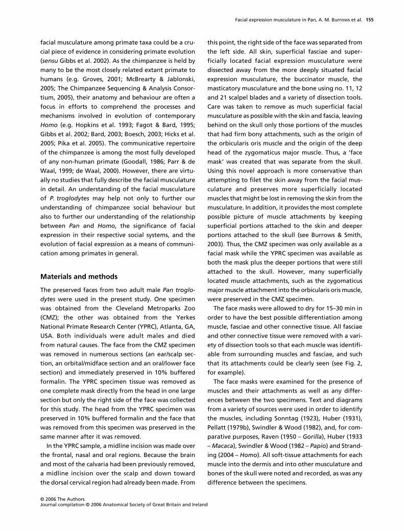
facial musculature among primate taxa could be a cru-
cial piece of evidence in considering primate evolution
(
sensu
Gibbs et al. 2002). As the chimpanzee is held by
many to be the most closely related extant primate to
humans (e.g. Groves, 2001; McBrearty & Jablonski,
2005; The Chimpanzee Sequencing & Analysis Consor-
tium, 2005), their anatomy and behaviour are often a
focus in efforts to comprehend the processes and
mechanisms involved in evolution of contemporary
Homo
(e.g. Hopkins et al. 1993; Fagot & Bard, 1995;
Gibbs et al. 2002; Bard, 2003; Boesch, 2003; Hicks et al.
2005; Pika et al. 2005). The communicative repertoire
of the chimpanzee is among the most fully developed
of any non-human primate (Goodall, 1986; Parr & de
Waal, 1999; de Waal, 2000). However, there are virtu-
ally no studies that fully describe the facial musculature
in detail. An understanding of the facial musculature
of
P. troglodytes
may help not only to further our
understanding of chimpanzee social behaviour but
also to further our understanding of the relationship
between
Pan
and
Homo
, the significance of facial
expression in their respective social systems, and the
evolution of facial expression as a means of communi-
cation among primates in general.
Materials and methods
The preserved faces from two adult male
Pan troglo-
dytes
were used in the present study. One specimen
was obtained from the Cleveland Metroparks Zoo
(CMZ); the other was obtained from the Yerkes
National Primate Research Center (YPRC), Atlanta, GA,
USA. Both individuals were adult males and died
from natural causes. The face from the CMZ specimen
was removed in numerous sections (an ear/scalp sec-
tion, an orbital/midface section and an oral/lower face
section) and immediately preserved in 10% buffered
formalin. The YPRC specimen tissue was removed as
one complete mask directly from the head in one large
section but only the right side of the face was collected
for this study. The head from the YPRC specimen was
preserved in 10% buffered formalin and the face that
was removed from this specimen was preserved in the
same manner after it was removed.
In the YPRC sample, a midline incision was made over
the frontal, nasal and oral regions. Because the brain
and most of the calvaria had been previously removed,
a midline incision over the scalp and down toward
the dorsal cervical region had already been made. From
this point, the right side of the face was separated from
the left side. All skin, superficial fasciae and super-
ficially located facial expression musculature were
dissected away from the more deeply situated facial
expression musculature, the buccinator muscle, the
masticatory musculature and the bone using no. 11, 12
and 21 scalpel blades and a variety of dissection tools.
Care was taken to remove as much superficial facial
musculature as possible with the skin and fascia, leaving
behind on the skull only those portions of the muscles
that had firm bony attachments, such as the origin of
the orbicularis oris muscle and the origin of the deep
head of the zygomaticus major muscle. Thus, a ‘face
mask’ was created that was separate from the skull.
Using this novel approach is more conservative than
attempting to filet the skin away from the facial mus-
culature and preserves more superficially located
muscles that might be lost in removing the skin from the
musculature. In addition, it provides the most complete
possible picture of muscle attachments by keeping
superficial portions attached to the skin and deeper
portions attached to the skull (see Burrows & Smith,
2003). Thus, the CMZ specimen was only available as a
facial mask while the YPRC specimen was available as
both the mask plus the deeper portions that were still
attached to the skull. However, many superficially
located muscle attachments, such as the zygomaticus
major muscle attachment into the orbicularis oris muscle,
were preserved in the CMZ specimen.
The face masks were allowed to dry for 15–30 min in
order to have the best possible differentiation among
muscle, fasciae and other connective tissue. All fasciae
and other connective tissue were removed with a vari-
ety of dissection tools so that each muscle was identifi-
able from surrounding muscles and fasciae, and such
that its attachments could be clearly seen (see Fig. 2,
for example).
The face masks were examined for the presence of
muscles and their attachments as well as any differ-
ences between the two specimens. Text and diagrams
from a variety of sources were used in order to identify
the muscles, including Sonntag (1923), Huber (1931),
Pellatt (1979b), Swindler & Wood (1982), and, for com-
parative purposes, Raven (1950 –
Gorilla
), Huber (1933
–
Macaca
), Swindler & Wood (1982 –
Papio
) and Strand-
ing (2004 –
Homo
). All soft-tissue attachments for each
muscle into the dermis and into other musculature and
bones of the skull were noted and recorded, as was any
difference between the specimens.
Facial expression musculature in Pan, A. M. Burrows et al.
© 2006 The AuthorsJournal compilation © 2006 Anatomical Society of Great Britain and Ireland
156
Results
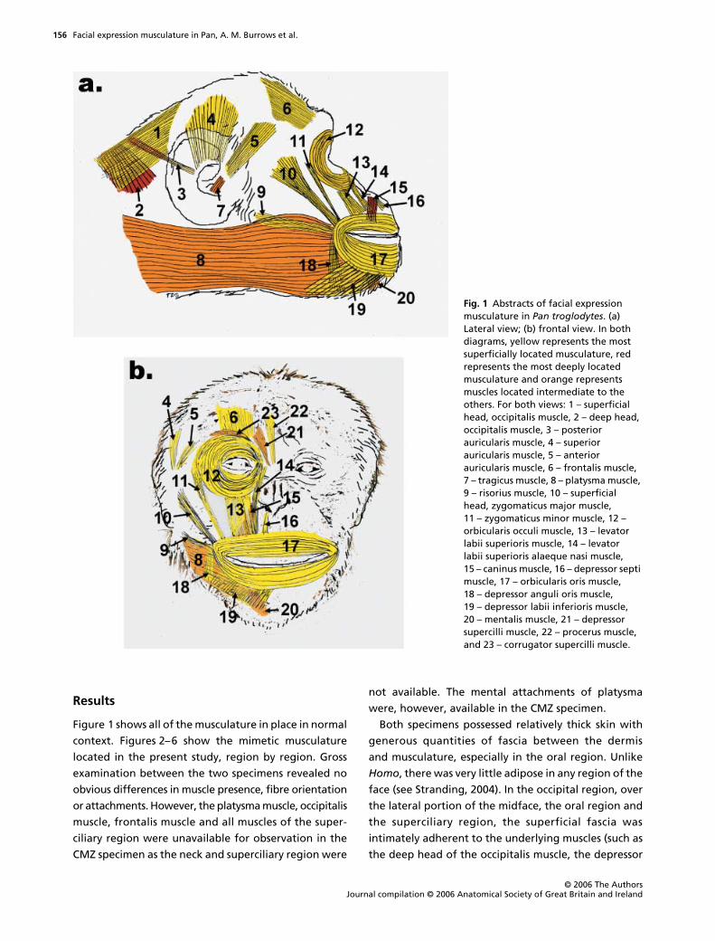
Figure 1 shows all of the musculature in place in normal
context. Figures 2–6 show the mimetic musculature
located in the present study, region by region. Gross
examination between the two specimens revealed no
obvious differences in muscle presence, fibre orientation
or attachments. However, the platysma muscle, occipitalis
muscle, frontalis muscle and all muscles of the super-
ciliary region were unavailable for observation in the
CMZ specimen as the neck and superciliary region were
not available. The mental attachments of platysma
were, however, available in the CMZ specimen.
Both specimens possessed relatively thick skin with
generous quantities of fascia between the dermis
and musculature, especially in the oral region. Unlike
Homo
, there was very little adipose in any region of the
face (see Stranding, 2004). In the occipital region, over
the lateral portion of the midface, the oral region and
the superciliary region, the superficial fascia was
intimately adherent to the underlying muscles (such as
the deep head of the occipitalis muscle, the depressor
Fig. 1 Abstracts of facial expression musculature in Pan troglodytes. (a) Lateral view; (b) frontal view. In both diagrams, yellow represents the most superficially located musculature, red represents the most deeply located musculature and orange represents muscles located intermediate to the others. For both views: 1 – superficial head, occipitalis muscle, 2 – deep head, occipitalis muscle, 3 – posterior auricularis muscle, 4 – superior auricularis muscle, 5 – anterior auricularis muscle, 6 – frontalis muscle, 7 – tragicus muscle, 8 – platysma muscle, 9 – risorius muscle, 10 – superficial head, zygomaticus major muscle, 11 – zygomaticus minor muscle, 12 – orbicularis occuli muscle, 13 – levator labii superioris muscle, 14 – levator labii superioris alaeque nasi muscle, 15 – caninus muscle, 16 – depressor septi muscle, 17 – orbicularis oris muscle, 18 – depressor anguli oris muscle, 19 – depressor labii inferioris muscle, 20 – mentalis muscle, 21 – depressor supercilli muscle, 22 – procerus muscle, and 23 – corrugator supercilli muscle.

Facial expression musculature in Pan, A. M. Burrows et al.
© 2006 The Authors Journal compilation © 2006 Anatomical Society of Great Britain and Ireland
157
supercilli muscle and the zygomaticus major muscle),
such that the scalpel blade frequently had to be used to
free the muscle fascicles from the overlying fascia. The
more superficially located muscles (such as the super-
ficial head of the occipitalis muscle, the zygomaticus
minor muscle and the risorius muscle) were frequently
intermingled with fascia. The remaining muscles were
robust, attached to discrete portions of the dermis or
cartilaginous pinna, skull and/or into other muscles.
In general, the muscles associated with the scalp and
pinna were fairly gracile whereas the muscles associ-
ated with the oral region were the thickest and largest.
Muscles associated with the orbital region were inter-
mediate in size (see Figs 2–6).
Individual muscles (see Table 1 and Figs 1–6)
Platysma muscle (Figs 1 and 3)
This muscle is flat, thin and broad with fibres running
horizontally from the cervical region, passing inferior
to the pinna and attaching partially into the oral
modiolus. More inferiorly located fibres pass along the
ventral aspect of the neck and attach into the mental
region, mingling with fibres of the orbicularis oris
muscle of the lower lip. This muscle is directly deep to the
skin with only weak attachments to the skin itself. It lies
deep to the risorius muscle but superficial to the deep
head of the occipitalis muscle, the mentalis muscle, and
the depressors anguli and labii inferioris muscles. It has
a firm attachment to the superficial head of the occipitalis
muscle. Huber (1931) describes this muscle in
P. troglodytes
as having lost its occipital and cervical portions, but
these are quite robust in the present specimens.
Sphincter colli muscle (Figs 1, 2 and 6)
This muscle is consistently noted in the prosimian pri-
mates but has not been described for anthropoids.
Here, there is a thickened layer of the superficial fascia
along the lateral portion of the face stretching from the
region of the oral commissure to the inferior border of
the mandible, passing caudally to the skin of the region
of the mandibular ramus. The fibres here are fleeting
and sparse, located superficial to the platysma muscle.
Occipitalis muscle, superficial head (Figs 1, 2 and 4)
This is a small, flat muscle embedded within the
superficial fascia associated with the occipital region.
Horizontal fibres pass from the fascia in the occipital
region to the deep head of the occipitalis muscle, to
which it fuses. Where the superficial head terminates
over the calvaria, the galea aponeurotica begins.
Pellatt (1979b) described this muscle as only a thin, fibrous
sheet, but it is a quite distinct, muscular structure in the
specimens studied here.
Fig. 2 Right side of facial mask from Pan troglodytes. This is a view of the deep surface of the face as dissected away from the skull.

Facial expression musculature in Pan, A. M. Burrows et al.
© 2006 The AuthorsJournal compilation © 2006 Anatomical Society of Great Britain and Ireland
158
Occipitalis muscle, deep head (Figs 1 and 3)
As noted by Pellatt (1979b), this is a thick, robust muscle
deep to the platysma muscle and the superficial head of
the occipitalis muscle. It has an extensive bony origin from
the superior nuchal crest immediately lateral to the tra-
pezius muscle but medial to the posterior auricularis
muscle. It passes inferolaterally, deep to the platysma muscle,
to which it fuses. Sonntag (1923) described only a single
occipitalis muscle, attaching to the bony landmarks cor-
responding to those found here for the deep head.
Frontalis muscle (Figs 1 and 2)
This is a flat, very thin sheet of muscle composed of
fibres that run from a caudal attachment at the galea
Table 1 Muscles of facial expression in Pan
Muscle Attachments
platysma occipitalis superficial m., modiolus, inferior aspect of orbicularis oris m., skin inferior to pinna back to occipital region and forward to zygomatic arch region and ventrally over the neck
sphincter colli fleeting fibres from oral commissure and slightly inferior to region of opening for ear canal over the area of the mandibular ramus
occipitalis (superficial belly) fleeting fibres mixed with superficial fascia, attached to the skin of the posterolateral scalp and to the platysma muscle and the occipitalis muscle deep belly
occipitalis (deep belly) large robust fibres from the superior nuchal crest next to the insertion of the trapezius muscle to attach into the deep surface of the caudal fibres of platysma
muscle frontalis galea aponeurotica of the scalp to the skin of the superciliary region as a flat sheetanterior auricularis anterolateral portion of scalp to the auricular cartilage near the junction of the helix and antihelix as one
large fan of fibres the auricular cartilage near the base of the antihelix as a broad, flat sheetposterior auricularis from the occipital bone at the superior nuchal crest to the posteiror portion of the pinna at the base of the
antihelix as one robust set of fibrestragicus skin over the lateral aspect of the midface close to the zygomatic arch region and the tragusorbicularis occuli gracile, sphincter-like fibres attached to the skin of the eyebrow, eyelid, and around orbital opening
(orbital part); attached to zygomaticus minor, levator labii superioris, and depressor supercilli muscles and to the frontal and lacrimal bones via the medial palpebral ligament
corrugator supercilli deep to orbicularis occuli muscle fibres; attached to skin of superciliary region and to medial portion of the bony orbit near the palpebral ligament
depressor supercilli on same level with corrugator supercilli m.; attached to skin over lateral aspect of nose, medial to orbicularis occuli muscle, to skin of medial portion of eyebrow proce on same level with orbicularis occuli m and deep to depressor supercilli m.; attached to skin over lateral aspect of nose, medial to orbicularis occuli muscle, to skin superior to eyebrow
zygomaticus major by two heads: deep head from lateral portion of zygomatic arch; superficial head from skin over zygomatic arch; heads join about half of the way down and attach into orbicularis oris muscle at the modiolus
zygomaticus minor small fibres from skin superficial to zygoma and from the lateral portion of orbicularis occuli muscle to orbicularis oris muscle, medial to zygomaticus major muscle
levator labii superioris large set of flat fibres from skin of midface and from the inferior fibres of orbicularis occuli muscle to skin of upper lip lateral to insertion of levator labii superioris alaeque nasi muscle and to the orbicularis oris muscle
levator labii superioris alaeque nasi
medial to levator labii superioris muscle, from medial part of the bony orbit to skin of upper lip just medial to insertion of levator labii superioris muscle and to this muscle itself
depressor septi small set of fibres from skin around the nares to the orbicularis oris muscle caninus deep to levator labii superioris muscle; wide, flat set of fibres from maxilla to skin of upper lip and orbicularis oris muscle
risorius fleeting horizontal fibres attached to the orbicularis oris muscle to skin over inferolateral portion of face; superficial to and separate from platysma muscle
orbicularis oris multilayered set of sphincter fibres attached to the alveolar margins of the maxilla and mandible and to the skin of the lips; attachments to levator labii superioris alaeque nasi, levator labii superioris, caninus, and zygomaticus major and minor muscles superiorly and to platysma, risorius, mentalis, and depressor anguli and labii inferioris muscles inferiorly
depressor anguli oris superficial to mentalis platysma muscles; from inferior portion of orbicularis oris muscle near the modiolus to skin near inferior border of mandible
depressor labii inferioris superficial to mentalis and platysma muscles; from inferior border of orbicularis oris muscle to skin near inferior border of mandible
mentalis short, thick fibres superficial to platysma muscle attached to inferior portion of orbicularis oris muscle and to the skin over the mental region

Facial expression musculature in Pan, A. M. Burrows et al.
© 2006 The Authors Journal compilation © 2006 Anatomical Society of Great Britain and Ireland
159
aponeurotica to a cranial attachment into the skin
associated with the superciliary region, just caudal to
the eyebrow. These fibres are separated from the
superior edge of the orbicularis occuli muscle by a
narrow cleft, contrasting with the findings of Sonntag
(1923). The frontalis muscle is superficial to the corru-
gator and depressor supercilli muscles but on the same
level as the orbicularis occuli muscle.
Anterior auricularis muscle (Figs 1 and 2)
This is a flat, fan-shaped set of fibres that passes infero-
laterally from the skin over the lateral margin of the
orbit to the cartilaginous pinna at the anterior portion
of the junction between the helix and antihelix.
Superior auricularis muscle (Figs 1, 2 and 4)
This is a flat but thick collection of expansive fibres
from the skin of the superolateral portion of the scalp.
These fibres run inferolaterally to the superior portion
of the junction between the helix and antihelix. Pellatt
(1979b) described the anterior and superior auricularis
muscles as appearing to be one large sheet of muscle
attaching to the pinna in a nearly convergent manner.
However, in the present specimens they are distinct
muscles separated by fascia and attaching to the pinna
at distinct points.
Posterior auricularis muscle (Figs 1, 3 and 4)
This muscle is the smallest of the auricularis group but is
the thickest. It has a discrete bony attachment to the
lateral aspect of the superior nuchal crest, superolateral
to the deep head of the occipitalis muscle. These fibres
are oblique and attach into the posterior portion of the
base of the antihelix. Whereas Pellatt (1979b) shows this
muscle as consisting of two separate bands in
P. troglo-
dytes
, it is represented as a single muscle here.
Tragicus muscle (Figs 1 and 2)
This is a small, fan-shaped muscle located along the
inferior aspect of the pinna. It passes in a superocranial
direction from the tragus to the skin over the lateral-
most portion of the zygomatic arch. The tragicus
muscle lies deep to the auricularis muscles. Pellatt
(1979b) did not describe this muscle.
Orbicularis occuli muscle (Figs 1, 2, 5 and 6)
This is a thin, sphincter-like muscle with a large orbital
part and a small, transversely arranged palpebral
part over the eyelid. It is firmly attached to the skin
surrounding the orbit but it does not extend caudally
beyond the eyebrow. Its inferior extent is much longer,
to the skin approximately one-third of the way to the
upper lip. There is a firm bony origin from the lacrimal
and frontal bones via the medial palpebral ligament.
It lies superficial to the corrugator and depressor super-
cilli muscles but is on the same level as the procerus
muscle. Inferiorly, it is attached to the levator labii
superioris muscle; medially, it is attached to the levator
labii superioris alaeque nasi muscle; laterally it bears an
attachment to the zygomaticus minor muscle.
Fig. 3 Right side of head from Pan troglodytes. (a) Dissection of the platysma muscle and its attachments to the orbicularis oris and occipitalis (deep head) muscles. The portion of the zygomaticus major muscle shown here is the superficial head. (b) Dissection of the facial mask away from the deeper structures. Note the more caudal fibres of risorius muscle and the deep and superficial heads of zygomaticus major muscle.

Facial expression musculature in Pan, A. M. Burrows et al.
© 2006 The AuthorsJournal compilation © 2006 Anatomical Society of Great Britain and Ireland
160
Fig. 4 Composite figure of the (a) right pinna, (b) right occipital region from a caudal view, and (c) right scalp and pinna region from the deep surface of the facial mask.

Facial expression musculature in Pan, A. M. Burrows et al.
© 2006 The Authors Journal compilation © 2006 Anatomical Society of Great Britain and Ireland
161
Corrugator supercilli muscle (Figs 1 and 5)
This large muscle lies deep to the orbicularis occuli
muscle, attaching inferomedially to the frontal bone
at the medial root of the superciliary arch. From this
attachment, four separate flat, fan-shaped bundles
diverge superolaterally and attach into the skin of the
superior border of the eyebrow, superior and deep to
the orbicularis occuli muscle. Pellatt (1979b) described
the corrugator as being barely distinguishable in
P. troglodytes
.
Depressor supercilli muscle (Figs 1 and 5)
The depressor is a set of vertically orientated fibres
located medial to the corrugator supercilli muscle,
Fig. 6 Composite figure of the (a) right midfacial and orbital regions, (b) orbicularis oris muscle and associated musculature, and (c) right lower lip and mental regions. All figures are of the deep surface of the facial masks. Note the especially thick and expansive orbicularis oris muscle in Pan troglodytes.
Fig. 5 Right side of face from Pan troglodytes with superciliary region shown in dissection. This is a skin flap from the frontal region pulled down to the level of the superciliary arch.
Facial expression musculature in Pan, A. M. Burrows et al.
© 2006 The AuthorsJournal compilation © 2006 Anatomical Society of Great Britain and Ireland
162
attaching inferiorly to the skin over the nasal bone
and ascending to attach into the skin of the medial
portion of the eyebrow. This muscle lies deep to the
procerus muscle and was not described by Pellatt (1979b).
Procerus muscle (Figs 1 and 5)
The procerus is a flat, thin and vertically orientated
sheet of fibres passing from an inferior attachment to
the skin over the nasal bone, but slightly lateral to the
depressor supercilli muscle. It attaches superiorly to the
skin over the frontal bone, superior to the eyebrow
but stopping inferior to the frontalis muscle. This muscle
is grossly similar to that illustrated in Pellatt (1979b).
Zygomaticus major muscle (Figs 1–3 and 6)
Unlike in previous descriptions (Sonntag, 1923;
Pellatt, 1979b), this muscle possesses a deep head,
attached caudally to the zygomatic arch, and a super-
ficial head, attached throughout to the skin over the
superolateral portion of the face. The deep head fibres
are arranged more transversely whereas the fibres of
the superficial head are more oblique. The heads fuse
approximately half of the way through their courses
and attach together into the lateral-most portion of the
orbicularis oris muscle with a brief attachment into the
corresponding skin. This muscle (both heads) is lateral
to the zygomaticus minor muscle.
Zygomaticus minor muscle (Figs 1, 2 and 6)
This muscle lies medial to the zygomaticus major
muscle and is a highly gracile, superficially located
collection of fibres. It is attached superiorly to the skin
over the zygoma near its junction with the zygomatic
arch. Here, it is attached to the orbicularis occuli muscle
but is clearly a distinct muscle. Inferiorly, it is attached
to the orbicularis oris muscle at the medial edge of
the attachment for the zygomaticus major muscle. It is
described in Pellatt (1979b) as incipient, being merely
part of the orbicularis occuli muscle.
Levator labii superioris muscle (Figs 1 and 6)
This is a large, flat muscle taking up most of the
midface. It has a broad superior attachment to the
orbicularis occuli muscle and to the skin over the maxilla.
Inferiorly it attaches into the skin of the upper lip
and into the orbicularis oris muscle, between the
levator labii superioris alaeque nasi and the zygomati-
cus minor muscles.
Levator labii superioris alaeque nasi muscle (Figs 1 and 6)
This narrow muscle is composed of vertically orientated
fibres medial to the levator labii superioris muscle.
Its superior attachment is to the skin over the region of
the medial palpebral ligament and from the lacrimal
bone medially. Inferiorly, it is attached into the skin
around the lateral margin of the nares and into the
medial edge of the levator labii superioris muscle.
Depressor septi muscle (Figs 1 and 6)
This muscle is small and vertically orientated, attaching
superiorly to the skin around the inferolateral boundary
of the nares. Inferiorly, it attaches into the orbicularis oris
muscle of the upper lip. It was not described by Pellatt
(1979b), and Sonntag (1923) stated that it is absent.
Caninus muscle (Figs 1 and 6)
The caninus is located deep to the depressor septi,
levator labii superioris alaeque nasi and levator labii
superioris muscles. It is attached superiorly to the
maxilla at a level midway down the piriform crest.
Inferiorly, it is attached to the skin of the upper lip and
to the orbicularis oris muscle. It was not described by
Pellatt (1979b).
Risorius muscle (Figs 1–3 and 6)
This is a thin set of fibres that passes horizontally from
a cranial attachment at the junction of the orbicularis
oris and depressor anguli oris muscles, caudally to the
skin superficial to the platysma muscle. The risorius
muscle stops approximately halfway over the masseter
region. It was not described at all by Pellatt (1979b)
but is described as part of the platysma muscle by
Sonntag (1923). Here, it is completely divorced from
the platysma muscle.
Orbicularis oris muscle (Figs 1, 2 and 6)
The orbicularis oris is an exceptionally thick, multi-
layered sphincter muscle surrounding the opening of
the oral cavity. Its superior extent is not as great as its

Facial expression musculature in Pan, A. M. Burrows et al.
© 2006 The Authors Journal compilation © 2006 Anatomical Society of Great Britain and Ireland
163
inferior extent, which reaches a level approximately
half of the way to the skin over the inferior border of
the mandible. It is attached superficially into the skin of
the lips. The deeper fibres are attached to the alveolar
margins of the maxilla and mandible. The superficial
and deep fibres are firmly attached to one another and
decussate at the modiolar region, receiving there parts
of the zygomaticus major, the depressor anguli oris
and the risorius muscles. The maxillary portion of the
orbicularis oris muscle receives part of the zygomaticus
major and minor muscles, the levator labii superioris,
levator labii superioris alaeque nasi, caninus and depressor
septi muscles. The mandibular portion receives the
platysma muscle and the depressor labii inferioris
muscle. The mandibular portion also holds collections
of labial glands.
Depressor anguli oris muscle (Figs 1 and 6)
This is a robust set of fibres passing lateral to medial.
It is attached superiorly to the modiolar region of the
orbicularis oris muscle and inferiorly to the skin about
two-thirds of the way to the level of the inferior border
of the mandible. The depressor anguli oris muscle lies
lateral and deep to the depressor labii inferioris muscle
and superficial to the platysma muscle. It is only weakly
interlaced with the platysma muscle, contrary to the
description in Pellatt (1979b).
Depressor labii inferioris muscle (Figs 1 and 6)
This is a flat, broad muscle attached superiorly to the
inferior border of the orbicularis oris muscle almost to
the midline of the face. It is attached to the skin over
the mandibular body to a level approximately two-
thirds of the way to the region of the inferior border of
the mandible. It is superficial to and clearly distinct
from the platysma muscle, contrary to the description
given by Pellatt (1979b).
Mentalis muscle (Figs 1 and 6)
The mentalis muscle is a small but robust muscle
composed of fan-shaped fibres. Inferiorly, it is attached
to the skin over the midline of the mandible at a relatively
concise point. The fibres diverge in a fan-like fashion and
attach superiorly to the skin over the inferior border of
the depressor labii inferioris muscle. It lies deep to the
depressor labii inferioris muscle.
Discussion
The present study described a variety of facial muscles
in
Pan troglodytes
that have not been previously
described or have been debated as to their existence
(Sonntag, 1923; Pellatt, 1979b). These include the risorius
muscle, the depressor septi muscle, the corrugator
supercilli and depressor supercilli muscles, the sphincter
colli muscle, and the caninus muscle. Additionally,
this study located deep and superficial heads of the
zygomaticus major muscle. Clearly, the muscles of facial
expression in
P. troglodytes
are far more complex than
previously described and are far more similar to the
arrangement seen in
Homo
than previously reported.
Part of the explanation for the greater number of
muscles located in the present study may be due to the
methodology. This study removed the face from the
skull, preserving and separating the superficially located
musculature (e.g. the risorius and zygomaticus minor
muscles) from the more deeply located musculature
(e.g. the levator labii superioris muscle). By using this
methodology instead of the more traditional method
of removing the skin and attempting to leave behind
all of the musculature with the skull, a greater number
of muscles may have been preserved.
In the scalp/pinna region, the present study confirms
the findings of Pellatt (1979b) in locating a superficial
and a deep head of the occipitalis muscle, but here a
firm fusion of these muscles was located cranially.
Whereas Huber (1931) described the occipitalis
muscle as being nearly vestigial, it is likely that he was
describing only the superficial head of this muscle.
Similarly, Sonntag (1923) described the occipitalis
muscle only as being attached to the occipital bone;
it is probable that he was describing only the deep
head. In contrast to the findings of Pellatt (1979b), we
found the superior and anterior auricularis muscles to
be distinct from one another. Whereas Huber (1931)
described a robust tragohelicis muscle, none was found
in the present study. However, and perhaps more
importantly, a robust tragicus muscle was found in
the present study, which Huber (1931) cites as being
unique to
Homo
.
In the superciliary/orbital region, robust corrugator
and depressor supercilli muscles were found, in agree-
ment with Huber (1931). This is of interest given differ-
ing reports of the presence of frowning in chimpanzees
(Pellatt, 1979b; Ladygina-Kohts, 2002; Parr et al. 2002).
However, the procerus muscle is completely divorced
Facial expression musculature in Pan, A. M. Burrows et al.
© 2006 The AuthorsJournal compilation © 2006 Anatomical Society of Great Britain and Ireland
164
from the frontalis muscle, contrary to the findings of
Huber (1931). In the midface, the zygomaticus major
muscle was found to possess both a deep head and
a large superficial head, contrary to previous studies
(Sonntag, 1923; Huber, 1931; Pellatt, 1979b). Pellatt
(1979b) described a zygomaticus major muscle and a
separate, medially located malaris muscle. However,
he showed these muscles as being arranged in a lateral
to medial relationship and separate throughout
their paths to the upper lip. Thus, it is unlikely that the
arrangement of the zygomaticus major muscle found
in the present study represents the separate muscles
described in Pellatt (1979b). The present study failed
to locate, however, a separate malaris muscle. Pellatt
(1979a) described a bifid zygomaticus major muscle
in
Papio ursinus
; however, this muscle has a common
superior attachment to the temporal bone, later
diverging near the upper lip. Finally, the present study
located a distinct risorius muscle, a character often
considered to be unique to humans (Huber, 1931), but
previously described by Sonntag (1923) as being merely
a slip from the platysma muscle.
One of the major differences found in the present
study between
P. troglodytes
and humans was the firm
fusion and, often, intimate infiltration of the superficial
fascia into some of the muscles, such as the deep head
of the occipitalis muscle, the zygomaticus major muscle,
and the depressors anguli oris and labii inferioris
muscles. In these muscles, the superficial fascia was
firmly blended with the muscle fascicles and slowed
progression of the dissection. This is very different
from the relationship between the superficial fascia
and musculature in human faces (e.g. Stranding, 2004)
where the superficial fascia typically lies only loosely
on top of the muscle. Whereas the present study has
revealed a generally greater anatomical similarity in
the facial muscles between chimpanzees and humans,
it has long been held that chimpanzees do not have as
varied a facial signalling repertoire as seen in humans
(e.g. van Hooff, 1972, 1973; Preuschoft, 2000). It is
possible that the differential arrangement of the super-
ficial fascia over the face may affect the ability of the facial
muscle in question to contract in
P. troglodytes
, potentially
reducing the resultant mobility of the facial mask in any
given region. Further investigation into the histological
arrangement of the fascia with the muscle fascicles is
needed in order to answer this question, however.
As in
Homo
, the facial expression musculature in
P. troglodytes
was thickest and most numerous in the
area of the oral cavity.
P. troglodytes
lives in loose
multi-male/multi-female fission–fusion communities
where the large group may frequently break out into
numerous small groups, interact with other groups
and then reunite (Nishida, 1979). Males are generally
dominant with a clear dominance hierarchy and frequent
territorial disputes (Goodall, 1986). In these intricate
social settings, a variety of vocal, chemical and visual
communication modes are employed to send informa-
tion on social intentions, emotional states, and various
aspects of an individual such as age, sex and repro-
ductive status (de Waal & Aureli, 1996; Parr, 2003).
P. troglodytes
is reported to use a number of facial
expressions to communicate various intentions (van
Hooff, 1972, 1973; de Waal & van Roosmalen, 1979;
Goodall, 1986; Parr et al. 1998). Many of these facial
expressions in social contexts are reported to feature
movements of the lips such as the silent bared-teeth
and relaxed open-mouth displays (van Hooff, 1972;
Parr et al. 1998; Waller & Dunbar, 2005), and very few
displays are noted to include movements of the orbital
region, scalp or pinna. The preponderance of muscula-
ture associated with the oral region may indeed reflect
these behavioural observations for
P. troglodytes
.
Comparative and phylogenetic considerations
Gross muscle findings from the present study provide
some insight into both the behavioural aspects of
P. troglodytes
facial expression and the evolution of
primate facial expression and its associated muscula-
ture. Most work into primate facial expression, both
anatomical and behavioural, has used as a foundation
the notion that the complexity of muscles increases
from the most primitive primates, the lorisoids (Prosimii:
Lorisiformes), to the catarrhines (Anthropoidea: Catar-
rhini), and on into the hominoids, with the highest level
of complexity being found in the hominines (Catarrhini:
Hominoidea: Hominidae: Homininae),
Homo
being
situated at the apex of the scale (Murie & Mivart,
1872; Ruge, 1885; Gregory, 1929; Huber, 1931; Schultz,
1969).
Recent work, however, has called this foundational
framework into question (Burrows & Smith, 2003),
finding far greater complexity in lorisoid musculature
than previously reported. In addition, facial musculature
found in
P. troglodytes
in the present study similarly
is more complex than previously reported. Indeed,
the musculature found here in
P. troglodytes
shows

Facial expression musculature in Pan, A. M. Burrows et al.
© 2006 The Authors Journal compilation © 2006 Anatomical Society of Great Britain and Ireland
165
only minimal difference from that of
Homo
(e.g. the
presence of deep and superficial heads of occipitalis
muscle). The presence of deep and superficial heads
of zygomaticus major muscle is not particularly surpris-
ing, given the great variation in the structure of this
muscle in
Homo
(e.g. Stranding, 2004). Aside from
the minor variations, there is no foundation for
claiming greater complexity in
Homo
facial expression
musculature.
The muscles of the scalp and pinna regions are greatly
reduced in
P. troglodytes
compared with those of a
typical lorisoid,
Otolemur
(greater galago) (Burrows &
Smith, 2003).
Otolemur
possesses a number of muscles
that connect the scalp to the pinna (e.g. attrahens aurem
and occipitofrontalis muscles) and small, discrete
muscles that move the pinna (e.g. atollens aurem and
retrahens aurem muscles). In
Otolemur
, the majority
of facial expression musculature is located around the
pinna and in connections between the pinna and the
lips (e.g. the auriculolabialis muscles) (Burrows & Smith,
2003). Very little musculature is located in the midface
and only a small number of muscles are connected to
the lips, contrary to the scenario in
P. troglodytes
and
Homo
. Behavioural studies report the frequency of pinna
movements in
Otolemur
, both for hunting purposes
and in social contexts (Charles-Dominique, 1977; Ankel-
Simons, 2000), while the connections between the
pinna and lips may represent mechanisms for drawing
back the lips in use of the vomeronasal organ, which is
quite large in
Otolemur
(Smith et al. 2001, 2002; Dennis
et al. 2004), similar to the behaviour seen in
Lemur
catta
(Bailey, 1978).
As facial expression musculature is examined in
catarrhines from cercopithecoids up to
Homo
(Huber,
1933; Pellatt, 1979a), the discrete individual muscles
associated with the pinna and the connections between
the pinna and lips in lorisoids is no longer apparent.
Instead, there is a concentration on musculature associ-
ated with the upper lip (Huber, 1931, 1933; Andrew,
1963; van Hooff, 1973; Swindler & Wood, 1982). Indeed,
many of the facial displays of catarrhines, including
Homo
, concentrate on movements of the upper lip and
midface in general (e.g. Preuschoft, 2000; Ekman et al.
2002; Waller & Dunbar, 2005) with a corresponding
decrease in relative size of the vomeronasal organ (Smith
et al. 2001, 2002). Given the results of the present study,
Burrows & Smith (2003) and Sherwood et al. (2003,
2005), the traditional ‘phylogenetic framework’,
sensu
Huber, seems to be a questionable model for under-
standing primate facial expression and its evolution.
Given the call for increased emphasis on soft-tissue
anatomy in phylogenetic analyses (Gibbs et al. 2002),
future studies using both a wider taxonomic sample
along with functional and developmental data may
indeed shed light on the phylogenetic basis of primate
facial musculature.
Acknowledgements
This study was supported by the Leverhulme Trust,
grant number F/00678/E. We wish to thank Kim A. Bard
for germinating the seeds of the current study in her
project to develop a facial action coding system for
chimpanzees from the Leverhulme Trust. We also wish
to thank the three reviewers for providing numerous
helpful suggestions. Figure 1 was drawn by Tim D. Smith.
References
Andrew RJ
(1963) The origin and evolution of the calls andfacial expressions of the primates.
Behaviour
20
, 1–109.
Ankel-Simons F
(2000)
Primate Anatomy
, 2nd edn. San Diego:Academic Press.
Bailey K
(1978) Flehmen in the ring-tailed lemur (
Lemur catta
).
Behaviour
65, 309–319.Bard KA (2003) Development of emotional expressions in
chimpanzees (Pan troglodytes). Ann NY Acad Sci 1000, 88–90.
Bearder SK, Honess PE, Ambrose L (1995) Species diversityamong galagos, with special reference to mate recognition.In: Creatures of the Dark: the Nocturnal Prosimians (edsAlterman L, Doyle GA, Izard MK), pp. 331–352. Pittsburgh:University of Pittsburgh Press.
Boesch C (2003) Is culture a golden barrier between humansand chimpanzees? Evol Anthropol 12, 82–91.
Burrows AM, Smith TD (2003) Muscles of facial expressionin Otolemur, with a comparison to Lemuroidea. Anat Rec274A, 827–836.
Charles-Dominique P (1977) Ecology and Behavior ofNocturnal Primates. New York: Columbia UniversityPress.
Darwin CR (1872) The Expression of Emotions in Man andAnimals. London: J. Murray.
Dennis JC, Smith TD, Bhatnagar KP, Burrows AM, Bonar CJ,Morrison EE (2004) Expression of neuron-specific markers bythe vomeronasal neuroepithelium in six primate species.Anat Rec 281, 1190–1200.
Duchenne de Boulogne C-B (1862 – translated 1990) TheMechanism of Human Facial Expression (ed. CuthbertsonRA). Cambridge: Cambridge University Press.
Ekman P (1973) Darwin and Facial Expression; a Century ofResearch in Review. New York: Academic Press.
Ekman P, Oster H (1979) Facial expressions of emotion. AnnRev Psych 20, 527–554.
Facial expression musculature in Pan, A. M. Burrows et al.
© 2006 The AuthorsJournal compilation © 2006 Anatomical Society of Great Britain and Ireland
166
Ekman P, Friesen WV, Hager JC (2002) Facial Action CodingSystem. Salt Lake City: Research Nexus.
Fagot J, Bard KA (1995) Asymmetrical-grasping response inneonate chimpanzees (Pan troglodytes). Inf Beh Dev 18,253–255.
Gibbs S, Collard M, Wood B (2002) Soft-tissue anatomy ofthe extant hominoids: a review and phylogenetic analysis.J Anat 200, 3–49.
Goodall J (1986) The Chimpanzees of Gombe: Patterns ofBehavior. Cambridge: Harvard University Press.
Gregory WK (1929) Our Face from Fish to Man. New York:G.P. Putnam’s Sons.
Groves C (2001) Primate Taxonomy. Washington, DC: Smithso-nian Institution.
Hicks TC, Fouts RS, Fouts DH (2005) Chimpanzee (Pan troglo-dytes troglodytes) tool use in the Ngotto Forest, CentralAfrican Republic. Am J Primatol 65, 221–237.
van Hooff JARAM (1962) Facial expressions in higher primates.Symp Zool Soc London 8, 97–125.
van Hooff JARAM (1972) A comparative approach to thephylogeny of laughter and smile. In: Nonverbal Communica-tion (ed. Hinde RA), pp. 209–241. Cambridge: CambridgeUniversity Press.
van Hooff JARAM (1973) A structural analysis of the socialbehaviour in a semi-captive group of chimpanzees. In:Expressive Movement and Nonverbal Communication (edsVon Cranach M, Vine I), pp. 75–161. London: AcademicPress.
Hopkins WD, Bard KA, Jones A, Bales S (1993) Chimpanzeehand preference for throwing and infant cradling: implica-tions for the origin of human handedness. Curr Anthropol34, 786–790.
Huber E (1930a) Evolution of facial musculature and cutane-ous field of trigeminus. Part I. Quart Rev Biol 5, 133–188.
Huber E (1930b) Evolution of facial musculature and cutaneousfield of trigeminus. Part II. Quart Rev Biol 5, 389–437.
Huber E (1931) Evolution of Facial Musculature and Expression.Baltimore: The Johns Hopkins University Press.
Huber E (1933) The facial musculature and its innervation. In:Anatomy of the Rhesus Monkey (eds Hartman CG, Straus WLJr), pp. 176–188. New York: Hafner Publishing Co.
Kaiser S (2002) Facial expressions as indicators of ‘functional’and ‘dysfunctional’ emotional processes. In: The Human Face:Measurement and Meaning (ed. Katsikitis M), pp. 235–254.Dordrecht: Kluwer.
Ladygina-Kohts NN (2002) In: Infant Chimpanzee and HumanChild: A Classic 1935 Comparative Study of Ape Emotion andIntelligence (ed. de Waal FBM). New York: Oxford UniversityPress.
Lightoller GS (1928) The facial muscles of three orang utansand two ceropithecidae. J Anat 63, 19–81.
Lightoller GS (1934) The facial musculature of some lesserprimates and a Tupaia. Proc Zool Soc Lond 1934, 259–309.
Marler P (1965) Communication in monkeys and apes. In:Primate Behavior (ed. DeVore I), pp. 544–584. New York:Holt, Rinehart & Winston.
Marler P (1976) Social organization, communication, andgraded signals: the chimpanzee and the gorilla. In: GrowingPoints in Ethology (eds Bateson PPG, Hinde RA), pp. 239–279. London: Cambridge University Press.
McBrearty S, Jablonski NG (2005) First fossil chimpanzee.Nature 437, 105–108.
Murie J, Mivart St G (1872) On the anatomy of the Lemuroidea.Trans Zool Soc Lond 7, 1–113 + 6 pl.
Nishida T (1979) The social structure of chimpanzees ofthe Mahale Mountains. In: The Great Apes (eds HamburgDA, McCown ER), pp. 73–121. Menlo Park, CA: Benjamin-Cummings.
Parr LA, Hopkins WD, de Waal FBM (1998) The perception offacial expressions by chimpanzees, Pan troglodytes. EvolComm 2, 1–23.
Parr LA, de Waal FBM (1999) Visual kin recognition inchimpanzees. Nature 399, 147–648.
Parr LA, Preuschoft S, de Waal FBM (2002) Research onfacial emotion in chimpanzees: 75 years since Kohts.In: Infant Chimpanzee and Human Child: a Classic 1935Comparative Study of Ape Emotion and Intelligence (ed.de Waal FBM), pp. 411–452. New York: Oxford UniversityPress.
Parr LA (2003) The discrimination of faces and their emotionalcontent by chimpanzees (Pan troglodytes). Ann NY Acad Sci1000, 56–78.
Parr LA, Cohen M, de Waal FBM (2005) The influence ofsocial context on the use of blended and graded facialdisplays in chimpanzees (Pan troglodytes). Int J Primatol 26,73–103.
Pellatt A (1979a) The facial muscles of Papio ursinus. S Afr J Sci75, 30–37.
Pellatt A (1979b) The facial muscles of three African primatescontrasted with those of Papio ursinus. S Afr J Sci 75, 436–440.
Pika S, Liebal K, Tomasello M (2005) Gestural communicationin subadult bonobos (Pan paniscus): repertoire and use. AmJ Primatol 65, 39–61.
Preuschoft S (2000) Primate faces and facial expressions. SocRes 67, 245–271.
Preuschoft S, van Hooff JARAM (1995) Homologizing primatefacial displays: a critical review of methods. Folia Primatolog65, 121–137.
Raven HC (1950) Regional anatomy of the gorilla. In: TheAnatomy of the Gorilla (ed. Gregory WK), pp. 15–188. NewYork: Columbia University Press.
Ruge G (1885) Über die Gesichtsmuskulatur der Halbaffen.Morph Jahrb 11, 243–315.
Schmidt KL, Cohn JF (2001) Human facial expressions as adapta-tions: evolutionary questions in facial expression research.Yearb Phys Anthropol 44, 3–24.
Schultz AH (1969) The Life of Primates. New York: UniverseBooks.
Seiler R (1975) Die Fazialismuskeln von Perodicticus potto undNycticebus coucang. Folia Primatol 23, 275–289.
Sherwood CC, Holloway RL, Gannon PJ, et al. (2003) Neuro-anatomical basis of facial expression in monkeys, apes, andhumans. Ann NY Acad Sci 1000, 99–103.
Sherwood CC, Hof PR, Holloway RL, et al. (2005) Evolutionof the brainstem orofacial motor system in primates:a comparative study of trigeminal, facial, and hypglossalnuclei. J Hum Evol 48, 45–84.
Sisson S (1921) The Anatomy of the Domestic Animals.Philadelphia: W.B. Saunders Company.

Facial expression musculature in Pan, A. M. Burrows et al.
© 2006 The Authors Journal compilation © 2006 Anatomical Society of Great Britain and Ireland
167
Smith TD, Siegel MI, Bhatnagar KP (2001) Reappraisal ofthe vomeronasal system of catarrhine primates, ontogeny,morphology, functionality, and persisting questions. AnatRec (New Anat) 265, 176–192.
Smith TD, Bhatnagar KP, Shimp KL, et al. (2002) Histologicaldefinition of the vomeronasal organ in humans andchimpanzees, with a comparison to other primates. Anat Rec267, 827–836.
Sonntag CF (1923) On the anatomy, physiology, and patho-logy of the chimpanzee. Proc Zool Soc London 23, 323–429.
Stranding S (2004) Gray’s Anatomy, 39th edn. London:Churchill Livingstone.
Swindler DR, Wood CD (1982) An Atlas of Primate GrossAnatomy. Malabar, FL: Robert E. Krieger Publishing.
The Chimpanzee Sequencing and Analysis Consortium (2005)Initial sequence of the chimpanzee genome and comparisonwith the human genome. Nature 437, 69–87.
de Waal FBM, van Roosmalen A (1979) Reconciliation and
consolation among chimpanzees. Behav Ecol Sociobiol 5,55–66.
de Waal FBM, Aureli F (1996) Consolation, reconciliation, anda possible cognitive difference between macaque andchimpanzee. In: Reaching into Thought: the Minds of theGreat Apes (eds Russon AE, Bard KA, Parker ST), pp. 80–110.Cambridge: Cambridge University Press.
de Waal FMB (2000) Primates – a natural heritage of conflictresolution. Science 289, 586–590.
Walker WF Jr, Liem KF (1994) Functional Anatomy of the Ver-tebrates, 2nd edn. Fort Worth: Saunders College Publishing.
Waller BM, Dunbar RIM (2005) Differential behaviouraleffects of silent bared teeth display and relaxed openmouth display in chimpanzees (Pan troglodytes). Ethology111, 129–142.
Young JZ (1957) The Life of Mammals. Oxford: Clarendon Press.Young JZ (1962) The Life of Vertebrates. New York: Oxford
University Press.