Embed Size (px)
Citation preview
CASE REPORT Open Access
Muscle weakness and myalgia as the initialpresentation of serous ovarian carcinoma: a casereportKyung-Jin Min†, Yung-Taek Ouh†, Hye-Ri Hong, Kyeong-A So, Jin Hwa Hong* and Jae-Kwan Lee
Abstract
Introduction: Epithelial ovarian cancer (EOC) has one of the worst prognoses among gynecologic cancers. Anappropriate screening method is not available for EOC, and the initial symptoms such as abdominal pain orbloating, anorexia, and urinary urgency are vague. As a result, most cases of EOC are diagnosed at an advancedstage.
Case presentation: We report novel insights gained from the case of a 45-year-old, gravida 0, para 0 woman whopresented to the emergency department with complaints of general weakness, fatigue, and myalgia over theprevious two months. She reported progressive muscle weakness of the upper and lower extremities leading todifficulty walking. Serum muscle enzymes, such as creatine phosphokinase, were markedly elevated. No evidence ofmalignancy was detected upon imaging. A biopsy of the left vastus medialis muscle was performed, and the resultswere consistent with primary myopathy with myofibrillar disarray, suggesting paraneoplastic necrotizing myopathy.Explorative laparotomy was performed to evaluate these results, and histopathological analysis of the full specimenrevealed a grade 3 ovarian serous adenocarcinoma with direct invasion to the rectum.
Conclusions: Because of the lack of screening tools for EOC, any clinical findings suggesting its presence arevaluable, and the possibility of EOC should be considered in unknown primary malignancies with initial complaintsof muscle weakness or myalgia.
Keywords: Paraneoplastic necrotizing myopathy, Ovarian carcinoma
BackgroundEpithelial ovarian cancer (EOC) has the one of the worstprognoses among gynecologic cancers [1,2]. No appro-priate screening method is available for EOC, and theinitial symptoms are vague, including abdominal pain,abdominal bloating, anorexia, and urinary urgency [3].As a result, most cases of EOC are not diagnosed untilthe disease has reached an advanced stage.Often, cancers can present as paraneoplastic syn-
dromes, which are disorders associated with systemiccancer and are caused by mechanisms other than directinvasion or metastasis [4]. Among them, paraneoplasticnecrotizing myopathy (PNM) is a rare entity charac-terized by a rapidly progressive, symmetric, painful,
predominantly proximal muscle weakness leading tosevere disability [5]. PNM is mostly associated with pri-mary cancers of the lung, breast, and gastrointestinaltract [6-8], and until now, it has not been reported inEOC. Notably, the myositides associated with EOC areusually dermatomyositis or polymyositis [9,10], notPNM.We report a case of a patient with initial complaints of
muscle weakness and myalgia but none of the commonsymptoms of ovarian cancer. After a thorough workup,this patient was finally diagnosed with adenocarcinomaof the ovary with multiple metastases.
Case presentationA 45-year-old, gravida 0, para 0 woman presented to theemergency department with complaints of general weak-ness, fatigue, and myalgia over the previous two months.She described a progressive muscle weakness of the
* Correspondence: [email protected]†Equal contributorsDepartment of Obstetrics and Gynecology, Korea University Medical Center,Seoul, Korea
© 2014 Min et al.; licensee BioMed Central Ltd. This is an Open Access article distributed under the terms of the CreativeCommons Attribution License (http://creativecommons.org/licenses/by/2.0), which permits unrestricted use, distribution, andreproduction in any medium, provided the original work is properly credited. The Creative Commons Public DomainDedication waiver (http://creativecommons.org/publicdomain/zero/1.0/) applies to the data made available in this article,unless otherwise stated.
Min et al. Journal of Ovarian Research 2014, 7:43http://www.ovarianresearch.com/content/7/1/43
upper and lower extremities, leading to difficulty wal-king. The patient had no history of diabetes mellitus,alcoholism, toxin exposure, nutritional deficiency, ormedication use.Upon physical examination, the only notable finding
was muscle weakness with a preserved gag reflex and nolateralizing or extra-pyramidal signs. Laboratory studiesshowed liver enzyme levels of aspartate transaminase andalanine aminotransferase to be elevated to 283 and 318,respectively. Serum levels of the muscle enzymes creatinephosphokinase (CPK), lactate dehydrogenase (LDH),myoglobin, and creatine kinase-MB (CK-MB) were mar-kedly elevated (21311 IU/L, 3750 IU/L, 2707 ng/ml, and
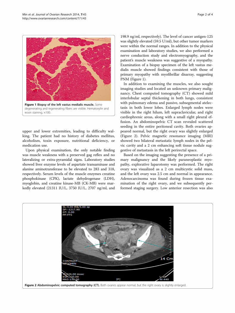
198.9 ng/ml, respectively). The level of cancer antigen-125was slightly elevated (59.5 U/ml), but other tumor markerswere within the normal ranges. In addition to the physicalexamination and laboratory studies, we also performed anerve conduction study and electromyography, and thepatient’s muscle weakness was suggestive of a myopathy.Examination of a biopsy specimen of the left vastus me-dialis muscle showed findings consistent with those ofprimary myopathy with myofibrillar disarray, suggestingPNM (Figure 1).In addition to examining the muscles, we also sought
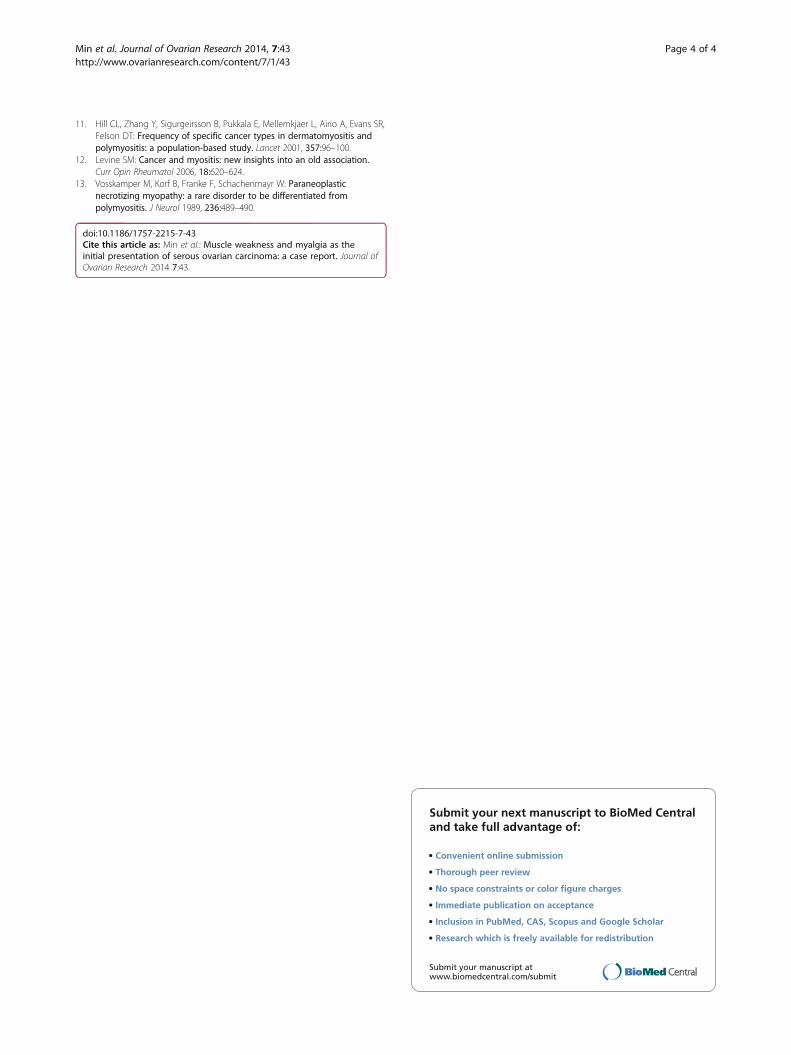
imaging studies and located an unknown primary malig-nancy. Chest computed tomography (CT) showed mildinterlobular septal thickening in both lungs, consistentwith pulmonary edema and passive, subsegmental atelec-tasis in both lower lobes. Enlarged lymph nodes werevisible in the right hilum, left supraclavicular, and rightcardiophrenic areas, along with a small right pleural ef-fusion. An abdominopelvic CT scan revealed scatteredseeding in the entire peritoneal cavity. Both ovaries ap-peared normal, but the right ovary was slightly enlarged(Figure 2). Pelvic magnetic resonance imaging (MRI)showed two bilateral metastatic lymph nodes in the pel-vic cavity and a 2 cm enhancing soft tissue nodule sug-gestive of metastasis in the left perirectal space.Based on the imaging suggesting the presence of a pri-
mary malignancy and the likely paraneoplastic myo-pathy, explorative laparotomy was performed. The rightovary was visualized as a 2 cm multicystic solid mass,and the left ovary was 2.5 cm and normal in appearance.Adenocarcinoma was found during frozen tissue exa-mination of the right ovary, and we subsequently per-formed staging surgery. Low anterior resection was also
Figure 1 Biopsy of the left vastus medialis muscle. Somedegenerating and regenerating fibers are visible. Hematoxylin andeosin staining, ×100.
Figure 2 Abdominopelvic computed tomography (CT). Both ovaries appear normal, but the right ovary is slightly enlarged.
Min et al. Journal of Ovarian Research 2014, 7:43 Page 2 of 4http://www.ovarianresearch.com/content/7/1/43

performed because of suspected rectal invasion of thecul-de-sac. No palpable nodules were observed on theliver surface, spleen, stomach, or appendix.After the laparotomy, histopathological analysis of the
entire specimen revealed a grade 3 ovarian serousadenocarcinoma. The right ovary measured 2 × 1.8 cm,and the left ovary measured 2.5 × 1.7 × 1.5 cm. Involve-ment of the right ovarian surface and fallopian tube wasfound, but the regional lymph nodes were spared. Theresected rectum revealed direct invasion of the serousadenocarcinoma and metastases in 12 of 17 pericoliclymph nodes. Postoperatively, the patient underwentadjuvant chemotherapy with paclitaxel and carboplatin.Muscle weakness improved, and CPK levels graduallydecreased.
ConclusionsOur report describes a patient who experienced muscleweakness and myalgia as the initial presenting symptomsof a primary ovarian malignancy. In this case, there wasno evidence of alternative causes of myopathy, such asother malignancies or medications. To our knowledge,this is the first report of PNM associated with EOC.Malignancy-associated myopathy has been anecdotally
reported in the past, and among these reported myopa-thies, idiopathic inflammatory myopathies (IIMs), mainlydermatomyositis and polymyositis, were found to be asso-ciated with cancer [5]. Specifically, the most commonhistological type of IIM-related cancer is adenocarcinoma[5]. According to a population-based study, dermato-myositis is more strongly associated with ovarian cancerpatients than with the general population [11], but theclinical correlation between cancer and inflammatorymyopathy can vary. A malignancy may occur before or inparallel with the diagnosis of inflammatory myopathy.Usually, cancer is found within three years of a myositisdiagnosis, but the risk of cancer is highest at the time ofdiagnosis [5,11,12]. The relationship between cancer andmyositis was explained by humoral immunologic mecha-nisms [12], and it is thought that the immune-mediateddestruction of muscle may be a type of paraneoplasticmanifestation of the immune system’s response to thecancer.One paraneoplastic syndrome, PNM, is characterized
by a symmetric and proximal myopathy accompanied byincreased levels of serum muscle enzymes such as crea-tine kinase. Furthermore, nerve conduction studies andelectromyography findings in PNM are suggestive of my-opathy. PNM is uniquely distinguishable from othermyositides such as dermatomyositis or polymyositis inthree ways. First, PNM is characterized by predominantnecrosis of muscle fibers with limited inflammation.Additionally, glucocorticoids are the treatment of choicein dermatomyositis, and PNM may not respond to
corticosteroid therapy. Lastly, although dermatomyositismay have a clinical course independent of the cancertreatment, the prognosis of PNM depends on the under-lying malignancy. Consequently, the prognosis for PNMis worse, with greater mortality [13]. Therefore, diag-nosis of the underlying malignancy is critical to themanagement of these patients. The successful treatmentof the malignancy is the mainstay of relieving the myo-sitis symptoms.In this study, we presented a case of ovarian serous
adenocarcinoma with concomitant PNM. Although neverreported until now, the possibility of EOC should be con-sidered in unknown primary malignancies with initialcomplaints of muscle weakness or myalgia.
Consent statementWritten informed consent was obtained from the patientfor publication of this case report.
AbbreviationsEOC: Epithelial ovarian cancer; PNM: Paraneoplastic necrotizing myopathy;CPK: Creatine phosphokinase; LDH: Lactate dehydrogenase; CK-MB: Creatinekinase-MB; CT: Computed tomography; MRI: Magnetic resonance imaging;IIMs: Idiopathic inflammatory myopathies.
Competing interestsThe authors declare that they have no competing interests.
Authors’ contributionsKJM, YTO and JHH participated in the care of the patient and wrote thearticle. KAS participated in the care of the patient. HRH and JKL: participatedin the writing of article. KJM and JHH validated content and form of thearticle. All authors read and approved the final manuscript.
Received: 3 December 2013 Accepted: 20 April 2014Published: 23 April 2014
References1. Ferlay J, Shin HR, Bray F, Forman D, Mathers C, Parkin DM: Estimates of
worldwide burden of cancer in 2008: GLOBOCAN 2008. Int J Cancer J Intdu Cancer 2010, 127:2893–2917.
2. Jung KW, Won YJ, Kong HJ, Oh CM, Seo HG, Lee JS: Prediction of cancerincidence and mortality in Korea, 2013. Cancer Res Treat 2013, 45:15–21.
3. Kim MK, Kim K, Kim SM, Kim JW, Park NH, Song YS, Kang SB: A hospital-based case–control study of identifying ovarian cancer using symptomindex. J Gynecol Oncol 2009, 20:238–242.
4. Darnell RB, Posner JB: Paraneoplastic Syndrome. New York: Oxford UniversityPress, Inc.; 2011.
5. Danko K, Ponyi A, Molnar AP, Andras C, Constantin T: Paraneoplasticmyopathy. Curr Opin Rheumatol 2009, 21:594–598.
6. Samuels N, Applbaum YH, Esayag Y: Paraneoplastic necrotizing myopathyand dermatomyositis in a patient with rectosigmoid carcinoma.Rheumatol Int 2013, 33:1619–1621.
7. Acciavatti A, Avolio T, Rappuoli S, Foderi L, Soldati V, Franchi M, Volpi N,Nuti R: Paraneoplastic necrotizing myopathy associated withadenocarcinoma of the lung - a rare entity with atypical onset: a casereport. J Med Case Rep 2013, 7:112.
8. Silvestre J, Santos L, Batalha V, Del Rio A, Lima C, Carvalho A, Martins A,Miranda H, Cabral F, Felix A, Aleixo A: Paraneoplastic necrotizingmyopathy in a woman with breast cancer: a case report. J Med Case Rep2009, 3:95.
9. Chao LW, Wei LH: Dermatomyositis as the initial presentation of ovariancancer. Taiwan J Obstet Gynecol 2009, 48:178–180.
10. Ghosh A, Malak TM, Pool AJ: Polymyositis and ovarian carcinoma: a casereport. Arch Gynecol Obstet 2007, 275:195–197.
Min et al. Journal of Ovarian Research 2014, 7:43 Page 3 of 4http://www.ovarianresearch.com/content/7/1/43
11. Hill CL, Zhang Y, Sigurgeirsson B, Pukkala E, Mellemkjaer L, Airio A, Evans SR,Felson DT: Frequency of specific cancer types in dermatomyositis andpolymyositis: a population-based study. Lancet 2001, 357:96–100.
12. Levine SM: Cancer and myositis: new insights into an old association.Curr Opin Rheumatol 2006, 18:620–624.
13. Vosskamper M, Korf B, Franke F, Schachenmayr W: Paraneoplasticnecrotizing myopathy: a rare disorder to be differentiated frompolymyositis. J Neurol 1989, 236:489–490.
doi:10.1186/1757-2215-7-43Cite this article as: Min et al.: Muscle weakness and myalgia as theinitial presentation of serous ovarian carcinoma: a case report. Journal ofOvarian Research 2014 7:43.
Submit your next manuscript to BioMed Centraland take full advantage of:
• Convenient online submission
• Thorough peer review
• No space constraints or color figure charges
• Immediate publication on acceptance
• Inclusion in PubMed, CAS, Scopus and Google Scholar
• Research which is freely available for redistribution
Submit your manuscript at www.biomedcentral.com/submit
Min et al. Journal of Ovarian Research 2014, 7:43 Page 4 of 4http://www.ovarianresearch.com/content/7/1/43