Embed Size (px)
Citation preview

Muscle strength measurements of the Hand Ton AR Schreuders, JW Brandsma, HJ Stam
“Is it nothing to have the mind awakened to the perception of the numerous proofs of design which present themselves in the study of the Hand- to be brought to the conviction that everything in its structure is orderly and systematic, and that the most perfect mechanism, the most minute and curious apparatus, and sensibilities the most delicate and appropriate, are all combined in operation that we may move the hand?” The Hand - Its Mechanism and Vital Endowments as Evincing Design. Sir Charles Bell (1833).
Introduction
Together with the brain, the hand is
the most important organ for accomplishing tasks of adaptation, exploration, prehension, perception and
manipulation, unique to humans.1 To study the anatomy and kinetic chains of the hand and the complex interplay of more than 40 muscles that control its movements requires an appreciation of the biomechanics of the hand and its dexterity.2
The muscles of the lower arm and hand can be conveniently arranged according to innervation and localisation. Usually the muscles are divided into extrinsic ones, where muscles have their origin proximal to the hand and intrinsic muscles which have their origin and insertion within the hand (Table 1). Sterling Bunnell3 wrote that “the intrinsic muscles of the hand, though tiny, are important because, with the long extensors and long flexors, they complete the muscle balance in the hand”.
Table 1. Extrinsic and intrinsic muscles listed by innervation.
Extrinsic Intrinsic Elbow Wrist Fingers Thumb Fingers Thumb
FCU (flexor carpi ulnaris)
FDP (dig 4,5) (flexor dig. prof.)
PI Palmar interossei
AP (adductor pollicis)
DI Dorsal interossei
FPB (part) (flexor pollicis brevis)
Ulnar
Lumbricals dig. 4,5 Hypothenar muscles
PT (pronator teres)
FCR (flexor carpi radialis)
FDP (dig 2,3) (flexor dig. prof.)
FPL (flexor pollicis longus)
Lumbricals dig. 2,3 APB (abd. pollicis brevis)
PL (palmaris longus)
FDS (dig 2-5) (flexor dig. sup.)
OP (opponens pollicis) Median
PQ (pronator quadratus)
FPB (part) (flexor pollicis brevis)
BR (brachio-radialis)
ECRL (ext. carpi rad. longus)
EDC (ext. dig. communis)
APL (abd. pollicis longus)
Supinator
ECRB (ext. carpi rad. brevis)
EDQ (ext. dig. quinti)
EPB (ext. pollicis brevis)
Radial ECU
(ext. carpi ulnaris) EIP (ext. indicis prop.)
EPL (ext. pollicis longus)
The intrinsic muscles are
sometimes referred to as tiny or small muscles of the hand, but this should not be interpreted as weak because some intrinsic muscles (especially the index finger DI and AP) have a cross-sectional area similar to several strong extrinsic
muscles.4 The term “tiny” or “small” is therefore correct only in that the intrinsic muscles are short.
This thesis focuses on the intrinsic muscles of the hand. Loss of intrinsic muscle strength of the fingers can cause severe loss of hand function, e.g. firmly
Ton A.R. Schreuders, JW Brandsma, HJ Stam
2
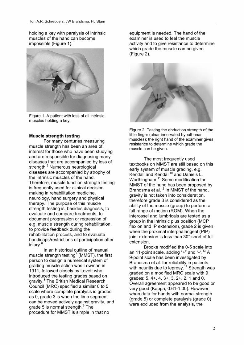
holding a key with paralysis of intrinsic muscles of the hand can become impossible (Figure 1).
Figure 1. A patient with loss of all intrinsic muscles holding a key. Muscle strength testing
For many centuries measuring muscle strength has been an area of interest for those who have been studying and are responsible for diagnosing many diseases that are accompanied by loss of strength.5 Numerous neurological diseases are accompanied by atrophy of the intrinsic muscles of the hand. Therefore, muscle function strength testing is frequently used for clinical decision making in rehabilitation medicine, neurology, hand surgery and physical therapy. The purpose of this muscle strength testing is, besides diagnosis, to evaluate and compare treatments, to document progression or regression of e.g. muscle strength during rehabilitation, to provide feedback during the rehabilitation process, and to evaluate handicaps/restrictions of participation after injury.6
In an historical outline of manual muscle strength testing7 (MMST), the first person to design a numerical system of grading muscle action was Lowman in 1911, followed closely by Lovett who introduced the testing grades based on gravity.8 The British Medical Research Council (MRC) specified a similar 0 to 5 scale where complete paralysis is graded as 0, grade 3 is when the limb segment can be moved actively against gravity, and grade 5 is normal strength.9 The procedure for MMST is simple in that no
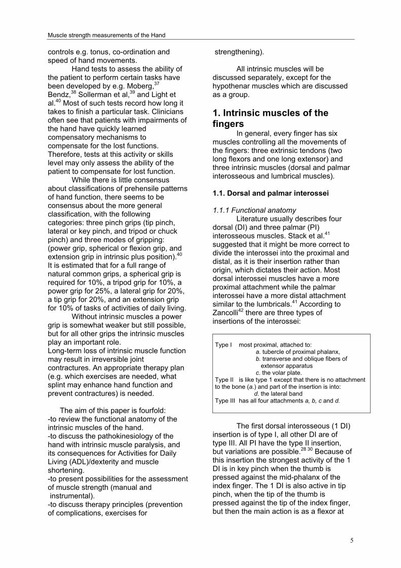
equipment is needed. The hand of the examiner is used to feel the muscle activity and to give resistance to determine which grade the muscle can be given (Figure 2).
Figure 2. Testing the abduction strength of the little finger (ulnar innervated hypothenar muscles); the right hand of the examiner gives resistance to determine which grade the muscle can be given.
The most frequently used textbooks on MMST are still based on this early system of muscle grading, e.g. Kendall and Kendall10 and Daniels L. Worthingham.11 Some modification for MMST of the hand has been proposed by Brandsma et al.12 In MMST of the hand, gravity is not taken into consideration, therefore grade 3 is considered as the ability of the muscle (group) to perform a full range of motion (ROM). When the interossei and lumbricals are tested as a group in the intrinsic plus position (MCP flexion and IP extension), grade 2 is given when the proximal interphalangeal (PIP) joint extension is less than 30° short of full extension.
Brooke modified the 0-5 scale into an 11-point scale, adding “+” and “-“.13 A 9-point scale has been investigated by Brandsma et al. for reliability in patients with neuritis due to leprosy.14 Strength was graded on a modified MRC scale with 9 grades: 5, 4+, 4, 3+, 3, 2+, 2, 1 and 0. Overall agreement appeared to be good or very good (Kappa; 0.61-1.00). However, when data for hands with normal strength (grade 5) or complete paralysis (grade 0) were excluded from the analysis, the

Muscle strength measurements of the Hand
3
reliability of the remaining mid-range scale was not acceptable. Limitations of MMST a) Although the textbooks usually present
the muscle tests as if muscles can be tested in isolation, clinicians should be aware that usually a muscle group is tested rather than just one muscle. Some have suggested labelling the movement rather than the muscle, e.g. grading the palmar abduction movement of the thumb instead of abductor pollicis brevis (APB), because several muscles are active when testing the palmar abduction of the thumb. Only a few muscles can be graded in isolation, e.g. flexor pollicis longus, flexor digitorum profundus and first dorsal interosseous (1DI).
b) The MRC uses a 6-point numeric scale (grades 0-5) and seems to indicate a constant distance between points. However, it is an ordinal scale with disproportional distances between grades; e.g. grade 4 is not twice as strong as grade 2. It might have been more appropriate to use terms such as normal, good, fair, trace and paralysed.
c) Another important comment concerning MMST was made in the American Society of Handtherapists (ASHT) recommendations,7 that its most appropriate use is in cases of extreme muscle deterioration. MMST is not appropriate for higher-level muscle function due to lack of sensitivity and precision, and should be used in conjunction with other evaluation tools. We contend that MMST is most useful for weak muscles with grades of 1, 2 and 3, but not for the higher grades.
d) MMST is dependent on the examiner’s ability to assess the pressure as a parameter for strength. Experience of the examiner is important for reliable measurements.
History of dynamometers for the hand
One of the first dynamometers for measuring hand strength was the Graham-Desaguliers dynamometer, which was developed in London in 1763. The
Regnier dynamometer was invented in Paris in 1798 to measure the traction properties of artillery horses, but was designed as an all-purpose instrument to measure specific human muscle groups as well.8
In the past decades many different dynamometers have been introduced, e.g. cable tensiometers, sphygmomanometers, vigorimeters,15 isokinetic dynamometers and strain gauge dynamometers. In response to the confusion generated by the many commercial and experimental grip strength instruments, the California Medical Association in 1956 evaluated the most commonly used instruments.16 They found the Jamar, first introduced by Bechtol in 1954,17 to be the most accurate. In 1978 the American Society for Surgery of the Hand recommended that the second position of the Jamar should be used and in 1981 the ASHT made additional recommendations, e.g. concerning posture and verbal instructions during measurements.7 Grip strength measurements with a dynamometer have become popular and have been studied extensively. Less studies have been conducted to investigate pinch strength measurements.
Van der Ploeg et al.18 noticed the shortcoming of the MMST method by giving an example of strength measurements of the biceps muscle as an elbow flexor. The biceps needed 5 N of its normal strength (250 N) to overcome gravity; thus grade 3 corresponds with only 2% of the full strength of the biceps muscle. Dynamometers for intrinsic muscle strength measurements
In his thesis van der Ploeg noted that most dynamometers have a scale far too crude for measuring forces in very small muscles like the abductor digiti quinti. However, assessing the strength of these muscles is of great importance in clinical neurology in the evaluation of mono- and poly-neuropathies. He noted that there is a need for an accurate device for these muscles.19
One of the first to develop a dynamometer for the intrinsic muscle strength was Mannerfelt, who later

Ton A.R. Schreuders, JW Brandsma, HJ Stam
4
manufactured a new device called the Intrins-o-meter.20 In 1997 he reported a study in 48 patients with ulnar nerve compression.21 Rosen et al. noted that assessing muscle function using the Intrinsi-o-meter was difficult due to the extremely small forces, and the instrument was difficult to handle and read. They suggested using MMST and grip strength measurements to evaluate nerve function. Interestingly they found a poor recovery of the intrinsic muscle strength with the Mannerfelt instrument and good grip strength recovery.22 23
Several others have developed instruments mainly to assess the abduction of the thumb.24-26 Some needed a specially constructed jig, e.g. to measure wrist, finger (metacarpo-phalangeal joints) and thumb extension strength.27 Rotterdam Intrinsic Hand Myometer (RIHM) (Figure 3)
In 1995 inventories were made at our department to establish which clinical evaluation instruments were available to assess the outcome after peripheral nerve surgery. Three methods were often used to assess the recovery of muscle strength: MMST and grip and pinch strength dynamometers.
Having encountered several patients with good grip strength but poor recovery of the intrinsic muscles strength, we questioned whether grip strength measurements were appropriate. We acknowledged the need for a dynamometer to measure the intrinsic muscles in isolation. Such a dynamometer should be easy to handle, e.g. portable and with an ergonomical design. It should also have the possibility to measure the opposition force of the thumb. Reliability should be good with acceptable measurement error making it possible to detect reasonably small changes in muscle strength. Figure 3 RIHM dynamometer
The Intrinsic Muscles of the Hand: Function, Assessment and Therapy principles Introduction
There have been many valuable studies concerning the anatomy28-30, mechanics,2 31 32 and architectural design,33 of the intrinsic muscles of the hand. Understanding the mechanics of human dexterity requires an appreciation of the kinetic chains that comprise the hand, and the intricate interplay of muscles and ligaments that control its movements.2 In these chains, the intrinsic muscles of the hand are of paramount importance for efficient hand function.33
There is a considerable decrease in functional efficiency in hands with loss of the intrinsic muscles function, often referred to as the clawhand or intrinsic minus hand (Figure 1).34-36
A comprehensive analysis of hand function should include assessment of the strength and length of the intrinsic muscles. This will provide important information and assist the assessor in e.g. determining nerve function, deciding which muscles need to be strengthened, what splint is needed, what surgery needs to be considered (tendon transfer), etc.
Although assessment of muscle strength and length are important elements of hand function other functions, e.g. mobility, sensibility and central properties of the brain, are equally or more important for hand function. The latter
Muscle strength measurements of the Hand
5
controls e.g. tonus, co-ordination and speed of hand movements.
Hand tests to assess the ability of the patient to perform certain tasks have been developed by e.g. Moberg,37 Bendz,38 Sollerman et al,39 and Light et al.40 Most of such tests record how long it takes to finish a particular task. Clinicians often see that patients with impairments of the hand have quickly learned compensatory mechanisms to compensate for the lost functions. Therefore, tests at this activity or skills level may only assess the ability of the patient to compensate for lost function.
While there is little consensus about classifications of prehensile patterns of hand function, there seems to be consensus about the more general classification, with the following categories: three pinch grips (tip pinch, lateral or key pinch, and tripod or chuck pinch) and three modes of gripping: (power grip, spherical or flexion grip, and extension grip in intrinsic plus position).40 It is estimated that for a full range of natural common grips, a spherical grip is required for 10%, a tripod grip for 10%, a power grip for 25%, a lateral grip for 20%, a tip grip for 20%, and an extension grip for 10% of tasks of activities of daily living.
Without intrinsic muscles a power grip is somewhat weaker but still possible, but for all other grips the intrinsic muscles play an important role. Long-term loss of intrinsic muscle function may result in irreversible joint contractures. An appropriate therapy plan (e.g. which exercises are needed, what splint may enhance hand function and prevent contractures) is needed.
The aim of this paper is fourfold: -to review the functional anatomy of the intrinsic muscles of the hand. -to discuss the pathokinesiology of the hand with intrinsic muscle paralysis, and its consequences for Activities for Daily Living (ADL)/dexterity and muscle shortening. -to present possibilities for the assessment of muscle strength (manual and instrumental). -to discuss therapy principles (prevention of complications, exercises for
strengthening).
All intrinsic muscles will be discussed separately, except for the hypothenar muscles which are discussed as a group. 1. Intrinsic muscles of the fingers
In general, every finger has six muscles controlling all the movements of the fingers: three extrinsic tendons (two long flexors and one long extensor) and three intrinsic muscles (dorsal and palmar interosseous and lumbrical muscles). 1.1. Dorsal and palmar interossei 1.1.1 Functional anatomy
Literature usually describes four dorsal (DI) and three palmar (PI) interosseous muscles. Stack et al.41 suggested that it might be more correct to divide the interossei into the proximal and distal, as it is their insertion rather than origin, which dictates their action. Most dorsal interossei muscles have a more proximal attachment while the palmar interossei have a more distal attachment similar to the lumbricals.41 According to Zancolli42 there are three types of insertions of the interossei: Type I most proximal, attached to:
a. tubercle of proximal phalanx, b. transverse and oblique fibers of extensor apparatus c. the volar plate.
Type II is like type 1 except that there is no attachment to the bone (a.) and part of the insertion is into:
d. the lateral band Type III has all four attachments a, b, c and d.
The first dorsal interosseous (1 DI)
insertion is of type I, all other DI are of type III. All PI have the type II insertion, but variations are possible.28 30 Because of this insertion the strongest activity of the 1 DI is in key pinch when the thumb is pressed against the mid-phalanx of the index finger. The 1 DI is also active in tip pinch, when the tip of the thumb is pressed against the tip of the index finger, but then the main action is as a flexor at

Ton A.R. Schreuders, JW Brandsma, HJ Stam
6
the metacarpo-phalangeal (MCP) joint. The first palmar interosseous (1 PI) muscle is more active in tip pinch activities.
In Type II, the insertion into the lateral band of the extensor apparatus (d.) is responsible for an important extension force at the proximal interphalangeal (PIP) joints. The first palmar interosseous also produces some supination of the index finger to get good approximation of the pulps. In this respect we might consider the 1 PI as an “opponens indices” muscle in tip pinch activities.
Without interossei the finger is
unstable and will collapse into the “claw” (i.e. intrinsic minus) position of flexed IP joints and (hyper-) extension of the MCP joint: therefore the interosseous are sometimes referred to as the “anti-claw” muscles.35 The primary function of the interossei is MCP flexion/stabilisation with extension of the interphalangeal (IP) joints. This is especially evident during pinch in which the collapse of the index PIP joint is apparent in, often > 90°, flexion. This is a sign of interosseous muscle weakness and sometimes referred to as the Mannerfelt sign.20 (Figure 2).
FIGURE 2. Mannerfelt and Froment sign on left hand of patient with ulnar nerve paralysis.
Recording the moments of the intrinsic muscles that were generated after electrical stimulation Lauer et al.43 found that the dorsal interossei muscles were strong abductors of the fingers and generated a significant moment in MCP joint flexion and IP joint extension. Similarly, Ketchum et al.34 found that the
interossei muscles of the index finger contribute 73% to the overall moment for flexion of the MCP joint.34 Li et al.44 investigated the role of the intrinsic finger flexor muscles during finger flexion tasks. When an external force is applied proximally to the PIP joint, the extensor mechanism (intrinsic muscle group) is the largest component (70%) on force production of all flexors.
Thus the interossei muscles are important flexors of the MCP joint together with the long flexors: flexor digitorum profundus (FDP) and flexor digitorum superficiales (FDS). However, at the PIP joint level the long flexors, primarily the FDS, and the interossei are antagonists.
When the PIP joint is flexed, some reduction of the extension moment of the interossei takes place, due to the volar displacement of the lateral bands (of about 4-mm) at the PIP joint level. At full flexion of both the PIP and distal interphalangeal (DIP) joints, the lateral bands approach the flexion-extension axis of the PIP joint, thereby minimising the extensor moment.2
The action of the interossei
muscles can be studied separately from the extensor digitorum communis (EDC) muscles in patients with a radial nerve paralysis. If the patient is asked to extend the fingers, no extension of the MCP will occur, but the finger IP joints will extend because of the ulnar and median nerve innervated interosseous and lumbrical muscles. However, in some patients the fingers, especially the index, can be extended in the MCP joint to some degree, especially with the wrist flexed; this is probably because the angle of attachment of the dorsal interossei muscles is only 0°-5°. When the tension on the extensor tendon is increased due to the flexed wrist, this angle will go beyond the 0° and thus the interosseous muscle becomes an extensor of the MCP joint. The angle of approach to the extensor mechanism for the palmar interossei muscles is 20°-25° and for the lumbrical muscles 35°.28
The index finger is deprived of all long flexors and the lumbrical muscles in patients with a high median nerve paralysis. The only active muscles are the long extensors and the two interossei. In
Muscle strength measurements of the Hand
7
an attempt to flex the fingers the index finger will remain extended, and is accurately described as the “pointing finger” where the IP joints are fully extended and the MCP joint flexed. In high median nerve paralysis the long finger is also deprived of the long flexor, but will usually flex in grip. This is due to the attachments between the FDP tendons of the long finger and the ring finger and is called the Quadriga phenomena.45 46 This term suggests a symmetrical organisation of the four tendons; however, the connections between the FDP of the index and middle finger are usually absent, and a strong connection between the flexor pollicis longus (FPL) and the FDP of the index finger can exist. 1.1.2 Pathokinesiology (paralyses, consequences for prehension/ADL, shortening)
The deficiency mentioned in textbooks due to loss of interossei is usually that of controlled abduction and adduction of the fingers. The loss of this action is important for those who play musical instruments or operate keyboards, but in most patients this is not recognised as a severe deficit. The loss of the interossei function of MCP flexion and PIP extension is a much more significant loss. In acute paralysis of the interossei muscles it will sometimes only be visible as a mild hyperextension at the MCP joints and slight flexed position of the PIP joints when the patient is asked to extend the fingers. However, this deformity usually progresses depending on the natural joint laxity of the hands, i.e. long, thin and hypermobile fingers will develop a so-called claw hand much quicker than thick, stiff fingers. In the mobile hands the volar ligamentous structures stretch more easily, causing increased hyperextension and subsequently less PIP extension. Extension of the PIP joints is only possible by contraction of the EDC when the MCPs are 'blocked', either actively (internally) through muscle contraction or passively (externally) through the examiners hand or a splint, preventing hyperextension of the MCP joints. In the latter situation this is called assisted extension, which is used
as a test to assess the integrity of the extensor apparatus.
Other factors that may contribute to the development of a claw hand are: hand dominance, continued use of the hand (or the lack of), compliance of the patient to perform the routine exercises to prevent joint stiffness, and use of (night) splints.
Without interosseous the hand can
still make a full fist, but the pattern of movement is changed (the MCP flexion takes place later than usual). The contribution of the interossei muscles in grip strength measurements will be discussed later. In patients with a “high” ulnar lesion, the FDP muscles of the 4th and 5th fingers are paralysed and less clawing is often visible because the flexion moment at the PIP joints is decreased compared to a “low” ulnar palsy. We might conclude from this observation that clawing is also the result of visco-elastic tension of FDP. When the ulnar nerve recovers, the 4th and 5th finger will show an increasing clawing. Maintaining the length of the FDPs, i.e. preventing flexor tightness, therefore also helps to prevent clawing. Another sign of interossei muscle weakness was first described by Andre-Thomas in 1917 and is called the Thomas sign.47 (p. 518) (Figure 3). This is the tendency (compensation) of a patient with weak interossei muscles of the fingers to automatically flex the wrist in an attempt to gain a better opening of the hand, i.e. MCP extension, by means of increasing the pull on the EDC. This in fact increases the hyperextension of the MCP joints and adds to the progress of the development of the claw hand. This “trick” movement is adopted very quickly and even when the interossei have regained their strength, will often take a long time to “un-learn”.
In longstanding paralyses of the interossei, e.g. when the ulnar nerve could not be repaired or did not recover, the PIP joints are continuously in a flexed position. This will cause a gradual stretching of the dorsal expansion (sometimes called the tri-angular ligament) over the PIP joint, which secures the lateral bands of the extensor tendon in their dorsal position. In

Ton A.R. Schreuders, JW Brandsma, HJ Stam
8
the normal finger, the lateral bands shift dorsally and towards the central position of the finger when the PIP joint is extended. Whereas when flexing the PIP joint the dorsal expansion needs to allow the lateral bands to move volarly towards the flexion-extension axis of movement. When this dorsal expansion is elongated, the lateral bands are too much volarly, resulting in a loss of PIP joint extension. Consequently, the oblique retinacular ligament (ORL) or Landsmeers ligament is slack most of the time and will adjust to this new situation by shortening and this may result in hyperextension of the DIP joint. This is a similar progression of changes as in an extensor tendon central slip injury, causing a Boutonnière deformity.4
FIGURE 3. Thomas sign: compensation movement when interossei muscles are weak; flexion of the wrist in an attempt to gain a better opening of the hand, i.e. by means of increasing the pull on the EDC.
Another impairment in longstanding
paralyses of the interossei muscles is related to hyperextension of the MCP joint. This causes an upward pull of the EDC tendons (bow stringing), stretching the sagittal bands. The laxity of the sagittal bands will result in an inability to maintain the EDC tendon on top of the MCP joint. The drop of the luxating tendon into the groove between the MCPs is especially observable when flexing the MCP joint. This is sometimes called “guttering” because the tendon drops into the “gutter” between the MCP joints.
The longstanding flexed position of the IP joints will result in a physiological shortening of the long flexors. This flexor
tightness will increase the PIP flexion position and can cause a deterioration of the PIP flexion contractures. This is an additional argument to maintain the length of the extrinsic flexors in patients with intrinsic muscle weakness.
Shortening of the interossei
muscles is called intrinsic tightness (IT), which can be caused by a trauma of the hand which can precipitate a cascade of events. The interossei are situated in rather tight compartments, therefore oedema/swelling will cause an increase of pressure in these compartments.
As a result blood circulation will be hampered causing anoxia and muscle fiber death, which results in fibrosis of the muscle and shortening. This is identical to the process which causes Volkmann's ischemic contracture in the forearm.48
The IT test consists of two parts. First, passive PIP flexion is tested with the MCP joint extended and, secondly, passive PIP flexion is tested again but now with the MCP joint flexed. If there is a large difference in PIP flexion between the two MCP positions, intrinsic tightness is present. The long-term complications of IT can result in decreased MCP extension and a swan neck finger, i.e. hyperextension of the PIP joint. A longstanding swan neck deformity might result in a painful snapping of the lateral bands at PIP level when the finger is flexed.
In rheumatoid arthritis a different process can also lead to IT. The role of the intrinsic muscles in producing MCP subluxation in the rheumatoid hand has been documented.49 50
Another intricacy sometimes
observed is what we call interosseous plus, which is a paradoxal extension: the harder the patient tries to bend the finger, the more the finger will extend in the PIP joint. This phenomena is sometimes seen in patients in which the interossei have been the only flexor of the finger for some time e.g. in high median nerve palsy, or in case of adhered flexor tendons. Although the flexors are active and can bend the finger, when a stronger grip is required the finger will extend.

Muscle strength measurements of the Hand
9
The explanation is that in a non-resistant grip of a normal functioning hand there is only minor activity of the interossei muscles. However, in a strong grip, the stronger the flexors are pulling, the more the intrinsic muscles become active to stabilise the PIP joints and prevent luxation. If the long flexor is weak or poorly activated, the interossei will overpower the flexor causing PIP extension. This paradoxal extension appears to be similar to the lumbrical plus phenomena (see lumbricals) and might be called interosseous plus. The patient has to be taught to, gently, contract the long flexors without the action of the interossei muscles. 1.1.3 Assessment possibilities (manual and instrumental)
Although the interosseous muscles have short fiber length, some are strong and have physiological cross-sectional areas comparable to the FDS muscles.4 In standard textbooks on muscle testing, the tests suggested are usually: abduction for dorsal and adduction for palmar interossei muscles.10 51 These tests are useful for isolated (specific) testing of the interossei. For example, patients with an ulnar nerve paralysis can not move their middle finger sideways, which has been called the Egawa sign.47 Functionally, it is much more meaningful to test the interossei muscles in the intrinsic plus position, by giving resistance to flexion of the MCP joints and extension of the PIP joint.52 53 (Figure 4). FIGURE 4. Testing the strength of the intrinsic muscles in the fingers.
Some have suggested that
dynamometry of the interossei muscles is possible, indirectly, by measuring the grip strength of the hand with e.g. a Jamar dynamometer. Janda et al. advocated to use the smallest handle position of the Jamar because the intrinsic muscles were most active in that position.54 Kozin et al.36 tested 21 healthy persons who underwent median and ulnar nerve blocks at the wrist level; the average decrease in grip strength was 38% after ulnar nerve block.
Pinch data in the study by Kozin et al. revealed a significant decrease in key pinch of 77% after ulnar block and 60% after median block.36 For evaluating and monitoring the motor function of the ulnar nerve, pinch strength measurements seem more meaningful than grip strength measurements.
Specific measurements of the first dorsal interosseous muscle can be done with dynamometers such as the RIHM,55 the Intrins-o-meter of Mannerfelt,21 or the Preston pinch gauge device.56 1.1.4 Therapy principles (prevention complications, strengthening, ADL)
Prevention of contractures is directed at the PIP joints. A (night) splint with the MCP joints in flexion and IPs in extension is advisable. These splints can help to prevent PIP flexion contractures and are especially important in longstanding problems. Increased hyperextension of the MCP joints, due to stretching of the volar plate, is also prevented in this position.
During the day the so-called “knuckle-bender” can assist the patient in some ADL functions. This splint will also help to move the PIP joint into full extension during the day and will maintain the integrity of the extensor mechanism. When patients choose not to wear a splint, they need to be taught how to do assisted extension exercises by blocking the MCP joints with their hands and routinely massaging to extend the IP joints (Figure 5).

Ton A.R. Schreuders, JW Brandsma, HJ Stam
10
FIGURE 5. Prevention of IP flexion contractures by massaging the fingers.
Exercises to strengthen the interossei muscles are all aimed at movements which flex the MCP and extend the IP joints. Therefore, exercises for the interossei muscles are all activities in intrinsic plus position: e.g. grasping a book, plate or a cylindrical object like a large bottle. Specific training of the first dorsal and palmar interossei are activities for which key and tip pinch activities are required.
To correct the long flexor tightness, the patient is taught to stretch the flexors by holding the hand flat on e.g. a table and by moving the forearm towards an angle perpendicular to the table. In a similar fashion, the hand can be placed on the seat of the chair while the patient sits on the hand and pulls the forearm towards the body. A night splint with the fingers and hand in extension might be necessary in severe cases. 1.2 Lumbricals 1.2.1 Functional anatomy
The lumbrical muscles are unique muscles in several aspects. They connect two extrinsic antagonistic muscles. Proximally the lumbricals are attached to the FDP and distally they are inserted into the lateral band of the extensor tendon. The third and fourth lumbricals also connect, by their bi-penal origin, two adjacent FDP tendons.
The function of the lumbrical muscles is much debated and some even
considered these muscles to be redundant. Brand suggested that the lumbrical muscles are not relevant for MCP flexion. He explained this with an illustration of a father carrying a child; it does not matter what the child (i.e. lumbrical) is carrying, the father (i.e. FDP) has to carry it anyway.4 Therefore, the lumbrical muscles have a unique ability to contract without adding flexion torque at the MCP joint, in contrast with the interosseous muscles which, when extending the IPs, need a stronger contraction of the EDC to counteract the flexion moment at the MCP joint level. The lumbricals provide a more efficient source for IP extension than the interossei.
Any contraction of the lumbrical muscle for IP extension simultaneously, reduces the visco-elastic force of the FDP tending to flex the IP joints. Accordingly the lumbrical can be regarded as a deflexor of the PIP joint.57 Its direct contribution to MCP flexion is small and in the flexed finger may be non-existent, but its indirect contribution to IP joint extension by decreasing the flexion torque is quite substantial.58
With the smallest physiological
cross-sectional area, it is certainly not a strong muscle. The lumbricals have a very long fiber length (40-48 mm) which indicates that they are designed for long excursions. If the lumbrical fiber length was short, FDP excursion could stretch the lumbrical sarcomeres to a point that they were unable to generate active force.33
The lumbrical muscles are richly endowed with muscle spindles, their passive stretch by contraction of the FDP might both inhibit finger extensors and facilitate wrist extensors.58-61 For this reason the lumbrical muscles have been called “tensiometers” between long flexors and extensors.62 Leijnse and Kalker46 concluded that the lumbricals are in an optimal position for proprioceptic feedback concerning the PIP-DIP joint mechanism.
These unique properties of the lumbricals indicate that they are probably important in fast, alternating movements, e.g. in typing and playing musical instruments.63

Muscle strength measurements of the Hand
11
1.2.2 Pathokinesiology (paralyses, consequences for prehension/ADL, shortening)
In low median nerve injuries the lumbrical muscles of the index and middle finger are paralysed. In these hands it is difficult to discover any problems in the motion of these fingers.64 A mildly diminished extension of the DIP joint has been noticed in a few patients, which might be explained by the decreased extension force on the extensor apparatus.
The “lumbrical plus” is a situation in which there is a FDP tendon rupture distal of the lumbrical origin, or in the situation where a too long graft has been used. The FDP now pulls through the lumbrical muscle rather than through its tendon, causing PIP extension.65 1.2.3 Assessment possibilities (manual and instrumental)
Manual muscle strength testing (MMST) of the lumbricals is practically impossible because of the synergistic action of the interossei muscles. Strength testing is most likely less relevant than evaluating the co-ordination and dexterity in e.g. a tapping test when the lumbrical is paralysed and normal function. However, no studies have been found in which this has been utilised.
The strength can be measured in isolation of the interossei muscles in patients with an ulnar nerve lesion where the index and long fingers have only the lumbrical to maintain the intrinsic plus position. One study measured a mean MCP joint flexion strength in the index and long finger of 0.8 kg (range 0.3-1.5) compared with 6.4 kg (range 4.6-7.9) in the non-involved hand. Thus, the affected fingers have only about 12% of the strength of those of the non-involved hand.64 1.2.4. Therapy principles (prevention complications, strengthening, ADL)
No specific training/splinting program for lumbrical paralysis has been advocated. Strength training is similar to interossei muscle training, with perhaps more focus on speed and co-ordination.
2. Thenar muscles The median nerve innervates the
intrinsic thumb muscles that make the hand a “human” hand. These muscles oppose the thumb to the fingers: abductor pollicis brevis (APB), opponens pollicis (OP) and the flexor pollicis brevis (FPB). All these muscles originate from the flexor retinaculum and the trapezium carpal bone. Located in the thumb web the adductor pollicis (AP) is sometimes also considered a thenar muscle and will be discussed separately. 2.1 Abductor Pollicis Brevis (APB) 2.1.1 Functional anatomy
The insertion of the APB is at the radial side of the proximal phalanx of the thumb. Even though the APB is the smallest intrinsic thenar muscle, atrophy is quickly noticed as it is the most superficial muscle. The main function of the APB is moving the thumb away from the palm, in a perpendicular direction to the palm of the hand, when grasping objects. This is usually called palmar abduction of thumb, but in more recent terminology anteposition of the thumb.66 The muscle is relatively weak as the action of abduction of the thumb is not one that is usually done against resistance; the APB “only” positions the thumb for action. This movement in the carpo-metacarpophalangeal (CMC) joint of the thumb is a synergistic action with the OP and, especially, the FPB.67
An extrinsic synergist of the APB is
the abductor pollicis longus (APL). The pull of the APL on the CMC joint of the thumb causes palmar abduction, especially when the wrist is in flexion. Flexion of the wrist increases the moment arm of the APL due to bow stringing.4
Since the APB has a dual insertion into the base of the proximal phalanx and into the extensor tendon expansion, it also has an extension moment on the IP joint. After loss of the extensor pollicis longus (EPL) the APB together with the oblique head of the AP may provide complete extension of the IP joint of the thumb. This is clinically important when evaluating

Ton A.R. Schreuders, JW Brandsma, HJ Stam
12
extension of the thumb and the EPL function.68 2.1.2. Pathokinesiology (paralyses, consequences for prehension/ADL, shortening)
Loss of the APB sometimes has little effect because the ulnar nerve innervated part of the FPB, the superficial head, can often move the thumb in palmar abduction by itself.69 Frykman et al.70 estimated that 50% of patients with a median nerve lesion would have satisfactory thumb opposition if no median nerve re-innervation occurred.
When the thumb can not be palmar
abducted (anteposition) there will be problems with manipulating small objects and tip-tip pinch activities, but also in positioning the thumb to grasp larger objects. Rosen71 found in 15 median nerve injured patients that of the 20 tasks of the Sollerman test, only a few tasks were particularly difficult: picking up coins from a purse, picking up nuts, putting on bolts, and fastening of buttons. 2.1.3 Assessment possibilities (manual and instrumental)
Because it is one of the few intrinsic muscles innervated by the median nerve, it is an important muscle in clinical practice e.g. to evaluate motor function impairment in median nerve compression in carpal tunnel syndrome. Unfortunately manual muscle strength testing in isolation is often hindered by the many synergists. Especially in the thumb, most movements are the product of multiple synergists.11 30
72 When testing the palmar abduction of the thumb, i.e. APB muscle strength, APL substitution can be diminished to a certain level by holding the wrist in extension. For strength testing, pressure is applied perpendicular to the palm of the hand at the MCP joint of the thumb52 (Figure 6).
FIGURE 6. Strength test of the abductor pollicis brevis (APB). Several dynamometers that specifically measure the abduction strength of the thumb have been developed.24-26 73 If these are not available the strength of the APB can be evaluated indirectly with a pinch dynamometer. We found a strong correlation between the pinch strength and the strength of the APB.74 Weakness of the APB can cause a diminished pinch strength, mainly because the strong thumb muscles cannot be put into action to exert their full strength when the APB is not able to position the thumb.55 2.1.4 Therapy principles (prevention of complications, exercises for strengthening) If the abduction of the thumb is weak (MRC < grade 3), there is a danger for thumb web (adduction) contractures, especially when ADL activities and/or work do not require opposition/abduction of the thumb. A night splint with thumb in maximum abduction is usually advised together with instructions how to maintain the mobility of the thumb web by pushing the thumb metacarpal away from the index, taking care not to push distal of the MCP joint (Figure 7). To assist in ADL several aids have been suggested: a weight to pull the thumb away from the index when the arm is in supination has been suggested by Wynn Parry.75 An elastic sling, pulling the thumb into palmar abduction and a static or semi-rigid (neoprene) splint, which maintains the thumb web in a wide position can be tried out. These splints are usually not well accepted by the patient,
Muscle strength measurements of the Hand
13
due to the restrictions they cause and the cosmetic aspects.
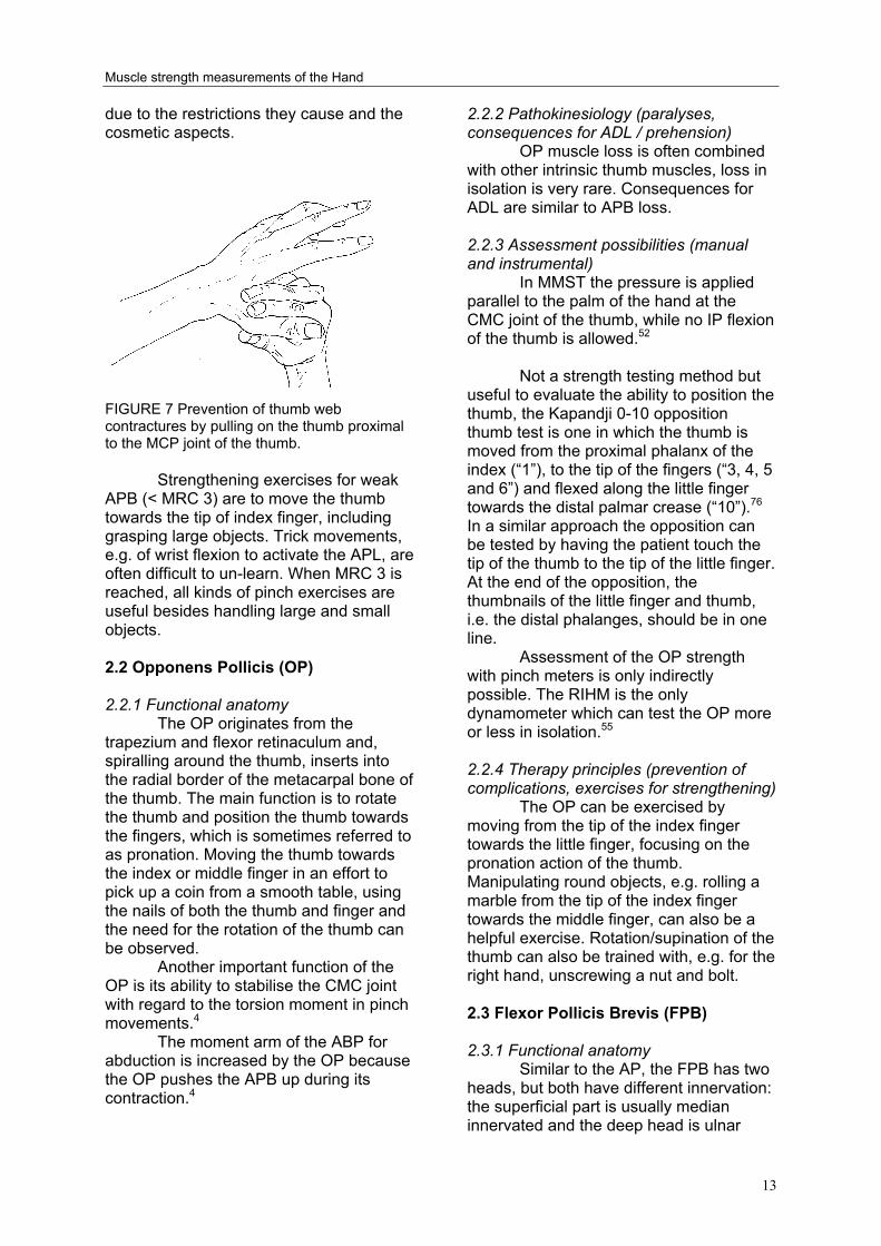
FIGURE 7 Prevention of thumb web contractures by pulling on the thumb proximal to the MCP joint of the thumb.
Strengthening exercises for weak
APB (< MRC 3) are to move the thumb towards the tip of index finger, including grasping large objects. Trick movements, e.g. of wrist flexion to activate the APL, are often difficult to un-learn. When MRC 3 is reached, all kinds of pinch exercises are useful besides handling large and small objects. 2.2 Opponens Pollicis (OP) 2.2.1 Functional anatomy
The OP originates from the trapezium and flexor retinaculum and, spiralling around the thumb, inserts into the radial border of the metacarpal bone of the thumb. The main function is to rotate the thumb and position the thumb towards the fingers, which is sometimes referred to as pronation. Moving the thumb towards the index or middle finger in an effort to pick up a coin from a smooth table, using the nails of both the thumb and finger and the need for the rotation of the thumb can be observed.
Another important function of the OP is its ability to stabilise the CMC joint with regard to the torsion moment in pinch movements.4
The moment arm of the ABP for abduction is increased by the OP because the OP pushes the APB up during its contraction.4
2.2.2 Pathokinesiology (paralyses, consequences for ADL / prehension)
OP muscle loss is often combined with other intrinsic thumb muscles, loss in isolation is very rare. Consequences for ADL are similar to APB loss. 2.2.3 Assessment possibilities (manual and instrumental)
In MMST the pressure is applied parallel to the palm of the hand at the CMC joint of the thumb, while no IP flexion of the thumb is allowed.52
Not a strength testing method but
useful to evaluate the ability to position the thumb, the Kapandji 0-10 opposition thumb test is one in which the thumb is moved from the proximal phalanx of the index (“1”), to the tip of the fingers (“3, 4, 5 and 6”) and flexed along the little finger towards the distal palmar crease (“10”).76 In a similar approach the opposition can be tested by having the patient touch the tip of the thumb to the tip of the little finger. At the end of the opposition, the thumbnails of the little finger and thumb, i.e. the distal phalanges, should be in one line.
Assessment of the OP strength with pinch meters is only indirectly possible. The RIHM is the only dynamometer which can test the OP more or less in isolation.55 2.2.4 Therapy principles (prevention of complications, exercises for strengthening)
The OP can be exercised by moving from the tip of the index finger towards the little finger, focusing on the pronation action of the thumb. Manipulating round objects, e.g. rolling a marble from the tip of the index finger towards the middle finger, can also be a helpful exercise. Rotation/supination of the thumb can also be trained with, e.g. for the right hand, unscrewing a nut and bolt. 2.3 Flexor Pollicis Brevis (FPB) 2.3.1 Functional anatomy
Similar to the AP, the FPB has two heads, but both have different innervation: the superficial part is usually median innervated and the deep head is ulnar
Ton A.R. Schreuders, JW Brandsma, HJ Stam
14
innervated. The origin for the FPB is the flexor retinaculum and the trapezium, respectively. Comparable to the APB, the FPB inserts into the extensor tendon and the lateral sesamoid bone, and assists in extension of the IP joint of the thumb.
The proximal fibers of the FPB are continuous with the OP, therefore, both act on the CMC joint of the thumb and flex the metacarpal. The major effect of the FPB is in sequence with the AP, in that both flex the MCP of the thumb, although the FPB pronates and the AP supinates the thumb.4 2.3.2 Pathokinesiology (paralyses, consequences for ADL/ prehension)
Isolated weakness of the FPB is difficult to assess, not only because of the variations in innervation, but mainly because of all the synergists in flexion of the MCP joint and sometimes the lack of mobility in the MCP joint of the thumb. Isolated loss of the FPB might go unnoticed, but loss in combination with loss of the AP, e.g. in ulnar nerve palsy, will cause significant loss of pinch strength (see AP).
Positioning of the thumb in pinch activities is a median nerve muscle function, but the strength of the pinch grip is derived from the ulnar nerve innervated muscles. 2.3.3. Assessment possibilities (manual and instrumental)
In MMST the strength of the FPB is evaluated by the assessment of flexion at the MCP joint of the thumb without flexion of the IP joint, which is the FPL action. A strength test aiming to diminish the FPL action has been studied but without convincing results.77
Measurements of the strength of the FPB with pinch dynamometers in isolation is not possible. In the dynamometry of the pinch grip, the FPB together with the AP contribute significantly to pinch strength. 2.3.4. Therapy principles (prevention of complications, exercises for strengthening)
All pinch activities can be exercised, in which the tendency to (hyper-) extend the MCP joint is a sign of
improper activation of the FPB and AP and needs to be corrected. A pinch whereby the MCP and IP joint is slightly flexed is also advantageous regarding the optimum (mid) position of the sarcomers of the intrinsic muscles of the thumb and for least tension on the soft tissues (ligaments, volar plate) of the thumb joints. 2.4 Adductor Pollicis (AP) 2.4.1 Functional anatomy
The ulnar innervated AP is a fanshaped muscle with two heads: an oblique part with its origin at the 2nd and 3rd metacarpals and the transverse part with its origin at the anterior surface of the 3rd metacarpal. The insertion of both heads is into proximal phalanx of the thumb and the sesamoid bone.
It is the most volar muscle in the thumb web, making atrophy visible in the palm of the hand. Of all the intrinsic muscles working on the thumb, the AP, working together with the FPB, has the largest flexion moment arm at the CMC joint. Therefore, the AP, together with the 1 DI, are the most important pinching muscle of the thumb, while other thumb muscles are just positioners and synergists.4 p 229 The synergists for adduction of the thumb are EPL, FPL and the first dorsal interosseus.78 2.4.2 Pathokinesiology (paralyses, consequences for prehension/ADL, shortening)
Direct lesions of the AP sometimes occur after injuries into the thumb web, e.g. knife wounds. Paralyses of the AP usually occur after ulnar nerve lesion and therefore there is also weakness of the other important muscle for pinch; the 1 DI.
In ulnar palsy there may be enough median nerve innervated FPB to position the thumb for pinch, but when power is needed, the diminished AP strength usually results in hyperflexion of the IP joint of the thumb (Froment’s sign) and sometimes in hyperextension of the MCP joint (Jeanne’s sign).4 p 54
A patient who is bedridden for a prolonged time with little activity of the hand (coma etc.) can develop a thumb web contracture due to shortening of the
Muscle strength measurements of the Hand
15
AP. After a trauma of the hand, similar to the IT, shortening of the AP can take place. The AP rests in a compartment and therefore muscle tightness can cause severe thumb web contractures. Spasticity, causing a strong pull of the thumb into the palm can be seen in patients with cerebral palsy and who have suffered a stroke. The CMC joint is adducted, making it difficult to grasp or hold but also to release larger objects. In a surgical procedure releasing a considerable part of the AP is often necessary. 2.4.3 Assessment possibilities (manual and instrumental)
Tests are often described in which plain adduction of the thumb is tested. Due to the many synergists, this usually does not provide any useful information. Weakness or paralyses will result in loss of pinch strength and show the so-called Froment’s sign. (Figure 2). This was first described by Jules Froment, who was watching a train commuter reading his newspaper with one thumb flexed and the other straight.79
In examining the pinch strength of both hands, e.g. pulling on a piece of paper, the IP joint angle is observed: if more flexion occurs at the involved hand, the Froment sign is positive. When the MCP joint of the thumb has some laxity (hypermobile) into extension, hyperextension of the MCP joint of the thumb will take place in pinch, which is called Jeanne’s sign.20 Both are signs of a reduced flexion moment at the MCP joint of the thumb, i.e. weak or paralysed AP. Grading is not possible, besides a classification of a positive or negative sign. Instrumental assessment of the strength of the AP can be done, indirectly, with pinch dynamometers. 2.4.4 Therapy principles (prevention of complications, exercises for strengthening)
Muscle strengthening exercises are all movements/activities where a pinch is needed, especially the key pinch or lateral pinch. If possible, pinch strength is trained with the thumb IP and MCP joints in slight flexion.
3. Hypothenar muscles
The muscles of the hypothenar are from ulnar to radial: abductor digiti minimi (ADM), flexor digiti minimi (brevis) (FDM), opponens digiti minimi (ODM). The palmaris brevis (PB) is the most superficial muscle overlying these muscles transversely and originates from the aponeurosis palmaris. All the hypothenar muscles are ulnar innervated. Isolated paralysis is rare and because there are functionally only minor differences between these muscles, they are discussed here as a group. 3.1 Functional anatomy
The most ulnar situated muscles (especially ADM) have the strongest ulnar abduction action of the little finger. The ODM, attached to the 5th metacarpal bone, has an important role in the opposition of 4th and 5th rays. All the hypothenar muscles are active in the intrinsic plus position of the fingers, except for the ODM and the PB. In this position, similarly to the interossei of the fingers, they flex the MCP joint of the little finger and extend the IP joints. 3.2 Pathokinesiology (paralyses, consequences for prehension/ADL, shortening)
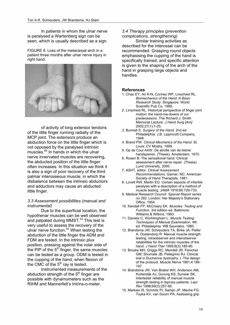
The 4th and 5th metacarpals are much more mobile in the CMC joints as compared to the 2nd and 3rd. This makes it possible to adjust the hand around a round object, but also e.g. the handle of a hammer. Flattening of the palmar arch of the hand (MCP joints) is another sign of weakness of the interossei and hypothenar muscles. The flexion of the 4th and 5th metacarpal bones at the CMC joints is diminished. This results in a flattening of the arch of the hand and in a weaker and less secure grip of the hand. (Figure 8). The loss of cupping function of the hand can go unnoticed in many patients but, e.g. for people accustomed to eating with their hands, can cause some trouble.
Ton A.R. Schreuders, JW Brandsma, HJ Stam
16
In patients in whom the ulnar nerve is paralysed a Wartenberg sign can be seen, which is usually described as a sign
FIGURE 8. Loss of the metacarpal arch in a patient three months after ulnar nerve injury in right hand.
of activity of long extensor tendons
of the little finger running radially of the MCP joint. The extensors produce an abduction force on the little finger which is not opposed by the paralysed intrinsic muscles.80 In hands in which the ulnar nerve innervated muscles are recovering, the abducted position of the little finger often increases. In this situation we think it is also a sign of poor recovery of the third palmar interosseous muscle, in which the disbalance between the intrinsic abductors and adductors may cause an abducted little finger. 3.3 Assessment possibilities (manual and instrumental)
Due to the superficial location, the hypothenar muscles can be well observed and palpated during MMST.53 This test is very useful to assess the recovery of the ulnar nerve function.53 When testing the abduction of the little finger the ADM and FDM are tested. In the intrinsic plus position, pressing against the volar side of the PIP of the 5th finger, the same muscles can be tested as a group. ODM is tested in the cupping of the hand, when flexion of the CMC of the 5th ray is tested.
Instrumented measurements of the abduction strength of the 5th finger are possible with dynamometers such as the RIHM and Mannerfelt’s Intrins-o-meter.
3.4 Therapy principles (prevention complications, strengthening)
Similar training activities as described for the interossei can be recommended. Grasping round objects emphasising the cupping of the hand is specifically trained, and specific attention is given to the shaping of the arch of the hand in grasping large objects and handles. References 1. Chao EY, An K-N, Conney WP, Linscheid RL.
Biomechanics of the Hand: A Basic Research Study. Singapore: World Scientific Pub Co, 1989.
2. Linscheid RL. Historical perspective of finger joint motion: the hand-me-downs of our predecessors. The Richard J. Smith Memorial Lecture. J Hand Surg [Am] 2002;27(1):1-25.
3. Bunnell S. Surgery of the Hand. 2nd ed. Philadelphia: J.B. Lippincott Company, 1948.
4. Brand PW. Clinical Mechanics of the Hand. St. Louis: CV Mosby, 1999.
5. Op de Coul AAW. De atrofie van de kleine handspieren. (Thesis). Amsterdam, 1970.
6. Rosen B. The sensational hand. Clinical assessment after nerve repair. (Thesis). Lund University, 2000.
7. ASHT, editor. Clinical Assessment Recommendations. Garner, NC: American Society of Hand Therapists, 1981.
8. Lovett RW, Martin EG. Certain aspects of infantile paralysis with a description of a method of muscle testing. JAMA 1916;66:729-733.
9. Medical Research Council. Special Report series no 282. London: Her Majesty's Stationary Office, 1954.
10. Kendall FP, McCreary EK. Muscles: Testing and Function. 3rd edition ed. Baltimore: Williams & Wilkins, 1983.
11. Daniels C, Worhtingham L. Muscle Testing: Techniques of Manual Examination. 4th ed. Philadelphia: WB Saunders, 1980.
12. Brandsma JW, Schreuders TA, Birke JA, Piefer A, Oostendorp R. Manual muscle strength testing: intraobserver and interobserver reliabilities for the intrinsic muscles of the hand. J Hand Ther 1995;8(3):185-90.
13. Brooke MH, Griggs RC, Mendell JR, Fenichel GM, Shumate JB, Pellegrino RJ. Clinical trial in Duchenne dystrophy. I. The design of the protocol. Muscle Nerve 1981;4:186-197.
14. Brandsma JW, Van Brakel WH, Anderson AM, Kortendijk AJ, Gurung KS, Sunwar SK. Intertester reliability of manual muscle strength testing in leprosy patients. Lepr Rev 1998;69(3):257-66.
15. Merkies IS, Schmitz PI, Samijn JP, Meche FG, Toyka KV, van Doorn PA. Assessing grip

Muscle strength measurements of the Hand
17
strength in healthy individuals and patients with immune-mediated polyneuropathies. Muscle Nerve 2000;23(9):1393-1401.
16. Kirkpatrick JE. Evaluation of grip loss; a factor of permanent partial disability in California. Ind Med Surg 1957;26(6):285-289.
17. Bechtol C. Grips test: the use of a dynamometer with adjustable handle spacing. J Bone Joint Surg 1954;36:820.
18. van der Ploeg RJ, Oosterhuis HJ, Reuvekamp J. Measuring muscle strength. J Neurol 1984;231(4):200-3.
19. van der Ploeg RJO. Hand-held dynamometry. (Thesis), 1992.
20. Mannerfelt L. Studies on the hand in ulnar nerve paralysis. A clinical-experimental investigation in normal and anomalous innervation. Acta Orthop Scand 1966(87):61-86.
21. Mannerfelt LG. Studies on ulnar nerve compression neuropathies with a new computerised instrument--the intrins-o-meter. Scand J Plast Reconstr Surg Hand Surg 1997;31(3):251-60.
22. Rosen B, Lundborg G. A model instrument for the documentation of outcome after nerve repair. J Hand Surg [Am] 2000;25(3):535-43.
23. Rosen B, Dahlin LB, Lundborg G. Assessment of functional outcome after nerve repair in a longitudinal cohort. Scand J Plast Reconstr Surg Hand Surg 2000;34(1):71-8.
24. Trumble TE, Kahn U, Vanderhooft E, Bach AW. A technique to quantitate motor recovery following nerve grafting. J Hand Surg [Am] 1995;20(3):367-72.
25. Boatright JR, Kiebzak GM, O'Neil DM, Peindl RD. Measurement of thumb abduction strength: normative data and a comparison with grip and pinch strength. J Hand Surg [Am] 1997;22(5):843-8.
26. Liu F, Carlson L, Watson HK. Quantitative abductor pollicis brevis strength testing: reliability and normative values. J Hand Surg [Am] 2000;25(4):752-9.
27. Richards RR, Gordon R, Beaton D. Measurement of wrist, metacarpophalangeal joint, and thumb extension strength in a normal population. J Hand Surg [Am] 1993;18(2):253-61.
28. Eyler DL, Markee JE. The anatomy and function of the intrinsic musculature of the fingers. J Bone Joint Surg 1954;36-A:1-10.
29. Eladoumikdachi F, Valkov PL, Thomas J, Netscher DT. Anatomy of the intrinsic hand muscles revisited: part II. Lumbricals. Plast Reconstr Surg 2002;110(5):1225-31.
30. Eladoumikdachi F, Valkov PL, Thomas J, Netscher DT. Anatomy of the intrinsic hand muscles revisited: part I. Interossei. Plast Reconstr Surg 2002;110(5):1211-24.
31. Landsmeer JM, Long C. The mechanism of finger control, based on electromyograms and location analysis. Acta Anat 1965;60(3):330-47.
32. Srinivasan H. Patterns of movement of totally intrinsic-minus fingers based on a study of
one hundred and forty-one fingers. J Bone Joint Surg Am 1976;58(6):777-85.
33. Jacobson MD, Raab R, Fazeli BM, Abrams RA, Botte MJ, Lieber RL. Architectural design of the human intrinsic hand muscles. J Hand Surg [Am] 1992;17(5):804-9.
34. Ketchum LD, Thompson D, Pocock G, Wallingford D. A clinical study of forces generated by the intrinsic muscles of the index finger and the extrinsic flexor and extensor muscles of the hand. J Hand Surg [Am] 1978;3(6):571-8.
35. Brandsma JW. Intrinsic minus hand: (patho) kinesiology, rehabilitation and reconstruction. (Thesis) [Thesis]. University of Utrecht, 1993.
36. Kozin SH, Porter S, Clark P, Thoder JJ. The contribution of the intrinsic muscles to grip and pinch strength. J Hand Surg [Am] 1999;24(1):64-72.
37. ASHT, editor. Clinical Assessment Recommendations. 2nd ed. Garner, NC: American Society of Hand Therapists, 1992.
38. Bendz P. Systematization of the grip of the hand in relation to finger motor systems. A kinesiologic study using a new method for recording finger joint motions. Scand J Rehabil Med 1974;6(4):158-65.
39. Sollerman C, Sperling L. Evaluation of ADL-function--especially hand function. Scand J Rehabil Med Suppl 1978;6:139-43.
40. Light CM, Chappell PH, Kyberd PJ. Establishing a standardized clinical assessment tool of pathologic and prosthetic hand function: normative data, reliability, and validity. Arch Phys Med Rehabil 2002;83(6):776-83.
41. Stack HG. Muscle function in the fingers. J Bone and J Surg 1962;44B:899-909.
42. Zancolli EA. Structural and Dynamic Bases of Hand Surgery. 2nd ed. Philadelphia: Lippincott, 1979.
43. Lauer RT, Kilgore KL, Peckham PH, Bhadra N, Keith MW. The function of the finger intrinsic muscles in response to electrical stimulation. IEEE Trans Rehabil Eng 1999;7(1):19-26.
44. Li ZM, Zatsiorsky VM, Latash ML. Contribution of the extrinsic and intrinsic hand muscles to the moments in finger joints. Clin Biomech (Bristol, Avon) 2000;15(3):203-11.
45. Neu BR, Murray JF, MacKenzie JK. Profundus tendon blockage: quadriga in finger amputations. J Hand Surg [Am] 1985;10:878-83.
46. Leijnse JN, Kalker JJ. A two-dimensional kinematic model of the lumbrical in the human finger. J Biomech 1995;28(3):237-49.
47. Tubiana R. The Hand Vol II. Philadelphia: WB Saunders, 1985:518.
48. Del Pinal F, Herrero F, Jado E, Garcia-Bernal FJ, Cerezal L. Acute hand compartment syndromes after closed crush: a

Ton A.R. Schreuders, JW Brandsma, HJ Stam
18
reappraisal. Plast Reconstr Surg 2002;110(5):1232-9.
49. Hueston JT, Wilson WF. The role of the intrinsic muscles in the production of metacarpophalangeal subluxation in the rheumatoid hand. Plast Reconstr Surg 1973;52(4):342-5.
50. Smith RJ. Non-ischemic contractures of the intrinsic muscles of the hand. J Bone Joint Surg Am 1971;53(7):1313-31.
51. Daniels L, Worthingham C. Muscle testing Techniques of Manual Examination. Philadelphia: W.B. Saunders Co, 1986.
52. Brandsma JW, Schreuders TAR, Birke JA, Piefer A, Oostendorp R. Manual muscle strength testing: intraobserver and interobserver reliabilities for the intrinsic muscles of the hand. J Hand Ther 1995;8(3):185-90.
53. Brandsma JW, Schreuders TA. Sensible manual muscle strength testing to evaluate and monitor strength of the intrinsic muscles of the hand: a commentary. J Hand Ther 2001;14(4):273-8.
54. Janda DH, Geiringer SR, Hankin FM, Barry DT. Objective evaluation of grip strength. J Occup Med 1987;29(7):569-71.
55. Schreuders TAR, Roebroeck ME, Goumans J, van Nieuwenhuijzen JF, Stijnen TH, Stam HJ. Measurement error in grip and pinch force measurements in patients with hand injuries. Phys Ther 2003;83(9):806-15.
56. Fischer T, Nagy L, Buechler U. Restoration of pinch grip in ulnar nerve paralysis: extensor carpi radialis longus to adductor pollicis and abductor pollicis longus to first dorsal interosseus tendon transfers. J Hand Surg [Br] 2003;28(1):28-32.
57. Thomas DH, Long C, Landsmeer JMF. Biomechanical Considerations of Lumbricalis Behavior in the Human Finger. J Biomech 1968:107-115.
58. Ranney D, Wells R. Lumbrical muscle function as revealed by a new and physiological approach. Anat Rec 1988;222(1):110-4.
59. Ranney DA, Wells RP, Dowling J. Lumbrical function: interaction of lumbrical contraction with the elasticity of the extrinsic finger muscles and its effect on metacarpophalangeal equilibrium. j Hand Surg (Am) 1987;12(4):110-114.
60. Backhouse KM, Catton WT. An experimental study of the function of the lumbrical muscles in the human hand. J Anat 1954;88(5):133-141.
61. Devanandan MS, Ghosh S, John KLT. A quantitative study of the muscle spindles and tendon organs in some intrinsic muscles of the hand. Anat Rec 1983;207:263-266.
62. Rabischong P. [Basic problems in the restoration of prehension]. Ann Chir 1971;25(19):927-33.
63. Leijnse JN. Why the lumbrical muscle should not be bigger--a force model of the lumbrical in the unloaded human finger. J Biomech 1997;30(11-12):1107-14.
64. Schreuders TAR, Stam HJ. Strength measurements of the lumbrical muscles. J Hand Ther 1996;9(4):303-5.
65. Parkes A. The 'lumbrical plus' finger. J Bone Joint Surg Br 1971;53(2):236-9.
66. (IFSSH) IFoSfSotH. Terminology for Hand Surgery: Harcourth Health Sciences, 2001.
67. Basmajian JV, DeLuca CJ. Muscles Alive: Their function Revealed by Electromyography. 5th ed. Baltimore: Williams and Wilkins, 1985.
68. Lemmen MH, Schreuders TA, Stam HJ, Hovius SE. Evaluation of restoration of extensor pollicis function by transfer of the extensor indicis. J Hand Surg [Br] 1999;24(1):46-9.
69. Vanderhooft E. Functional outcomes of nerve grafts for the upper and lower extremities. Hand Clin 2000;16(1):93-104, ix.
70. Strickland JW, Idler, R.S., DelSignore, J.L. Ulnar Nerve Repair. In: Gelberman RH, editor. Operative Nerve Repair and Reconstruction. Philadelphia: J.B. Lippincott Comp, 1991:425-436.
71. Rosen B. Recovery of sensory and motor function after nerve repair. A rationale for evaluation. J Hand Ther 1996;9(4):315-27.
72. Sunderland S. The actions of the extensor digitorum communis, interosseus and lumbrical muscles. J Anat 1945;77:189-209.
73. An KN, Chao EY, Askew LJ. Hand strength measurement instruments. Arch Phys Med Rehabil 1980;61(8):366-8.
74. Schreuders TAR, Roebroeck ME, Jaquet J-B, Hovius SER, Stam HJ. Longterm outcome of muscle strength in patients with ulnar and median nerve injury: Comparing manual muscle strength testing, grip and pinch strength dynamometers and a new intrinsic muscle strength dynamometer. J Rehabil Med 2004;36.
75. Wynn Parr CB. Rehabilitation of the hand. 4th ed. London: Butterworth, 1981.
76. Kapandji AI. Clinical evaluation of the thumb's opposition. J Hand Ther 1992;5(2):102-6.
77. Howell JW, Rothstein JM, Lamb RL, Merritt WH. An experimental investigation for the extensor pollicis longus and flexor pollicis brevis muscles. J Hand Ther 1989;2(2):20-28.
78. Kaufman KR, An KN, Litchy WJ, Cooney WP, 3rd, Chao EY. In-vivo function of the thumb muscles. Clin Biomech (Bristol, Avon) 1999;14(2):141-50.
79. Froment, J. La prehension dans les paralysies du nerf cubital et le signe du pouce. La presse medicale, Paris. 1915;23:409.
80. Blacker GJ, Lister GD, Kleinert MD. The abducted little finger in low ulnar nerve palsy. J Hand Surg [Am] 1976;1(3):190-6.