Embed Size (px)
Citation preview
Muscle PhysiologyHuman Anatomy and Physiology
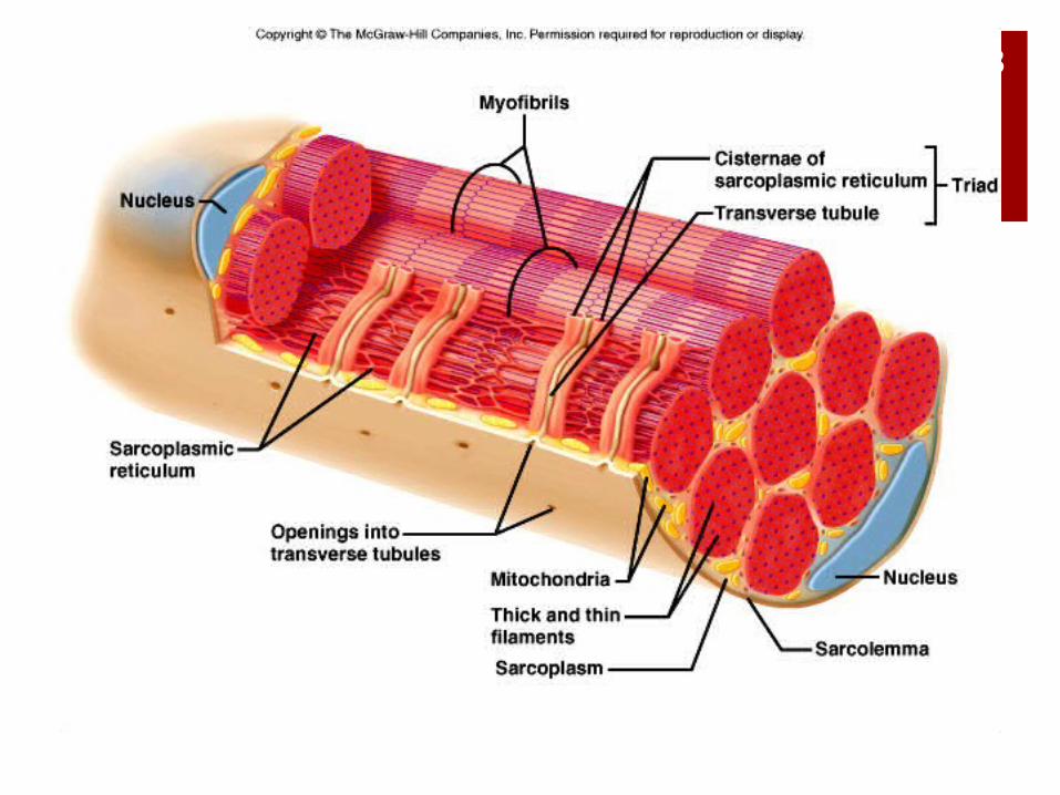
2 Beneath the sarcolemma of a
muscle fiber lies the sarcoplasmic reticulum (endoplasmic reticulum), which is associated with transverse (T) tubules (invaginations of the sarcolemma).
Each T tubule lies between two cisternae of the sarcoplasmic reticulum and is open to the outside of the muscle fiber.
The sarcoplasmic reticulum and transverse tubules activate the muscle contraction mechanism when the fiber is stimulated.
CopyrightThe McGraw-Hill Companies, Inc. Permission required for reproduction or display.
3
4Neuromuscular JunctionThe site where the motor neuron
and muscle fiber meet is the neuromuscular junction.
The muscle fiber membrane forms a motor end plate in which the sarcolemma is tightly folded and where nuclei and mitochondria are abundant.
The cytoplasm of the motor neuron contains numerous mitochondria and synaptic vesicles storing neurotransmitters.
CopyrightThe McGraw-Hill Companies, Inc. Permission required for reproduction or display.
5
Motor Units
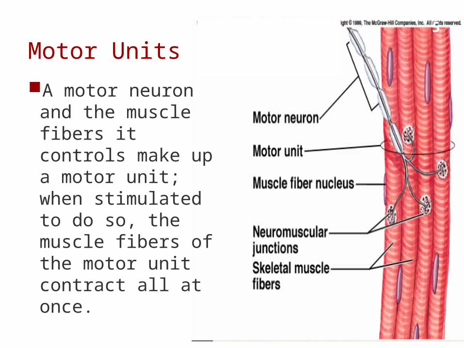
A motor neuron and the muscle fibers it controls make up a motor unit; when stimulated to do so, the muscle fibers of the motor unit contract all at once.

6
7Skeletal Muscle Contraction
Muscle contraction involves several components that result in the shortening of sarcomeres, and the pulling of the muscle against its attachments.
CopyrightThe McGraw-Hill Companies, Inc. Permission required for reproduction or display.
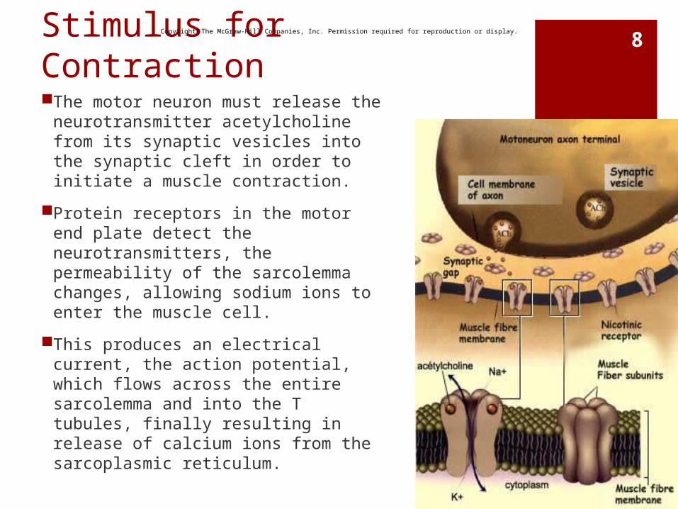
8Stimulus for ContractionThe motor neuron must release the
neurotransmitter acetylcholine from its synaptic vesicles into the synaptic cleft in order to initiate a muscle contraction.
Protein receptors in the motor end plate detect the neurotransmitters, the permeability of the sarcolemma changes, allowing sodium ions to enter the muscle cell.
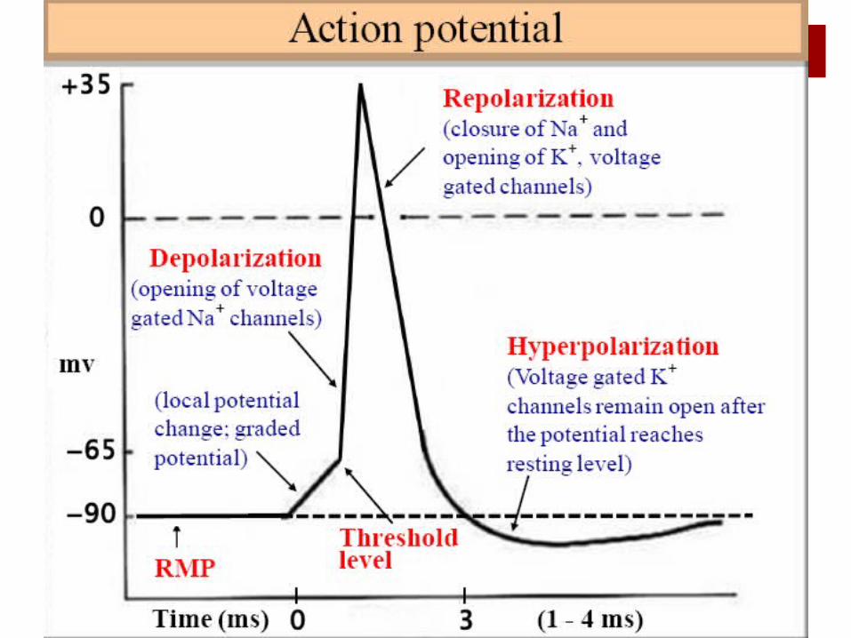
This produces an electrical current, the action potential, which flows across the entire sarcolemma and into the T tubules, finally resulting in release of calcium ions from the sarcoplasmic reticulum.
CopyrightThe McGraw-Hill Companies, Inc. Permission required for reproduction or display.
10
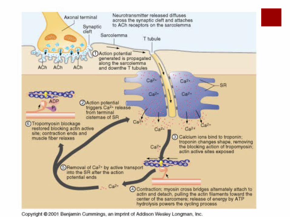
Upon receipt of the action potential, the sarcoplasmic reticulum releases its stored calcium to the sarcoplasm of the muscle fiber.
The high concentration of calcium in the sarcoplasm interacts with the troponin and tropomyosin molecules, which move aside, exposing the myosin binding sites on the actin filaments.
Myosin cross-bridges now bind and pull on the actin filaments, causing the sarcomeres to shorten.
After the nervous impulse has been received, acetylcholinesterase rapidly decomposes the acetylcholine.
Then, calcium is returned to the sarcoplasmic reticulum, and the linkages between myosin and actin are broken.
CopyrightThe McGraw-Hill Companies, Inc. Permission required for reproduction or display.
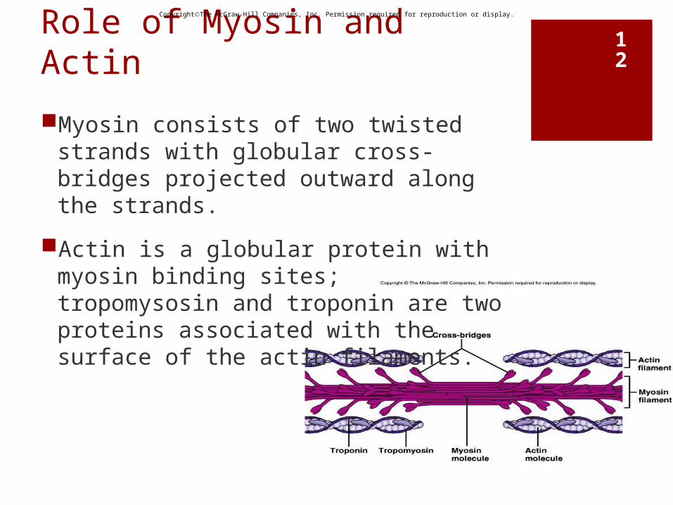
12Role of Myosin and Actin
Myosin consists of two twisted strands with globular cross-bridges projected outward along the strands.
Actin is a globular protein with myosin binding sites; tropomysosin and troponin are two proteins associated with the surface of the actin filaments.
CopyrightThe McGraw-Hill Companies, Inc. Permission required for reproduction or display.
13
Sliding Filament Theory
According to the sliding filament theory of muscle contraction, the myosin crossbridge attaches to the binding site on the actin filament and bends, pulling on the actin filament; it then releases and attaches to the next binding site on the actin, pulling again.
Energy from the conversion of ATP to ADP is provided to cross-bridges from the enzyme ATPase, causing them to be in a “cocked” position.
CopyrightThe McGraw-Hill Companies, Inc. Permission required for reproduction or display.
Sliding Filament TheoryVideo clips of sliding filament theory
http://www.sci.sdsu.edu/movies/actin_myosin.html http://media.pearsoncmg.com/bc/bc_campbell_biol
ogy_6/cipl/ins/49/HTML/source/71.html http://www.wisc-online.com/objects/index_tj.asp?o
bjID=AP2904


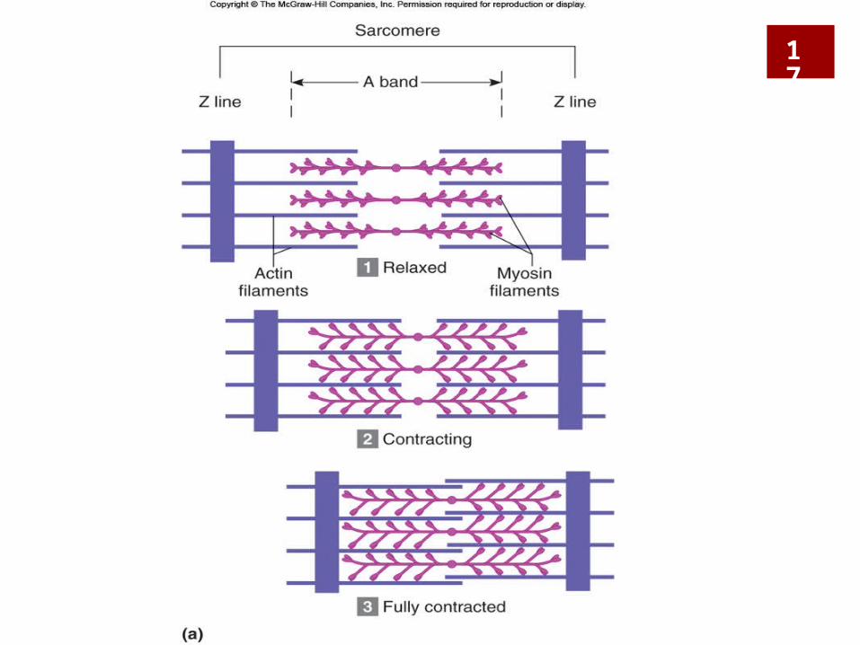
Sliding Filament Theory
In the contracted sarcomere, the light H zone in the
center of the A band has disappeared,
the Z lines are closer to the thick filaments,
and I bands have nearly disappeared.
The A bands move closer together but do not change in length
17
Sliding Filament TheorySarcomere Shortening Animation