Embed Size (px)
Citation preview
Murine Model of Autoimmune Hemolytic Anemia: Red Blood Cell Clearance Mechanisms and
Treatment Efficacy
by
Xi Chen
A thesis submitted in conformity with the requirements for the degree of Masters of Science
Graduate Department of Laboratory Medicine and Pathobiology University of Toronto
© Copyright by Xi Chen 2012
ii
Murine Model of Autoimmune Hemolytic Anemia: Red Blood
Cell Clearance Mechanisms and Treatment Efficacy
Xi Chen
Masters of Science
Graduate Department of Laboratory Medicine and
University of Toronto
2012
Abstract
Antibodies against erythrocyte-specific antigens can cause immune red blood cell (RBC)
destruction, resulting in autoimmune hemolytic anemia (AHA). Therapy for patients with
AHA remains a challenge, and better knowledge of pathophysiological mechanisms is
needed. In this thesis, the mechanisms of RBC clearance mediated by a panel of monoclonal
anti-mouse RBC antibodies were characterized in a murine model of passive AHA, and
efficacy of treatments was evaluated. It is demonstrated herein that mechanisms in addition
to the classical macrophage Fcγ receptor (FcγR)- and/or complement-mediated hemolysis
may be contributing to the development and/or persistence of anemia. The efficacy of
intravenous immunoglobulin (IVIg) therapy varies in relation to the mechanism of anemia in
mice with FcγR-mediated anemia being highly related to IVIg responsiveness. In addition,
this thesis shows that monoclonal RBC-specific antibodies can ameliorate thrombocytopenia
in a murine model of passive immune thrombocytopenia (ITP) analogous to the use of anti-D
in ITP patients.
iii
Acknowledgments
I would first like to thank my supervisor, Dr. Alan Lazarus, for having always believed in
and encouraged me. I feel so lucky to have shared his incredible knowledge, wisdom and
optimism both in science and in life, and will be forever indebted to him for his support
professionally, financially and personally over the years.
A heart-felt thanks to all the past and present members of the Lazarus lab: Andrew, Sara,
Nancy, Zhongwei, Joan, Pat, Honghui, Lidice and Alaa for your help and support to me, and
most importantly, for all the laughter we shared in and out of the lab.
Sincere thanks to the members of my advisory committee, Dr. Heyu Ni and Dr. John Semple,
for their expertise and encouragement throughout my graduate program. A special thank you
goes to my colleagues in the Ni lab and Semple lab for being fantastic neighbors and never
hesitating to help.
I also want to thank my collaborators, especially Dr. Hasan Ghaffar from St. Michael’s
Hospital, for sacrificing his own time to help me with my project.
For all the people cited above, and those I did not mention but who have helped and
supported me along the journey, I wish you all the best of success in your future endeavors!
Last but not least, I want to thank my friends and family for their constant love and generous
support, without which this thesis would not have happened.
iv
Table of Contents
Acknowledgments ................................................................................................................... iii
Table of Contents ..................................................................................................................... iv
List of Tables ......................................................................................................................... viii
List of Figures .......................................................................................................................... ix
List of Appendices .................................................................................................................. xii
List of Abbreviations ............................................................................................................. xiii
Chapter 1 Introduction .............................................................................................................. 1
Chapter 2 Literature Review ..................................................................................................... 6
2.1 Mechanisms of RBC clearance ................................................................................ 7
2.1.1 Overview ...................................................................................................... 7
2.1.2 Extravascular hemolysis .............................................................................. 8
2.1.3 Intravascular hemolysis ............................................................................... 9
2.1.4 Multivalency-mediated hemagglutination ................................................... 9
2.1.5 Anti-glycophorin A antibody-mediated RBC hemolysis ........................... 10
2.1.6 Anti-Kell antibody-mediated inhibition/destruction of erythroid progenitor
cells……………………………………………………………………………….11
2.2 Mouse models of AHA .......................................................................................... 12
2.2.1 Overview .................................................................................................... 12
2.2.2 Mouse IgG-FcγR system ............................................................................ 12
2.2.3 Mouse AHA models .................................................................................. 16
Chapter 3 Rationale, Hypothesis and Specific Aims .............................................................. 17
v
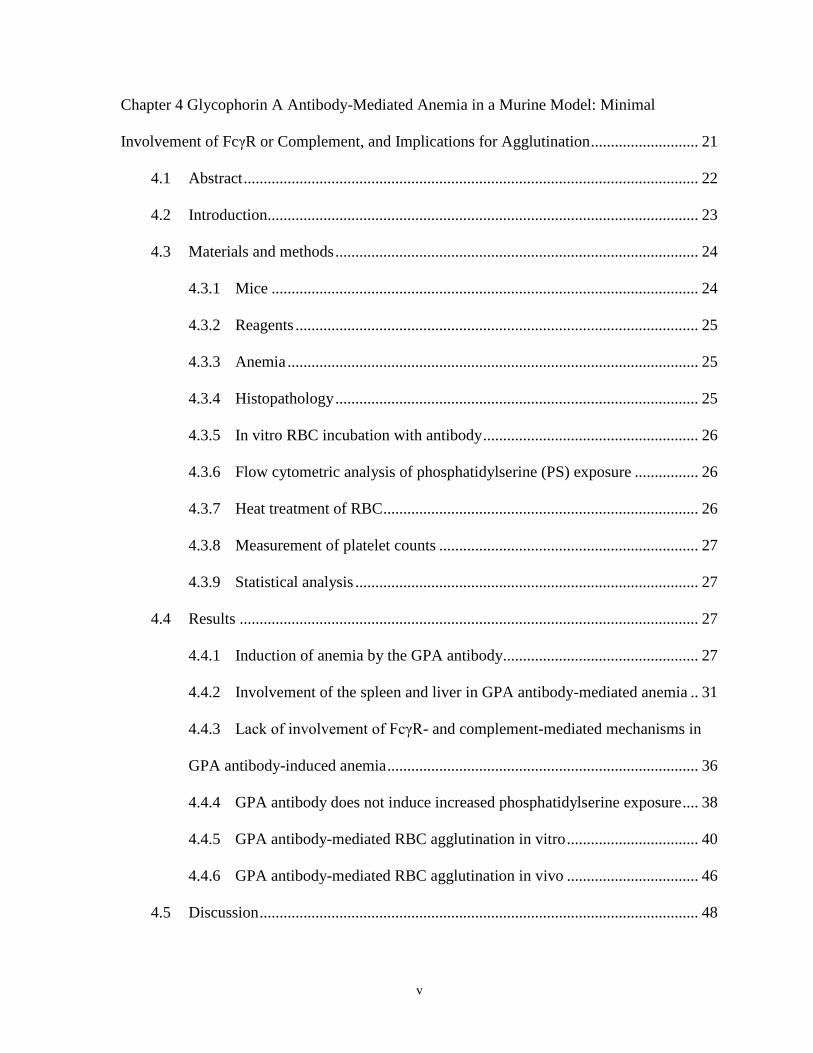
Chapter 4 Glycophorin A Antibody-Mediated Anemia in a Murine Model: Minimal
Involvement of FcγR or Complement, and Implications for Agglutination ........................... 21
4.1 Abstract .................................................................................................................. 22
4.2 Introduction............................................................................................................ 23
4.3 Materials and methods ........................................................................................... 24
4.3.1 Mice ........................................................................................................... 24
4.3.2 Reagents ..................................................................................................... 25
4.3.3 Anemia ....................................................................................................... 25
4.3.4 Histopathology ........................................................................................... 25
4.3.5 In vitro RBC incubation with antibody ...................................................... 26
4.3.6 Flow cytometric analysis of phosphatidylserine (PS) exposure ................ 26
4.3.7 Heat treatment of RBC ............................................................................... 26
4.3.8 Measurement of platelet counts ................................................................. 27
4.3.9 Statistical analysis ...................................................................................... 27
4.4 Results ................................................................................................................... 27
4.4.1 Induction of anemia by the GPA antibody................................................. 27
4.4.2 Involvement of the spleen and liver in GPA antibody-mediated anemia .. 31
4.4.3 Lack of involvement of FcγR- and complement-mediated mechanisms in
GPA antibody-induced anemia .............................................................................. 36
4.4.4 GPA antibody does not induce increased phosphatidylserine exposure .... 38
4.4.5 GPA antibody-mediated RBC agglutination in vitro ................................. 40
4.4.6 GPA antibody-mediated RBC agglutination in vivo ................................. 46
4.5 Discussion .............................................................................................................. 48
vi
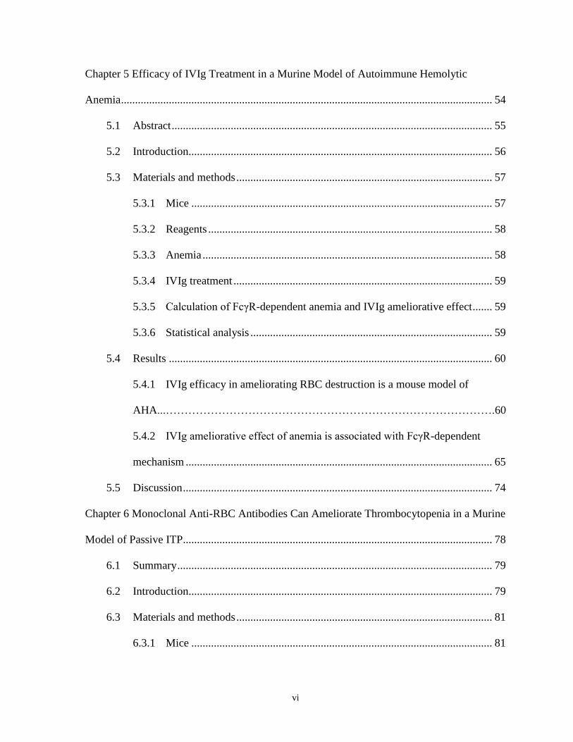
Chapter 5 Efficacy of IVIg Treatment in a Murine Model of Autoimmune Hemolytic
Anemia .................................................................................................................................... 54
5.1 Abstract .................................................................................................................. 55
5.2 Introduction............................................................................................................ 56
5.3 Materials and methods ........................................................................................... 57
5.3.1 Mice ........................................................................................................... 57
5.3.2 Reagents ..................................................................................................... 58
5.3.3 Anemia ....................................................................................................... 58
5.3.4 IVIg treatment ............................................................................................ 59
5.3.5 Calculation of FcγR-dependent anemia and IVIg ameliorative effect ....... 59
5.3.6 Statistical analysis ...................................................................................... 59
5.4 Results ................................................................................................................... 60
5.4.1 IVIg efficacy in ameliorating RBC destruction is a mouse model of
AHA...…………………………………………………………………………….60
5.4.2 IVIg ameliorative effect of anemia is associated with FcγR-dependent
mechanism ............................................................................................................. 65
5.5 Discussion .............................................................................................................. 74
Chapter 6 Monoclonal Anti-RBC Antibodies Can Ameliorate Thrombocytopenia in a Murine
Model of Passive ITP.............................................................................................................. 78
6.1 Summary ................................................................................................................ 79
6.2 Introduction............................................................................................................ 79
6.3 Materials and methods ........................................................................................... 81
6.3.1 Mice ........................................................................................................... 81
vii
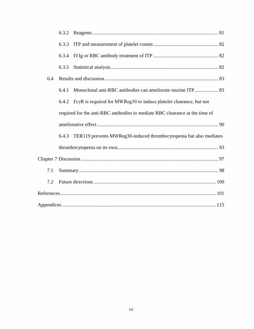
6.3.2 Reagents ..................................................................................................... 81
6.3.3 ITP and measurement of platelet counts .................................................... 82
6.3.4 IVIg or RBC antibody treatment of ITP .................................................... 82
6.3.5 Statistical analysis ...................................................................................... 82
6.4 Results and discussion ........................................................................................... 83
6.4.1 Monoclonal anti-RBC antibodies can ameliorate murine ITP ................... 83
6.4.2 FcγR is required for MWReg30 to induce platelet clearance, but not
required for the anti-RBC antibodies to mediate RBC clearance at the time of
ameliorative effect ................................................................................................. 90
6.4.3 TER119 prevents MWReg30-induced thrombocytopenia but also mediates
thrombocytopenia on its own................................................................................. 93
Chapter 7 Discussion .............................................................................................................. 97
7.1 Summary ................................................................................................................ 98
7.2 Future directions .................................................................................................. 100
References............................................................................................................................. 101
Appendices ........................................................................................................................... 115
viii
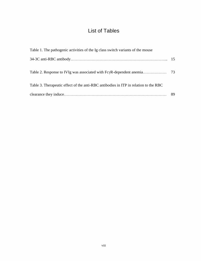
List of Tables
Table 1. The pathogenic activities of the Ig class switch variants of the mouse
34-3C anti-RBC antibody……………………………………………………………….. 15
Table 2. Response to IVIg was associated with FcγR-dependent anemia……………… 73
Table 3. Therapeutic effect of the anti-RBC antibodies in ITP in relation to the RBC
clearance they induce…………………………………………………………………… 89
ix
List of Figures
Figure 1. The family of mouse Fc receptors for IgG…………………………………... 14
Figure 2. Sensitization of peripheral RBC with the anti-GPA antibody………………. 28
Figure 3. Induction of anemia by the anti-GPA antibody…………………………….. 29
Figure 4. Induction of hemoglobinuria by the anti-GPA antibody……………………. 30
Figure 5. Induction of splenomegaly by the GPA antibody…………………………… 32
Figure 6. Spleen pathologies mediated by the GPA antibody…………………………. 33
Figure 7. Liver pathologies mediated by the GPA antibody…………………………... 34
Figure 8. Role of spleen in the development of anemia mediated by GPA antibody…. 35
Figure 9. GPA antibody-induced anemia in Fcγ-/-
, C3-/-
, and C5-deficient mice……… 37
Figure 10. Detection of PS exposure on GPA antibody-treated RBC………………… 39
Figure 11. GPA antibody incubation with mouse RBC in vitro induces dose-
dependent hemolysis…………………………………………………………………... 42
Figure 12. Flow cytometry scatter plot of mouse RBC incubated with the GPA
antibody………………………………………………………………………………… 43
x
Figure 13. GPA antibody-induced in vitro hemolysis is dependent on presence of
extracellular ions……………………………………………………………………….
44
Figure 14. GPA antibody-induced RBC agglutination in vitro……………………….. 45
Figure 15. GPA antibody-induced RBC agglutination in vivo………………………… 47
Figure 16. Efficacy of IVIg therapy for band 3 antibody-mediated anemia…………… 61
Figure 17. Efficacy of IVIg therapy for CD24 antibody-mediated anemia……………. 62
Figure 18. Efficacy of IVIg therapy for GPA antibody-mediated anemia…………….. 63
Figure 19. Efficacy of IVIg therapy for Kell antibody-mediated anemia……………… 64
Figure 20. Anemia in complement C3-deficient mice…………………………………. 66
Figure 21. Band 3 antibody-mediated anemia in FcγR-deficient mice………………... 68
Figure 22. CD24 antibody-mediated anemia in FcγR-deficient mice………………… 69
Figure 23. GPA antibody-mediated anemia in FcγR-deficient mice………………….. 70
Figure 24. Kell antibody-mediated anemia in FcγR-deficient mice…………………… 71
Figure 25. Efficacy of the GPA antibody in the amelioration of a murine model of
passive ITP. …………………………………………………………………………… 84
Figure 26. Efficacy of the band 3 antibody in the amelioration of a murine model of
passive ITP…………………………………………………………………………….. 85
xi
Figure 27. Efficacy of the Kell antibody in the amelioration of a murine model of
passive ITP……………………………………………………………………………..
86
Figure 28. Efficacy of the CD24 antibody in the amelioration of a murine model of
passive ITP…………………………………………………………………………….. 87
Figure 29. Fcγ-/-
mice were fully protected from thrombocytopenia induced by
MWReg30……………………………………………………………………………… 91
Figure 30. Degree of anemia in Fcγ-/-
mice……………………………………………. 92
Figure 31. Effect of anti-GPA on platelet counts……………………………………… 95
xii
List of Appendices
Appendix 1. Induction of thrombocytopenia by the anti-GPA antibody………………. 116
Appendix 2. Antibody-mediated anemia in surgically splenectomized mice………….. 117
Appendix 3. IVIg rescues CD1 mice from anti-GPA antibody-induced death………... 118
Appendix 4. Sensitization of mouse RBC with the GPA antibody in vivo…………….. 119
Appendix 5. Sensitization of mouse RBC with the band 3 antibody in vivo. …………. 120
Appendix 6. Sensitization of mouse RBC with the Kell antibody in vivo……………... 121
Appendix 7. Peripheral reticulocyte counts increase exponentially in response to
treatment with the GPA antibody……………………………………………………… 122
Appendix 8. IVIg does not cause hemolysis in mice………………………………….. 123
Appendix 9. The CD24 antibody fails to ameliorate thrombocytopenia induced by
MWReg30 regardless of the manufacturer…………………………………………….. 124
Appendix 10. The anti-CD44 antibody KM114 does not ameliorate passive AHA
mediated by the band 3 antibody in mice regardless of treatment regime……………... 125
Appendix 11. Transfusion of aged or heat-damaged RBC was associated with modest
increase in platelet counts following induction of ITP by MWReg30………………… 126
xiii
List of Abbreviations
AHA: autoimmune hemolytic anemia
CA: cold agglutinin
CAS: cold agglutinin syndrome
CR: complement receptor
CPDA: citrate-phosphate-dextrose solution with adenine
DC: dendritic cells
FcγR: Fc γ receptor
GPA: glycophorin A
GPC: glycophorin C
HDN: hemolytic diseases of the newborn
HSA: heat stable antigen
Ig: immunoglobulin
IHA: immune hemolytic anemia
ITAM: immunoreceptor tyrosine based activation motif
ITIM: Immunoreceptor tyrosine based activation motif
ITP: immune thrombocytopenia
IVIg: intravenous immunoglobulin
mAb: monoclonal antibody
MAC: membrane attack complex
MBL: mannose-binding lectin
MFI: mean fluorescence intensity
xiv
NK: natural killer
NOD: non-obese diabetic
NZB: New Zealand Black
PCH: paroxysmal cold hemoglobinuria
PE: phosphatidylethanolamine
PRP: platelet-rich plasma
PS: phosphatidylserine
RBC: red blood cell
RES: reticuloendothelial system
RT: room temperature
SEM: standard error of the mean
SLE: systemic lupus erythematosus
TRALI: transfusion-related acute lung injury

1
Chapter 1
Introduction
2
1 Introduction
Autoimmune hemolytic anemia (AHA) is an acquired immunologic disease characterized by
destruction of red blood cells (RBC) as a result of production of antibodies directed against self
RBC antigens. AHA is arguably the oldest recognized autoimmune condition in humans [1, 2],
yet a relatively uncommon disease with annual incidence estimated at 1~3 per 100,000 [3]. The
majority of cases of AHA are mediated by warm-reactive autoantibodies, defined by antibodies
that exhibit maximal RBC reactivity at 37˚C, and therefore referred to as warm AHA (wAHA).
In contrast, cold agglutinin syndrome (CAS) is caused by antibodies that display optimal RBC
reactivity at 4˚C, Patients with warm and cold antibodies detected in their sera that satisfy the
diagnostic criteria for both wAHA and CAS are said to have mixed AHA. Another AHA subtype
is paroxysmal cold hemoglobinuria (PCH). PCH is distinct from CAS in that the causative
antibody requires exposure to cold temperature followed by exposure to 37˚C environment in
order for the hemolysis to occur. Finally, cases have been reported where ingestion of a drug
causes the development of RBC autoantibodies; such cases are termed drug-induced AHA.
Alternatively, AHA can be classified as being primary (idiopathic) if it is not associated with an
underlying disorder and secondary if an association with an additional disease is established.
The distinction between different types of AHA is important for prognosis and management
purposes. The most common type of AHA, wAHA represents 70~90% of all cases [4, 5], and is
associated with the poorest prognosis with significant mortality [6]. Typically of
immunoglobulin G (IgG) isotype, warm autoantibodies used to be considered nonspecific and
bind to all the common types of human RBC in blood bank test panels. Recent reports, however,
suggest that binding is specific but to “public” antigens common to almost all human RBC and
thereby appear to be panreactive [6]. It has been demonstrated that autoantibody specificities in
3
about half of the patients are predominantly directed against Rh antigens such as e, E or C, and
other specificities identified include epitopes on glycophorins, membrane band 3 protein, the
Kell, Kid, Duffy, and ABO systems [7, 8].
In addition to antibody specificities, other characteristics of the bound antibody including
quantity, ability to fix complement and ability to bind to tissue macrophages are all determinants
of the degree and mechanism of hemolysis. The current understanding of mechanisms of
immune RBC destruction is that two distinct types of hemolysis exist. The term intravascular
hemolysis describes complement-mediated RBC lysis directly in the blood stream resulting in
hemoglobinemia and likely hemoglobinuria, whereas the term extravascular hemolysis is used
when RBC is instead destroyed within the reticuloendothelial system (RES) as a result of the
recognition of the RBC-bound antibodies or complement components by their respective
receptors expressed by macrophages of the RES. Despite the fact that intra- and extra-vascular
hemolysis are considered the principle immune RBC destruction mechanisms, there are
emerging clinical findings that seem to challenge the absolute requirement for complement or
macrophage classical phagocytosis pathways [9, 10]. Indeed, several novel mechanisms have
been proposed to explain individual cases yet are hard to reproduce due to the rare but potentially
very heterogeneous nature of the disease. These mechanisms are discussed in subsequent
chapters of this thesis.
Parallel to an incomplete understanding of the RBC destruction mechanisms, management of
AHA remains regrettably experience-based in the era of evidence-based medicine [11]. Newly
diagnosed primary wAHA is managed with first-line treatment of glucocorticoids (steroids), to
which approximately 70-80% of patients will respond, but only 15-20% will achieve complete
remission and remain steroid free. Patients who become refractory to steroid therapy are
4
considered for second-line treatment that includes the options of splenectomy, cytotoxic drugs,
and in recent years, the anti-CD20 antibody rituximab. Most if not all of these therapies have low
curative potential, but severe and usually long term side effects [7, 11].
The efficacy of intravenous immunoglobulin (IVIg) treatment of immune thrombocytopenia
(ITP), which is an autoimmune condition characterized by platelet destruction mediated by
platelet specific autoantibodies, was established in the 1980s [12, 13] and suggested that the IVIg
treatment of AHA might offer an alternative to the conventional treatments but with considerable
lower risk of side effects. However, reported efficacy has been inconsistent [14-17], comparing
unfavorably with IVIg treatment of ITP and of autoimmune neutropenia, and with other
treatment modalities for AHA [18]; as a result, IVIg remains off-label for this indication [19]. In
order to identify patient variables useful for identifying responders to IVIg, a controlled clinical
trial will be required; yet such studies are generally lacking with AHA patients due to the low
incidence of the disease and numerous confounding factors such as additional underlying
disorder (in the case of secondary AHA) and the fact that most patients have received or are
receiving multiple concurrent treatments.
In summary, AHA is an autoimmune disorder with potentially severe consequences. The
pathological mechanisms are not fully understood, and the current treatment for AHA patients is
not satisfactory. In this thesis, we developed a mouse model of passively induced AHA with a
panel of monoclonal mouse RBC-specific antibodies, and used this model as a platform to study
the mechanism of RBC destruction and efficacy of treatment. These results where we examined
the pathologic effects of these RBC-specific antibodies are presented in Chapters 4 and 5. In
Chapter 6 we explored the therapeutic potential of RBC clearance, and studied these monoclonal

5
RBC antibodies for their ability to ameliorate thrombocytopenia in a murine model of passively
induced immune thrombocytopenia (ITP).

6
Chapter 2
Literature Review
7
2 Literature review
2.1 Mechanisms of RBC clearance
2.1.1 Overview
One’s total RBC volume can only be decreased rapidly by two mechanisms: bleeding (blood loss)
or RBC hemolysis. A cessation of RBC production in a patient with a normal RBC life span, on
the other hand, can only result in a decrease in RBC count of a hardly detectable 1% per day,
since the normal RBC life span is ~120 days in the circulation in human [20]. The precise
mechanism of recognition of senescent and injured RBC is not fully understood. One theory
suggests that erythrocyte aging may lead to binding of autologous IgG recognizing a senescent
antigen residing on the band 3 membrane protein and subsequent removal of the IgG-bound RBC
through phagocytosis mediated by the macrophages of the RES [21]. Alternatively, some
propose a mechanism of caspase-independent RBC apoptosis, or “eryptosis”, where the aged red
cells experience a series of changes including increased Ca2+
permeability, cell shrinkage,
reduced CD47 expression, and exposure of the inner membrane phospholipids that are
recognized by macrophages equipped with the corresponding receptors [22, 23].
RBC clearance as a result of autoantibody-coated RBC in AHA is mediated by different
mechanisms than in senescent RBC clearance. The current understanding of mechanisms of
immune RBC destruction is that two distinct, but not exclusive, types of hemolysis exist [6]. The
dominant type of extravascular hemolysis describes the macrophage-mediated RBC destruction
that predominantly takes place in macrophages of the RES in the spleen and liver. Intravascular
hemolysis, on the other hand, describes the complement-mediated direct lysis of antibody-coated
8
RBC in the blood stream. These two mechanisms are reviewed in detail in the following sub-
sessions. Despite the fact that intra- and extra-vascular hemolysis are considered the principle
immune RBC destruction mechanisms, there are emerging clinical findings that seem to
challenge the absolute requirement for and involvement of complement or macrophage classical
phagocytosis pathways [24]. Indeed, several novel mechanisms have been proposed and will also
be discussed.
2.1.2 Extravascular hemolysis
The principle mechanism of hemolysis in IHA is considered to be the opsonization of the red
cells by erythrocyte-specific antibodies as well as complement components, followed by
recognition by the Fc- and complement receptors, respectively, expressed by the splenic
macrophages and liver Kupffer cells, leading to phagocytosis of these cells and consequently,
extravascular hemolysis [25]. Both IgG and IgM antibodies can be involved, although likely via
different mechanisms. IgM in its pentameric form engages the classical complement pathway
most efficiently among all immunoglobulins. As the early complement components C3b and
iC3b are activated, they act to opsonize the IgM-sensitized red cells and mediate the subsequent
phagocytosis [26]. On the other hand, the IgG antibodies, considered relatively poor activators of
the complement pathway, are readily recognized by the Fc receptors on the phagocyte surface to
initiate erythrophagocytosis. Interestingly, there is evidence that the IgG antibody and
complement fragments may act in a synergistic manner to enhance phagocytosis when both are
present on the RBC [27-29].

9
2.1.3 Intravascular hemolysis
Intravascular hemolysis in AHA is caused by complement-mediated immune hemolysis. Among
the three pathways of complement activation: classical, the alternative and the mannose-binding
lectin (MBL) pathways [30], the classical pathway appears to be the most relevant pathway in
causing intravascular hemolysis in immune mediated RBC destruction, whereas evidence
indicating a role of the other two pathways is rare [6]. Activation of late components of the
complement cascade is required, most likely by IgM antibodies, the membrane attack complex
(MAC, composed of complement C5-C9) is formed, and the target RBC are lysed within the
circulation, therefore causing intravascular hemolysis. The lysed RBC then release hemoglobins
into the plasma which are filtered through the glomerulus, and the patient is presented with
hemoglobinemia and hemoglobinuria.
2.1.4 Multivalency-mediated hemagglutination
Work done in a passive mouse model of AHA has revealed yet another possible mechanism of
multivalency-mediated hemagglutination. Using monoclonal anti-RBC autoantibodies derived
from the New Zealand Black (NZB) mouse, Shibata et al [31] showed that the IgM
autoantibodies derived from these mice did not cause significant phagocytosis, nor did they
stimulate complement-mediated lysis. Instead, these IgM monoclonal antibodies markedly
agglutinated mouse RBC both in vitro and in vivo, and caused severe anemia. Baudino et al [32]
extended these findings by reporting that polymeric forms of IgM and IgA class-switch variants
of the same antibody caused anemia as a result of hemagglutination and subsequent sequestration
of the target RBC in the spleen, whereas their monomeric counterparts were free of any
pathogenic effects. They also confirmed the lack of complement contribution in this process by
10
showing the similar degree of anemia developed in the C3-deficient mice. Furthermore, an IgA-
mediated human AHA case was reported recently [10], where it was shown that
hemagglutination in the spleen induced by the polymeric IgA antibody was likely responsible for
the anemia, which was found to be independent of complement activation and Fc-mediated
erythrophagocytosis.
2.1.5 Anti-glycophorin A antibody-mediated RBC hemolysis
Recently Brain et al [9, 33, 34] presented a series of case reports of life-threatening AHA due to
autoantibodies that had a specificity for determinants on the RBC membrane glycoprotein,
glycophorin A (GPA). They went on to show that in vitro incubation of erythrocytes with the
GPA-specific antibody from the patients lead to hemolysis not via complement but via an
enhanced RBC membrane permeability to the Na+ and Ca
2+ cations, accompanied by increased
phosphatidylethanolamine (PE) exposure. GPA is one of the 2 most abundant integral proteins in
the erythrocyte membrane (the other is band 3), present at an estimated 1 million copies per cell
[35], and carries several clinically relevant blood group antigens including M, N, Pr, and Ena.
The exact function of GPA remains unclear, although it carries the majority of the negative
charges of RBC due to its high sialic acid content, and thereby likely contributing to the
electrostatic repulsion that prevents RBC aggregation [36]. Of interest, reported AHA cases due
to GPA-specific antibodies are relatively rare but usually severe, and Brain et al.’s findings
suggest that mechanisms other than the canonical macrophage FcR/complement-mediated
hemolysis may be involved.
11
2.1.6 Anti-Kell antibody-mediated inhibition/destruction of erythroid progenitor cells
The Kell blood-group system is one of the major antigenic systems in human RBC, and anti-Kell
antibodies account for 10% of all cases of immune mediated severe fetal anemia in hemolytic
disease of the new-born (HDN), second only to HDN mediated by anti-D [37]. Interestingly, the
mechanism of fetal anemia caused by anti-Kell antibodies seem to differ from that of classic
HDN associated with anti-D. Affected Kell-alloimmunized fetuses usually present with lower
numbers of circulating reticulocytes and normoblasts, compared with anti-D-affected fetuses,
despite an elevated level of erythropoietin released, and the inverse relationship between
hemoglobin level and reticulocyte counts found in anti-D-affected cases was missing in anti-
Kell-affected fetuses. These observations led to the hypothesis that anti-Kell antibodies target
RBC precursor cells instead of mature RBC in the circulation. Consistent with this hypothesis, it
was found that anti-Kell-affected cases have low serum bilirubin levels perhaps as a result of
hemolysis of erythroid precursor cells which are non- or in-completely hemaglobinized [38]. In
addition, Vaughan et al [39] demonstrated that monoclonal IgG and IgM antibodies are capable
of specifically inhibiting the growth of Kell-positive (but not Kell-negative) RBC precursor cells
in a dose-dependent manner, whereas anti-D had no such effect. Their findings were furthered by
Daniels et al [40] who showed that Kell antigen appears on erythroid progenitor cells early in
erythropoiesis, prior to the expression of glycophorin A and band 3. The authors went on to show
that Kell-positive progenitor cells sensitized with anti-Kell sera from pregnant women elicited a
strong response from monocytes in vitro, and proposed that anti-Kell but not anti-D may cause
fetal anemia by promoting immune destruction of the Kell-positive erythroid early progenitor
cells by macrophages in the fetal liver.
12
2.2 Mouse models of AHA
2.2.1 Overview
The best way to understand human disease is to study human patients, yet this is particularly
difficult with AHA patients due to the low number of cases and the highly heterogeneous nature
of the disease. As a result, most published reports contain information from very small
populations, and results are often inconsistent between studies. In studies aimed to evaluate
efficacy of treatments, this is further complicated by the fact that most AHA patients have
received or are receiving multiple therapies for their anemia, making it difficult to determine the
effect of individual medications.
Mouse models are widely used to understand basic pathogenic mechanisms of antibody-mediated
RBC clearance [41, 42], and are the choice of animal model in this report. Mice are not humans,
and there are some differences that need to be considered when extrapolating mouse data to the
human system. For example, the mouse complement has been shown to be relatively inefficient
in lysing erythrocyte targets compared to humans and other species [43, 44], and the IgG
subclasses and Fc receptors are different in mice and humans [45, 46]. The mouse IgG-FcγR
system will be briefly reviewed next, followed by an introduction to the various mouse models of
AHA.
2.2.2 Mouse IgG-FcγR system
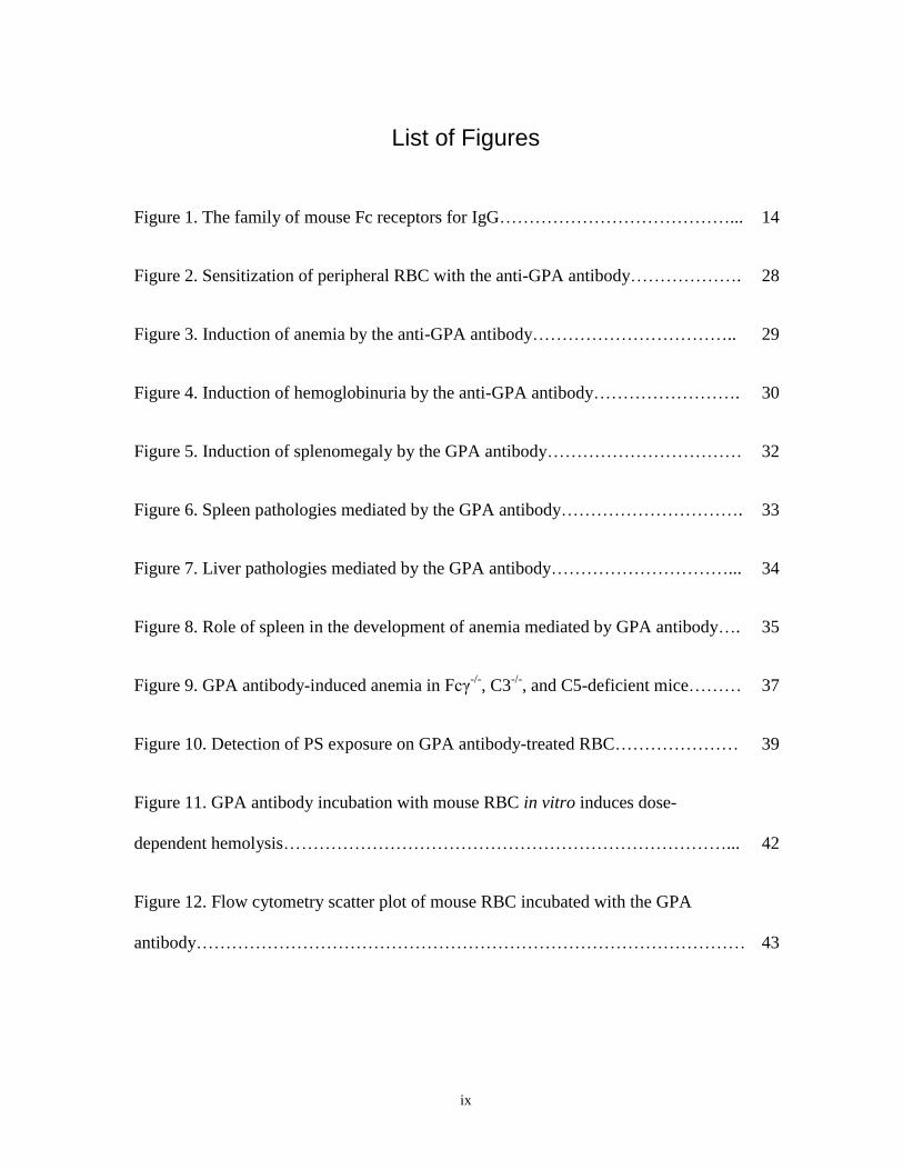
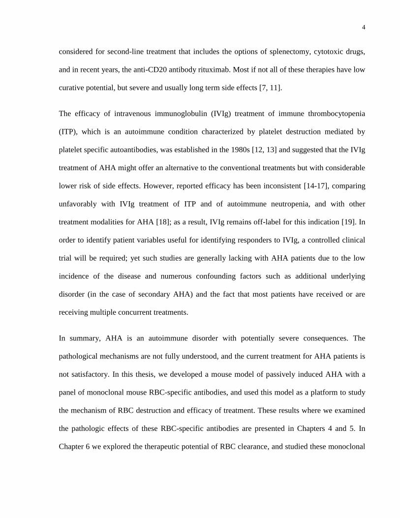
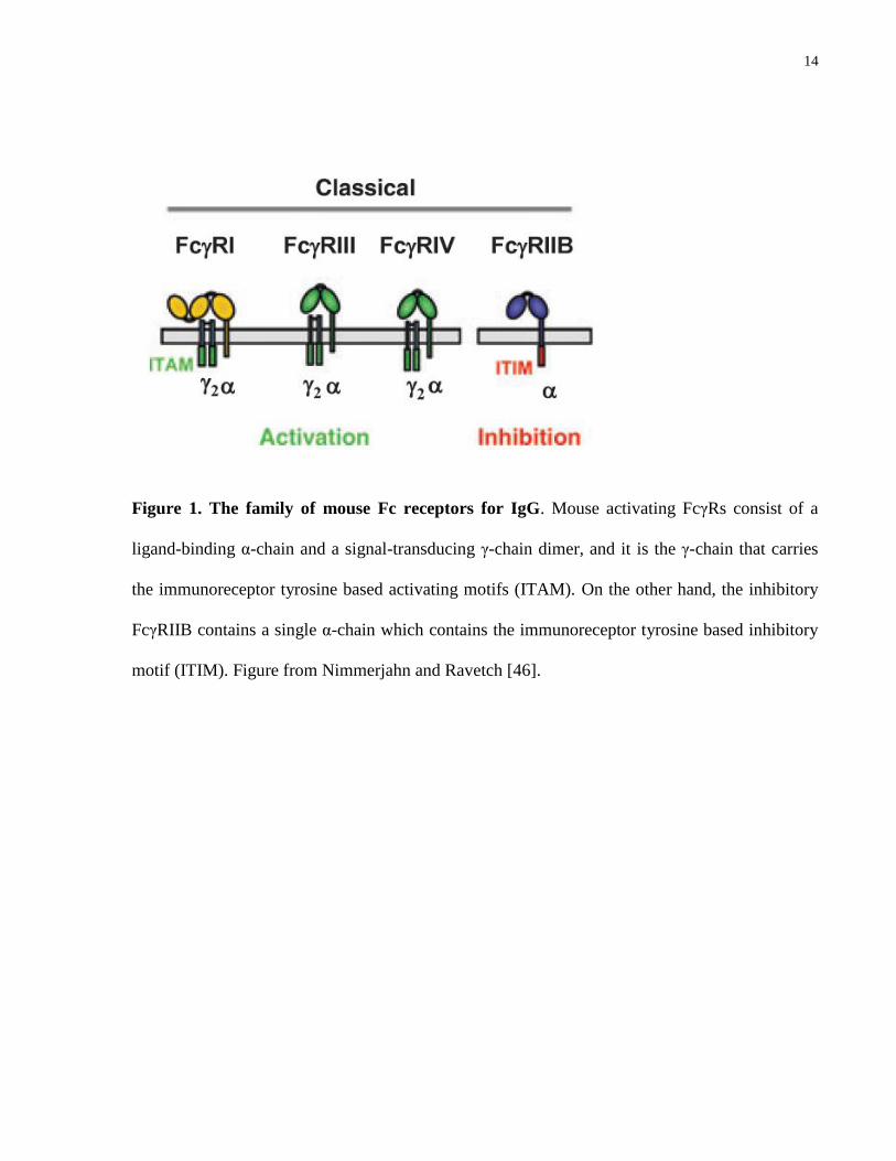
In mice there are four IgG subclasses: IgG1, 2a, 2b, and 3, and four classes of Fcγ receptors
including the activating Fc receptors FcγRI, FcγRIII, FcγRIV, and an inhibitory receptor
FcγRIIB (Fig. 1). The high affinity FcγRI is specific for IgG2a, the low affinity FcγRIII for IgG1,
13
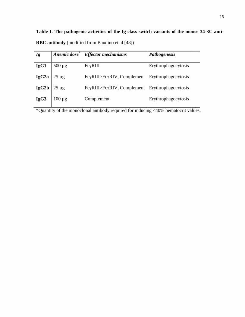
2a, and 2b, and FcγRIV binds to IgG2a and 2b with intermediate affinity [47]. Using Ig class
switch variants of an anti-mouse RBC antibody, Baudino et al [47, 48] documented the role of
each activating Fc receptor in effector mechanisms mediated by different IgG subclass in a
mouse model of AHA. Summary of their findings is presented in Table 1. All three activating
FcRs share a common signal-transducing γ-chain (FcRγ), which is absent in the inhibitory
FcγIIB; the γ-chain harbors the immunoreceptor tyrosine-based activation motif (ITAM) and is
required for cell activation [46]. Both activating and inhibitory Fc receptors are expressed by the
innate immune effector cells including monocytes, macrophages and dendritic cells (DC),
whereas natural killer cells (NK) only express FcγIII, and the B cells solely express the
inhibitory FcγIIB [49, 50]. For the purpose of dissecting the role(s) of each of the Fc receptors in
the Fc-dependent RBC clearance mediated by different IgG subclasses, knockout mice deficient
in the FcRγ or FcγRIIB, in combination with blocking antibodies directed against one or several
of the FcγRs have proved powerful tools [41, 48].
14
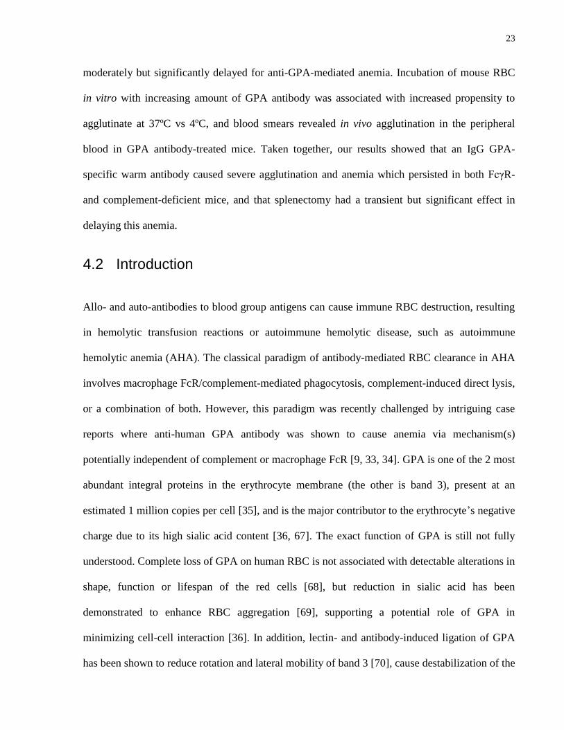
Figure 1. The family of mouse Fc receptors for IgG. Mouse activating FcγRs consist of a
ligand-binding α-chain and a signal-transducing γ-chain dimer, and it is the γ-chain that carries
the immunoreceptor tyrosine based activating motifs (ITAM). On the other hand, the inhibitory
FcγRIIB contains a single α-chain which contains the immunoreceptor tyrosine based inhibitory
motif (ITIM). Figure from Nimmerjahn and Ravetch [46].
15
Table 1. The pathogenic activities of the Ig class switch variants of the mouse 34-3C anti-
RBC antibody (modified from Baudino et al [48])
Ig Anemic dose*
Effector mechanisms Pathogenesis
IgG1 500 µg FcγRIII Erythrophagocytosis
IgG2a 25 µg FcγRIII>FcγRIV, Complement Erythrophagocytosis
IgG2b 25 µg FcγRIII>FcγRIV, Complement Erythrophagocytosis
IgG3 100 µg Complement Erythrophagocytosis
*Quantity of the monoclonal antibody required for inducing <40% hematocrit values.
16
2.2.3 Mouse AHA models
AHA can be induced in mice after introduction of xenogeneic RBCs. One classical example of
this is the model developed by Playfair and Marshall-Clarke [51] that involves repeated
immunization with rat RBCs. In these mice anti-mouse RBC autoantibodies eventually appear as
well as the rat-specific xenoantibodies.
On the other hand, AHA can be readily studied in spontaneous mouse models such as the New
Zealand Black (NZB) and the non-obese diabetic (NOD) mice [52, 53]. For example, the NZB
mouse strain is genetically predisposed to develop a spontaneous and severe form of AHA,
usually by 6 months of age, caused by pathogenic anti-mouse RBC autoantibodies [52]. One
caveat of the use of these spontaneous models is that these mice also develop other autoimmune
diseases as they age that also involve autoantibody formation (e.g., systemic lupus erythematosus
or SLE in the NZB mice [54], and autoimmune insulin-dependent diabetes in the NOD mice
[53]). Therefore, an alternative passive model has been developed and preferred, in which
transfusion of the anti-RBC antibodies, or implantation of anti-RBC hybridomas are used to
induce AHA with greater control [41]. For example, a passive model of monoclonal anti-RBC
autoantibodies, derived from the NZB mice, and their Ig class switch variants has been used
extensively to study the effector mechanism of IgM- or IgG-coated RBC clearance [32, 47, 48,
55-59]. Unlike in humans where autoantibodies against the RBC anion channel protein, band 3,
are only produced in a minority of patients, band 3 is the dominant autoantigen in the NZB mice
[60]. The most widely used monoclonal antibody derived from these mice, 34-3C, an IgG2a, has
been consistently shown to cause FcγR-mediated erythrophagocytosis [47, 48].
17
Chapter 3
Rationale, Hypothesis and Specific Aims
18
3 Rationale, hypothesis and specific aims
1. Rationale
Current paradigm dictates that RBC clearance in immune-mediated hemolytic anemia
primarily involves extravascular hemolysis (macrophage FcR/complement-mediated
phagocytosis), intravascular hemolysis (complement-induced direct lysis), or a
combination of both. However, recent clinical reports suggest that previous unrecognized
mechanisms might be contributing to severe hemolysis [10, 24], in particular, in cases
where GPA-specific antibodies are implicated [9, 10, 24, 33, 34], yet most of the clinical
investigations are defined by in vitro work.
Hypothesis
In this thesis, it is hypothesized that different anti-RBC antibodies may mediate different
RBC clearance mechanism(s), and that mechanism(s) in addition to the classical extra-
and intra-vascular hemolysis may be involved in GPA antibody-mediated anemia.
Aims
The thesis aims to first optimize a murine model of passive AHA, and with this model, to
examine RBC destruction attributable to the classical extra- and intra-vascular hemolysis
mechanisms caused by a panel of monoclonal mouse erythrocyte-specific antibodies
(chapter 4 and 5). Particular interest was placed on a GPA antibody to take advantage of
both in vitro and in vivo system in the mouse model to elucidate potential novel
pathological mechanism(s) (chapter 4).
2. Rationale
19
IVIg is used to effectively treat autoimmune diseases targeting blood cells such as
immune thrombocytopenia (ITP) and autoimmune neutropenia (AIN), yet its use in the
treatment of AHA remains controversial due to inconsistent efficacy, with no established
explanation as to why some patients respond while others do not. Controlled clinical
trials are needed to determine patient variables associated with treatment success yet such
trials are extremely difficult because of the low number of patients and the heterogeneous
patient population due to high prevalence of secondary conditions and concurrent
medications.
Hypothesis
In this thesis, it is hypothesized that mechanisms of anemia induced by AHA-causing
antibodies may be an important factor in determining response to IVIg therapy.
Aims
The thesis aims to test IVIg efficacy in ameliorating a murine model of passive AHA
caused by a panel of monoclonal mouse erythrocyte-specific antibodies, and to determine
if specific RBC destruction mechanism(s) is associated with IVIg responsiveness
(Chapter 5).
3. Rationale
Another interest of the monoclonal mouse erythrocyte-specific antibodies lies in their use
in the treatment of ITP. Polyclonal anti-D has long been established as an effective
alternative to IVIg in the treatment of ITP in D-positive patients [61]. If monoclonal
erythrocyte-specific antibodies prove effective in the amelioration of ITP, they would
provide a cheaper and potentially safer substitution for IVIg and anti-D. However, a small
20
pilot study [62] reported a lack of beneficial response in 6 out of 7 patients tested, making
it unethical for further trials in patients. Nevertheless, interest in monoclonal anti-D (or
anti-D-like antibodies) was rekindled by recent findings in mouse studies where two
monoclonal anti-mouse RBC antibodies were shown capable of ameliorating immune-
mediated thrombocytopenia in a murine model of ITP [63]. However, controversy arises
in one of these two antibodies as its efficacy was confirmed in some of the later
publications [64, 65] but questioned in others [66]. In addition, the therapeutic
mechanism of these erythrocyte-specific antibodies in the amelioration of ITP awaits
further elucidation.
Hypothesis
In this thesis, it is hypothesized that monoclonal mouse erythrocyte-specific antibodies
are of potential value in the treatment of thrombocytopenia in a murine model of ITP, and
that they may work by different therapeutic mechanisms.
Aims
The thesis aims to evaluate the efficacy of a panel of mouse erythrocyte-specific
antibodies in the amelioration of thrombocytopenia in a murine model of passive ITP,
and investigate if the ability to mediate FcγR-mediated RBC clearance is required for
efficacy (Chapter 6).
21
Chapter 4
Glycophorin A Antibody-Mediated Anemia in a Murine Model:
Minimal Involvement of FcγR or Complement,
and Implications for Agglutination
22
4 Glycophorin A Antibody-Mediated Anemia in a Murine
Model: Minimal Involvement of FcγR or Complement,
and Implications for Agglutination
4.1 Abstract
Autoimmune hemolytic anemia (AHA) is characterized by premature destruction of red blood
cell (RBC) mediated by erythrocyte-specific antibodies. Therapy for patients with AHA is
challenging and better knowledge of the involved pathophysiological mechanisms is needed. The
classical paradigm of antibody-mediated RBC clearance in AHA involves macrophage Fc
receptor (FcR)/complement-mediated phagocytosis, complement-induced direct lysis, or a
combination of both. However, this paradigm has been challenged by intriguing case reports
where anti-human GPA antibody was shown to cause hemolysis via mechanism(s) potentially
independent of complement or macrophage FcR. In this study, using a murine model of immune
hemolytic anemia (IHA), we investigated the pathophysiological mechanisms of GPA antibody-
mediated anemia with a mouse GPA-specific antibody (TER119). The anemic effect of the IgG
GPA-specific antibody was studied in vivo, by injecting the antibody into normal vs mice
genetically deficient for the Fc γ-chain (required for activating FcγR function), complement C3,
mice naturally deficient in complement C5 and splenectomized mice. In vivo pathologies were
assessed by histological examination of peripheral blood and the spleen and liver of the
reticuloendothelial system (RES). In addition, direct RBC destruction was evaluated in vitro by
incubation of mouse RBC with the GPA antibody. We report that the GPA antibody induced
significant anemia and an acute episode of hemoglobinuria in mice. It caused a similar degree of
anemia in Fcγ-/-
mice, C3-/-
mice and C5-deficient mice, in comparison to control mice. Spleen
weight was found correlated with the degree of RBC destruction, and splenectomized mice were
23
moderately but significantly delayed for anti-GPA-mediated anemia. Incubation of mouse RBC
in vitro with increasing amount of GPA antibody was associated with increased propensity to
agglutinate at 37ºC vs 4ºC, and blood smears revealed in vivo agglutination in the peripheral
blood in GPA antibody-treated mice. Taken together, our results showed that an IgG GPA-
specific warm antibody caused severe agglutination and anemia which persisted in both FcγR-
and complement-deficient mice, and that splenectomy had a transient but significant effect in
delaying this anemia.
4.2 Introduction
Allo- and auto-antibodies to blood group antigens can cause immune RBC destruction, resulting
in hemolytic transfusion reactions or autoimmune hemolytic disease, such as autoimmune
hemolytic anemia (AHA). The classical paradigm of antibody-mediated RBC clearance in AHA
involves macrophage FcR/complement-mediated phagocytosis, complement-induced direct lysis,
or a combination of both. However, this paradigm was recently challenged by intriguing case
reports where anti-human GPA antibody was shown to cause anemia via mechanism(s)
potentially independent of complement or macrophage FcR [9, 33, 34]. GPA is one of the 2 most
abundant integral proteins in the erythrocyte membrane (the other is band 3), present at an
estimated 1 million copies per cell [35], and is the major contributor to the erythrocyte’s negative
charge due to its high sialic acid content [36, 67]. The exact function of GPA is still not fully
understood. Complete loss of GPA on human RBC is not associated with detectable alterations in
shape, function or lifespan of the red cells [68], but reduction in sialic acid has been
demonstrated to enhance RBC aggregation [69], supporting a potential role of GPA in
minimizing cell-cell interaction [36]. In addition, lectin- and antibody-induced ligation of GPA
has been shown to reduce rotation and lateral mobility of band 3 [70], cause destabilization of the
24
phospholipid bilayer [33], and make RBC more resistant to deformation [71] that may result in
splenic entrapment [72]. Of interest, reported AHA cases caused by antibody directed against
epitopes on or associated with GPA (eg., anti-M,-N,-Pr,-Ena) are relatively rare but usually
severe [24].
In this study, we modeled anti-GPA antibody-mediated anemia with a monoclonal GPA-specific
antibody, TER119, in a murine model of immune hemolytic anemia (IHA). This antibody is of
interest because it can ameliorate passive immune thrombocytopenia (ITP) in mice analogous to
the use of anti-D in ITP patients [63-65]. TER119 is widely used as a specific marker for the
erythroid lineage in studies of mouse hematopoiesis [73], and is, to our knowledge, the only
antibody specific for the extracellular domain of mouse GPA that is currently available. We
report herein that infusion of this IgG GPA antibody caused development of anemia in mice
deficient for the FcγR- or complement C3/C5-mediated mechanisms, and provide in vitro and in
vivo evidence consistent with a non-classical mechanism of anemia likely involving RBC
agglutination and subsequent splenic sequestration.
4.3 Materials and methods
4.3.1 Mice
C57BL/6, BALB/c, CD1 and DBA/2 mice were from Charles River Laboratories, FcR γ-chain-
deficient mice (B6.129P2-Fcer1gtm1Rav
N12) were from Taconic Farms, and C3-deficient mice
(B6.129S4-C3tm1Crr/J) were from The Jackson Laboratory. Surgically splenectomized
C57BL/6 mice and control C57BL/6 mice were from the Jackson Laboratory. All mice were
used at 6-12 weeks of age. All animal experiments were approved by the Animal Care and Use
Committee of St. Michael’s Hospital.
25
4.3.2 Reagents
Normal rat IgG and FITC-conjugated goat F(ab’)2 fragments of rat IgG-specific antibody was
purchased from Caltag, and the anti-CD41 (MWReg30), anti-GPA (TER119), anti-CD24
(M1/69) antibodies, rat IgG2b κ isotype control (A95-1) antibodies, and the Annexin V-FITC
apoptosis detection kit were from BD Biosciences.
4.3.3 Anemia
Anemia was induced in mice by intravenous injection of 45 μg of anti-GPA antibody TER119 in
200 µL PBS. At indicated times post injection, 10 µL of whole blood was drawn by saphenous
bleeding and diluted 1: 100 in PBS/EDTA and another 1: 1,000 in PBS. The diluted blood (500
µL) was then acquired on a flow cytometer (Guava EasyCyte Mini System) for RBC counts.
Detection of antibody binding was done as described [74]. Briefly, mouse blood was washed
with PBS for 3 times (400g; 10 minutes), and incubated with FITC-conjugated goat F(ab’)2 anti-
rat IgG at room temperature for 30 min in dark, then washed with PBS to remove unbound
secondary antibody, and analyzed on a Guava EasyCyte Mini System.
4.3.4 Histopathology
Mouse spleen and liver tissues were obtained at autopsy, processed for histological examination,
and stained with hematoxylin and eosin (H&E) as described [10]. Tissue slides were viewed with
a Nikon Eclipse 80i microscope, and pictures were taken with a Nikon Digital Sight camera and
acquired on Nis Element D3.00 SP5 software.
26
4.3.5 In vitro RBC incubation with antibody
Mouse blood was washed with PBS for 3 times (400g; 10 minutes), the buffy coat and plasma
removed, and 200 μL was incubated with the indicated concentration of GPA antibody at 4ºC,
room temperature, or 37ºC for 1 hour. Unbound antibody was washed away after the incubation.
For detection of microagglutination, diluted RBC was observed on a hemocytometer with a
Nikon Eclipse TS100 microscope. Pictures were taken with a Nikon Coolpix 4500 camera.
4.3.6 Flow cytometric analysis of phosphatidylserine (PS) exposure
The level of PS exposure on mouse erythrocytes was measured using an Annexin V-FITC assay
on a Guava EasyCyte Mini System according to manufacturer’s instructions. Briefly, mouse
blood was washed with cold PBS for 3 times (400g; 10 minutes), the buffy coat and plasma
removed, and cells were resuspended in 1X Binding Buffer at 1 x 10⁶ cells/mL. Add 5 µL of
FITC Annexin V to 100 µL of the cell solution (1 x 10⁵ cells) and incubate for 15 min at room
temperature in dark. At the end of incubation, add 400 µL of 1X Binding Buffer and analyze by
flow cytometry (Guava EasyCyte Mini System). Flow cytometric data were analyzed on FlowJo
software (version X).
4.3.7 Heat treatment of RBC
Heat treatment of RBC was performed as described [75]. Briefly, whole blood was drawn by
retro-orbital bleeding and diluted in citrate phosphage dextrose adenine (CPDA). Blood was
washed 3 times with PBS (400g; 10 minutes), and buffy coat and plasma was removed. Dilute
the washed blood in warm (50°C) PBS to 50% hematocrit (Hct), and incubated in 50°C water
bath for 30 minutes. Then wash the cells 3 times with PBS (400g; 10 minutes).
27
4.3.8 Measurement of platelet counts
Platelet counts were determined as described [74]. Briefly, 10 µL of whole blood was drawn by
saphenous bleeding and diluted 1: 100 in PBS/EDTA. The diluted blood was centrifuged (170g;
2 minutes; brake off) to separate the platelet-rich plasma (PRP). The PRP was diluted 1: 100 in
isoton and platelet counts were determined on a Beckman Z2 Coulter Counter.
4.3.9 Statistical analysis
Data are presented as mean plus or minus standard error of the mean (SEM). Note, in some cases
where the error bars are not apparent, this is due to a minimal SEM. Statistical analysis was
performed with the Student t test using GraphPad Prism software. P<0.05 was considered
significant.
4.4 Results
4.4.1 Induction of anemia by the GPA antibody
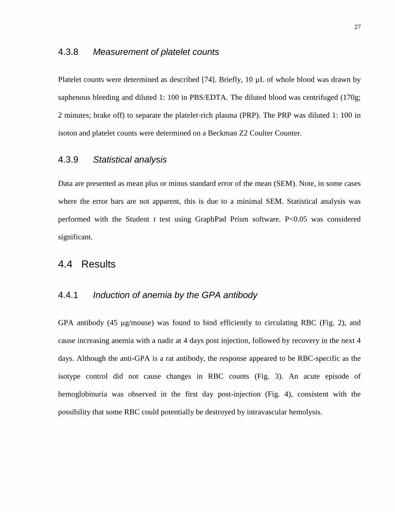
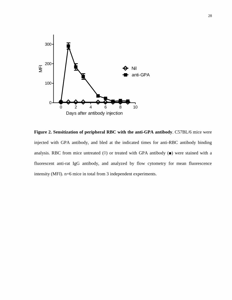
GPA antibody (45 μg/mouse) was found to bind efficiently to circulating RBC (Fig. 2), and
cause increasing anemia with a nadir at 4 days post injection, followed by recovery in the next 4
days. Although the anti-GPA is a rat antibody, the response appeared to be RBC-specific as the
isotype control did not cause changes in RBC counts (Fig. 3). An acute episode of
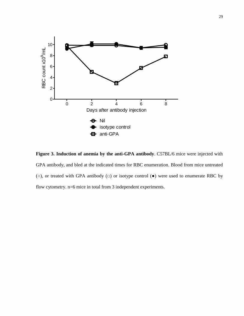
hemoglobinuria was observed in the first day post-injection (Fig. 4), consistent with the
possibility that some RBC could potentially be destroyed by intravascular hemolysis.
28
0 2 4 6 8 100
100
200
300
anti-GPA
Nil
Days after antibody injection
MF
I
Figure 2. Sensitization of peripheral RBC with the anti-GPA antibody. C57BL/6 mice were
injected with GPA antibody, and bled at the indicated times for anti-RBC antibody binding
analysis. RBC from mice untreated (◊) or treated with GPA antibody (■) were stained with a
fluorescent anti-rat IgG antibody, and analyzed by flow cytometry for mean fluorescence
intensity (MFI). n=6 mice in total from 3 independent experiments.
29
0 2 4 6 80
2
4
6
8
10
anti-GPA
Nil
isotype control
Days after antibody injection
RB
C c
ount x1
09/m
L
Figure 3. Induction of anemia by the anti-GPA antibody. C57BL/6 mice were injected with
GPA antibody, and bled at the indicated times for RBC enumeration. Blood from mice untreated
(○), or treated with GPA antibody (□) or isotype control (●) were used to enumerate RBC by
flow cytometry. n=6 mice in total from 3 independent experiments.
30
Figure 4. Induction of hemoglobinuria by the anti-GPA antibody. C57BL/6 mice were
injected with GPA antibody. Upper panel: urine accumulated from mice untreated (left) or
treated with GPA antibody (right) in the first day post-injection. Shown are typical pictures from
3 independent experiments. Lower panel: Urine samples from mice transfused with the GPA
antibody displayed absorbance peaks at ~414 nm, confirming that the pigment was free
hemoglobin [42].
31
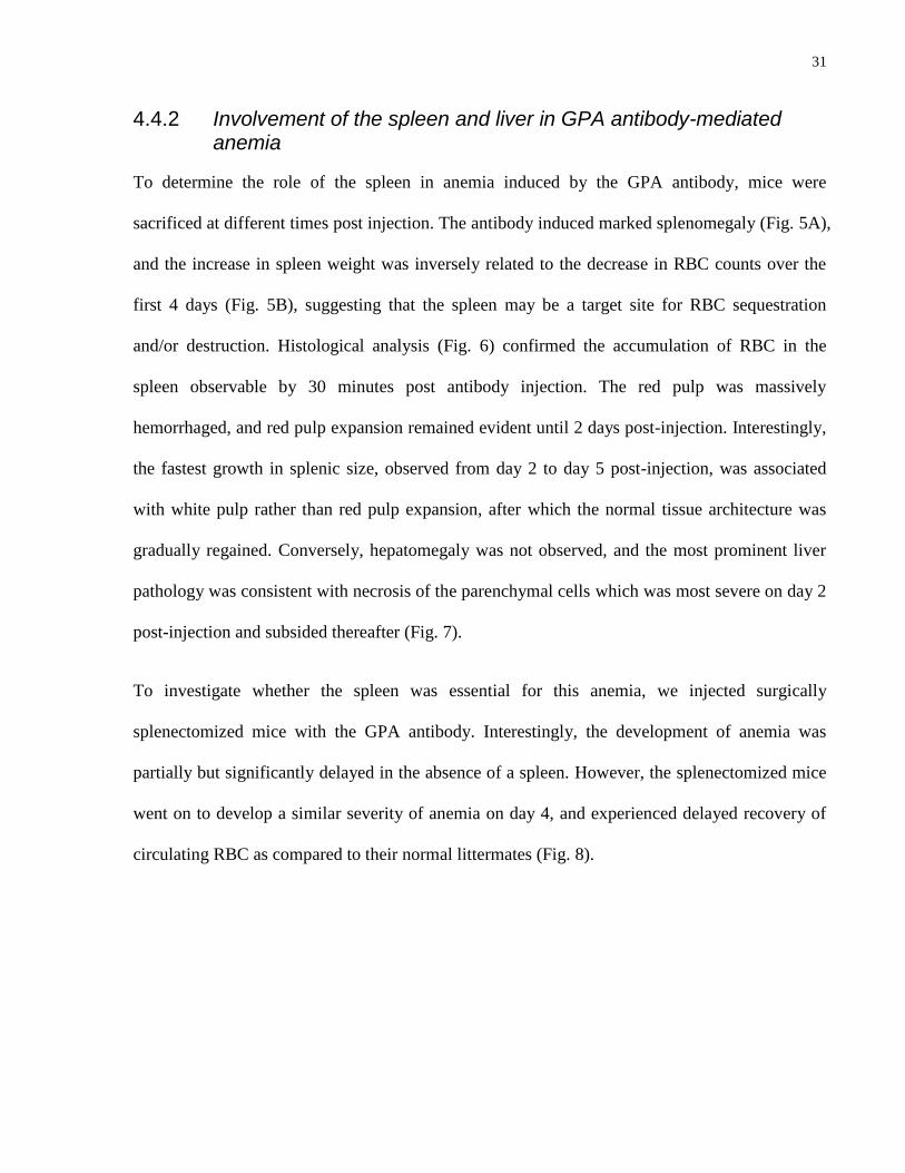
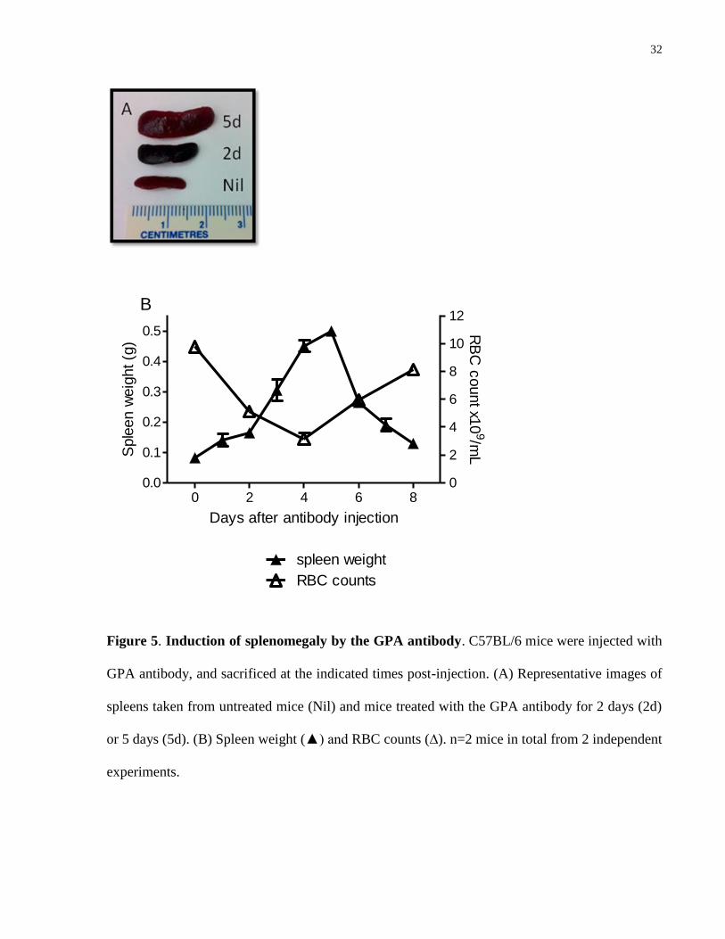
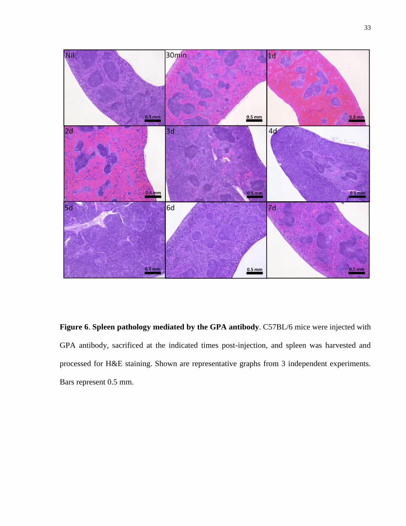
4.4.2 Involvement of the spleen and liver in GPA antibody-mediated anemia
To determine the role of the spleen in anemia induced by the GPA antibody, mice were
sacrificed at different times post injection. The antibody induced marked splenomegaly (Fig. 5A),
and the increase in spleen weight was inversely related to the decrease in RBC counts over the
first 4 days (Fig. 5B), suggesting that the spleen may be a target site for RBC sequestration
and/or destruction. Histological analysis (Fig. 6) confirmed the accumulation of RBC in the
spleen observable by 30 minutes post antibody injection. The red pulp was massively
hemorrhaged, and red pulp expansion remained evident until 2 days post-injection. Interestingly,
the fastest growth in splenic size, observed from day 2 to day 5 post-injection, was associated
with white pulp rather than red pulp expansion, after which the normal tissue architecture was
gradually regained. Conversely, hepatomegaly was not observed, and the most prominent liver
pathology was consistent with necrosis of the parenchymal cells which was most severe on day 2
post-injection and subsided thereafter (Fig. 7).
To investigate whether the spleen was essential for this anemia, we injected surgically
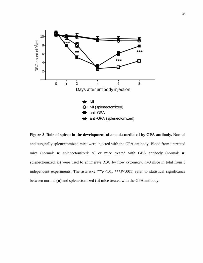
splenectomized mice with the GPA antibody. Interestingly, the development of anemia was
partially but significantly delayed in the absence of a spleen. However, the splenectomized mice
went on to develop a similar severity of anemia on day 4, and experienced delayed recovery of
circulating RBC as compared to their normal littermates (Fig. 8).
32
0 2 4 6 80.0
0.1
0.2
0.3
0.4
0.5
0
2
4
6
8
10
12
spleen weight
RBC counts
B
Days after antibody injection
Sple
en w
eig
ht (g
)
RB
C c
ount x1
09/m
L
Figure 5. Induction of splenomegaly by the GPA antibody. C57BL/6 mice were injected with
GPA antibody, and sacrificed at the indicated times post-injection. (A) Representative images of
spleens taken from untreated mice (Nil) and mice treated with the GPA antibody for 2 days (2d)
or 5 days (5d). (B) Spleen weight (▲) and RBC counts (∆). n=2 mice in total from 2 independent
experiments.
33
Figure 6. Spleen pathology mediated by the GPA antibody. C57BL/6 mice were injected with
GPA antibody, sacrificed at the indicated times post-injection, and spleen was harvested and
processed for H&E staining. Shown are representative graphs from 3 independent experiments.
Bars represent 0.5 mm.
34
Figure 7. Liver pathology mediated by the GPA antibody. C57BL/6 mice were injected with
GPA antibody, sacrificed at the indicated times post-injection, and liver was harvested and
processed for H&E staining. Shown are representative graphs from 3 independent experiments.
Bars represent 0.5 mm.
35
0 2 4 6 8
2
4
6
8
10
anti-GPA
Nil
anti-GPA (splenectomized)
Nil (splenectomized)
**
***
***
Days after antibody injection
1
***
RB
C c
ount x1
09/m
L
Figure 8. Role of spleen in the development of anemia mediated by GPA antibody. Normal
and surgically splenectomized mice were injected with the GPA antibody. Blood from untreated
mice (normal: ●; splenectomized: ○) or mice treated with GPA antibody (normal: ■;
splenectomized: □) were used to enumerate RBC by flow cytometry. n=3 mice in total from 3
independent experiments. The asterisks (**P<.01, ***P<.001) refer to statistical significance
between normal (■) and splenectomized (□) mice treated with the GPA antibody.
36
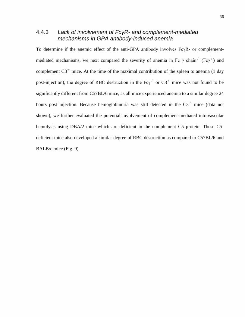
4.4.3 Lack of involvement of FcγR- and complement-mediated mechanisms in GPA antibody-induced anemia
To determine if the anemic effect of the anti-GPA antibody involves FcγR- or complement-
mediated mechanisms, we next compared the severity of anemia in Fc γ chain-/-
(Fcγ-/-
) and
complement C3-/-
mice. At the time of the maximal contribution of the spleen to anemia (1 day
post-injection), the degree of RBC destruction in the Fcγ-/-
or C3-/-
mice was not found to be
significantly different from C57BL/6 mice, as all mice experienced anemia to a similar degree 24
hours post injection. Because hemoglobinuria was still detected in the C3-/-
mice (data not
shown), we further evaluated the potential involvement of complement-mediated intravascular
hemolysis using DBA/2 mice which are deficient in the complement C5 protein. These C5-
deficient mice also developed a similar degree of RBC destruction as compared to C57BL/6 and
BALB/c mice (Fig. 9).
37
C57BL/6 Fc -/- C3-/- DBA/2 BALB/c 0
20
40
60
80
100
anti-GPA: - + - + - + - + - +
% Initia
l R
BC
counts
Figure 9. GPA antibody-induced anemia in Fcγ-/-
, C3-/-
, and C5-deficient mice. C57BL/6,
Fcγ-/-
, C3-/-
, DBA/2 (C5-deficient) and BALB/c mice were injected with the GPA antibody, and
bled at 24 hours post-injection for RBC enumeration. RBC counts were expressed as a
percentage of initial (baseline) counts. n=4 mice in total from 4 independent experiments.
38
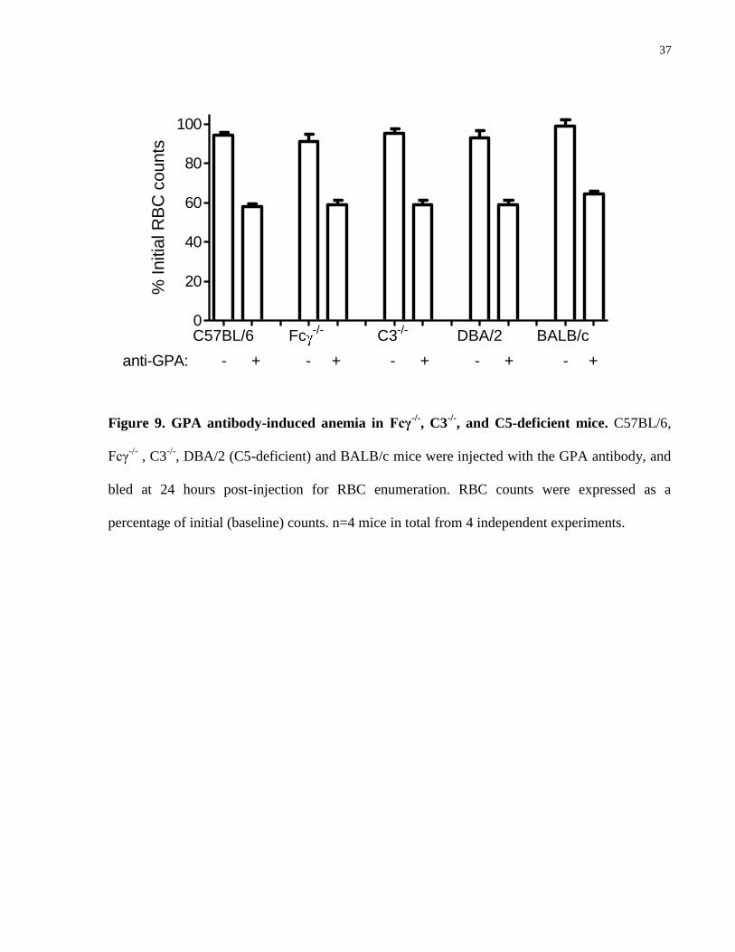
4.4.4 GPA antibody does not induce increased phosphatidylserine exposure
Because of evidence in the literature suggesting an indispensible role of macrophages in the
induction of anemia by the anti-GPA antibody [76], yet we did not find FcγR- or complement-
mediated phagocytosis to be a major mechanism, we tested the possibility that the GPA-specific
antibody induces phosphatidylserine (PS) exposure which then activates macrophage PS
receptor-mediated phagocytosis, using a PS flow cytometry detection kit. RBC were drawn from
mice injected with anti-GPA antibody 24 hours post injection, and did not seem to have
increased PS expression, whereas heat-damaged RBC had a distinct population with increased
PS expression (Fig. 10).
39
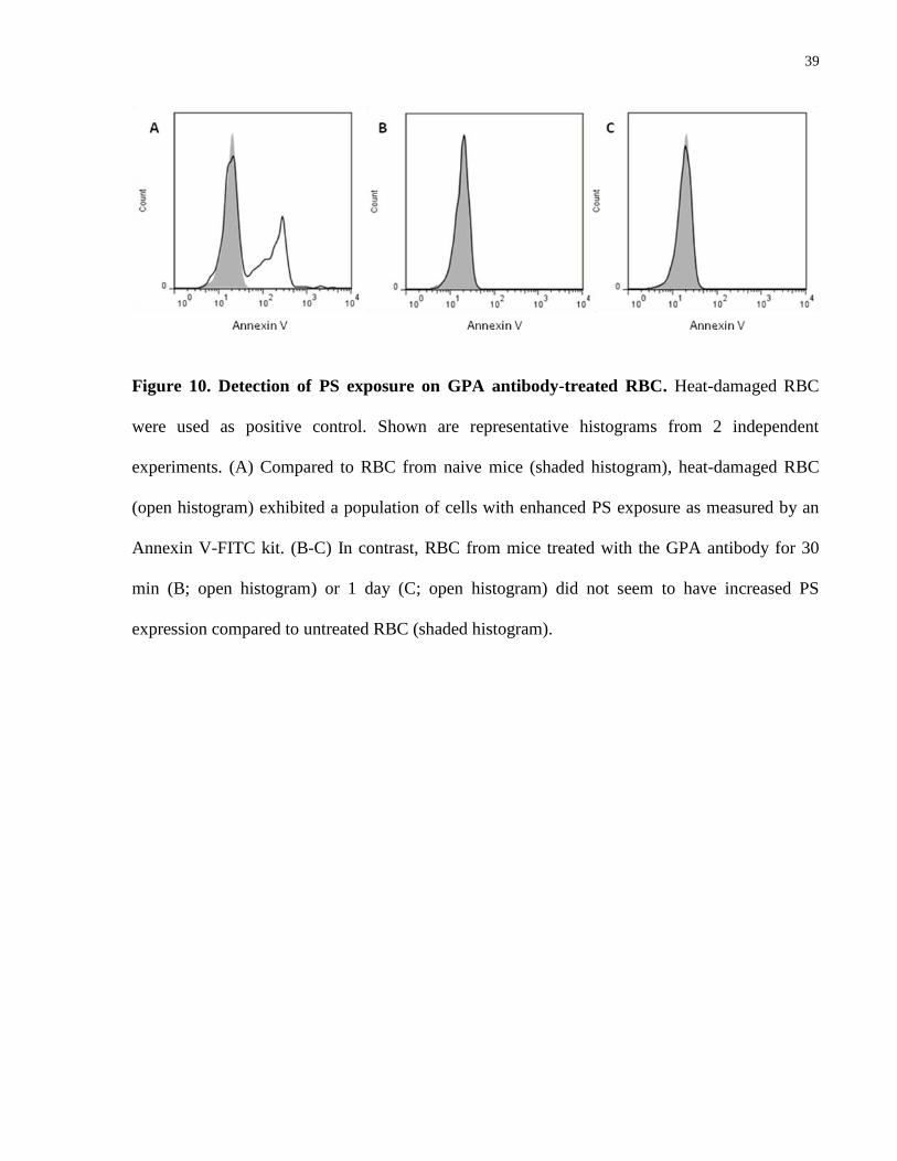
Figure 10. Detection of PS exposure on GPA antibody-treated RBC. Heat-damaged RBC
were used as positive control. Shown are representative histograms from 2 independent
experiments. (A) Compared to RBC from naive mice (shaded histogram), heat-damaged RBC
(open histogram) exhibited a population of cells with enhanced PS exposure as measured by an
Annexin V-FITC kit. (B-C) In contrast, RBC from mice treated with the GPA antibody for 30
min (B; open histogram) or 1 day (C; open histogram) did not seem to have increased PS
expression compared to untreated RBC (shaded histogram).
40
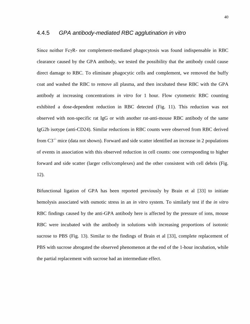
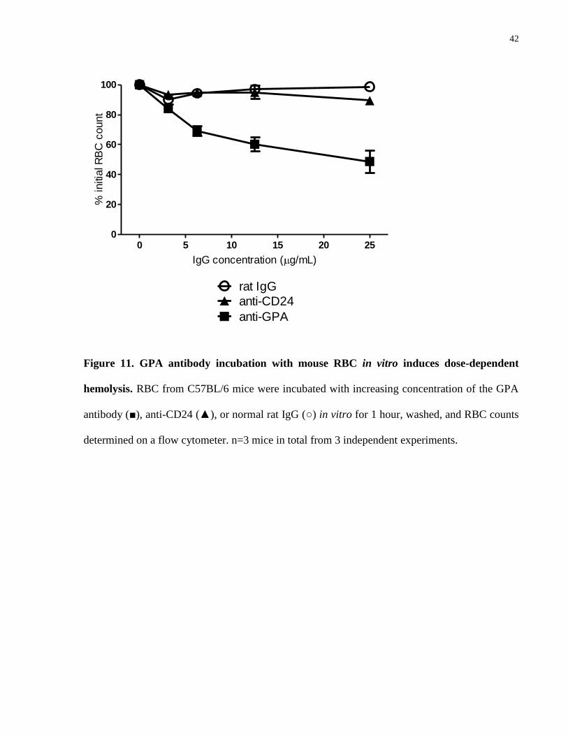
4.4.5 GPA antibody-mediated RBC agglutination in vitro
Since neither FcγR- nor complement-mediated phagocytosis was found indispensable in RBC
clearance caused by the GPA antibody, we tested the possibility that the antibody could cause
direct damage to RBC. To eliminate phagocytic cells and complement, we removed the buffy
coat and washed the RBC to remove all plasma, and then incubated these RBC with the GPA
antibody at increasing concentrations in vitro for 1 hour. Flow cytometric RBC counting
exhibited a dose-dependent reduction in RBC detected (Fig. 11). This reduction was not
observed with non-specific rat IgG or with another rat-anti-mouse RBC antibody of the same
IgG2b isotype (anti-CD24). Similar reductions in RBC counts were observed from RBC derived
from C3-/-
mice (data not shown). Forward and side scatter identified an increase in 2 populations
of events in association with this observed reduction in cell counts: one corresponding to higher
forward and side scatter (larger cells/complexes) and the other consistent with cell debris (Fig.
12).
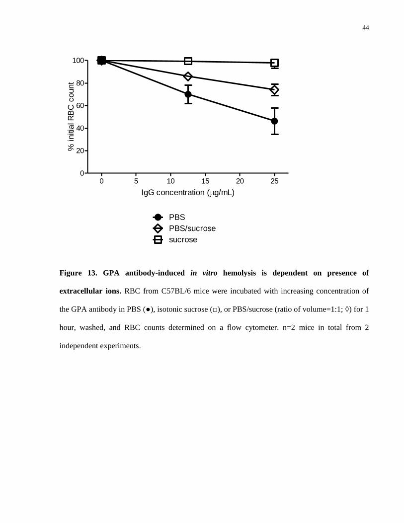
Bifunctional ligation of GPA has been reported previously by Brain et al [33] to initiate
hemolysis associated with osmotic stress in an in vitro system. To similarly test if the in vitro
RBC findings caused by the anti-GPA antibody here is affected by the pressure of ions, mouse
RBC were incubated with the antibody in solutions with increasing proportions of isotonic
sucrose to PBS (Fig. 13). Similar to the findings of Brain et al [33], complete replacement of
PBS with sucrose abrogated the observed phenomenon at the end of the 1-hour incubation, while
the partial replacement with sucrose had an intermediate effect.
41
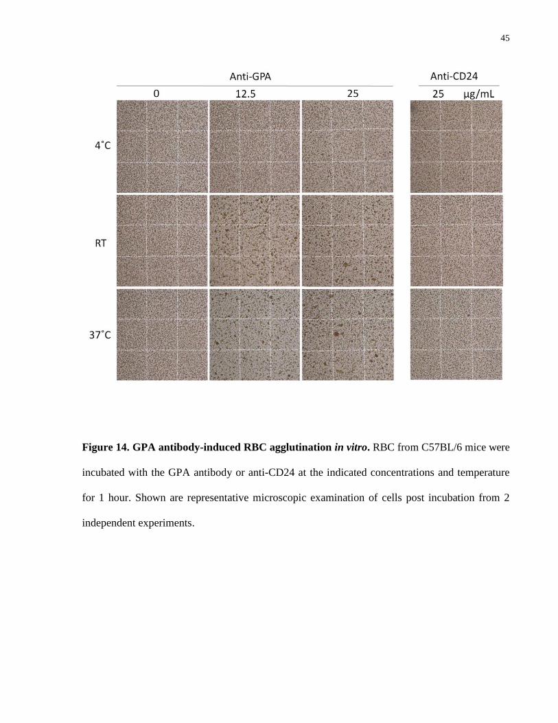
To determine if the decreased RBC cell counting by flow cytometry could be due to RBC
agglutination, samples were observed by microscopy (Fig. 14). Incubation with GPA antibody at
room temperature was associated with a dose-dependent increase in the proportion of cells in
agglutinated form. This antibody-associated agglutination was enhanced at 37ºC, and delayed at
4ºC. No agglutination was found with cells incubated with normal rat IgG (data not shown) or
the anti-CD24 antibody at any temperature tested.
42
0 5 10 15 20 250
20
40
60
80
100
rat IgG
anti-GPA
anti-CD24
IgG concentration ( g/mL)
% initia
l R
BC
count
Figure 11. GPA antibody incubation with mouse RBC in vitro induces dose-dependent
hemolysis. RBC from C57BL/6 mice were incubated with increasing concentration of the GPA
antibody (■), anti-CD24 (▲), or normal rat IgG (○) in vitro for 1 hour, washed, and RBC counts
determined on a flow cytometer. n=3 mice in total from 3 independent experiments.
43
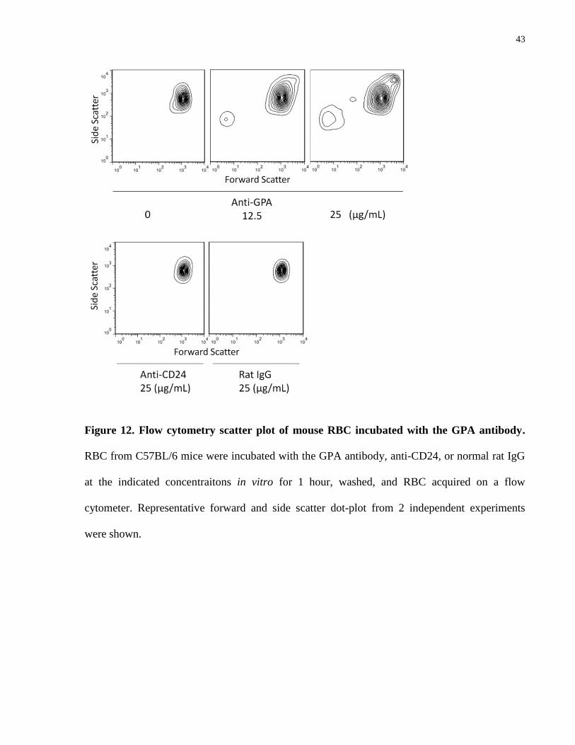
Figure 12. Flow cytometry scatter plot of mouse RBC incubated with the GPA antibody.
RBC from C57BL/6 mice were incubated with the GPA antibody, anti-CD24, or normal rat IgG
at the indicated concentraitons in vitro for 1 hour, washed, and RBC acquired on a flow
cytometer. Representative forward and side scatter dot-plot from 2 independent experiments
were shown.
44
0 5 10 15 20 250
20
40
60
80
100
PBS
PBS/sucrose
sucrose
IgG concentration ( g/mL)
% initia
l R
BC
count
Figure 13. GPA antibody-induced in vitro hemolysis is dependent on presence of
extracellular ions. RBC from C57BL/6 mice were incubated with increasing concentration of
the GPA antibody in PBS (●), isotonic sucrose (□), or PBS/sucrose (ratio of volume=1:1; ◊) for 1
hour, washed, and RBC counts determined on a flow cytometer. n=2 mice in total from 2
independent experiments.
45
Figure 14. GPA antibody-induced RBC agglutination in vitro. RBC from C57BL/6 mice were
incubated with the GPA antibody or anti-CD24 at the indicated concentrations and temperature
for 1 hour. Shown are representative microscopic examination of cells post incubation from 2
independent experiments.
46
4.4.6 GPA antibody-mediated RBC agglutination in vivo
To determine if the GPA antibody-mediated anemia was attributable to agglutination in vivo,
blood films were prepared with whole blood from mice injected with the antibody.
Microagglutinates were clearly visible in mice 30 min post injection of GPA antibody, and
persisted in these mice at 24 hours-post injection. Individual RBC appeared to have visible
variation in sizes on the first 2 days post-injection, and polychromasia was observed at later stage
of the anemia (Fig. 15).
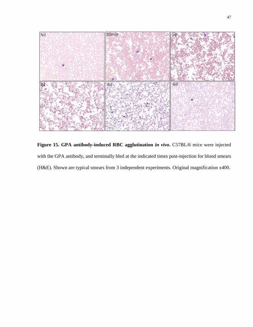
47
Figure 15. GPA antibody-induced RBC agglutination in vivo. C57BL/6 mice were injected
with the GPA antibody, and terminally bled at the indicated times post-injection for blood smears
(H&E). Shown are typical smears from 3 independent experiments. Original magnification x400.
48
4.5 Discussion
Allo- and auto-antibodies to human glycophorin A (GPA)-associated antigens can mediate
immune RBC destruction, leading to transfusion reactions, hemolytic disease of the newborn
(HDN), and autoimmune hemolytic anemia (AHA) [77]. Anti-GPA antibodies encountered in
AHA, albeit relatively rare, are frequently associated with severe hemolysis unexplained by
routine serological findings [24]. As such, treatment remains experience-based and better
understanding of the pathophysiologies is needed [9, 78]. In the current study, we initiated work
to model anti-GPA antibody-mediated anemia in mice using the monoclonal anti-mouse GPA
antibody TER119 [36].
The GPA antibody caused rapid and severe anemia in mice, inducing an acute episode of
hemoglobinuria and dropping peripheral RBC counts to less than 50% of baseline level within 2
days after injection. In an attempt to determine the mechanism(s) of RBC destruction and/or
clearance, we first considered the two principle mechanisms of antibody-mediated hemolysis in
AHA, namely extravascular hemolysis primarily executed by splenic macrophages and liver
Kupffer cells and, more rarely, intravascular hemolysis as a result of complement-induced lysis
[25]. Surprisingly, mice genetically deficient in the FcR γ chain, which are deficient in the
functions of all 3 activating Fc receptors in mice (FcγRI, III and IV) [50, 79], were not protected
from anemia caused by the anti-GPA antibody, and neither were mice genetically deficient in
complement C3. Because hemoglobinuria was observed in both the C57BL/6 and C3-/-
mice
treated with the antibody, to rule out the possibility that intravascular hemolysis may have
occured via C3-indepednent C5 generation [80], we repeated the experiment with DBA/2 mice
which are naturally deficient in C5 [81], and showed that similar degree of anemia persisted in
the absence of C5. Although C4b (upstream from C3 in complement cascade) is also capable of
49
binding to macrophage complement receptor (CR) for initiation of phagocytosis [82], this seems
unlikely because C4-deficient mice were not protected from anemia following injection with the
same anti-GPA antibody (D. R. Branch, personal communication, 2011).
The lack of prominent involvement of FcR- or complement-dependent hemolysis was further
supported by in vitro finding where in the absence of phagocytic cells and complement
components, the anti-GPA antibody was still able to cause dose-dependent RBC damage
attributable to cell aggregation. Temperature analysis revealed that the anti-GPA does not behave
like a typical cold agglutinin; in fact, agglutination was more efficient at 37˚C than room
temperature, but prevented at 4˚C, suggesting that the antibody is more consistent with a “warm
agglutinin” to rapidly bind and agglutinate circulating RBC. In addition, unlike cold agglutinin-
mediated hemolytic anemia in which the major mechanism of hemolysis is complement-
dependent [83], our in vitro and in vivo results consistently showed that neither agglutination nor
the associated hemoglobinuria required complement.
We next sought to determine if agglutination was contributing to the development of anemia in
vivo. Peripheral blood smears revealed that microagglutinates formed as soon as 30 min
following intravenous injection of the anti-GPA antibody. Subsequently, reduction in peripheral
RBC counts was associated with splenomegaly due to massive accumulation of red cells mainly
in the red pulp of the spleen. The GPA antibody may have pathogenic consequences beyond
anemia. Red cell agglutination was accompanied by liver injury, which progressed to
pronounced necrosis which may be secondary to reduced delivery of oxygen to hepatocytes as a
consequence of the extensive agglutination. This had been described by reports where antibody-
mediated RBC aggregates were thought to contribute to fatal anemia as a result of hepatic failure
[84, 85]. Notably, antibodies specific for human GPA determinants were also found in some of
50
these reported patient cases [86]. In addition, we found that the antibody-mediated pathologies
may have additional hemostatic effect. In particular, despite no binding to mouse platelet in vitro
or in vivo (data not shown), we observed that, in addition to anemia, injection of the anti-mouse
GPA was associated with a ~30-40% drop in peripheral platelet counts by 1 day post-injection
(Appendix 1A), and like the anemia, this thrombocytopenic effect persisted in both the FcγR-
and the complement C3-deficient mice (Appendix 1B). Interestingly, Ott and colleagues [69]
recently reported that platelet-rich plasma (PRP) obtained from suspensions containing
agglutinated human RBC had lower platelet counts, which they suggested was likely due to the
avid binding of platelets and formation of RBC-platelet aggregates. Therefore one possibility
consistent with our observation is that in the anti-GPA-treated mice, platelets could have been
trapped in/attracted to the microagglutinates as they form rapidly in the blood stream, and thus
resulting in transient thrombocytopenia. Alternatively, platelet pooling in an enlarged spleen is
long known to contribute to thrombocytopenia in splenomegalic patients [87, 88]. Given the
massively enlarged and congested spleen we observed in GPA antibody-treated mice, the
accompanied thrombocytopenia could have been secondary to splenomegaly.
Of interest, the role of the spleen in the progression of anemia seems two-fold. Initially, it could
serve as a primary target site for the filtering and sequestration of agglutinated RBC in the red
pulp in the first 2 days post-injection. This period was when the blood smears revealed individual
RBC with visibly variable sizes, suggesting that an adaptive volume regulation mechanism may
have been triggered as a result of splenic entrapment [72]. Accordingly, consistent with previous
reports showing correlation between spleen sequestration/clearance and response to splenectomy
in patients [10, 89], we report a transient but significant effect of splenectomy in delaying the
anemia at this stage. On the other hand, the most striking increment in spleen weight occurring
between day 3 and day 5 was a result of massive white pulp expansion likely due to replacement
51
of previously hemorrhaged areas with mitotically active mononuclear cells. Data in
splenectomized mice during the same period of time (day 3-8) revealed that the absence of
spleen was associated with sustained anemia, supporting the interpretation that the spleen may be
involved in facilitating the recovery stage of the anemia, which is consistent with the general
belief that the murine spleen constitutes a major hematopoietic organ [90-92], especially in
response to acute anemia [93]. Interestingly, a similar phenomenon was observed in all other
RBC-specific antibodies tested (anti-CD24 and anti-band 3; Appendix 2), where we also report a
delay in the recovery in splenectomized mice as compared to their normal littermates.
Multivalency-mediated hemagglutination, which requires the polymeric forms of antibodies [32],
has been shown to be responsible for IgM- and IgA-mediated severe anemia both in mouse and
human [10, 31]. However, IgG antibodies, as is the anti-GPA antibody TER119 used in the
current study, are monomers seldom known to directly crosslink RBC in causing immune
hemolytic anemia [6]. Instead, an indirect mechanism could have been afforded by the IgG
TER119 by binding of GPA and thereby inducing changes in cell aggregability. Interestingly,
GPA with its high sialic acid content is the major contributor to the negative cell surface charge
believed to be responsible for the electrostatic repulsion that prevents RBC aggregation [36].
Reduction in the sialic acid, achieved by neuraminidase-mediated hydrolysis, for instance, has
been shown to enhance RBC aggregation [69]. Similarly, the binding of GPA by the antibody
may have rendered the RBC more prone to agglutination by inducing membrane destabilization.
Support for this interpretation is provided by published studies of anti-human GPA cold
agglutinin (CA) where the authors proposed a novel mechanism of alteration in membrane
permeability to explain the severe hemolysis in absence of significant complement participation
[9, 33, 34]. In the current study we also tested their hypothesis that osmotic stress is contributing
to the antibody-mediated cell damage. By subjecting isolated mouse RBC to incubation with the
52
antibody in isotonic sucrose, we report delayed agglutination than if incubation was performed in
the cation-loaded PBS. It should be noted that, despite the antibodies in the cited human studies
being cold agglutinins, agglutination was minimized by the authors so as to demonstrate cation
permeability as a novel mechanism of hemolysis. Our results in a mouse model, in addition to
supporting their interpretation that antibody binding to GPA can affect membrane permeability,
seemed to suggest that agglutination itself can also be influenced by the presence of cations.
Finally, despite a lack of evidence supporting a prominent involvement of FcγR- or complement-
dependent phagocytosis, it is likely that phagocytes may still play a role in mediating GPA
antibody-mediated anemia. Recently it was reported that liposomal clodronate, a potent anti-
macrophage agent, had efficacy in preventing anemia induced by the same anti-GPA antibody in
mice [76]. In addition, pretreatment with an inhibitor of the syk protein tyrosine kinase, which is
involved in signal transduction in phagocytes, was also found to be partially protective of this
anti-GPA antibody-mediated anemia [94]. These observations suggest that macrophages might
still contribute to elimination of antibody-targeted RBC [95]; though judging from our data, their
involvement are likely subsequent/secondary to the rather rapid action of agglutination. This
interpretation is further supported by a recently proposed two-phased RBC clearance mechanism
in a mouse model of hemolytic transfusion reaction [96], where it was suggested that a first
phase of sequestration of incompatible RBC was followed by a second phase of terminal
clearance dependent on phagocytes.
In conclusion, using a mouse model of IHA, we showed that an IgG GPA-specific antibody
caused severe anemia that persisted in both FcγR- and complement-deficient mice, and our in
vitro and in vivo results support a novel mechanism of IgG-mediated RBC warm agglutination
and subsequent sequestration and destruction in the spleen. Consistent with previous reports

53
showing a correlation between spleen sequestration and response to splenectomy in patients [10,
89], we reported a transient but significant effect of splenectomy in delaying the GPA antibody-
induced anemia.
54
Chapter 5
Efficacy of IVIg Treatment in a Murine Model of Autoimmune
Hemolytic Anemia
55
5 Efficacy of IVIg Treatment in a Murine Model of
Autoimmune Hemolytic Anemia
5.1 Abstract
Intravenous immunoglobulin (IVIg) is used to effectively treat autoimmune diseases including
immune thrombocytopenia (ITP), yet its use in the treatment of autoimmune hemolytic anemia
(AHA) remains controversial due to inconsistent efficacy, with no established explanation as to
why some patients respond while others do not. AHA is characterized by red blood cell (RBC)
destruction by antibodies against RBC-specific antigens, and the current paradigm suggests that
RBC clearance is primarily caused by macrophage/complement-mediated phagocytosis and/or
lysis. In this study, we tested the efficacy of IVIg therapy in a murine model of immune
hemolytic anemia (IHA) induced by 4 IgG mouse erythrocyte-specific antibodies: anti-band 3,
-CD24, -GPA, and -Kell. We then evaluated the degree of RBC destruction attributable to FcγR-
vs complement-dependent mechanisms using mice genetically deficient for the Fc receptor γ
chain (Fcγ-/-
) or complement C3 (C3-/-
), respectively. Consistent with previous reports, we found
a minimal role for complement-mediated mechanism in anemia caused by these antibodies.
When injected with the anti-band 3 or anti-CD24 antibody, the Fcγ-/-
mice were found to develop
less than 50% of the anemia observed in C57BL/6 mice, suggesting a major role of FcγR-
mediated RBC clearance mechanisms induced by these 2 antibodies. In contrast, only a minor
degree of anemia caused by the anti-GPA was attributable to FcγR-dependent mechanism(s).
Interestingly, the Fcγ-/-
mice seemed to develop a similar degree of anemia in response to the
anti-Kell antibody but have accelerated recovery as compared to C57BL/6 mice. When IVIg was
infused into AHA mice, a significant response was observed in anti-band 3 and anti-CD24
antibody treated mice, but not in anti-GPA treated mice, and IVIg was also associated with
56
accelerated recovery in anti-Kell treated mice. In conclusion, similar to observations in human
patients, our findings demonstrate a varied efficacy of high dose IVIg treatment in a murine
model of IHA. With the use of 4 anti-mouse RBC antibodies we found that response to IVIg
treatment was associated with the presence of FcγR-dependent mechanism(s) of anemia.
5.2 Introduction
Intravenous immunoglobulin (IVIg) is an immunoglobulin (Ig) G fraction prepared from large
pools of plasma, typically from thousands of blood donors. Initially an antibody replacement
therapy for patients with immune deficiency [97], and still is, IVIg was first reported to be an
effective treatment for autoimmune diseases when Imbach et al [13] demonstrated in 1981 that
high dose administration of IVIg was associated with a rapid reversal of thrombocytopenia in
children. Discoveries of efficacy in other autoimmune conditions soon followed [98-101], and
IVIg is currently FDA-approved for indications in immune thrombocytopenia (ITP) among other
diseases, and also used off-label in numerous other disorders. One of the off-label uses is in the
treatment of autoimmune hemolytic anemia (AHA), a relatively uncommon autoimmune disease
characterized by autoantibodies that target and attack autologous red blood cell (RBC) , leading
to anemia which can be at times life-threatening [102, 103]. Treatment of AHA with IVIg was
first attempted in the early 1980s in a series of studies usually involving less than 10 patients,
and the results were inconsistent: limited studies reporting efficacy [16, 17, 104] were balanced
by others showing no beneficial response [14, 15]. A mega-analysis was later published,
reporting an overall response rate of 40% in 73 patients [105]. In addition, the authors were able
to identify 1) a low pre-treatment hemoglobin level; and 2) presence of hepatomegaly to be
57
strongly associated with success of IVIg treatment. However, their results did not allow a firm
conclusion on the impact of a specific RBC clearance mechanism on treatment efficacy. It was
acknowledged that such questions require a controlled trial to answer, which is extremely hard
due to the highly heterogeneous population of this relatively rare disease, and the fact that most
patients were on multiple concurrent medications in all the IVIg-AHA studies cited.
Therefore, in the current study we took advantage of a murine model of passively induced
immune hemolytic anemia (IHA) that allowed us to study the efficacy of IVIg therapy. Similar to
observations in patients, we found a varied efficacy of IVIg in the treatment of mouse IHA with
a panel of four monoclonal RBC-specific antibodies including an anti-band 3, an anti-CD24, an
anti-GPA, and an anti-Kell. In an attempt to identify disease variables correlated with treatment
success, we evaluated the degree of anemia caused by each antibody attributable to Fcγ receptor
(FcγR)- versus complement C3-dependent mechanisms, and report a potential association
between IVIg efficacy and FcγR-dependent anemia.
5.3 Materials and methods
5.3.1 Mice
C57BL/6 and CD1 mice were purchased from Charles River Laboratories, FcR γ-chain-deficient
mice (B6.129P2-Fcer1gtm1Rav
N12) were from Taconic Farms, and C3-deficient mice (B6.129S4-
C3tm1Crr/J) were from The Jackson Laboratory. All mice were used at 6-10 weeks of age. All
animal experiments were approved by the Animal Care and Use Committee of St. Michael’s
Hospital.
58
5.3.2 Reagents
Normal rat IgG and FITC-conjugated goat F(ab’)2 fragments of rat IgG-specific antibody was
purchased from Caltag Laboratories, FITC-conjugated goat F(ab’)2 anti-mouse IgG(H+L) was
from Jackson ImmunoResearch Laboratories, the anti-CD41 (MWReg30, IgG1), anti-GPA
(TER119, IgG2b), and anti-CD24 (M1/69, IgG2b) antibodies were from BD Biosciences, the
anti-band 3 (34-3C, IgG2a) was from Hycult Biotech, and anti-Kell antibody (MIMA87, IgG2a)
was from Dr Gregory Halverson (New York Blood Center). IVIg was either Gamunex 10%
(Talecris Biotherapeutics) or Privigen 10% (CSL Behring).
5.3.3 Anemia
Unless otherwise indicated, anemia was induced in mice by intravenous injection of 45 μg of
anti-GPA antibody (TER119), 70 μg of anti-CD24 antibody (M1/69), 50 μg of anti-band 3
antibody (34-3C), or 50 μg of anti-Kell antibody (MIMA87) in 200 µL PBS. At indicated times
post injection, 10 µL of whole blood was drawn by saphenous bleeding and diluted 1: 100 in
PBS/EDTA and another 1: 1,000 in PBS. The diluted blood (500 µL) was then acquired on a
flow cytometer (Guava EasyCyte Mini System) for RBC counts. Detection of antibody binding
was done as described [74]. Briefly, mouse blood was washed with PBS for 3 times (400g; 10
minutes), and incubated with FITC-conjugated secondary antibody at room temperature for 30
min in dark, then washed with PBS to remove unbound secondary antibody, and analyzed on a
Guava EasyCyte Mini System.
59
5.3.4 IVIg treatment
IVIg was infused into mice using a protocol modified from Pottier et al [106]. Briefly, the
indicated groups of mice were treated with a total of 80 mg/mouse (generally equivalent to 4g/kg)
IVIg by intravenous injection, including 40 mg/mouse (~2g/kg) injected 24 hours before
injection of anti-RBC antibody, and another 2 injections of 20 mg/mouse (~1g/kg) at 1 day and 3
days post injection of anti-RBC antibody.
5.3.5 Calculation of FcγR-dependent anemia and IVIg ameliorative effect
Calculation was done as follows. FcγR-dependent anemia = 1- (RBC counts in nil Fcγ-/-
mice -
RBC counts in antibody treated Fcγ-/-
mice)/(RBC counts in nil C57BL/6 mice - RBC counts in
antibody treated C57BL/6 mice). IVIg ameliorative effect = 1- (RBC counts in nil C57BL/6 mice
- RBC counts in antibody treated C57BL/6 mice with IVIg treatment)/(RBC counts in nil
C57BL/6 mice - RBC counts in antibody treated C57BL/6 mice).
5.3.6 Statistical analysis
Data are presented as mean plus or minus standard error of the mean (SEM). Note, in some cases
where the error bars are not apparent, this is due to a minimal SEM. Statistical analysis was
performed with the Student t test using GraphPad Prism software. P<.05 was considered
significant.
60
5.4 Results
5.4.1 IVIg efficacy in ameliorating RBC destruction is a mouse model of AHA
Reported efficacy of high dose IVIg therapy is inconsistent in the treatment of immune mediated
anemia in AHA patients [105]. To investigate if IVIg can ameliorate anemia in a mouse model of
AHA, C57BL/6 mice were injected with one of 4 monoclonal IgG anti-mouse RBC antibodies,
and efficacy of IVIg was determined. All 4 antibodies induced a significant drop in RBC counts
with a nadir reported on day 4 post injection (Fig. 16~19). With the anti-band 3 (Fig. 16), anti-
CD24 (Fig. 17) or anti-GPA (Fig. 18) antibody, the peripheral RBC counts started to increase
from day 4 onwards, whereas anemia caused by the anti-Kell antibody was characterized by
sustained low RBC counts and no recovery until day 8 (Fig. 19).
IVIg therapy was administered over a five-day period on alternate days: first injection of 2g/kg
was given 1 day before the anti-RBC antibody injection, and a second and a third injection of
1g/kg each after 1 and 3 days of antibody injection, respectively. The total dose of 4g/kg IVIg
was associated with significant increase in RBC counts in anti-band 3 (Fig. 16) and anti-CD24
(Fig. 17) antibody treated mice, raising RBC counts on day 4 by 56% and 38%, respectively,
suggesting a therapeutic effect of IVIg in ameliorating anemia caused by these 2 antibodies. In
contrast, no increase in RBC counts was detected in the anti-GPA treated mice at any time (Fig.
18). Interestingly, IVIg had no effect in RBC counts in the “clearance phase” (day 0 to 4) in anti-
Kell treated mice, yet was associated with accelerated recovery in the later phase of anemia (day
4 to 8; Fig. 19).
61
-2 -1 0 1 2 3 4 5 6 7 8
2
4
6
8
10
anti-band 3 only
4g/kg IVIg-anti-band 3
Nil
Days after antibody injection
**
** *
RB
C c
ou
nt
x10
9/m
L
Figure 16. Efficacy of IVIg therapy for band 3 antibody-mediated anemia. C57BL/6 mice
were injected with the band 3 antibody, and the indicated group also received treatment with a
total of 4g/kg IVIg as described in “materials and methods”. Each arrow indicates an infusion of
IVIg. Shown are peripheral RBC counts in untreated mice (○), and mice treated with anti-RBC
antibody only (□) or IVIg + anti-RBC antibody (■). n=4 in total from 4 independent experiments.
The asterisks (*P<.05, **P<.01) refer to statistical significance between antibody-induced
anemia with (■) and without (□) IVIg treatment at the same time points.
62
-2 -1 0 1 2 3 4 5 6 7 8
6
8
10
anti-CD24 only
4g/kg IVIg-anti-CD24
Nil
Days after antibody injection
**
RB
C c
ou
nt
x10
9/m
L
Figure 17. Efficacy of IVIg therapy for CD24 antibody-mediated anemia. C57BL/6 mice
were injected with the CD24 antibody, and the indicated group also received treatment with
4g/kg IVIg as described in “materials and methods”. Each arrow indicates an infusion of IVIg.
Shown are peripheral RBC counts in untreated mice (○), and mice treated with anti-RBC
antibody only (□) or IVIg + anti-RBC antibody (■). n=4 in total from 4 independent experiments.
The asterisks (*P<.05) refer to statistical significance between antibody-induced anemia with (■)
and without (□) IVIg treatment at the same time points.
63
-2 -1 0 1 2 3 4 5 6 7 8
2
4
6
8
10
anti-GPA only
4g/kg IVIg-anti-GPA
Nil
Days after antibody injection
RB
C c
ou
nt
x10
9/m
L
Figure 18. Efficacy of IVIg therapy for GPA antibody-mediated anemia. C57BL/6 mice
were injected with the GPA antibody, and the indicated group also received treatment with 4g/kg
IVIg as described in “materials and methods”. Each arrow indicates an infusion of IVIg. Shown
are peripheral RBC counts in untreated mice (○), and mice treated with anti-RBC antibody only
(□) or IVIg + anti-RBC antibody (■). n=4 in total from 4 independent experiments.
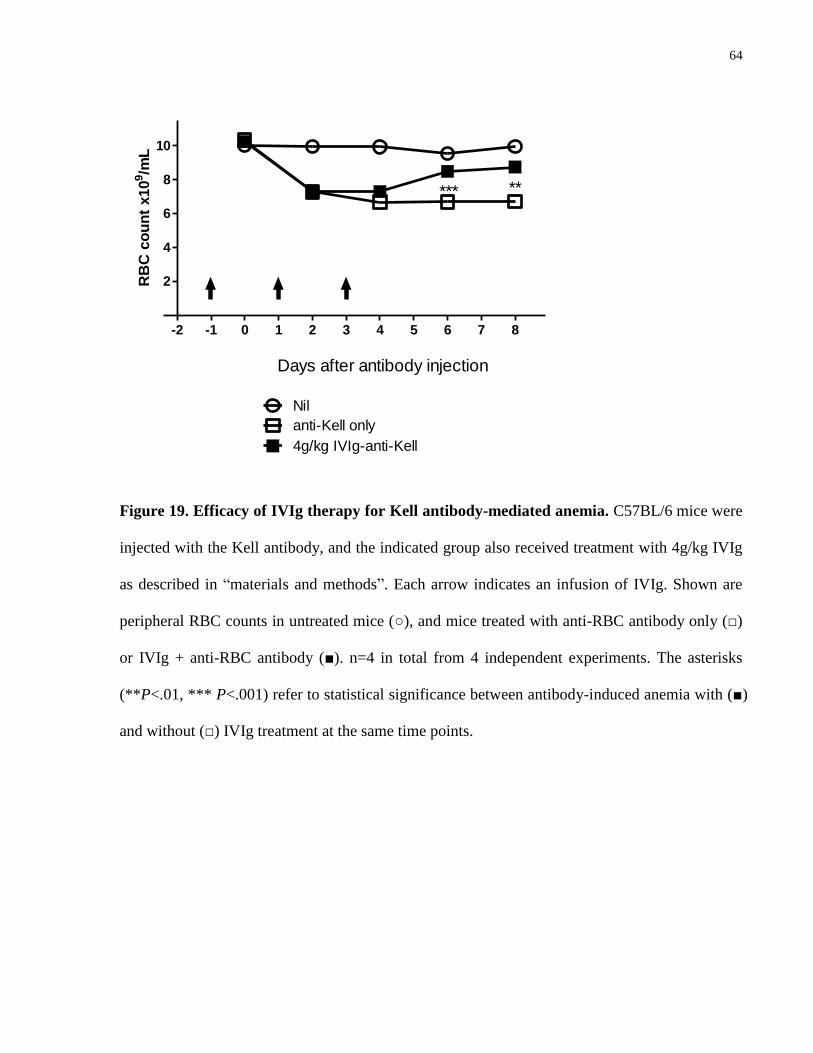
64
-2 -1 0 1 2 3 4 5 6 7 8
2
4
6
8
10
anti-Kell only
4g/kg IVIg-anti-Kell
Nil
Days after antibody injection
*** **
RB
C c
ou
nt
x10
9/m
L
Figure 19. Efficacy of IVIg therapy for Kell antibody-mediated anemia. C57BL/6 mice were
injected with the Kell antibody, and the indicated group also received treatment with 4g/kg IVIg
as described in “materials and methods”. Each arrow indicates an infusion of IVIg. Shown are
peripheral RBC counts in untreated mice (○), and mice treated with anti-RBC antibody only (□)
or IVIg + anti-RBC antibody (■). n=4 in total from 4 independent experiments. The asterisks
(**P<.01, *** P<.001) refer to statistical significance between antibody-induced anemia with (■)
and without (□) IVIg treatment at the same time points.
65
5.4.2 IVIg ameliorative effect of anemia is associated with FcγR-dependent mechanism
In an attempt to characterize the mechanism of anemia caused by each anti-RBC antibody, we
determined the degree of anemia attributable to FcγR-dependent versus complement-mediated
mechanism(s) using mice genetically deficient for the Fcγ chain (Fcγ-/-
) or complement
component C3 (C3-/-
), respectively (except for the anti-band 3 antibody, which was only tested in
Fcγ-/-
mice; however, this antibody has been tested in C3-/-
mice in published works [107]).
A similar degree of anemia was found in C3-/-
mice compared to C57BL/6 mice treated with anti-
CD24 (Fig. 20A), anti-GPA (Fig. 20B), or anti-Kell (Fig. 20C) antibody. Although not tested in
the current study, the anti-band 3 antibody at the same dose of 50 µg per mouse has been shown
by Azeredo da Silveira and coworkers [107] to cause similar degree of anemia in C3-/-
and
C57BL/6 mice at the same dose used in this study (50 µg/mouse). Altogether, these results
suggest a minimal role, if any, for complement-mediated mechanism in anemia caused by these
antibodies.
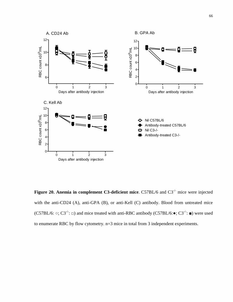
66
0 1 2 3
6
8
10
12
A. CD24 Ab
Days after antibody injection
RB
C c
ount x1
09/m
L
0 1 2 30
2
4
6
8
10
12
B. GPA Ab
Days after antibody injection
RB
C c
ount x1
09/m
L
0 1 2 30
2
4
6
8
10
12
Nil C57BL/6
Antibody-treated C57BL/6
Nil C3-/-
Antibody-treated C3-/-
C. Kell Ab
Days after antibody injection
RB
C c
ount x1
09/m
L
Figure 20. Anemia in complement C3-deficient mice. C57BL/6 and C3-/-
mice were injected
with the anti-CD24 (A), anti-GPA (B), or anti-Kell (C) antibody. Blood from untreated mice
(C57BL/6: ○; C3-/-
: □) and mice treated with anti-RBC antibody (C57BL/6:●; C3-/-
: ■) were used
to enumerate RBC by flow cytometry. n=3 mice in total from 3 independent experiments.
67
On the other hand, when the Fcγ-/-
mice were injected with the anti-band 3 (Fig. 21) and anti-
CD24 (Fig. 22) antibody, the degree of anemia was considerably less than what was observed in
the C57BL/6 mice on day 4, suggesting a role of FcγR-mediated mechanisms induced by these 2
antibodies. In contrast, in Fcγ-/-
mice treated with the anti-GPA antibody, only a minor
proportion of anemia was prevented as compared to the C57BL/6 mice (Fig. 23). Interestingly,
following injection with the anti-Kell antibody, the Fcγ-/-
mice seemed to develop similar degree
of anemia as in the C57BL/6 mice, but have accelerated recovery in the period between day 4
and day 8 post-injection (Fig. 24). In addition, anti-Kell-treated Fcγ-/-
mice were found no longer
respond to IVIg treatment (data not shown).
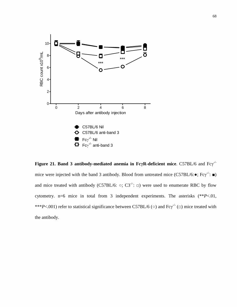
68
0 2 4 6 80
2
4
6
8
10
C57BL/6 Nil
C57BL/6 anti-band 3
Fc-/-
Nil
Fc-/-
anti-band 3
******
**
Days after antibody injection
RB
C c
ount x1
09/m
L
Figure 21. Band 3 antibody-mediated anemia in FcγR-deficient mice. C57BL/6 and Fcγ-/-
mice were injected with the band 3 antibody. Blood from untreated mice (C57BL/6:●; Fcγ-/-
: ■)
and mice treated with antibody (C57BL/6: ○; C3-/-
: □) were used to enumerate RBC by flow
cytometry. n=6 mice in total from 3 independent experiments. The asterisks (**P<.01,
***P<.001) refer to statistical significance between C57BL/6 (○) and Fcγ-/-
(□) mice treated with
the antibody.
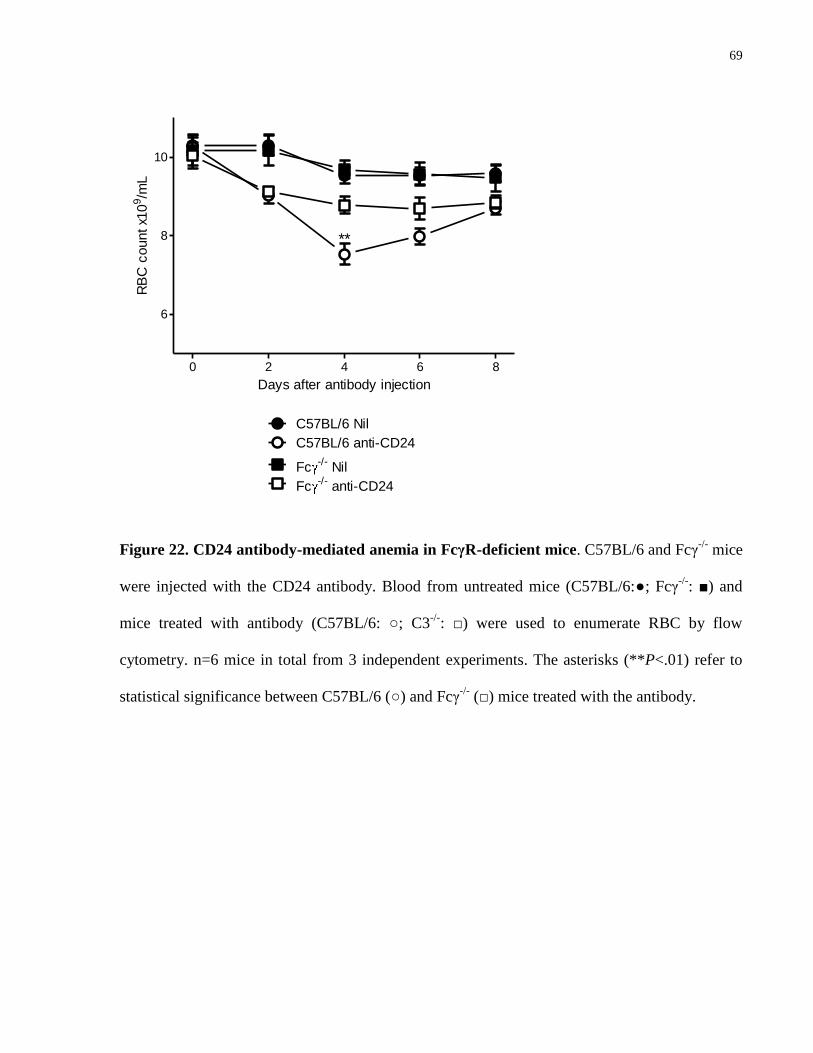
69
0 2 4 6 8
6
8
10
C57BL/6 Nil
C57BL/6 anti-CD24
Fc-/-
Nil
Fc-/-
anti-CD24
**
Days after antibody injection
RB
C c
ount x1
09/m
L
Figure 22. CD24 antibody-mediated anemia in FcγR-deficient mice. C57BL/6 and Fcγ-/-
mice
were injected with the CD24 antibody. Blood from untreated mice (C57BL/6:●; Fcγ-/-
: ■) and
mice treated with antibody (C57BL/6: ○; C3-/-
: □) were used to enumerate RBC by flow
cytometry. n=6 mice in total from 3 independent experiments. The asterisks (**P<.01) refer to
statistical significance between C57BL/6 (○) and Fcγ-/-
(□) mice treated with the antibody.
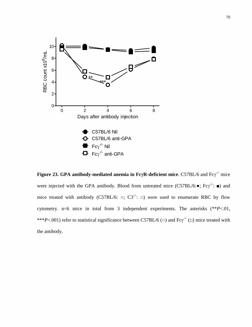
70
0 2 4 6 80
2
4
6
8
10
C57BL/6 Nil
C57BL/6 anti-GPA
Fc-/-
Nil
Fc-/-
anti-GPA
*****
Days after antibody injection
RB
C c
ount x1
09/m
L
Figure 23. GPA antibody-mediated anemia in FcγR-deficient mice. C57BL/6 and Fcγ-/-
mice
were injected with the GPA antibody. Blood from untreated mice (C57BL/6:●; Fcγ-/-
: ■) and
mice treated with antibody (C57BL/6: ○; C3-/-
: □) were used to enumerate RBC by flow
cytometry. n=6 mice in total from 3 independent experiments. The asterisks (**P<.01,
***P<.001) refer to statistical significance between C57BL/6 (○) and Fcγ-/-
(□) mice treated with
the antibody.
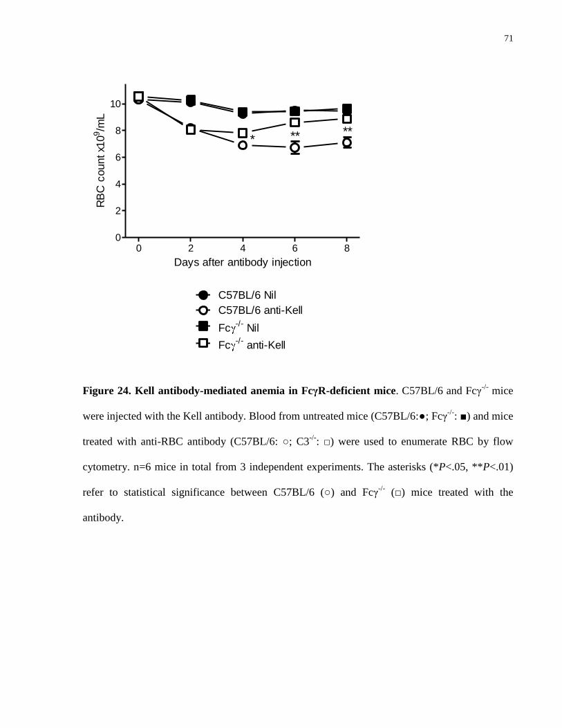
71
0 2 4 6 80
2
4
6
8
10
C57BL/6 Nil
C57BL/6 anti-Kell
Fc-/-
Nil
Fc-/-
anti-Kell
** ***
Days after antibody injection
RB
C c
ount x1
09/m
L
Figure 24. Kell antibody-mediated anemia in FcγR-deficient mice. C57BL/6 and Fcγ-/-
mice
were injected with the Kell antibody. Blood from untreated mice (C57BL/6:●; Fcγ-/-
: ■) and mice
treated with anti-RBC antibody (C57BL/6: ○; C3-/-
: □) were used to enumerate RBC by flow
cytometry. n=6 mice in total from 3 independent experiments. The asterisks (*P<.05, **P<.01)
refer to statistical significance between C57BL/6 (○) and Fcγ-/-
(□) mice treated with the
antibody.
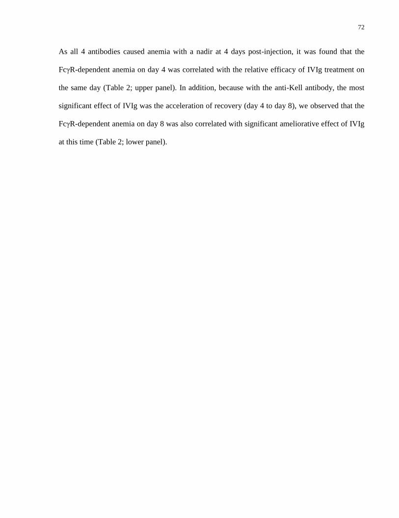
72
As all 4 antibodies caused anemia with a nadir at 4 days post-injection, it was found that the
FcγR-dependent anemia on day 4 was correlated with the relative efficacy of IVIg treatment on
the same day (Table 2; upper panel). In addition, because with the anti-Kell antibody, the most
significant effect of IVIg was the acceleration of recovery (day 4 to day 8), we observed that the
FcγR-dependent anemia on day 8 was also correlated with significant ameliorative effect of IVIg
at this time (Table 2; lower panel).
73
Table 5-1. Response to IVIg was associated with FcγR-dependent anemia. *P<.05, **P<.01,
*** P<.001; ns= not significant.
Antibody
specificity
FcγR-dependent anemia on day 4 IVIg ameliorative effect on day 4
Band 3 61%*** 56%**
CD24 54%*** 38%*
GPA 21%*** 9% ns
Kell 37%* 19% ns
Antibody
specificity
FcγR-dependent anemia on day 8 IVIg-ameliorative effect on day 8
Kell 70%** 62%**
74
5.5 Discussion
Treatment for patients with AHA remains a challenging task as the current therapeutic regime is
still essentially experience-based [11]. Despite its established efficacy in other autoimmune
cytopenias (eg., immune thrombocytopenia and autoimmune neutropenia) and low risk of side
effects compared to other mainstay therapies of AHA (eg., glucocorticoids and cytotoxic drugs),
IVIg remains an off-label indication for the treatment of AHA due in part to inconsistent
responses [105], with no established explanation as to why some patients respond but others do
not. In the present study, we examined the mechanism of anemia as a potential factor in
determining the efficacy of IVIg in a murine model of passively induced immune mediated
hemolytic anemia (IHA).
Of the 4 IgG anti-mouse RBC antibodies used, three recognize RBC antigens that are of
relevance to the human disease (band 3, GPA and Kell), but CD24 is not expressed on human
erythrocytes [108]. All 4 antibodies induced significant anemia in mice, although two of the
antibodies (anti-CD24 and anti-GPA) are rat antibodies, the response appears to be RBC specific
as normal rat IgG does not cause changes in RBC counts (data not shown).
Similar to observations in human patients, our findings demonstrated differential efficacy of high
dose IVIg treatment in the IHA mice. In the “clearance phase” of anemia, which is the period
from the injection of the anti-RBC antibody to when the anemia reaches its nadir (day 0 to day
4), treatment with IVIg (4g/kg) was associated with significantly less anemia in mice injected
with the anti-band 3 and anti-CD24 antibody. In contrast, whereas no significant efficacy of IVIg
was detected in the clearance phase in anti-Kell antibody-treated mice, IVIg therapy was
associated with accelerated recovery in peripheral RBC counts, indicating a likely role of IVIg in
75
facilitating this recovery phase of anti-Kell-mediated anemia. Taken together, these results
suggested that not only is the efficacy of IVIg different in anemia caused by different antibodies,
the underlying therapeutic mechanism could also be different.
Consistent with previous reports [31, 109], our results suggested a minor role, if any, of
complement-mediated mechanisms in anemia mediated by these mouse erythrocyte-specific
antibodies. As such, our data could not provide information as to the impact of IVIg treatment on
complement actions, yet suggested that therapeutic agents targeting complement may not be
effective in treating primary IHA in these mice.
In contrast, a significant component of FcγR-dependent anemia was identified 1) in the clearance
phase in anti-CD24 and anti-band 3 treated mice, and 2) in the recovery phase of anti-Kell
treated mice. Interestingly, in both cases, IVIg efficacy was found associated with FcγR-
dependent anemia. Similar associations have been identified in a mouse model of ITP [110, 111]
where it was showed that the anti-GPIbα antibodies which mediate predominantly Fc-
independent thrombocytopenia, responded poorly to IVIg, whereas anti-GPIIbIIIa antibodies that
cause Fc-dependent disease responded well.
Interestingly, in anemia caused by each antibody the initial/acute decrease in peripheral RBC
counts (day 0 to day 2) seemed to be unaccounted for by the 2 mechanisms examined, despite the
current understanding that antibody-mediated anemia is caused by FcR-mediated phagocytosis or
complement-mediated phagocytosis and/or lysis, or a combination of both [24, 112]. The
observations with the anti-GPA antibody were particularly striking, as this antibody was capable
of causing severe anemia in the absence of the FcγR or complement C3. Further investigation of
the mechanisms mediated by this antibody was presented in Chapter 4. Consistent with our
observed correlation between presence of FcγR-dependent anemia and response to IVIg therapy,
76
we did not find IVIg to improve RBC counts in C57BL/6 mice treated with this antibody, but
had an interesting observation in CD1 mice, where 50 μg of this anti-GPA antibody was
associated with 75% death within 1 day of injection. Surprisingly, despite no major improvement
in RBC counts, IVIg pretreatment was associated with 100% survival (Appendix 3). IVIg
therapy has been shown to be associated with prevention of death in other disease mouse models.
Chow et al [113] showed in an active model of ITP that bleeding mortality caused by antibody-
mediated thrombocytopenia was preventable with IVIg treatment. More recently, Semple et al
[114] demonstrated that IVIg prevents death in a mouse model of antibody-mediated transfusion-
related acute lung injury (TRALI). Interestingly, in both studies, as well as our observation, IVIg
was shown to prevent antibody-mediated death, despite different target of the antibody in each
case. Whether the life-saving property of IVIg in all 3 studies is mediated by a common
mechanism is a subject of future studies.
Another interesting antibody is the anti-Kell antibody. Similar to the other 3 antibodies, this anti-
Kell antibody caused increasing anemia with a nadir at 4 days post-injection, yet the recovery
was significantly delayed compared to anemia caused by the other antibodies. This could be due
to longer half-life of antibody-coated RBC, yet we found, if anything, the anti-Kell antibody was
associated with less antibody-positive circulating RBC, and the amount of bound antibodies was
also considerably lower than anti-GPA and anti-band 3 (Appendix 4~6; note the difference in the
scale of the y-axis). Anti-Kell antibodies are known as the second most common cause of severe
hemolytic diseases of the newborn (HDN), second only to HDN caused by anti-D. Affected Kell-
alloimmunized fetuses usually present with lower numbers of circulating reticulocytes and
normoblasts, compared with anti-D-affected fetuses, despite elevated level of erythropoietin
released [38]. Vaughan et al [39] demonstrated that monoclonal IgG and IgM antibodies
specifically inhibited growth of Kell-positive (but not Kell-negative) erythroid progenitor cells in
77
a dose-dependent manner, whereas anti-D had no such effect. Their findings were furthered by
Daniels et al [40] who showed that Kell antigen appears on erythroid progenitor cells early in
erythropoiesis, prior to the expression of GPA and band 3. The authors went on to show that
Kell-positive progenitor cells sensitized with anti-Kell sera from pregnant women elicited a
strong response from monocytes in vitro, and proposed that anti-Kell but not anti-D may cause
fetal anemia by promoting immune destruction of the Kell-positive erythroid early progenitor
cells by macrophages in the fetal liver.
Whether our results showing delayed recovery in anti-Kell antibody-mediated anemia are due to
a similar mechanism, namely inhibition of erythropoiesis by anti-Kell-mediated immune
destruction of Kell-expressing erythroid progenitor cells, remains to be explored in future studies.
Nevertheless, such a mechanism is not only consistent with the observed delay in recovery, but
may also explain the fact that in Fcγ-/-
mice this inhibition is reversed, consistent with the
possibility that Fcγ-/-
mice could not mediate macrophage FcγR-dependent phagocytosis of these
progenitor cells.
In conclusion, our results demonstrate that efficacy of IVIg in ameliorating IHA varies in relation
to the mechanism of anemia in mice, with FcγR-mediated anemia being highly related to
response to IVIg.
78
Chapter 6
Monoclonal Anti-RBC Antibodies Can Ameliorate
Thrombocytopenia in a Murine Model of Passive ITP
79
6 Monoclonal anti-RBC antibodies can ameliorate thrombocytopenia in a murine model of passive ITP
6.1 Summary
To explore the potential for monoclonal erythrocyte-specific antibodies as an alternative
treatment for immune thrombocytopenia (ITP) and to explore the mechanism of their therapeutic
action, we tested a panel of four monoclonal anti-mouse RBC antibodies in a murine model of
ITP. We report that three of these antibodies: one anti-glycophorin A (GPA), one anti-band 3 and
one anti-Kell antibody, were capable of ameliorating ITP to a similar extent as intravenous
immunoglobulin (IVIg) but at a potentially 3-log fold lower dosage as compared to IVIg.
Improvement in peripheral platelet counts was found to be positively associated with the severity
of anemia in mice injected with these RBC-specific antibodies. However, whether these anti-
RBC antibodies mediate the amelioration via Fcγ receptor (FcγR) blockade remains unclear.
6.2 Introduction
The beneficial effect of IVIg in ameliorating thrombocytopenia in ITP was first discovered by
Imbach et al [13] in 1981. Subsequent investigations in the early 80s [12, 115] suggested the
most likely mechanism of its therapeutic action to be a transitory blockade of the
reticuloendothelial system (RES). Salama et al [116] proposed that it is the IgG-coated
autologous RBC rather than IgG themselves that mediate the interaction with the Fc receptors
(FcR) on macrophages of the RES. Supporting Salama et al’s proposed mechanism, polyclonal
anti-D was later demonstrated to reverse thrombocytopenia in D-positive but not D-negative
patients [117, 118]; and polyclonal anti-D remains one of the first-line treatment for ITP in D-
positive patients to this date [61]. Because polyclonal anti-D is in limited supply and priority of
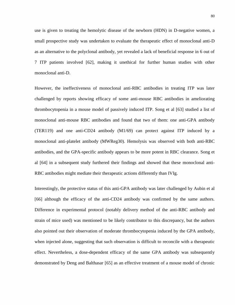
80
use is given to treating the hemolytic disease of the newborn (HDN) in D-negative women, a
small prospective study was undertaken to evaluate the therapeutic effect of monoclonal anti-D
as an alternative to the polyclonal antibody, yet revealed a lack of beneficial response in 6 out of
7 ITP patients involved [62], making it unethical for further human studies with other
monoclonal anti-D.
However, the ineffectiveness of monoclonal anti-RBC antibodies in treating ITP was later
challenged by reports showing efficacy of some anti-mouse RBC antibodies in ameliorating
thrombocytopenia in a mouse model of passively induced ITP. Song et al [63] studied a list of
monoclonal anti-mouse RBC antibodies and found that two of them: one anti-GPA antibody
(TER119) and one anti-CD24 antibody (M1/69) can protect against ITP induced by a
monoclonal anti-platelet antibody (MWReg30). Hemolysis was observed with both anti-RBC
antibodies, and the GPA-specific antibody appears to be more potent in RBC clearance. Song et
al [64] in a subsequent study furthered their findings and showed that these monoclonal anti-
RBC antibodies might mediate their therapeutic actions differently than IVIg.
Interestingly, the protective status of this anti-GPA antibody was later challenged by Aubin et al
[66] although the efficacy of the anti-CD24 antibody was confirmed by the same authors.
Difference in experimental protocol (notably delivery method of the anti-RBC antibody and
strain of mice used) was mentioned to be likely contributor to this discrepancy, but the authors
also pointed out their observation of moderate thrombocytopenia induced by the GPA antibody,
when injected alone, suggesting that such observation is difficult to reconcile with a therapeutic
effect. Nevertheless, a dose-dependent efficacy of the same GPA antibody was subsequently
demonstrated by Deng and Balthasar [65] as an effective treatment of a mouse model of chronic
81
passive ITP. Interestingly, Deng and Balthasar did not use or mention the anti-CD24 antibody,
despite recognizing anti-GPA antibody-associated severe hemolysis.
In this thesis, in addition to anti-GPA and anti-CD24, another 2 monoclonal IgG mouse
erythrocyte-specific antibodies were characterized in terms of therapeutic efficacy in murine ITP
in relation to their ability to cause RBC clearance. We report herein that 3 out of 4 of these
antibodies were capable of ameliorating ITP to a similar extent as 2.5g/kg (ie., 50 mg/mouse)
IVIg, although at much lower dosage. The ability to improve platelet counts seemed to associate
with the extent of RBC clearance, but association of efficacy with the presence of FcγR-
dependent RBC clearance mechanism, as previously asserted, was not supported. In addition, we
also observed the thrombocytopenic effect of the GPA antibody, which we did not find to
interfere with the ameliorative effect of this antibody in the amelioration of ITP.
6.3 Materials and methods
6.3.1 Mice
C57BL/6 mice were purchased from Charles River Laboratories, and FcR γ-chain-deficient mice
(B6.129P2-Fcer1gtm1Rav
N12) were from Taconic Farms. All mice were used at 6-10 weeks of
age. All animal experiments were approved by the Animal Care and Use Committee of St.
Michael’s Hospital.
6.3.2 Reagents
The anti-CD41 (MWReg30, IgG1, 2 µg/mouse), anti-GPA (TER119, IgG2b, 45 µg/mouse), and
anti-CD24 (M1/69, IgG2b, 70 µg/mouse) antibodies were purchased from BD Biosciences, the
anti-band 3 (34-3C, IgG2a, 50 µg/mouse) antibody was from Hycult Biotech, and the anti-Kell
82
(MIMA87, IgG2a, 50 µg/mouse) was from Dr Gregory Halverson (New York Blood Center).
IVIg was either Gamunex 10% (Talecris Biotherapeutics) or Privigen 10% (CSL Behring), and
used at 50 mg/mouse (equivalent to ~2.5g/kg).
6.3.3 ITP and measurement of platelet counts
Thrombocytopenia was induced in mice by intravenous injection of 2 μg of MWReg30 antibody
in 200 µL PBS. Platelet counts were determined as described [74]. Briefly, 10 µL of whole blood
was drawn by saphenous bleeding and diluted 1: 100 in PBS/EDTA. The diluted blood was
centrifuged (170g; 2 minutes; brake off) to separate the platelet-rich plasma (PRP). The PRP was
diluted 1: 100 in isoton and platelet counts were determined on a Beckman Z2 Coulter Counter.
6.3.4 IVIg or RBC antibody treatment of ITP
Pretreatment with IVIg or one of the monoclonal anti-RBC antibodies was given 24 hours prior
to the induction of ITP with the MWReg30 antibody. Platelet counts were determined 24 hours
post MWReg30 injection.
6.3.5 Statistical analysis
Data are presented as mean plus or minus standard error of the mean (SEM). Note, in some cases
where the error bars are not apparent, this is due to a minimal SEM. Statistical analysis was
performed with the Student t test using GraphPad Prism software. P<.05 was considered
significant
83
6.4 Results and discussion
6.4.1 Monoclonal anti-RBC antibodies can ameliorate murine ITP
To determine their efficacy in the amelioration of ITP, each of the 4 anti-RBC antibodies: anti-
GPA (TER119), anti-CD24 (M1/69), anti-band 3 (34-3C) and anti-Kell (MIMA87) was injected
intravenously into mice 24 hours before the induction of ITP by the anti-CD41 antibody
MWReg30. All but M1/69 was associated with improved platelet counts (Fig. 25~28). In
particular, the anti-GPA (45 µg/mouse; Fig. 25) and anti-band 3 (50 µg/mouse; Fig. 26) were
found to achieve a similar increase in platelet counts as 50 mg IVIg, and 50 µg anti-Kell (Fig. 27)
also delivered comparable efficacy as compared to 50 mg IVIg. Unexpectedly, 50 µg of the anti-
CD24 antibody, which was originally shown by Song et al [63] to be effective, failed to affect
platelet counts (data not shown), and even an increased dose of 70 µg seemed to still be
ineffective (Fig. 28).
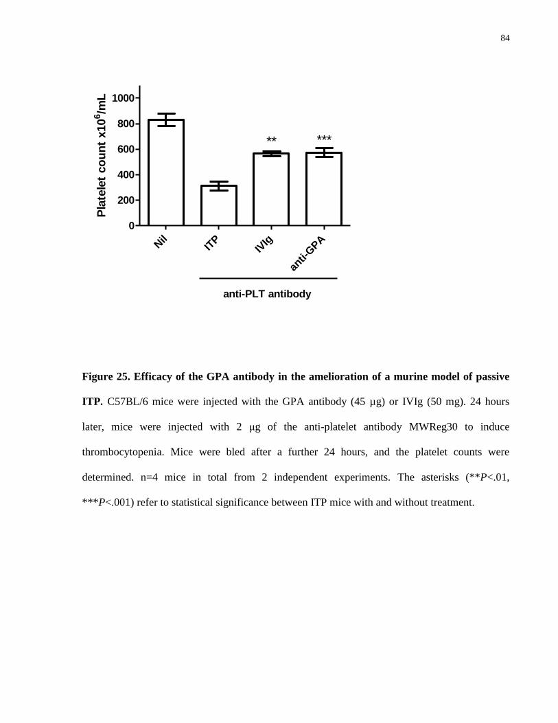
84
Nil
ITP
IVIg
anti-
GPA
0
200
400
600
800
1000
anti-PLT antibody
** ***
Pla
tele
t co
un
t x10
6/m
L
Figure 25. Efficacy of the GPA antibody in the amelioration of a murine model of passive
ITP. C57BL/6 mice were injected with the GPA antibody (45 µg) or IVIg (50 mg). 24 hours
later, mice were injected with 2 μg of the anti-platelet antibody MWReg30 to induce
thrombocytopenia. Mice were bled after a further 24 hours, and the platelet counts were
determined. n=4 mice in total from 2 independent experiments. The asterisks (**P<.01,
***P<.001) refer to statistical significance between ITP mice with and without treatment.
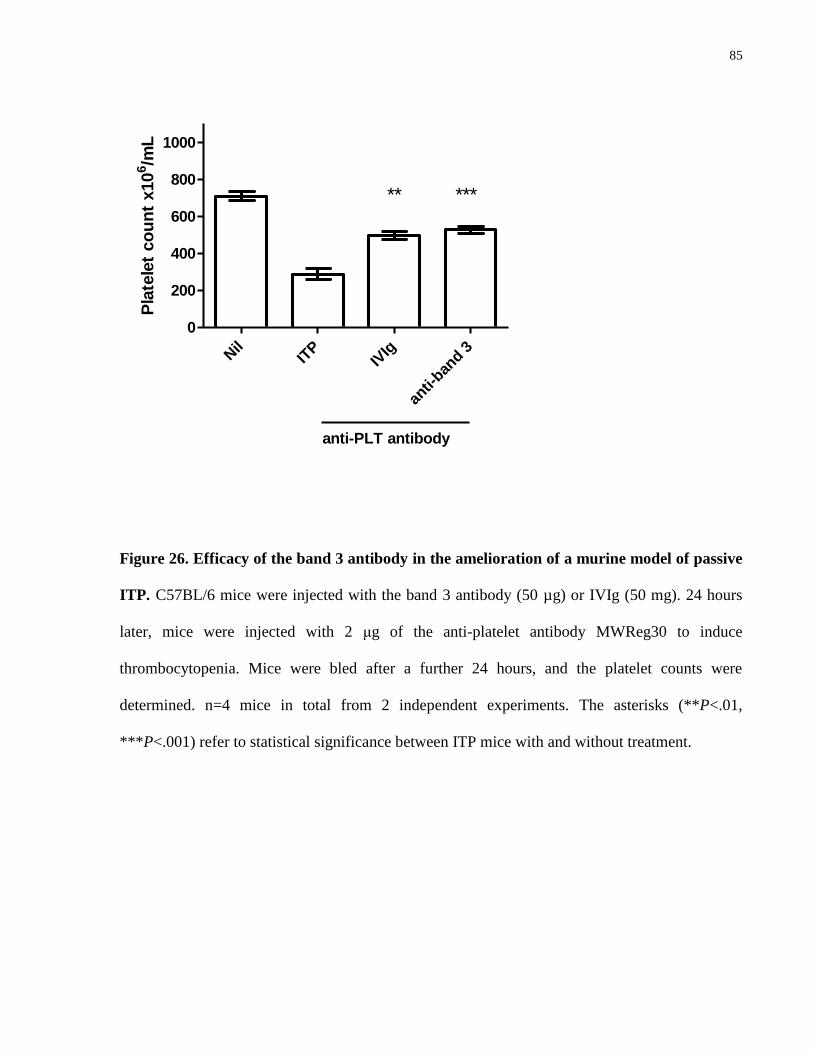
85
Nil
ITP
IVIg
anti-
band 3
0
200
400
600
800
1000
anti-PLT antibody
** ***
Pla
tele
t co
un
t x10
6/m
L
Figure 26. Efficacy of the band 3 antibody in the amelioration of a murine model of passive
ITP. C57BL/6 mice were injected with the band 3 antibody (50 µg) or IVIg (50 mg). 24 hours
later, mice were injected with 2 μg of the anti-platelet antibody MWReg30 to induce
thrombocytopenia. Mice were bled after a further 24 hours, and the platelet counts were
determined. n=4 mice in total from 2 independent experiments. The asterisks (**P<.01,
***P<.001) refer to statistical significance between ITP mice with and without treatment.
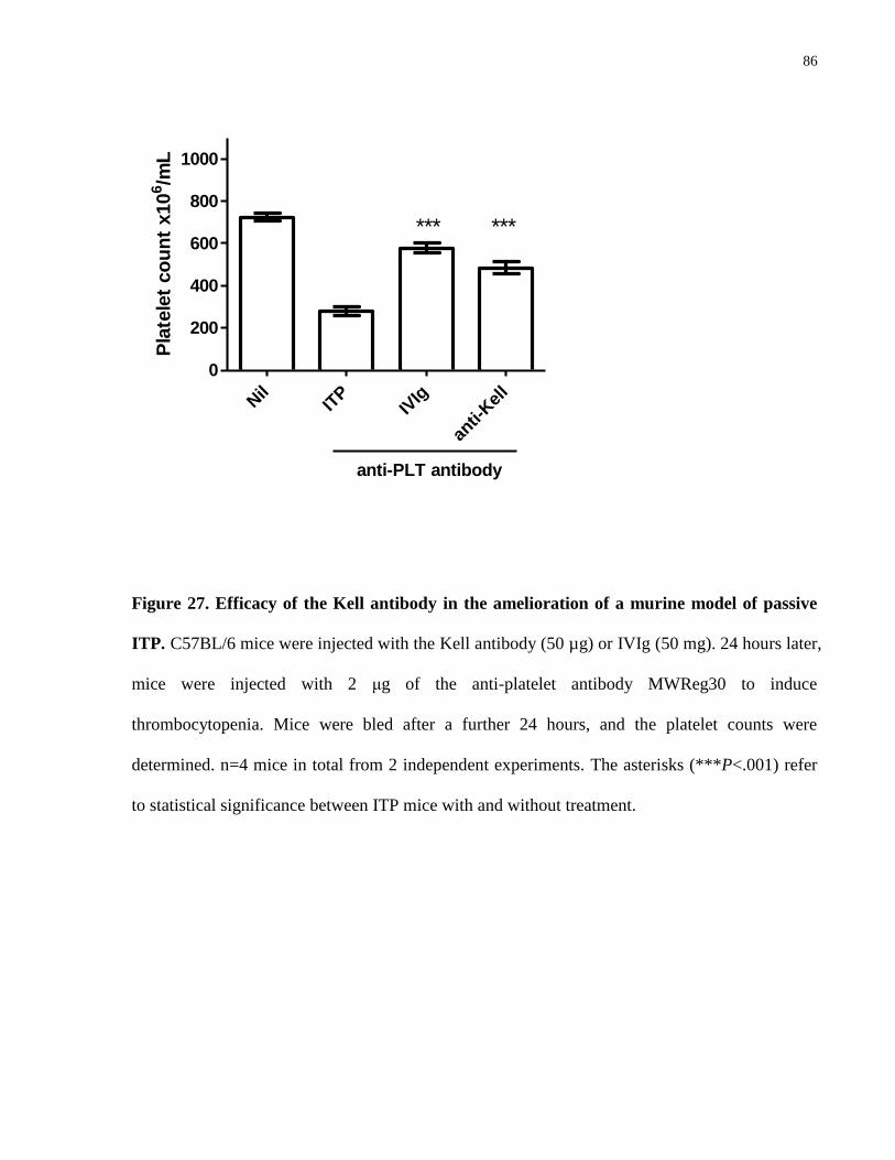
86
Nil
ITP
IVIg
anti-
Kel
l0
200
400
600
800
1000
anti-PLT antibody
*** ***
Pla
tele
t co
un
t x10
6/m
L
Figure 27. Efficacy of the Kell antibody in the amelioration of a murine model of passive
ITP. C57BL/6 mice were injected with the Kell antibody (50 µg) or IVIg (50 mg). 24 hours later,
mice were injected with 2 μg of the anti-platelet antibody MWReg30 to induce
thrombocytopenia. Mice were bled after a further 24 hours, and the platelet counts were
determined. n=4 mice in total from 2 independent experiments. The asterisks (***P<.001) refer
to statistical significance between ITP mice with and without treatment.
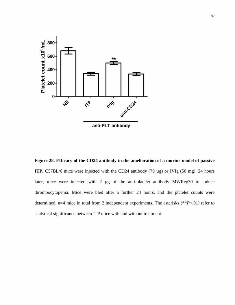
87
Nil
ITP
IVIg
anti-
CD24
0
200
400
600
800
anti-PLT antibody
**
Pla
tele
t co
un
t x10
6/m
L
Figure 28. Efficacy of the CD24 antibody in the amelioration of a murine model of passive
ITP. C57BL/6 mice were injected with the CD24 antibody (70 µg) or IVIg (50 mg). 24 hours
later, mice were injected with 2 μg of the anti-platelet antibody MWReg30 to induce
thrombocytopenia. Mice were bled after a further 24 hours, and the platelet counts were
determined. n=4 mice in total from 2 independent experiments. The asterisks (**P<.01) refer to
statistical significance between ITP mice with and without treatment.
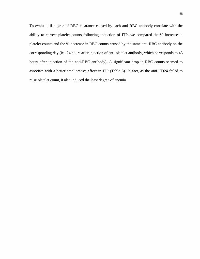
88
To evaluate if degree of RBC clearance caused by each anti-RBC antibody correlate with the
ability to correct platelet counts following induction of ITP, we compared the % increase in
platelet counts and the % decrease in RBC counts caused by the same anti-RBC antibody on the
corresponding day (ie., 24 hours after injection of anti-platelet antibody, which corresponds to 48
hours after injection of the anti-RBC antibody). A significant drop in RBC counts seemed to
associate with a better ameliorative effect in ITP (Table 3). In fact, as the anti-CD24 failed to
raise platelet count, it also induced the least degree of anemia.
89
Table 3. Therapeutic effect of the anti-RBC antibodies in ITP in relation to the RBC
clearance they induce. Max. ↓ % RBC was measured on day 4 post anti-RBC antibody injection
(see chapter 5).
Antibody specificity ↑% PLT Corresp.↓ % RBC Max. ↓% RBC
GPA 50% 49% 57%
CD24 0 15% 28%
Band 3 57% 27% 51%
Kell 46% 29% 35%
90
6.4.2 FcγR is required for MWReg30 to induce platelet clearance, but not required for the anti-RBC antibodies to mediate RBC clearance at the time of ameliorative effect
One of the most prominent theories of the therapeutic action of anti-D in the treatment of D-
positive ITP patients is the binding of anti-D coated RBC to the macrophage FcγR [119], thereby
preventing binding of antibody-coated platelet by a competitive mechanism. We first determined
if the development of ITP in our mouse model requires FcγR-mediated platelet clearance with
the use of Fc γ chain knock-out (Fcγ-/-
) mice, which are deficient in the functions of all 3 classes
of activating Fc receptors (FcγI, III and IV). The Fcγ-/-
mice were fully protected from
thrombocytopenia induced by MWReg30 (Fig. 29), demonstrating that MWReg30 requires the
FcγR to mediate platelet clearance.
As the Fcγ-/-
mice do not develop any thrombocytopenia due to MWReg30, it is difficult to
directly determine if FcγR is required for the anti-RBC antibodies to ameliorate ITP in this
mouse model. We therefore examined RBC clearance in Fcγ-/-
mice, instead, as an alternative
measure of the engagement of FcγR by the anti-RBC antibodies. We found that, none of the 3
effective antibodies (anti-GPA, anti-band 3 and anti-Kell) seemed to require the FcγR in
mediating anemia in the first 48 hours after injection of the anti-RBC antibody (Fig. 30),
suggesting that the RBC clearance responsible for the attenuation of ITP was largely independent
of FcγR-dependent mechanisms.
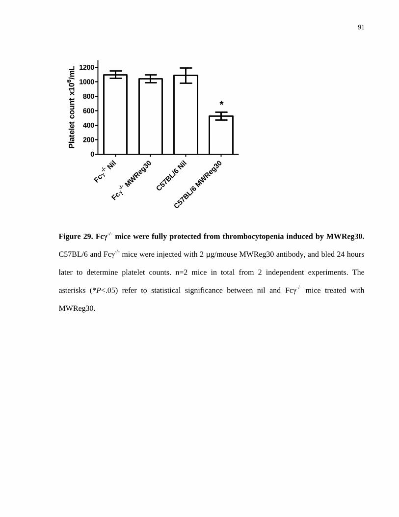
91
Nil
-/-
Fc
MW
Reg
30
-/-
FcC57
BL/6
Nil
C57
BL/6
MW
Reg
30
0
200
400
600
800
1000
1200
*
Pla
tele
t co
un
t x10
6/m
L
Figure 29. Fcγ-/-
mice were fully protected from thrombocytopenia induced by MWReg30.
C57BL/6 and Fcγ-/-
mice were injected with 2 µg/mouse MWReg30 antibody, and bled 24 hours
later to determine platelet counts. n=2 mice in total from 2 independent experiments. The
asterisks (*P<.05) refer to statistical significance between nil and Fcγ-/-
mice treated with
MWReg30.
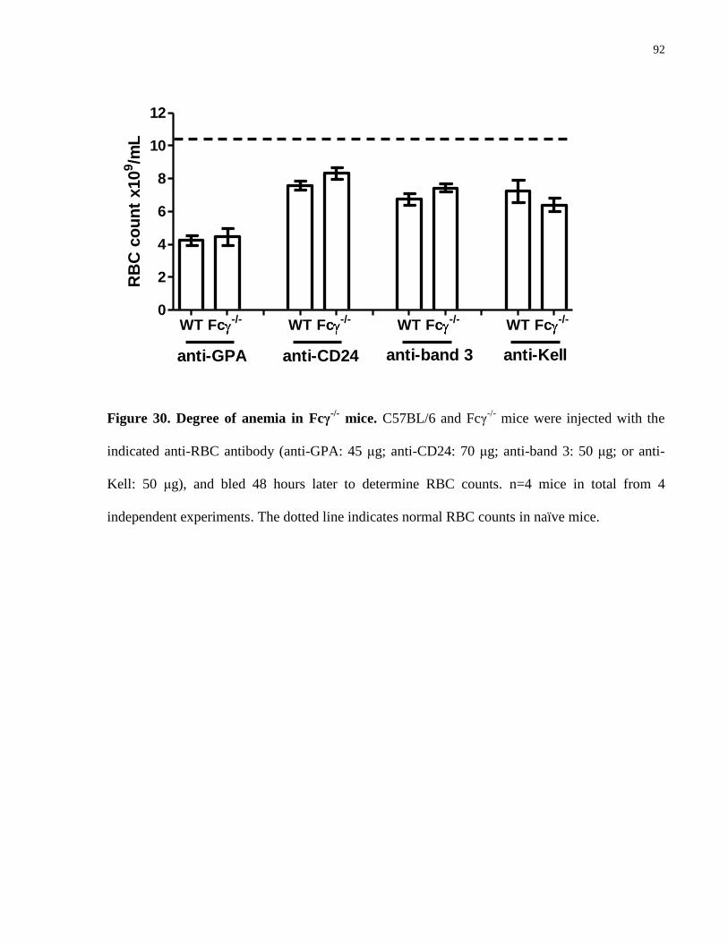
92
WT Fc-/-
WT Fc-/-
WT Fc-/-
WT Fc-/-
0
2
4
6
8
10
12
anti-GPA anti-CD24 anti-band 3 anti-Kell
RB
C c
ou
nt
x10
9/m
L
Figure 30. Degree of anemia in Fcγ-/-
mice. C57BL/6 and Fcγ-/-
mice were injected with the
indicated anti-RBC antibody (anti-GPA: 45 μg; anti-CD24: 70 μg; anti-band 3: 50 μg; or anti-
Kell: 50 μg), and bled 48 hours later to determine RBC counts. n=4 mice in total from 4
independent experiments. The dotted line indicates normal RBC counts in naïve mice.
93
Taken together, these results suggested that RBC clearance but not necessarily FcγR-mediated
RBC clearance is associated with the ameliorative effect of monoclonal anti-RBC antibodies in a
mouse model of ITP. It is likely that the antibody-coated RBC still compete with antibody-coated
platelet for phagocytosis, but via different receptor/mechanism for the engagement of the
macrophage.
6.4.3 TER119 prevents MWReg30-induced thrombocytopenia but also mediates thrombocytopenia on its own
Aubin et al [66] noticed decrease in platelet counts in anti-GPA (TER119)-treated BALB/c mice,
which, in their argument, complemented their data showing anti-GPA as “unprotective” in
mouse ITP induced by MWReg30. We also observed a transient thrombocytopenic effect of anti-
GPA in C57BL/6 and also the Fcγ-/-
mice (Appendix 1). The GPA antibody injected alone caused
30-40% drop in platelet counts by 24 hours post injection; after 24 hours, platelet counts started
to recover. Interestingly, if mice received MWReg30 24 hours after they received anti-GPA,
MWReg30 failed to cause any further decrease in platelet counts than what was already induced
by anti-GPA (Fig. 31). In other words, because anti-GPA caused less thrombocytopenia than
MWReg30, its thrombocytopenic effect did not interfere with its anti-thrombocytopenic effect.
It is not clear how anti-GPA mediates its thrombocytopenic effect; a more detailed discussion
can be found in Chapter 4. Our data seem to argue against the assertion of Aubin et al [66] by
showing that the seemingly paradoxical observations of pro- and anti-thrombocytopenic effects
can be reconciled. In fact, one of the possible mechanisms of therapeutic action of anti-GPA in
anti-platelet antibody-mediated ITP could be related to its thrombocytopenic action. As
demonstrated by Deng and Balthasar [65] in their chronic ITP model, therapeutic dose of anti-
GPA (50 µg in their case) was able to raise platelet counts to >200% of the initial platelet counts,
that is, >100% more than normal level. We also observed a “rebound” in platelet level 3-4 days
94
after anti-GPA injection (data not shown) although not as pronounced. This “rebound” in platelet
counts could potentially be due to compensatory action of the body’s hematopoietic system in
response to platelet (and RBC) clearance induced by anti-GPA. Further studies will help to
determine if a stimulation of thrombopoiesis plays a role in the therapeutic action of the GPA
antibody in murine ITP.
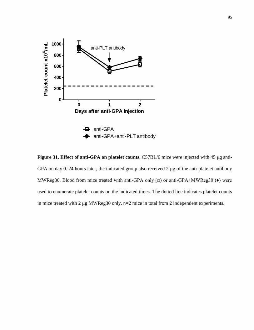
95
0 1 20
200
400
600
800
1000
anti-GPA
anti-GPA+anti-PLT antibody
anti-PLT antibody
Days after anti-GPA injection
Pla
tele
t co
un
t x10
6/m
L
Figure 31. Effect of anti-GPA on platelet counts. C57BL/6 mice were injected with 45 μg anti-
GPA on day 0. 24 hours later, the indicated group also received 2 μg of the anti-platelet antibody
MWReg30. Blood from mice treated with anti-GPA only (□) or anti-GPA+MWReg30 (♦) were
used to enumerate platelet counts on the indicated times. The dotted line indicates platelet counts
in mice treated with 2 μg MWReg30 only. n=2 mice in total from 2 independent experiments.
96
In conclusion, our results expanded the list of monoclonal anti-RBC antibodies that have an
“anti-D-like” effect in ameliorating thrombocytopenia in a mouse model of ITP. We showed that
an anti-band 3 and an anti-Kell, as well as the anti-GPA antibody previously identified by Song
et al [63], can prevent ITP to a similar extent as IVIg, but at a potentially 3-log fold lower dosage
as compared to IVIg. All 3 antibodies recognize “public” antigens expressed by almost all human
RBCs, and therefore could potentially be of advantage in the treatment of D-negative ITP
patients. Using Fcγ-/-
mice, we demonstrated that improvement of platelet counts coincided with
significant RBC clearance that appeared to be largely FcγR-independent, despite a complete
dependency on FcγR for development of thrombocytopenia by the platelet-specific antibody
MWReg30, arguing against a therapeutic mechanism of competition for FcγR binding. However,
it should be noted that these monoclonal anti-mouse RBC antibodies could behave very
differently from polyclonal anti-D in human patients. An example is offered by the anti-GPA
antibody: its ability to cause thrombocytopenia and ameliorate MWReg30-mediated
thrombocytopenia at the same time seemed paradoxical, but might in fact suggest a novel
therapeutic mechanism yet to be explored.

97
Chapter 7
Discussion
98
7 Discussion
7.1 Summary
Antibodies against erythrocyte-specific antigens are the focus of this thesis. A murine model of
passively induced autoimmune hemolytic anemia (AHA) was optimized for a panel of mouse
RBC-specific antibodies, and the mechanism(s) of RBC clearance were investigated. Data in this
thesis from the use of four different antibodies consistently showed that mechanisms in addition
to the classical macrophage Fc receptor (FcR)/complement-mediated extra- and intravascular
hemolysis may be contributing to the severe anemia observed in mice (Chapter 5). Many factors
can influence the pathogenicity of antibodies, including the antibody class, subclass, thermal
amplitude, complement/macrophage activating efficiency and so on [112], and results presented
in this thesis suggest that the antibody specificity may potentially be of importance. For instance,
the glycophorin A (GPA)-specific antibody-mediated RBC agglutination is unexpected for an
IgG antibody [6], but consistent with the role GPA plays in minimizing cell-cell interaction [36]
(Chapter 4). Moreover, the Kell-specific antibody-induced Fc-dependent inhibition of recovery is
also potentially related to the early expression of the Kell antigens on the erythroid precursor
cells [40] (Chapter 5). Nevertheless, the canonical mechanism of extravascular hemolysis was
well represented in this model, as two out of four of the antibodies (anti-band 3 and anti-CD24)
were found to mediate primarily an FcR-dependent mechanism of clearance (Chapter 5).
The treatment of AHA continues to pose a clinical problem [11], and the current treatment
regime remains experience-based as controlled clinical trials are extremely difficult due to the
low numbers of patients and high frequency of underlying secondary conditions [6, 11]. The
murine model therefore provides a platform for the evaluation and optimization of both known
99
and novel treatment regimes. In this thesis the efficacy of IVIg therapy, a well established
treatment for many autoimmune conditions (but with inconsistent efficacy in AHA patients), was
evaluated in murine AHA caused by the four mouse RBC-specific antibodies, and response to
treatment was found to correlate with FcγR-dependent anemia (Chapter 5). A similar correlation
of IVIg effect and FcγR-mediated cytopenia has been previously demonstrated in a murine
model of immune thrombocytopenia [110, 111], and this thesis, to our best knowledge, is the
first to demonstrate such a relationship in the treatment of AHA. In addition, a potential
treatment with CD44 antibodies was also evaluated in the murine model of AHA. An effective
treatment of FcγR-mediated murine ITP [63, 74], the anti-CD44 antibody KM114 was
hypothesized to be an alternative treatment for FcγR-mediated anemia in the current mouse
model, but was found ineffective in the amelioration of band 3 antibody-mediated anemia
(Appendix 10).
In addition to the elucidation of the pathophysiological effects of the RBC-specific antibodies,
this thesis also explored the therapeutic potential of these monoclonal antibodies in the treatment
of murine ITP analogous to the use of anti-D in ITP patients (Chapter 6). As three out of four of
the anemia-causing anti-RBC antibodies were found protective of passively induced ITP in mice,
these results add to the evidence that challenges the hypothesis that monoclonal antibodies are
not useful in treating immune forms of thrombocytopenia [62-64]. These three antibodies all
target public antigens (GPA, band 3 and Kell) expressed by almost all human RBC, suggesting
the possibility of an alternative to anti-D in the use of D-negative patients.
In conclusion, results in this thesis show in a murine model of AHA that antibody-mediated
mechanisms of anemia may be independent of complement or macrophage classical pathways,
and that the beneficial effect of IVIg is primarily displayed in FcγR-dependent anemia. Efficacy
100
of monoclonal RBC-specific antibodies was demonstrated in the treatment of immune-mediated
thrombocytopenia in mice, suggesting that they may provide an effective alternative to anti-D in
the treatment of ITP.
7.2 Future directions
In future experiments, more antibodies may be studied to investigate the mechanism(s) of anemia.
Multiple GPA-specific antibodies will be desired to determine if the agglutination mechanism is
a global observation or only specific to a subset of antibodies with certain characteristics (e.g.,
antibody isotype, specific epitope, etc). Data from the four antibodies assessed in this thesis
demonstrated for the first time a potential correlation between FcγR-dependent mechanism of
anemia and response to IVIg. Future experiments with more antibodies will be needed to further
evaluate this correlation, and most importantly, patient samples (if available) should be tested to
see if such a relationship is also valid in the human disease. Finally, this thesis established a
platform for the studies of monoclonal erythrocyte-specific antibodies in the amelioration of a
mouse model of ITP, and further investigation is warranted for the elucidation of their
therapeutic mechanisms of action.
101
References
1. Loutit JF and Mollison PL. Haemolytic icterus (acholuric jaundice) congenital and acquired. J
Pathol Bacteriol 1946;58:711-728.
2. Izui S. Autoimmune hemolytic anemia. Curr Opin Immunol 1994;6:926-930.
3. Bottiger LE and Westerholm B. Acquired haemolytic anaemia. I. Incidence and aetiology.
Acta Med Scand 1973;193:223-226.
4. Petz LD and Garratty G. Acquired immune hemolytic anemias. New York: Churchill
Livingstone; 1980.
5. Engelfriet CP, Overbeeke MA and von dem Borne AE. Autoimmune hemolytic anemia.
Semin Hematol 1992;29:3-12.
6. Petz LD and Garratty G. Immune hemolytic anemias 2nd
ed. Philadelphia: Churchill
Livingstone; 2004.
7. Gehrs BC and Friedberg RC. Autoimmune hemolytic anemia. Am J Hematol 2002;69:258-
271.
8. Packman CH. Hemolytic anemia due to warm autoantibodies. Blood Rev 2008;22:17-31.
9. Brain MC, Ruether B, Valentine K et al. Life-threatening hemolytic anemia due to an autoanti-
Pr cold agglutinin: evidence that glycophorin A antibodies may induce lipid bilayer exposure and
cation permeability independent of agglutination. Transfusion 2010;50:292-301.
102
10. Chadebech P, Michel M, Janvier D et al. IgA-mediated human autoimmune hemolytic
anemia as a result of hemagglutination in the spleen, but independent of complement activation
and FcalphaRI. Blood 2010;116:4141-4147.
11. Lechner K and Jager U. How I treat autoimmune hemolytic anemias in adults. Blood
2010;116:1831-1838.
12. Fehr J, Hofmann V and Kappeler U. Transient reversal of thrombocytopenia in idiopathic
thrombocytopenic purpura by high-dose intravenous gamma globulin. N Engl J Med
1982;306:1254-1258.
13. Imbach P, Barandun S, d'Apuzzo V et al. High-dose intravenous gammaglobulin for
idiopathic thrombocytopenic purpura in childhood. Lancet 1981;1:1228-1231.
14. Salama A, Mahn I, Neuzner J et al. IgG therapy in autoimmune haemolytic anaemia of warm
type. Blut 1984;48:391-392.
15. Mueller-Eckhardt C, Salama A, Mahn I et al. Lack of efficacy of high-dose intravenous
immunoglobulin in autoimmune haemolytic anaemia: a clue to its mechanism. Scand J Haematol
1985;34:394-400.
16. Bussel JB, Cunningham-Rundles C and Abraham C. Intravenous treatment of autoimmune
hemolytic anemia with very high dose gammaglobulin. Vox Sang 1986;51:264-269.
17. Macintyre EA, Linch DC, Macey MG and Newland AC. Successful response to intravenous
immunoglobulin in autoimmune haemolytic anaemia. Br J Haematol 1985;60:387-388.
103
18. Flores G, Cunningham-Rundles C, Newland AC and Bussel JB. Efficacy of intravenous
immunoglobulin in the treatment of autoimmune hemolytic anemia: results in 73 patients. Am J
Hematol 1993;44:237-242.
19. Anderson D, Ali K, Blanchette V et al. Guidelines on the use of intravenous immune
globulin for hematologic conditions. Transfus Med Rev 2007;21:S9-56.
20. Bocci V. Determinants of erythrocyte ageing: a reappraisal. Br J Haematol 1981;48:515-522.
21. Kay MM, Goodman SR, Sorensen K et al. Senescent cell antigen is immunologically related
to band 3. Proc Natl Acad Sci U S A 1983;80:1631-1635.
22. Lang KS, Lang PA, Bauer C et al. Mechanisms of suicidal erythrocyte death. Cell Physiol
Biochem 2005;15:195-202.
23. Bosman GJ, Willekens FL and Werre JM. Erythrocyte aging: a more than superficial
resemblance to apoptosis? Cell Physiol Biochem 2005;16:1-8.
24. Garratty G. A new mechanism for immune destruction of red blood cells? Transfusion
2010;50:274-277.
25. Dacie SJ. The immune haemolytic anaemias: a century of exciting progress in understanding.
Br J Haematol 2001;114:770-785.
26. Brown EJ. Complement receptors and phagocytosis. Curr Opin Immunol 1991;3:76-82.
27. Packman CH. Hemolytic anemia due to warm autoantibodies. Blood Rev 2008;22:17-31.
28. Kumar V, Ali SR, Konrad S et al. Cell-derived anaphylatoxins as key mediators of antibody-
dependent type II autoimmunity in mice. J Clin Invest 2006;116:512-520.
104
29. Schmidt RE and Gessner JE. Fc receptors and their interaction with complement in
autoimmunity. Immunol Lett 2005;100:56-67.
30. Walport MJ. Complement. First of two parts. N Engl J Med 2001;344:1058-1066.
31. Shibata T, Berney T, Reininger L et al. Monoclonal anti-erythrocyte autoantibodies derived
from NZB mice cause autoimmune hemolytic anemia by two distinct pathogenic mechanisms.
Int Immunol 1990;2:1133-1141.
32. Baudino L, Fossati-Jimack L, Chevalley C et al. IgM and IgA anti-erythrocyte autoantibodies
induce anemia in a mouse model through multivalency-dependent hemagglutination but not
through complement activation. Blood 2007;109:5355-5362.
33. Brain MC, Prevost JM, Pihl CE and Brown CB. Glycophorin A-mediated haemolysis of
normal human erythrocytes: evidence for antigen aggregation in the pathogenesis of immune
haemolysis. Br J Haematol 2002;118:899-908.
34. Brain MC, Pihl C, Robertson L and Brown CB. Evidence for a mechanosensitive calcium
influx into red cells. Blood Cells Mol Dis 2004;32:349-352.
35. Cartron JP and Rahuel C. Human erythrocyte glycophorins: protein and gene structure
analyses. Transfus Med Rev 1992;6:63-92.
36. Auffray I, Marfatia S, de Jong K et al. Glycophorin A dimerization and band 3 interaction
during erythroid membrane biogenesis: in vivo studies in human glycophorin A transgenic mice.
Blood 2001;97:2872-2878.
37. Vaughan JI, Warwick R, Letsky E et al. Erythropoietic suppression in fetal anemia because
of Kell alloimmunization. Am J Obstet Gynecol 1994;171:247-252.
105
38. Weiner CP and Widness JA. Decreased fetal erythropoiesis and hemolysis in Kell hemolytic
anemia. Am J Obstet Gynecol 1996;174:547-551.
39. Vaughan JI, Manning M, Warwick RM et al. Inhibition of erythroid progenitor cells by anti-
Kell antibodies in fetal alloimmune anemia. N Engl J Med 1998;338:798-803.
40. Daniels G, Hadley A and Green CA. Causes of fetal anemia in hemolytic disease due to anti-
K. Transfusion 2003;43:115-116.
41. Hod EA, Arinsburg SA, Francis RO et al. Use of mouse models to study the mechanisms and
consequences of RBC clearance. Vox Sang 2010;99(2):99-111.
42. Schirmer DA, Song SC, Baliff JP et al. Mouse models of IgG- and IgM-mediated hemolysis.
Blood 2007;109:3099-3107.
43. Ong GL and Mattes MJ. Mouse strains with typical mammalian levels of complement
activity. J Immunol Methods 1989;125:147-158.
44. Ish C, Ong GL, Desai N and Mattes MJ. The specificity of alternative complement pathway-
mediated lysis of erythrocytes: a survey of complement and target cells from 25 species. Scand J
Immunol 1993;38:113-122.
45. Gessner JE, Heiken H, Tamm A and Schmidt RE. The IgG Fc receptor family. Ann Hematol
1998;76:231-248.
46. Nimmerjahn F and Ravetch JV. Fcgamma receptors: old friends and new family members.
Immunity 2006;24:19-28.
106
47. Baudino L, Nimmerjahn F, Azeredo da Silveira S et al. Differential contribution of three
activating IgG Fc receptors (FcgammaRI, FcgammaRIII, and FcgammaRIV) to IgG2a- and
IgG2b-induced autoimmune hemolytic anemia in mice. J Immunol 2008;180:1948-1953.
48. Baudino L, Azeredo da Silveira S, Nakata M and Izui S. Molecular and cellular basis for
pathogenicity of autoantibodies: lessons from murine monoclonal autoantibodies. Springer
Semin Immunopathol 2006;28:175-184.
49. Packman CH. Hemolytic anemia due to warm autoantibodies. Blood Rev 2008;22:17-31.
50. Nimmerjahn F and Ravetch JV. Fcgamma receptors as regulators of immune responses. Nat
Rev Immunol 2008;8:34-47.
51. Playfair JH and Marshall-Clarke S. Induction of red cell autoantibodies in normal mice. Nat
New Biol 1973;243:213-214.
52. Scott BB, Sadigh S, Stow M et al. Molecular mechanisms resulting in pathogenic anti-mouse
erythrocyte antibodies in New Zealand black mice. Clin Exp Immunol 1993;93:26-33.
53. Baxter AG and Mandel TE. Hemolytic anemia in non-obese diabetic mice. Eur J Immunol
1991;21:2051-2055.
54. Helyer BJ and Howie JB. Spontaneous auto-immune disease in NZB/BL mice. Br J
Haematol 1963;9:119-131.
55. Meyer D, Schiller C, Westermann J et al. FcgammaRIII (CD16)-deficient mice show IgG
isotype-dependent protection to experimental autoimmune hemolytic anemia. Blood
1998;92:3997-4002.
107
56. Fossati-Jimack L, Reininger L, Chicheportiche Y et al. High pathogenic potential of low-
affinity autoantibodies in experimental autoimmune hemolytic anemia. J Exp Med
1999;190:1689-1696.
57. Fossati-Jimack L, Ioan-Facsinay A, Reininger L et al. Markedly different pathogenicity of
four immunoglobulin G isotype-switch variants of an antierythrocyte autoantibody is based on
their capacity to interact in vivo with the low-affinity Fcgamma receptor III. J Exp Med
2000;191:1293-1302.
58. Schiller C, Janssen-Graalfs I, Baumann U et al. Mouse FcgammaRII is a negative regulator
of FcgammaRIII in IgG immune complex-triggered inflammation but not in autoantibody-
induced hemolysis. Eur J Immunol 2000;30:481-490.
59. Izui S, Fossati-Jimack L, Azeredo da Silveira S and Moll T. Isotype-dependent pathogenicity
of autoantibodies: analysis in experimental autoimmune hemolytic anemia. Springer Semin
Immunopathol 2001;23:433-445.
60. de Sa Oliveira GG, Izui S, Ravirajan CT et al. Diverse antigen specificity of erythrocyte-
reactive monoclonal autoantibodies from NZB mice. Clin Exp Immunol 1996;105:313-320.
61. Neunert C, Lim W, Crowther M et al. The American Society of Hematology 2011 evidence-
based practice guideline for immune thrombocytopenia. Blood 2011;117:4190-4207.
62. Godeau B, Oksenhendler E, Brossard Y et al. Treatment of chronic autoimmune
thrombocytopenic purpura with monoclonal anti-D. Transfusion 1996;36:328-330.
63. Song S, Crow AR, Freedman J and Lazarus AH. Monoclonal IgG can ameliorate immune
thrombocytopenia in a murine model of ITP: an alternative to IVIG. Blood 2003;101:3708-3713.
108
64. Song S, Crow AR, Siragam V et al. Monoclonal antibodies that mimic the action of anti-D in
the amelioration of murine ITP act by a mechanism distinct from that of IVIg. Blood
2005;105:1546-1548.
65. Deng R and Balthasar JP. Comparison of the effects of antibody-coated liposomes, IVIG, and
anti-RBC immunotherapy in a murine model of passive chronic immune thrombocytopenia.
Blood 2007;109:2470-2476.
66. Aubin E, Lemieux R and Bazin R. Absence of cytokine modulation following therapeutic
infusion of intravenous immunoglobulin or anti-red blood cell antibodies in a mouse model of
immune thrombocytopenic purpura. Br J Haematol 2007;136:837-843.
67. Chasis JA and Mohandas N. Red blood cell glycophorins. Blood 1992;80:1869-1879.
68. Tanner MJ and Anstee DJ. The membrane change in En(a-) human erythrocytes. Absence of
the major erythrocyte sialoglycoprotein. Biochem J 1976;153:271-277.
69. Ott C, Lardi E, Schulzki T and Reinhart WH. The influence of erythrocyte aggregation on
induced platelet aggregation. Clin Hemorheol Microcirc 2010;45:375-382.
70. Knowles DW, Chasis JA, Evans EA and Mohandas N. Cooperative action between band 3
and glycophorin A in human erythrocytes: immobilization of band 3 induced by antibodies to
glycophorin A. Biophys J 1994;66:1726-1732.
71. Chasis JA, Mohandas N and Shohet SB. Erythrocyte membrane rigidity induced by
glycophorin A-ligand interaction. Evidence for a ligand-induced association between
glycophorin A and skeletal proteins. J Clin Invest 1985;75:1919-1926.
109
72. Safeukui I, Buffet PA, Deplaine G et al. Quantitative assessment of sensing and sequestration
of spherocytic erythrocytes by the human spleen. Blood 2012;120:424-430.
73. Ikuta K, Kina T, MacNeil I et al. A developmental switch in thymic lymphocyte maturation
potential occurs at the level of hematopoietic stem cells. Cell 1990;62:863-874.
74. Crow AR, Suppa SJ, Chen X et al. The neonatal Fc receptor (FcRn) is not required for IVIg
or anti-CD44 monoclonal antibody-mediated amelioration of murine immune thrombocytopenia.
Blood 2011;118:6403-6406.
75. Hendrickson JE, Hod EA, Cadwell CM et al. Rapid clearance of transfused murine red blood
cells is associated with recipient cytokine storm and enhanced alloimmunogenicity. Transfusion
2011;51:2445-2454.
76. Jordan MB, van Rooijen N, Izui S et al. Liposomal clodronate as a novel agent for treating
autoimmune hemolytic anemia in a mouse model. Blood 2003;101:594-601.
77. Issitt PD AD. Applied blood group serology. 4th ed. Durham, NC: Montgomery Scientific
Publications 1998.
78. Curtis BR, Lamon J, Roelcke D and Chaplin H. Life-threatening, antiglobulin test-negative,
acute autoimmune hemolytic anemia due to a non-complement-activating IgG1 kappa cold
antibody with Pra specificity. Transfusion 1990;30:838-843.
79. Bruhns P. Properties of mouse and human IgG receptors and their contribution to disease
models. Blood 2012;119:5640-5649.
80. Huber-Lang M, Sarma JV, Zetoune FS et al. Generation of C5a in the absence of C3: a new
complement activation pathway. Nat Med 2006;12:682-687.
110
81. van den Berg CW, Hofhuis FM, Rademaker PM and van Dijk H. Induction of active
immunological hypo/non-responsiveness to C5 in adult C5-deficient DBA/2 mice. Immunology
1991;74:380-385.
82. Kalli KR and Fearon DT. Binding of C3b and C4b by the CR1-like site in murine CR1. J
Immunol 1994;152:2899-2903.
83. Berentsen S and Tjonnfjord GE. Diagnosis and treatment of cold agglutinin mediated
autoimmune hemolytic anemia. Blood Rev 2012;26:107-115.
84. Waterbury L, Parnes H and Katz RS. Fatal warm antibody autoimmune hemolysis with
marked erythrocyte autoagglutination and liver necrosis. South Med J 1986;79:646-647.
85. Shirey RS, Kickler TS, Bell W et al. Fatal immune hemolytic anemia and hepatic failure
associated with a warm-reacting IgM autoantibody. Vox Sang 1987;52:219-222.
86. Garratty G, Arndt P, Domen R et al. Severe autoimmune hemolytic anemia associated with
IgM warm autoantibodies directed against determinants on or associated with glycophorin A.
Vox Sang 1997;72:124-130.
87. Hussein MA, Lee EJ and Schiffer CA. Platelet transfusions administered to patients with
splenomegaly. Transfusion 1990;30:508-510.
88. Aster RH. Pooling of platelets in the spleen: role in the pathogenesis of "hypersplenic"
thrombocytopenia. J Clin Invest 1966;45:645-657.
89. Silberstein LE, Berkman EM and Schreiber AD. Cold hemagglutinin disease associated with
IgG cold-reactive antibody. Ann Intern Med 1987;106:238-242.
111
90. Hara H and Ogawa M. Erthropoietic precursors in mice with phenylhydrazine-induced
anemia. Am J Hematol 1976;1:453-458.
91. Spivak JL, Toretti D and Dickerman HW. Effect of phenylhydrazine-induced hemolytic
anemia on nuclear RNA polymerase activity of the mouse spleen. Blood 1973;42:257-266.
92. Tan JK and O'Neill HC. Investigation of murine spleen as a niche for hematopoiesis.
Transplantation 2010;89:140-145.
93. Brodsky I, Dennis LH, Kahn SB and Brady LW. Normal mouse erythropoiesis. I. The role of
the spleen in mouse erythropoiesis. Cancer Res 1966;26:198-201.
94. Podolanczuk A, Lazarus AH, Crow AR et al. Of mice and men: an open-label pilot study for
treatment of immune thrombocytopenic purpura by an inhibitor of Syk. Blood 2009;113:3154-
3160.
95. Shibata T, Kindler V, Chicheportiche Y et al. Interleukin 3 perfusion prevents death due to
acute anemia induced by monoclonal antierythrocyte autoantibody. J Exp Med 1990;171:1809-
1814.
96. Liepkalns JS, Hod EA, Stowell SR et al. Biphasic clearance of incompatible red blood cells
through a novel mechanism requiring neither complement nor Fcgamma receptors in a murine
model. Transfusion 2012.
97. Shehata N, Palda V, Bowen T et al. The use of immunoglobulin therapy for patients with
primary immune deficiency: an evidence-based practice guideline. Transfus Med Rev 2010;24
Suppl 1:S28-50.
112
98. Pollack S, Cunningham-Rundles C, Smithwick EM et al. High-dose intravenous gamma
globulin for autoimmune neutropenia. N Engl J Med 1982;307:253.
99. Newburger JW, Takahashi M, Burns JC et al. The treatment of Kawasaki syndrome with
intravenous gamma globulin. N Engl J Med 1986;315:341-347.
100. van der Meche FG and Schmitz PI. A randomized trial comparing intravenous immune
globulin and plasma exchange in Guillain-Barre syndrome. Dutch Guillain-Barre Study Group.
N Engl J Med 1992;326:1123-1129.
101. Kaveri SV, Dietrich G, Hurez V and Kazatchkine MD. Intravenous immunoglobulins
(IVIg) in the treatment of autoimmune diseases. Clin Exp Immunol 1991;86:192-198.
102. Pirofsky B. Immune haemolytic disease: the autoimmune haemolytic anaemias. Clin
Haematol 1975;4:167-180.
103. Heisel MA and Ortega JA. Factors influencing prognosis in childhood autoimmune
hemolytic anemia. Am J Pediatr Hematol Oncol 1983;5:147-152.
104. Hilgartner MW and Bussel J. Use of intravenous gamma globulin for the treatment of
autoimmune neutropenia of childhood and autoimmune hemolytic anemia. Am J Med
1987;83:25-29.
105. Flores G, Cunningham-Rundles C, Newland AC and Bussel JB. Efficacy of intravenous
immunoglobulin in the treatment of autoimmune hemolytic anemia: results in 73 patients. Am J
Hematol 1993;44:237-242.
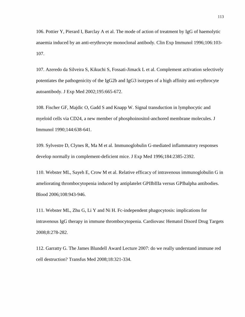
113
106. Pottier Y, Pierard I, Barclay A et al. The mode of action of treatment by IgG of haemolytic
anaemia induced by an anti-erythrocyte monoclonal antibody. Clin Exp Immunol 1996;106:103-
107.
107. Azeredo da Silveira S, Kikuchi S, Fossati-Jimack L et al. Complement activation selectively
potentiates the pathogenicity of the IgG2b and IgG3 isotypes of a high affinity anti-erythrocyte
autoantibody. J Exp Med 2002;195:665-672.
108. Fischer GF, Majdic O, Gadd S and Knapp W. Signal transduction in lymphocytic and
myeloid cells via CD24, a new member of phosphoinositol-anchored membrane molecules. J
Immunol 1990;144:638-641.
109. Sylvestre D, Clynes R, Ma M et al. Immunoglobulin G-mediated inflammatory responses
develop normally in complement-deficient mice. J Exp Med 1996;184:2385-2392.
110. Webster ML, Sayeh E, Crow M et al. Relative efficacy of intravenous immunoglobulin G in
ameliorating thrombocytopenia induced by antiplatelet GPIIbIIIa versus GPIbalpha antibodies.
Blood 2006;108:943-946.
111. Webster ML, Zhu G, Li Y and Ni H. Fc-independent phagocytosis: implications for
intravenous IgG therapy in immune thrombocytopenia. Cardiovasc Hematol Disord Drug Targets
2008;8:278-282.
112. Garratty G. The James Blundell Award Lecture 2007: do we really understand immune red
cell destruction? Transfus Med 2008;18:321-334.
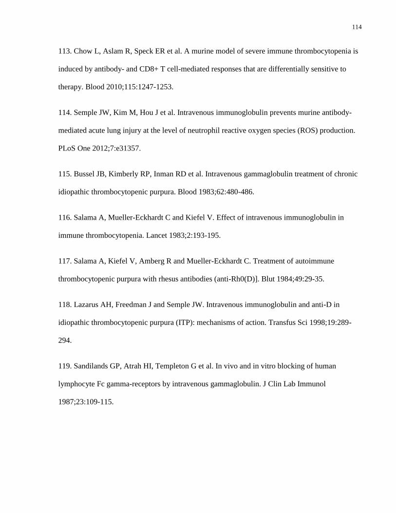
114
113. Chow L, Aslam R, Speck ER et al. A murine model of severe immune thrombocytopenia is
induced by antibody- and CD8+ T cell-mediated responses that are differentially sensitive to
therapy. Blood 2010;115:1247-1253.
114. Semple JW, Kim M, Hou J et al. Intravenous immunoglobulin prevents murine antibody-
mediated acute lung injury at the level of neutrophil reactive oxygen species (ROS) production.
PLoS One 2012;7:e31357.
115. Bussel JB, Kimberly RP, Inman RD et al. Intravenous gammaglobulin treatment of chronic
idiopathic thrombocytopenic purpura. Blood 1983;62:480-486.
116. Salama A, Mueller-Eckhardt C and Kiefel V. Effect of intravenous immunoglobulin in
immune thrombocytopenia. Lancet 1983;2:193-195.
117. Salama A, Kiefel V, Amberg R and Mueller-Eckhardt C. Treatment of autoimmune
thrombocytopenic purpura with rhesus antibodies (anti-Rh0(D)]. Blut 1984;49:29-35.
118. Lazarus AH, Freedman J and Semple JW. Intravenous immunoglobulin and anti-D in
idiopathic thrombocytopenic purpura (ITP): mechanisms of action. Transfus Sci 1998;19:289-
294.
119. Sandilands GP, Atrah HI, Templeton G et al. In vivo and in vitro blocking of human
lymphocyte Fc gamma-receptors by intravenous gammaglobulin. J Clin Lab Immunol
1987;23:109-115.

115
Appendices
116
0 1 2 30
200
400
600
800
1000
1200
Nil
anti-GPA
anti-CD24
anti-platelet
A
Days after antibody injection
Pla
tele
t count x1
06/m
L
C57BL/6 Fc -/- C3-/-0
20
40
60
80
100
anti-GPA: - + - + - +
B
% initia
l pla
tele
t count
Appendix 1. Induction of thrombocytopenia by the anti-GPA antibody. (A) C57BL/6 mice
were injected with the indicated antibody, and bled at the indicated times for platelet
enumeration. RBC from untreated mice (●), or mice treated with GPA antibody (45 µg/mouse;
□), another erythrocyte-specific antibody anti-CD24 (70 µg/mouse; ◊) or the anti-platelet
antibody MWReg30 (2 µg/mouse; ∆) were used to enumerate platelet by flow cytometry. n=3
mice in total from 3 independent experiments. (B) C57BL/6, Fcγ-/-
, and C3-/-
mice were injected
with the GPA antibody, and bled at 24 hours post-injection for platelet enumeration. Platelet
counts were expressed as a percentage of initial (baseline) counts. n=3 mice in total from 3
independent experiments.
117
0 2 4 6 80
2
4
6
8
10
Nil (splenectomized)
Nil
anti-CD24
anti-CD24(splenectomized)
Days after antibody injection
A
**
RB
C c
ou
nt
x10
9/m
L
0 2 4 6 80
2
4
6
8
10
Nil (splenectomized)
Nil
anti-band 3(splenectomized)
anti-band 3
Days after antibody injection
B
*
RB
C c
ou
nt
x10
9/m
L
Appendix 2. Antibody-mediated anemia in surgically splenectomized mice. Normal and
surgically splenectomized mice were injected with 70 µg/mouse anti-CD24 (A) or 50 µg/mouse
anti-band 3 (B) antibody. Blood from untreated mice (normal: ∆; splenectomized: ▲) or mice
treated with antibody (normal: □; splenectomized: ■) were used to enumerate RBC by flow
cytometry. n=2 mice in total from 2 independent experiments. The asterisks (*P<.05, **P<.01)
refer to statistical significance between normal (□) and splenectomized (■) mice treated with
antibody.
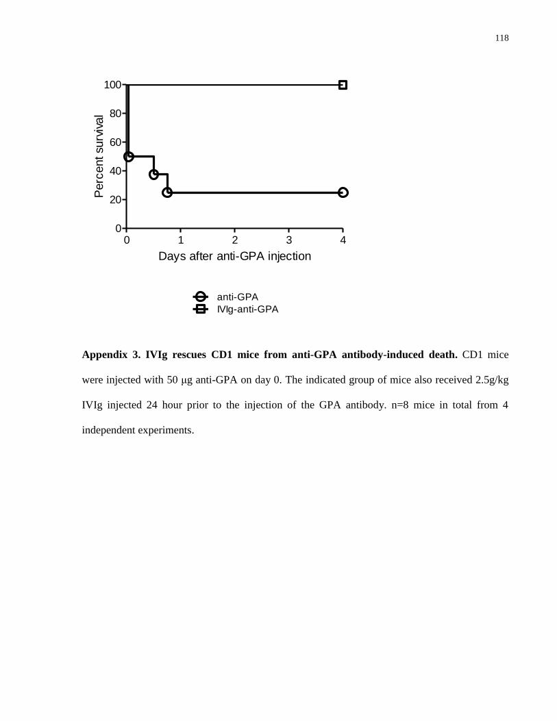
118
0 1 2 3 40
20
40
60
80
100
anti-GPA
IVIg-anti-GPA
Days after anti-GPA injection
Perc
ent surv
ival
Appendix 3. IVIg rescues CD1 mice from anti-GPA antibody-induced death. CD1 mice
were injected with 50 μg anti-GPA on day 0. The indicated group of mice also received 2.5g/kg
IVIg injected 24 hour prior to the injection of the GPA antibody. n=8 mice in total from 4
independent experiments.
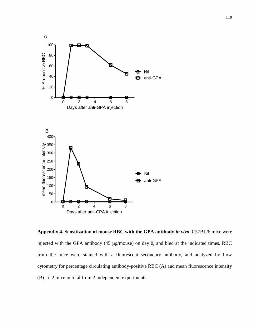
119
0 2 4 6 80
20
40
60
80
100
anti-GPA
Nil
A
Days after anti-GPA injection
% A
b-p
ositiv
e R
BC
0 2 4 6 80
50
100
150
200
250
300
350
400
anti-GPA
Nil
B
Days after anti-GPA injection
mean f
luore
scence inte
nsity
Appendix 4. Sensitization of mouse RBC with the GPA antibody in vivo. C57BL/6 mice were
injected with the GPA antibody (45 µg/mouse) on day 0, and bled at the indicated times. RBC
from the mice were stained with a fluorescent secondary antibody, and analyzed by flow
cytometry for percentage circulating antibody-positive RBC (A) and mean fluorescence intensity
(B). n=2 mice in total from 2 independent experiments.
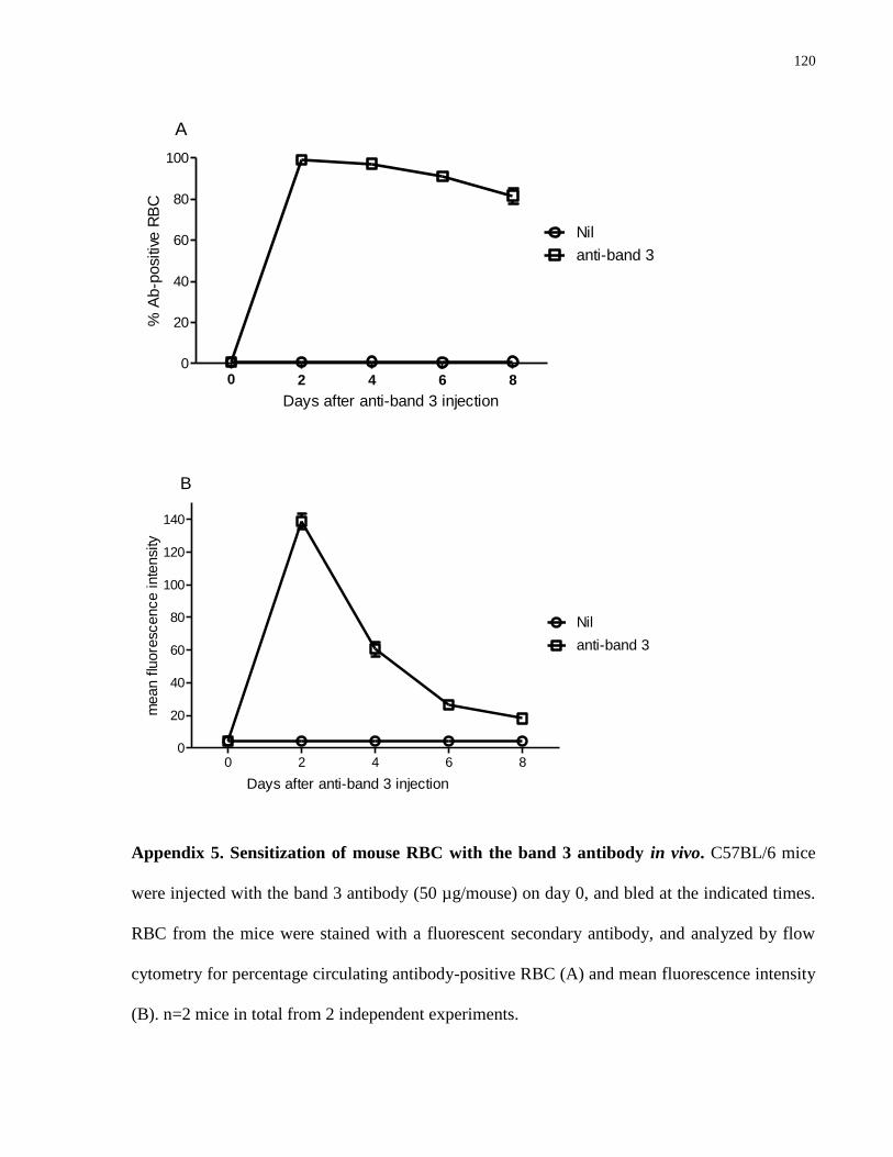
120
00
20
40
60
80
100
anti-band 3
Nil
2 4 6 8
Days after anti-band 3 injection
A
% A
b-p
ositiv
e R
BC
0 2 4 6 80
20
40
60
80
100
120
140
anti-band 3
Nil
Days after anti-band 3 injection
B
mean f
luore
scence inte
nsity
Appendix 5. Sensitization of mouse RBC with the band 3 antibody in vivo. C57BL/6 mice
were injected with the band 3 antibody (50 µg/mouse) on day 0, and bled at the indicated times.
RBC from the mice were stained with a fluorescent secondary antibody, and analyzed by flow
cytometry for percentage circulating antibody-positive RBC (A) and mean fluorescence intensity
(B). n=2 mice in total from 2 independent experiments.
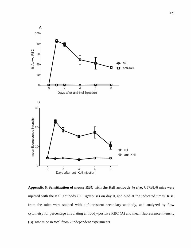
121
0 2 4 6 80
20
40
60
80
100
Nil
anti-Kell
Days after anti-Kell injection
A
% A
b+
ve R
BC
0 2 4 6 80
10
20
30
Nil
anti-Kell
Days after anti-Kell injection
B
mean f
luore
scence inte
nsity
Appendix 6. Sensitization of mouse RBC with the Kell antibody in vivo. C57BL/6 mice were
injected with the Kell antibody (50 µg/mouse) on day 0, and bled at the indicated times. RBC
from the mice were stained with a fluorescent secondary antibody, and analyzed by flow
cytometry for percentage circulating antibody-positive RBC (A) and mean fluorescence intensity
(B). n=2 mice in total from 2 independent experiments.
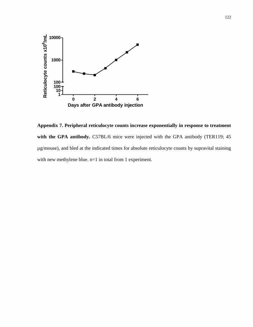
122
0 2 4 61
10100100
1000
10000
Days after GPA antibody injection
Reti
cu
locyte
co
un
ts x
10
6/m
L
Appendix 7. Peripheral reticulocyte counts increase exponentially in response to treatment
with the GPA antibody. C57BL/6 mice were injected with the GPA antibody (TER119; 45
µg/mouse), and bled at the indicated times for absolute reticulocyte counts by supravital staining
with new methylene blue. n=1 in total from 1 experiment.
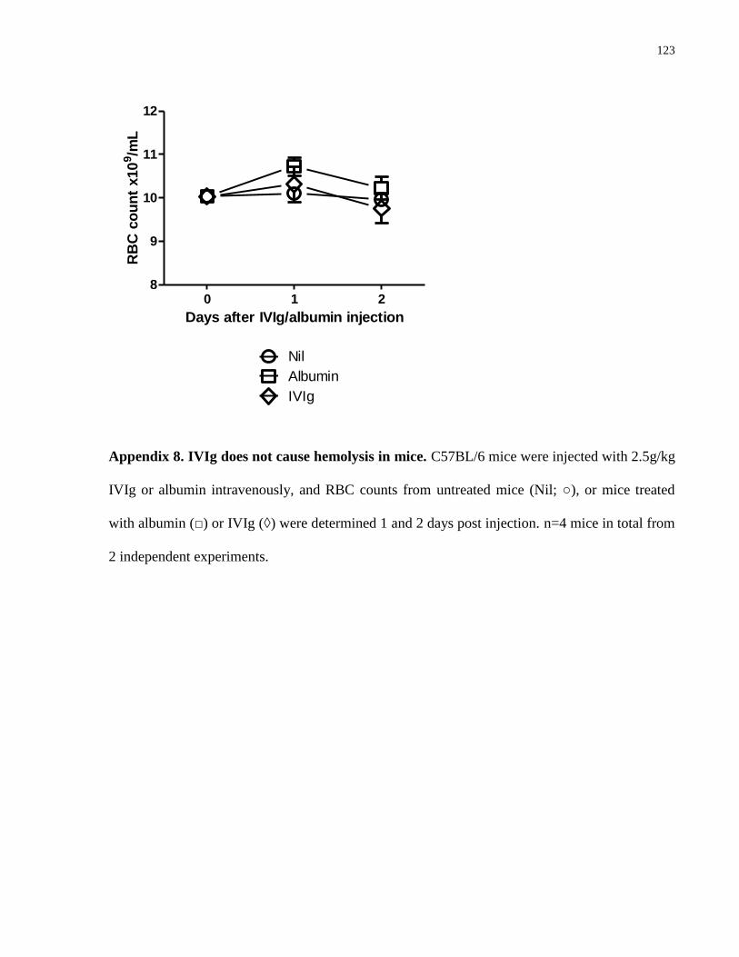
123
0 1 28
9
10
11
12
Nil
Albumin
IVIg
Days after IVIg/albumin injection
RB
C c
ou
nt
x10
9/m
L
Appendix 8. IVIg does not cause hemolysis in mice. C57BL/6 mice were injected with 2.5g/kg
IVIg or albumin intravenously, and RBC counts from untreated mice (Nil; ○), or mice treated
with albumin (□) or IVIg (◊) were determined 1 and 2 days post injection. n=4 mice in total from
2 independent experiments.
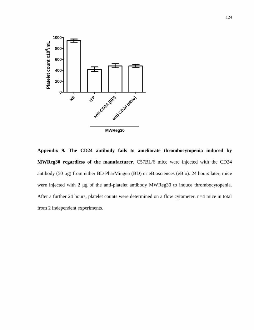
124
Nil
ITP
anti-
CD24
(BD)
anti-
CD24
(eBio
)
0
200
400
600
800
1000
MWReg30
Pla
tele
t co
un
t x10
6/m
L
Appendix 9. The CD24 antibody fails to ameliorate thrombocytopenia induced by
MWReg30 regardless of the manufacturer. C57BL/6 mice were injected with the CD24
antibody (50 µg) from either BD PharMingen (BD) or eBiosciences (eBio). 24 hours later, mice
were injected with 2 μg of the anti-platelet antibody MWReg30 to induce thrombocytopenia.
After a further 24 hours, platelet counts were determined on a flow cytometer. n=4 mice in total
from 2 independent experiments.
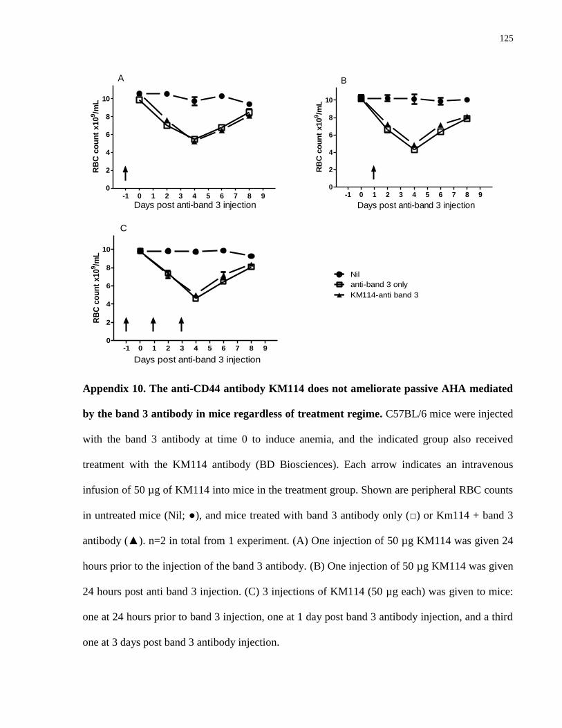
125
-1 0 1 2 3 4 5 6 7 8 90
2
4
6
8
10
Days post anti-band 3 injection
RB
C c
ou
nt
x10
9/m
L
A
-1 0 1 2 3 4 5 6 7 8 90
2
4
6
8
10
Days post anti-band 3 injection
RB
C c
ou
nt
x10
9/m
L
B
-1 0 1 2 3 4 5 6 7 8 90
2
4
6
8
10
Days post anti-band 3 injection
anti-band 3 only
Nil
KM114-anti band 3
C
RB
C c
ou
nt
x10
9/m
L
Appendix 10. The anti-CD44 antibody KM114 does not ameliorate passive AHA mediated
by the band 3 antibody in mice regardless of treatment regime. C57BL/6 mice were injected
with the band 3 antibody at time 0 to induce anemia, and the indicated group also received
treatment with the KM114 antibody (BD Biosciences). Each arrow indicates an intravenous
infusion of 50 µg of KM114 into mice in the treatment group. Shown are peripheral RBC counts
in untreated mice (Nil; ●), and mice treated with band 3 antibody only (□) or Km114 + band 3
antibody (▲). n=2 in total from 1 experiment. (A) One injection of 50 µg KM114 was given 24
hours prior to the injection of the band 3 antibody. (B) One injection of 50 µg KM114 was given
24 hours post anti band 3 injection. (C) 3 injections of KM114 (50 µg each) was given to mice:
one at 24 hours prior to band 3 injection, one at 1 day post band 3 antibody injection, and a third
one at 3 days post band 3 antibody injection.
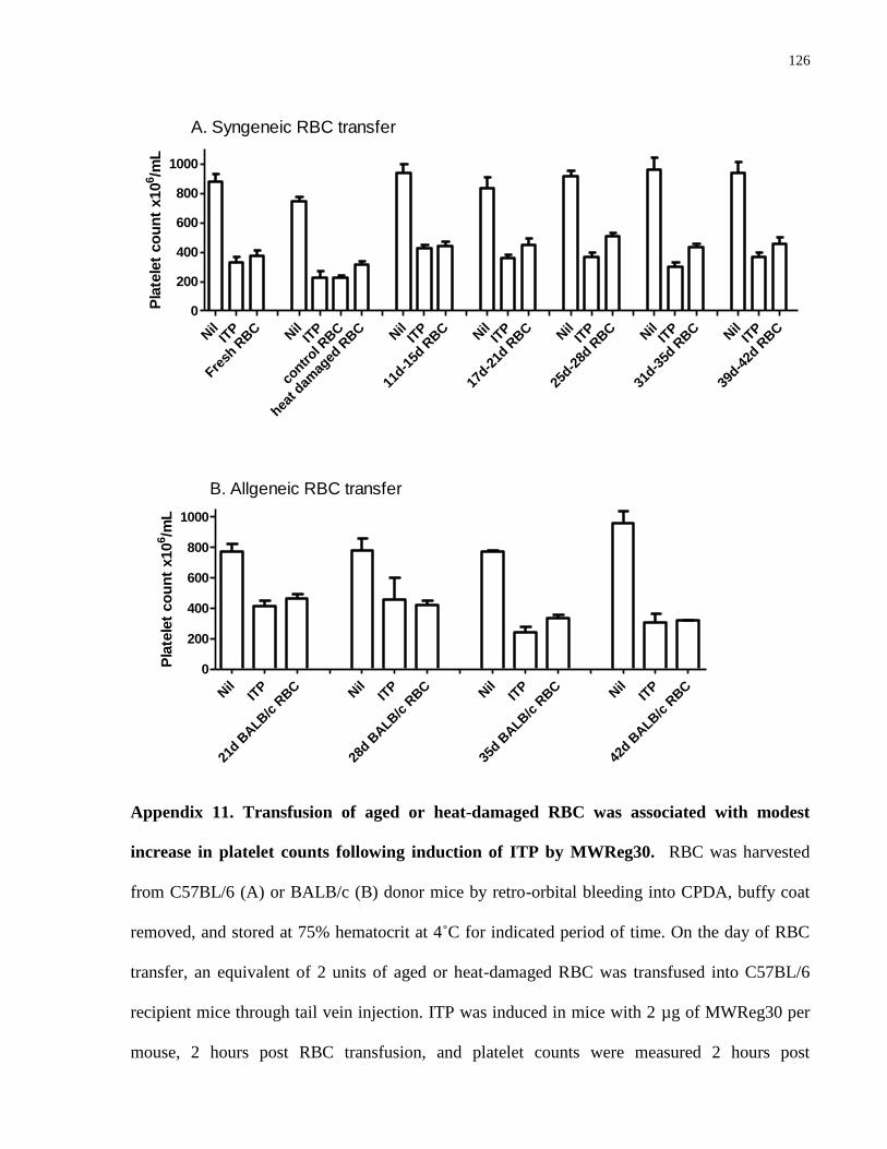
126
Nil
ITP
Fresh
RBC N
ilIT
P
contr
ol RBC
heat d
amag
ed R
BC N
ilIT
P
11d-1
5d R
BC N
ilIT
P
17d-2
1d R
BC N
ilIT
P
25d-2
8d R
BC N
ilIT
P
31d-3
5d R
BC N
ilIT
P
39d-4
2d R
BC
0
200
400
600
800
1000
A. Syngeneic RBC transfer
Pla
tele
t co
un
t x10
6/m
L
Nil
ITP
21d B
ALB
/c R
BC N
ilIT
P
28d B
ALB
/c R
BC N
ilIT
P
35d B
ALB
/c R
BC N
ilIT
P
42d B
ALB
/c R
BC
0
200
400
600
800
1000
B. Allgeneic RBC transfer
Pla
tele
t co
un
t x10
6/m
L
Appendix 11. Transfusion of aged or heat-damaged RBC was associated with modest
increase in platelet counts following induction of ITP by MWReg30. RBC was harvested
from C57BL/6 (A) or BALB/c (B) donor mice by retro-orbital bleeding into CPDA, buffy coat
removed, and stored at 75% hematocrit at 4˚C for indicated period of time. On the day of RBC
transfer, an equivalent of 2 units of aged or heat-damaged RBC was transfused into C57BL/6
recipient mice through tail vein injection. ITP was induced in mice with 2 µg of MWReg30 per
mouse, 2 hours post RBC transfusion, and platelet counts were measured 2 hours post

127
MWReg30 injection. (A) Syngeneic RBC transfer from C57BL/6 donor to C57BL/6 recipient
mice. n=4~8 in total from 2~4 independent experiments. B) Allogeneic RBC transfer from
BALB/c donor to C57BL/6 recipient mice. n=2 in total from 1 experiment.


![HISK 10 ANemia HEMOLITIK.ppt [Read-Only] - ocw.usu.ac.idocw.usu.ac.id/course/download/1110000096-hematology-and-immunology... · Autoimmune hemolytic anemia caused byAutoimmune hemolytic](https://img.pdfslide.us/doc/110x75/5c7c94c409d3f23a2a8b4fbf/hisk-10-anemia-read-only-ocwusuacidocwusuacidcoursedownload1110000096-hematology-and-immunology.jpg)