Embed Size (px)
Citation preview

Developmental Biology 356 (2011) 553–565
Contents lists available at ScienceDirect
Developmental Biology
j ourna l homepage: www.e lsev ie r.com/deve lopmenta lb io logy
Multipotent neural stem cells generate glial cells of the central complex throughtransit amplifying intermediate progenitors in Drosophila brain development
Gudrun Viktorin a,⁎, Nadia Riebli a, Anna Popkova b, Angela Giangrande b, Heinrich Reichert a
a Biozentrum, University of Basel, Basel, Switzerlandb Institut de Génétique et de Biologie Moleculaire et Cellulaire, CNRS/INSERM/ULP, C.U. de Strasbourg, France
⁎ Corresponding author at: Biozentrum, University o4056 BASEL, Switzerland. Fax: +41 61 267 1613.
E-mail address: [email protected] (G. Vikto
0012-1606/$ – see front matter © 2011 Elsevier Inc. Aldoi:10.1016/j.ydbio.2011.06.013
a b s t r a c t
a r t i c l e i n f oArticle history:Received for publication 16 December 2010Revised 27 May 2011Accepted 11 June 2011Available online 25 June 2011
Keywords:DrosophilaLarval brainNeuroblast lineagesIntermediate progenitor cellsNeuroglioblastGliogenesis
The neural stem cells that give rise to the neural lineages of the brain can generate their progeny directly orthrough transit amplifying intermediate neural progenitor cells (INPs). The INP-producing neural stem cells inDrosophila are called type II neuroblasts, and their neural progeny innervate the central complex, a prominentintegrative brain center. Here we use genetic lineage tracing and clonal analysis to show that the INPs of thesetype II neuroblast lineages give rise to glial cells aswell as neurons during postembryonic brain development. Ourdata indicate that two main types of INP lineages are generated, namely mixed neuronal/glial lineages andneuronal lineages. Genetic loss-of-function and gain-of-function experiments show that the gcm gene isnecessary and sufficient for gliogenesis in these lineages. The INP-derivedglial cells, like the INP-derivedneuronalcells, make major contributions to the central complex. In postembryonic development, these INP-derived glialcells surround the entire developing central complex neuropile, and once themajor compartments of the centralcomplex are formed, they also delimit each of these compartments. During this process, the number of these glialcells in the central complex is increased markedly through local proliferation based on glial cell mitosis. Takentogether, these findings uncover a novel and complex form of neurogliogenesis in Drosophila involving transitamplifying intermediate progenitors.Moreover, they indicate that type II neuroblasts are remarkablymultipotentneural stem cells that can generate both the neuronal and the glial progeny thatmakemajor contributions to oneand the same complex brain structure.
f Basel, Klingelbergstr. 70, CH-
rin).
l rights reserved.
© 2011 Elsevier Inc. All rights reserved.
Introduction
Neural stem cells are the primary progenitor cells at differentdevelopmental stages that initiate the neural lineages comprising thedifferentiated neurons and glia of the brain (Kriegstein and Alvarez-Buylla, 2009). Analysis of stem cell-dependent neurogenesis in severalmodel systems indicates that neural cells are not always produceddirectly from the primary progenitors, but can also be generated bysecondary progenitors of more restricted potential (Götz and Huttner,2005; Kriegstein et al., 2006). In mammals, neural stem cells cangenerate neural cells either directly through asymmetric division orindirectly through secondary progenitors which either divide only onceto produce two neural progeny or divide more than once to amplify thenumber of neural progeny further (Haubensak et al., 2004;Miyata et al.,2004; Noctor et al., 2004, 2007).
Remarkably similar findings have been obtained in studies ofneurogenesis in the developing CNS of Drosophila. There, neural stemcell-like primary progenitors called neuroblasts can also generateneurons directly through asymmetric division or, more typically,
indirectly through ganglion mother cells (GMC) which divide onlyonce to generate two postmitotic progeny (Doe, 2008; Knoblich, 2008;Skeath and Thor, 2003; Technau et al., 2006). Moreover, recently a thirdtype of neurogenesis has been discovered in theDrosophila central brainwhere identified neuroblasts generate intermediate neural progenitorcells (INPs)whichundergo several rounds of asymmetric divisions, eachof which results in INP self-renewal and generation of a GMC thatproduces two neural progeny (Bello et al., 2008; Boone and Doe, 2008;Bowmanet al., 2008). These INPshave alsobeen referred toas IPs, transitamplifying GMCs, or secondary neuroblasts (Weng and Lee, 2010.)Through the resulting amplification of proliferation these INP producing“type II” neuroblasts generate large neural lineages that contribute tomajor neuropile substructures of the brain such as the central complex(Bayraktar et al., 2010; Izergina et al., 2009; Pereanu et al., 2011).
In mammalian brain development, neural stem cells can give rise toboth neurons and glial cells, however the neurogenic phase and thegliogenic phase of these primary progenitors are generally separate(Miller and Gauthier, 2007; Miyata et al., 2010). Neural stem cells thatcan generate both neurons and glia are also found inDrosophila (VanDeBor and Giangrande, 2002). These so-called neuroglioblasts have beenidentified on a single-cell basis in the embryonic ventral nerve cord, andtogetherwith glioblasts, which only generate glial cells, they give rise toseveral subtypes of glial cells in the ventral CNS (Beckervordersandforth
554 G. Viktorin et al. / Developmental Biology 356 (2011) 553–565
et al., 2008; Bossing et al., 1996; Broadus et al., 1995; Schmidt et al.,1997). In the ventral nerve cord, gliogenesis is controlledby the glial cellsmissing (gcm) gene that acts as a key switch-like regulator in thedevelopment of all embryonic glial cells except those generated frommesectodermal precursors (Hosoya et al., 1995; Jones et al., 1995;Vincent et al., 1996).
Much less is knownabout the developmental origins of the glial cellsin the Drosophila larval brain. The glial cells of the early larval brain arethought to be generated by a fewneuroglioblasts during embryogenesis(Hartenstein et al., 1998). However, most of the glial cells in the adultcentral brain are generated postembryonically, and during larvaldevelopment a marked increase in glial cell number occurs (Pereanuet al., 2005).While someof these glial cells apparently arisebymitosis ofother glial cells, the bulk of the postembryonically added glial cells isthought to be generated through the proliferation of neuroglioblasts,which, however, have not yet been identified (Awasaki et al., 2008;Pereanu et al., 2005). This lack of identification of the postulatedpostembryonic neuroglioblasts has been a major obstacle for under-standing the mechanisms by which glial cells are generated duringpostembryonic development of the central brain.
Herewe show that identified type II neuroblasts have neuroglioblastfunction during postembryonic development of the central brain. Weuse genetic lineage tracing and clonal analysis to demonstrate that theINPs of type II neuroblast lineages can give rise to glial cells as well asneurons. Our data indicate that two main types of INP-derived lineagesare generated postembryonically, namelymixed neuronal/glial lineagesand neuronal lineages. Moreover, they show that INP-derived glial cells,like INP-derived neuronal cells from the same type II neuroblastlineages, make major contributions to the developing neuropile of thecentral complex. They surround the entire developing central complexneuropile, and once its major compartments are formed, they alsodelimit each of these. During this process, the population of INP-derivedglial cells associated with central complex undergoes clonal expansionthrough local proliferation. Taken together, these findings uncovernovel mechanisms for neurogliogenesis in Drosophila involving thetransit amplifying intermediate progenitors of type II lineages. Thus,type II neuroblasts are remarkably multipotent neural stem cells thatcan generate both the neuronal and the glial progeny of one and thesame complex brain structure.
Materials and methods
Fly stocks, MARCM, and G-Trace analysis
Flies were maintained on standard cornmeal medium at 25 °C unlessnoted otherwise. To visualize the potentially glial offspring of neuroblasts,we used insc-Gal4MZ1407, UAS-mCD8GFPLL5 homozygous flies, or matedthem to gcm-lacZrA87/CyO, act-gfpJMR1. For erm-Gal4 (R09D11; Pfeifferet al., 2008) G-Trace (flp-out and real-time; Evans et al., 2009) expressionpatterns we mated UAS-flp, ubiNstopNnGFP, UAS-RFP/CyO, act-gfpJMR1;erm-Gal4 to gcm-lacZrA87/CyO, act-gfpJMR1; erm-Gal4. For membranelabeling of erm-Gal4 progeny, we mated UAS-flp; UAS-mCD8GFPLL5; erm-GAL4 to UAS-mCD8GFPLL5; actN*,CD2NGal4 (Pignoni and Zipursky, 1997).To generate wild type MARCM (Lee and Luo, 1999) clones, we mated yw hs-flp1; tubP-Gal4, UAS-mCD8GFPLL5/CyO, act-gfpJMR1; FRT82B, tub-Gal80LL3 (Bello et al., 2003) to gcm-lacZrA87/CyO, act-gfpJMR1; FRT82Bmales. To generate clonesmisexpressing gcm, wemated elav-Gal4C155, hs-flp1; UAS-mCD8GFPLL5/CyO, act-gfpJMR1; FRT82B tub-Gal80LL3 to UAS-gcmm24a/CyO, act-gfpJMR1; FRT82B (UAS-gcmm24a from Bernardoni et al.,1998). To induce gcm loss-of-function clones, we mated y w hs-flp1;FRT40A, tubP-Gal80LL10/CyO, act-gfpJMR1; tubP-Gal4LL7, UAS-mCD8GFPLL6
(Bello et al., 2006) to FRT40A, gcmN7-4/CyO, act-gfpJMR1. Eggswere collectedfor 2–4 h, grown tofirst larval instar (22–30 hafter egg laying, AEL), platesimmersed in a 37 °C waterbath for 5 min (sparse wild type clones) or upto 30 min (other clones), and grown to the desired stage at 25 °C. Whenrecovering clones from wandering L3 larvae, some bottles were raised at
18 °C during third instar to delay development until dissection. Whenrecovering clones from earlier stages, larvae were grown at 25 °Cthroughout, and kept at a maximum density of 170 larvae per bottle toavoid developmental delay due to food competition and to ensure exactstaging.
Immunohistochemistry and in situ hybridization
Brains were dissected in ice-cold PBS and fixed in 2% Paraformal-dehyde for 30–60 min at room temperature, washed several times inPBS/0.5% Triton X-100, and preincubated in PBS containing 0.5% TritonX-100 and 10% normal goat serum. Antibodies were incubatedovernight at 4 °C. We used chicken anti-GFP 1:500 (ab13970, Abcam,Cambridge, UK), rabbit anti-beta-Galactosidase 1:500 (55976, MPBiomedicals, Solon, Ohio, USA), mouse anti-Neurotactin 1:20 (BP106,DSHB, Iowa City, Iowa, USA), rat anti-Elav 1:30 (7E8A10, DSHB), mouseanti-Repo 1:30 (8D12, DSHB), rabbit anti-Repo 1:400 (kindly providedbyVeronica Rodrigues), rabbit anti-phospho histoneH3 (Ser10, 06-570,Millipore, Temecula, CA, USA), rat anti-Deadpan and rabbit anti-Asense(both kind gifts from Cheng-Yu Lee), and Alexa-conjugated secondaryantibodies 1:300 (A11039, A21247, A11036, A11031, A21244, Molec-ular Probes, Eugene, OR, USA). In situ hybridization was performed asdescribed previously (Soustelle et al., 2007).
Identification of dorsomedial type II lineages
DM1 was uniquely identified in Nrt/BP106 staining as thedorsoanterior-most large lineage in late third instar brains. DM5was identified by a unique gcm-lacZ and Repo-positive cell clusterwithin the lineage (Figs. 1D, 5E). Other DM lineages were identified bytheir position relative to DM1 and DM5, their very large cell number(Bello et al., 2008), complex axon branching pattern (Izergina et al.,2009), and location at the posterior/medial brain surface. In youngerlarvae, DM1–3 lineages were recognized by their similar shapes andpositions compared to those in wandering larvae. In addition, theywere confirmed to be Type II lineages by weak or absent Asensestaining in the neuroblast, the presence of numerous Asense- orDeadpan-positive cells apart from the neuroblast, or equally numer-ous Prospero- and Elav-negative cells throughout the lineage.
Microscopy and image processing
Fluorescent images were recorded on a Leica TCS SP5 confocalmicroscope, and processed using Fiji (Schindelin, 2008) or ImageJ(Rasband, 1997–2008). All adjustments were linear and were per-formed on whole images. Cells were counted using the CellCounterplugin for Fiji/ImageJ (Kurt De Vos).
Results
Postembryonic development of neurons and glial cells in the central brain
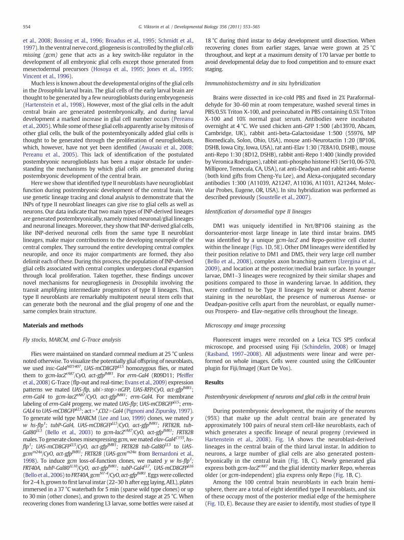
During postembryonic development, the majority of the neurons(95%) that make up the adult central brain are generated byapproximately 100 pairs of neural stem cell-like neuroblasts, each ofwhich generates a specific lineage of neural progeny (reviewed inHartenstein et al., 2008). Fig. 1A shows the neuroblast-derivedlineages in the central brain of the third larval instar. In addition toneurons, a large number of glial cells are also generated postem-bryonically in the central brain (Fig. 1B, C). Newly generated gliaexpress both gcm-lacZrA87 and the glial identity marker Repo, whereasolder (or gcm-independent) glia express only Repo (Fig. 1B, C).
Among the 100 central brain neuroblasts in each brain hemi-sphere, there are a total of eight identified type II neuroblasts, and sixof these occupy most of the posterior medial edge of the hemisphere(Fig. 1D, E). Because they are easier to identify, most studies of type II
gcm-lacZ Repo gcm-lacZ
CB
OL
insc>mCD8GFP
E2
0 m -23 m
A
D G -19 m
3
45
1
32
45
6
2
3
20 m
*
gcm-lacZ Repoinsc>mCD8GFP
F 0 m
1
Repoinsc>mCD8GFP
*
CB
*
Fig. 1. Central brain neuroblast lineages give rise to glial and neuronal progeny. (A–E) Wandering third instar larval brain (genotype insc-Gal4, UAS-mCD8GFP/gcm-lacZrA87, 80 μmmaximum intensity projection) labeled with Repo (blue) and gcm-lacZ (red). (A–C) Within the region of central brain neuroblast lineages (A; inscNmCD8GFP), many newly formedglia are present (B,C; magenta overlap) expressing both Repo and gcm-lacZ. They are positioned in the commissure (arrowheads) as well as medially (short arrows) and laterally(long arrows) in the central brain. Dotted line indicates border between central brain (CB) and optic lobe (OL). (D, E) DM1–DM6 lineages in the same brain; numbers indicateidentified DM lineages. (F, G) insc-Gal4, UAS-mCD8GFP homozygous larvae have Repo-positive cells (magenta) in GFP-labeled DM lineages (arrows). Numbers indicate tracts ofidentified DM lineages. Other glia (presumably surface, cortex, or astrocyte-like glia) are not GFP-labeled (asterisks). All figures present anterior to the top. Scale bars, 20 μm.
555G. Viktorin et al. / Developmental Biology 356 (2011) 553–565
neuroblasts have focused on the six posterior medially locatedlineages. These six lineages have been termed DM1-DM6 by Bello etal. (2008), and tentatively correspond to the DPMm1 (DM1),DPMpm1 (DM2), DPMpm2 (DM3), CM4 (DM4), CM3 (DM5), andCM1 (DM6) lineages (Pereanu et al., 2011; Pereanu and Hartenstein,2006). A previous analysis of the postmitotic cells generated in thesesix identified type II neuroblast lineages during postembryonicdevelopment indicates that the majority of these are neurons, butglial cells are also present (Izergina et al., 2009). This is supported bythe observation that some neuroblast lineage-specific Gal4 lines labelglial cells in DM lineages (Fig. 1F, G).
These findings suggest that type II neuroblasts may in fact functionas neuroglioblasts that produce glia in a novelmode involving INPs. Toinvestigate this, we first required a method for specific labeling of allINP-derived cells in type II neuroblast lineages.
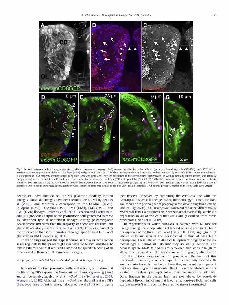
INP progeny are labeled by erm-Gal4-dependent lineage tracing
In contrast to other progenitor cells in the brain, all mature andproliferating INPs express the Drosophila Fezf homolog earmuff (erm)and can be reliably labeled by an erm-Gal4 line (Pfeiffer et al., 2008;Weng et al., 2010). Although the erm-Gal4 line labels all mature INPsof the type II neuroblast lineages, it does not reveal all of their progeny
(see below). However, by combining the erm-Gal4 line with theGal4/flp-out based cell lineage tracing methodology G-Trace, the INPsand their entire (clonal) set of progeny in the developing brain can belabeled (Fig. 2A, B). In G-Trace, two fluorescent reporters differentiallyreveal real-timeGal4 expression in precursor cells versusflp-out basedexpression in all of the cells that are clonally derived from theseprecursors (Evans et al., 2009).
In experiments in which erm-Gal4 is coupled with G-Trace forlineage tracing, three populations of labeled cells are seen in the brainhemispheres of the third instar larva (Fig. 2C, D). First, large groups oflabeled cells are seen at the dorsomedial midline of each brainhemisphere. These labeled midline cells represent progeny of the sixmedial type II neuroblasts. Because they are easily identified, andbecause sparse MARCM clones are recovered frequently enough toallow conclusions about the association with migratory glia derivedfrom them, these dorsomedial cell groups are the focus of thisinvestigation. Second, smaller groups of more laterally located cellsaremanifested in each brain hemisphere; they represent the progeny ofthe two lateral type II neuroblasts. Third, numerous labeled cells arelocated in the developing optic lobes; their precursors are unknown.Other lineages in the central brain are not labeled by erm-Gal4-dependent flp-out, indicating that few, if any, non-type II-derived cellsexpress erm-Gal4 in the central brain at the stages investigated.
**
*
VNC
OLOL
A B
C D
E F
Fig. 2. erm-Gal4-dependent lineage tracing labels INP-derived progeny of type IIneuroblast lineages. (A) Schematic of the type II neuroblast lineages in the larval centralbrain. (B) Expression of erm-Gal4 in G-Trace flies (UAS-flp, ubiN*NnGFP, UAS-RFP) leads toerm-Gal4 independent GFP expression in erm-Gal4 expressing cells and their progeny.Real-time expression of erm-Gal4 is reported by RFP expression. (C) erm-Gal4 drivenlineage tracing labels the six dorsomedial type II lineages, the two lateral type II lineages(asterisk) and cells in the optic lobe (OL; delimited by dashed lines). (D) Close-up of theDM lineages. (E, F) Close-up of the DM1 region. Real-time expression (red) is located nearthe cortical surface, flip-out based clonal expression (green) is located in cells that extendfurther from the cortical surface toward the commissure. Scale bars, 25 μm.
556 G. Viktorin et al. / Developmental Biology 356 (2011) 553–565
Previous studies have shown that INP cell bodies are clustered inthe vicinity of their parent type II neuroblasts near the cortical surfaceof the brain hemisphere, while the cell bodies of their neural progenyextend in a clustered array away from the INPs (and their neuroblast)toward the developing neuropile (Bello et al., 2008; Weng et al.,2010). Given this spatial array of the erm-positive INPs and their erm-negative neural progeny, spatially distinct expression of the two labelsin erm-Gal4 G-Trace experiments is expected. This is the case (Fig. 2E,F). Real-time erm-Gal4 expression (red/orange label) is concentratedin a small group of cell bodies clustered near the cortical surface (allINPs), while flip-out based clonal expression (green label) reveals amuch larger group of labeled cell bodies (most INPs and all of theirclonal progeny) that extend toward the developing neuropile.
These findings confirm previous observations on the spatiallocation of INPs versus their differentiated neural progeny in type IIlineages and also demonstrate that (real-time) erm-Gal4 only labels asubset of the total number of cells that comprise these lineages.
INP lineages can give rise to glial as well as neuronal progeny
During postembryonic development, the six type II neuroblastsgenerate large lineages, and most of the differentiated cells in these
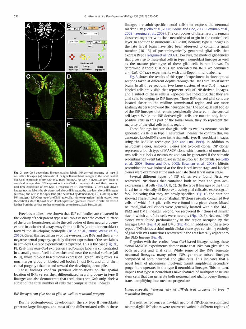
lineages are adult-specific neural cells that express the neuronalmarker Elav (Bello et al., 2008; Boone and Doe, 2008; Bowman et al.,2008; Izergina et al., 2009). The cell bodies of these neurons remainclustered together with their neuroblast of origin in the cortical celllayer. In addition to numerous (400–500) neurons, type II lineages inthe late larval brain have also been observed to contain a smallnumber (10–15) of postembryonically generated glial cells thatexpress Repo (Izergina et al., 2009). However, the mode of gliogenesisthat gives rise to these glial cells in type II neuroblast lineages as wellas the mature phenotype of these glial cells is not known. Todetermine if these glial cells are generated via INPs, we combinederm-Gal4 G-Trace experiments with anti-Repo immunolabeling.
Fig. 3 shows the results of this type of experiment in three opticalsections taken at different depths through the late third larval instarbrain. In all three sections, two large clusters of erm-Gal4 lineage-labeled cells are visible that represent cells of INP-derived lineages,and a subset of these cells is Repo-positive indicating that they areglial cells belonging to INP lineages. These INP-derived glial cells arelocated closer to the midline commissural region and are morespatially dispersed toward the neuropile than the non-glial cell bodiesof the INP lineages that remain peripherally clustered in the corticalcell layer. While the INP-derived glial cells are not the only Repo-positive cells in this part of the larval brain, they do represent themajority of the glial cells in this region.
These findings indicate that glial cells as well as neurons can begenerated via INPs in type II neuroblast lineages. To confirm this, wegenerated labeled INP clones in the sixmedial type II neuroblast lineagesusing the MARCM technique (Lee and Luo, 1999). In addition toneuroblast clones, single-cell clones and two-cell clones, INP clonesrepresent a fourth type of MARCM clone which consists of more thantwo cells but lacks a neuroblast and can be generated if the somaticrecombination event takes place in the neuroblast (for details, see Belloet al., 2008; Boone and Doe, 2008; Bowman et al., 2008). Mitoticrecombination was induced at the first larval instar stage and labeledclones were examined at the mid- and late third larval instar stage.
Several different types of INP clones were found. First, werecovered INP clones that contained both neurons and gcm-lacZ-expressing glial cells (Fig. 4A, B, C). (In the type II lineages of the thirdlarval instar, virtually all Repo-expressing glial cells also express gcm-lacZ, indicating that they are newly specified; Fig. 1B and data notshown.) These mixed neuronal/glial INP clones usually contained 8–9cells, of which 1–3 glial cells were found in a given clone. Mixedneuronal/glial cell clones were generally located within the DM1,DM2 and DM3 lineages. Second, we recovered INP clones of similarsize in which all of the cells were neurons (Fig. 4D, F). Neuronal INPclones were found predominantly in the region occupied by thelineages DM4 (Fig. 4D) and DM6 (Fig. 4F). In addition to these twotypes of INP clones, a third multicellular clone type consisting entirelyof glial cells was sometimes recovered in the area laterally adjacent tothe DM5 lineage (Fig. 4E).
Together with the results of erm-Gal4-based lineage tracing, theseclonal MARCM experiments demonstrate that INPs can give rise toboth neurons and glial cells. While some of the INPs generateneuronal lineages, many other INPs generate mixed lineagescomposed of both neuronal and glial cells. This indicates that anovel form of gliogenesis involving transit amplifying secondaryprogenitors operates in the type II neuroblast lineages. This, in turn,implies that type II neuroblasts have features of multipotent neuralstem cells that can generate both neuronal and glial progeny thoughtransit amplifying intermediate progenitors.
Lineage-specific heterogeneity of INP-derived progeny in type IIneuroblast lineages
The relative frequencywithwhichneuronal INP clones versusmixedneuronal/glial INP clones were recovered varied in different regions of
Repo
A''erm>{ubi>>nGFP}
A'
B
B'
B''
C
C'
C''
A -23 m-10 m0 m
Fig. 3. erm-Gal4 expressing INPs give rise to glial cells. (A–C) Three optical sections at different depths in the third larval instar brain of erm-Gal4 flp-out (UAS-flp, ubiN*NnGFP) drivennGFP expression show the DM1 regions of both hemispheres. Most glia (Repo, magenta) are flp-out progeny of erm-Gal4 expressing INPs (green; overlap white). erm-Gal4-derivedglia are indicated with white dotted outlines, glia not derived from erm-Gal4 with orange dotted outlines. Scale bar, 25 μm.
tub>gfp clone
gcm-lacZ
DM1 DM2 DM3 DM4
6MDlaretal5MD
A' B' C' D'
A B C D
E E' F F'
Fig. 4. Progeny of single INPs are composed of neurons and/or glia in a lineage-specific manner. tub-Gal4 UAS-mCD8GFP/gcm-lacZrA87 — labeled MARCM clones of INPs withinidentified DM lineages. (A–C) INP clones derived from DM1–3 contain 8–9 cells in total, including one or two glial cells. (D, F) INP clones from DM4 and DM6 contain a similarnumber of cells but no glia. (E) DM5-derived subclones are positioned in the center of the hemisphere and are composed of only glial cells. Scale bar, 20 μm.
557G. Viktorin et al. / Developmental Biology 356 (2011) 553–565
A
A'
B
B'
C
C'
D
D'
E
E'
F
50 m
F'
DM1 DM2 DM3 DM4 DM5 DM6
0 m-23 m
tub
>mC
D8g
fp M
AR
CM
gcm
-lac
Z in
clo
ne
/ ou
tsid
e o
f cl
on
e
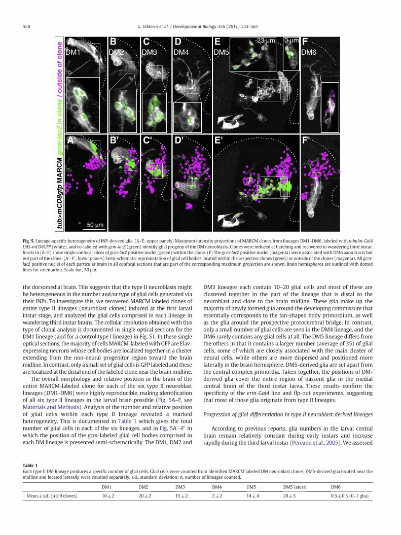
Fig. 5. Lineage-specific heterogeneity of INP-derived glia. (A–E; upper panels) Maximum intensity projections of MARCM clones from lineages DM1–DM6, labeled with tubulin-Gal4UAS-mCD8GFP (white), and co-labeled with gcm-lacZ (green) identify glial progeny of the DM neuroblasts. Clones were induced at hatching and recovered in wandering third instar.Insets in (A–E) show single confocal slices of gcm-lacZ positive nuclei (green) within the clone. (F) The gcm-lacZ positive nuclei (magenta) were associated with DM6 axon tracts butnot part of the clone. (A′–F′; lower panels) Semi-schematic representation of glial cell bodies locatedwithin the respective clones (green) or outside of the clones (magenta). All gcm-lacZ positive nuclei of each particular brain in all confocal sections that are part of the corresponding maximum projection are shown. Brain hemispheres are outlined with dottedlines for orientation. Scale bar, 50 μm.
558 G. Viktorin et al. / Developmental Biology 356 (2011) 553–565
the dorsomedial brain. This suggests that the type II neuroblasts mightbe heterogeneous in the number and/or type of glial cells generated viatheir INPs. To investigate this, we recovered MARCM labeled clones ofentire type II lineages (neuroblast clones) induced at the first larvalinstar stage, and analyzed the glial cells comprised in each lineage inwandering third instar brains. The cellular resolution obtainedwith thistype of clonal analysis is documented in single optical sections for theDM1 lineage (and for a control type I lineage) in Fig. S1. In these singleoptical sections, themajority of cellsMARCM-labeledwithGFP are Elav-expressing neurons whose cell bodies are localized together in a clusterextending from the non-neural progenitor region toward the brainmidline. In contrast, only a small set of glial cells is GFP labeled and theseare localized at the distal end of the labeled clonenear the brainmidline.
The overall morphology and relative position in the brain of theentire MARCM-labeled clone for each of the six type II neuroblastlineages (DM1-DM6) were highly reproducible, making identificationof all six type II lineages in the larval brain possible (Fig. 5A–F, seeMaterials and Methods). Analysis of the number and relative positionof glial cells within each type II lineage revealed a markedheterogeneity. This is documented in Table 1 which gives the totalnumber of glial cells in each of the six lineages, and in Fig. 5A′–F′ inwhich the position of the gcm-labeled glial cell bodies comprised ineach DM lineage is presented semi-schematically. The DM1, DM2 and
Table 1Each type II DM lineage produces a specific number of glial cells. Glial cells were counted fromidline and located laterally were counted separately. s.d., standard deviation; n, number
DM1 DM2 DM3
Mean±s.d. (n≥9 clones) 10±2 20±2 15±2
DM3 lineages each contain 10–20 glial cells and most of these areclustered together in the part of the lineage that is distal to theneuroblast and close to the brain midline. These glia make up themajority of newly formed glia around the developing commissure thatessentially corresponds to the fan-shaped body primordium, as wellas the glia around the prospective protocerebral bridge. In contrast,only a small number of glial cells are seen in the DM4 lineage, and theDM6 rarely contains any glial cells at all. The DM5 lineage differs fromthe others in that it contains a larger number (average of 35) of glialcells, some of which are closely associated with the main cluster ofneural cells, while others are more dispersed and positioned morelaterally in the brain hemisphere. DM5-derived glia are set apart fromthe central complex primordia. Taken together, the positions of DM-derived glia cover the entire region of nascent glia in the medialcentral brain of the third instar larva. These results confirm thespecificity of the erm-Gal4 line and flp-out experiments, suggestingthat most of those glia originate from type II lineages.
Progression of glial differentiation in type II neuroblast-derived lineages
According to previous reports, glia numbers in the larval centralbrain remain relatively constant during early instars and increaserapidly during the third larval instar (Pereanu et al., 2005).We assessed
m identified MARCM labeled DM neuroblast clones. DM5-derived glia located near theof lineages counted.
DM4 DM5 DM5 lateral DM6
2±2 14±4 20±5 0.3±0.5 (0–1 glia)
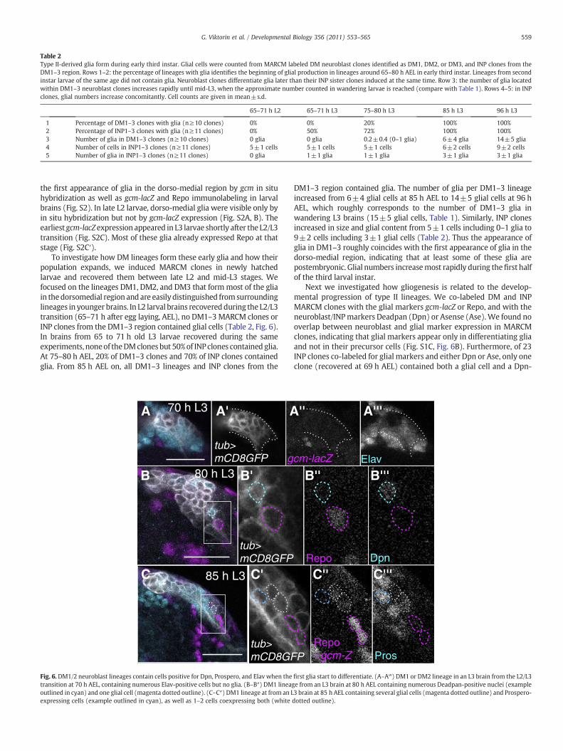
Table 2Type II-derived glia form during early third instar. Glial cells were counted from MARCM labeled DM neuroblast clones identified as DM1, DM2, or DM3, and INP clones from theDM1–3 region. Rows 1–2: the percentage of lineages with glia identifies the beginning of glial production in lineages around 65–80 h AEL in early third instar. Lineages from secondinstar larvae of the same age did not contain glia. Neuroblast clones differentiate glia later than their INP sister clones induced at the same time. Row 3: the number of glia locatedwithin DM1–3 neuroblast clones increases rapidly until mid-L3, when the approximate number counted in wandering larvae is reached (compare with Table 1). Rows 4–5: in INPclones, glial numbers increase concomitantly. Cell counts are given in mean±s.d.
65–71 h L2 65–71 h L3 75–80 h L3 85 h L3 96 h L3
1 Percentage of DM1–3 clones with glia (n≥10 clones) 0% 0% 20% 100% 100%2 Percentage of INP1–3 clones with glia (n≥11 clones) 0% 50% 72% 100% 100%3 Number of glia in DM1–3 clones (n≥10 clones) 0 glia 0 glia 0.2±0.4 (0–1 glia) 6±4 glia 14±5 glia4 Number of cells in INP1–3 clones (n≥11 clones) 5±1 cells 5±1 cells 5±1 cells 6±2 cells 9±2 cells5 Number of glia in INP1–3 clones (n≥11 clones) 0 glia 1±1 glia 1±1 glia 3±1 glia 3±1 glia
559G. Viktorin et al. / Developmental Biology 356 (2011) 553–565
the first appearance of glia in the dorso-medial region by gcm in situhybridization as well as gcm-lacZ and Repo immunolabeling in larvalbrains (Fig. S2). In late L2 larvae, dorso-medial glia were visible only byin situ hybridization but not by gcm-lacZ expression (Fig. S2A, B). Theearliest gcm-lacZexpression appeared in L3 larvae shortly after the L2/L3transition (Fig. S2C). Most of these glia already expressed Repo at thatstage (Fig. S2C′).
To investigate how DM lineages form these early glia and how theirpopulation expands, we induced MARCM clones in newly hatchedlarvae and recovered them between late L2 and mid-L3 stages. Wefocused on the lineages DM1, DM2, and DM3 that formmost of the gliain thedorsomedial region andare easily distinguished fromsurroundinglineages in younger brains. In L2 larval brains recoveredduring the L2/L3transition (65–71 h after egg laying, AEL), no DM1–3 MARCM clones orINP clones from the DM1–3 region contained glial cells (Table 2, Fig. 6).In brains from 65 to 71 h old L3 larvae recovered during the sameexperiments, noneof theDMclones but 50%of INPclones contained glia.At 75–80 h AEL, 20% of DM1–3 clones and 70% of INP clones containedglia. From 85 h AEL on, all DM1–3 lineages and INP clones from the
gtub>mCD8GFP
tub>mCD8GF
85 h L3
80 h L3
70 h L3
tub>mCD8GFP
A
B
A' A
B'
C'C
Fig. 6. DM1/2 neuroblast lineages contain cells positive for Dpn, Prospero, and Elav when thetransition at 70 h AEL, containing numerous Elav-positive cells but no glia. (B–B″) DM1 lineagoutlined in cyan) and one glial cell (magenta dotted outline). (C–C″) DM1 lineage at from an Lexpressing cells (example outlined in cyan), as well as 1–2 cells coexpressing both (white
DM1–3 region contained glia. The number of glia per DM1–3 lineageincreased from 6±4 glial cells at 85 h AEL to 14±5 glial cells at 96 hAEL, which roughly corresponds to the number of DM1–3 glia inwandering L3 brains (15±5 glial cells, Table 1). Similarly, INP clonesincreased in size and glial content from 5±1 cells including 0–1 glia to9±2 cells including 3±1 glial cells (Table 2). Thus the appearance ofglia in DM1–3 roughly coincides with the first appearance of glia in thedorso-medial region, indicating that at least some of these glia arepostembryonic. Glial numbers increasemost rapidly during thefirst halfof the third larval instar.
Next we investigated how gliogenesis is related to the develop-mental progression of type II lineages. We co-labeled DM and INPMARCM clones with the glial markers gcm-lacZ or Repo, and with theneuroblast/INPmarkers Deadpan (Dpn) or Asense (Ase).We found nooverlap between neuroblast and glial marker expression in MARCMclones, indicating that glial markers appear only in differentiating gliaand not in their precursor cells (Fig. S1C, Fig. 6B). Furthermore, of 23INP clones co-labeled for glial markers and either Dpn or Ase, only oneclone (recovered at 69 h AEL) contained both a glial cell and a Dpn-
cm-lacZ Elav
PRepo+gcm-Z Pros
DpnRepo
'' A'''
B'''B''
C'' C'''
first glia start to differentiate. (A–A‴) DM1 or DM2 lineage in an L3 brain from the L2/L3e from an L3 brain at 80 h AEL containing numerous Deadpan-positive nuclei (example3 brain at 85 h AEL containing several glial cells (magenta dotted outline) and Prospero-dotted outline).
gcm-lacZ Elavtub>mCD8GFP
Repo+gcmZ Pros
tub-mCD8GFP Repo+gcmZ Pros
A A' A'' A'''
B C
D
C'
D'
C''
D''
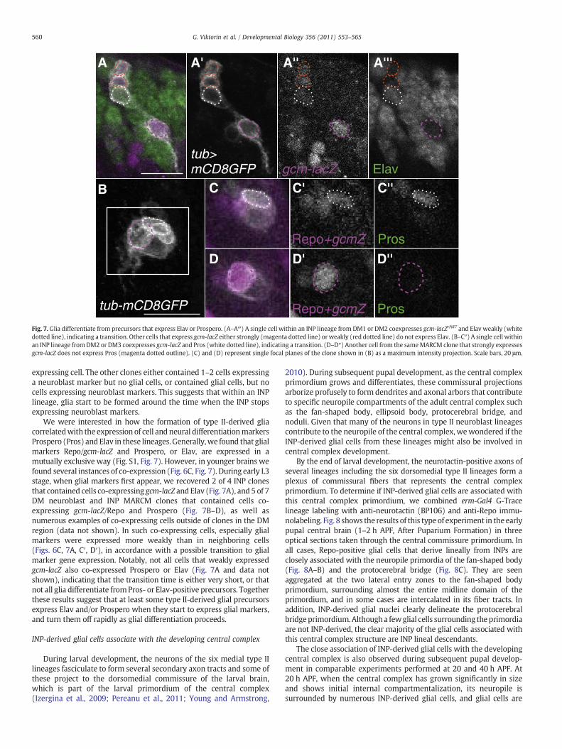
Fig. 7. Glia differentiate from precursors that express Elav or Prospero. (A–A‴) A single cell within an INP lineage from DM1 or DM2 coexpresses gcm-lacZrA87 and Elav weakly (whitedotted line), indicating a transition. Other cells that express gcm-lacZ either strongly (magenta dotted line) or weakly (red dotted line) do not express Elav. (B–C″) A single cell withinan INP lineage from DM2 or DM3 coexpresses gcm-lacZ and Pros (white dotted line), indicating a transition. (D–D″) Another cell from the sameMARCM clone that strongly expressesgcm-lacZ does not express Pros (magenta dotted outline). (C) and (D) represent single focal planes of the clone shown in (B) as a maximum intensity projection. Scale bars, 20 μm.
560 G. Viktorin et al. / Developmental Biology 356 (2011) 553–565
expressing cell. The other clones either contained 1–2 cells expressinga neuroblast marker but no glial cells, or contained glial cells, but nocells expressing neuroblast markers. This suggests that within an INPlineage, glia start to be formed around the time when the INP stopsexpressing neuroblast markers.
We were interested in how the formation of type II-derived gliacorrelatedwith the expression of cell and neural differentiationmarkersProspero (Pros) and Elav in these lineages. Generally,we found that glialmarkers Repo/gcm-lacZ and Prospero, or Elav, are expressed in amutually exclusive way (Fig. S1, Fig. 7). However, in younger brains wefound several instances of co-expression (Fig. 6C, Fig. 7). During early L3stage, when glial markers first appear, we recovered 2 of 4 INP clonesthat contained cells co-expressing gcm-lacZ and Elav (Fig. 7A), and 5 of 7DM neuroblast and INP MARCM clones that contained cells co-expressing gcm-lacZ/Repo and Prospero (Fig. 7B–D), as well asnumerous examples of co-expressing cells outside of clones in the DMregion (data not shown). In such co-expressing cells, especially glialmarkers were expressed more weakly than in neighboring cells(Figs. 6C, 7A, C′, D′), in accordance with a possible transition to glialmarker gene expression. Notably, not all cells that weakly expressedgcm-lacZ also co-expressed Prospero or Elav (Fig. 7A and data notshown), indicating that the transition time is either very short, or thatnot all glia differentiate fromPros- or Elav-positive precursors. Togetherthese results suggest that at least some type II-derived glial precursorsexpress Elav and/or Prospero when they start to express glial markers,and turn them off rapidly as glial differentiation proceeds.
INP-derived glial cells associate with the developing central complex
During larval development, the neurons of the six medial type IIlineages fasciculate to form several secondary axon tracts and some ofthese project to the dorsomedial commissure of the larval brain,which is part of the larval primordium of the central complex(Izergina et al., 2009; Pereanu et al., 2011; Young and Armstrong,
2010). During subsequent pupal development, as the central complexprimordium grows and differentiates, these commissural projectionsarborize profusely to form dendrites and axonal arbors that contributeto specific neuropile compartments of the adult central complex suchas the fan-shaped body, ellipsoid body, protocerebral bridge, andnoduli. Given that many of the neurons in type II neuroblast lineagescontribute to the neuropile of the central complex, wewondered if theINP-derived glial cells from these lineages might also be involved incentral complex development.
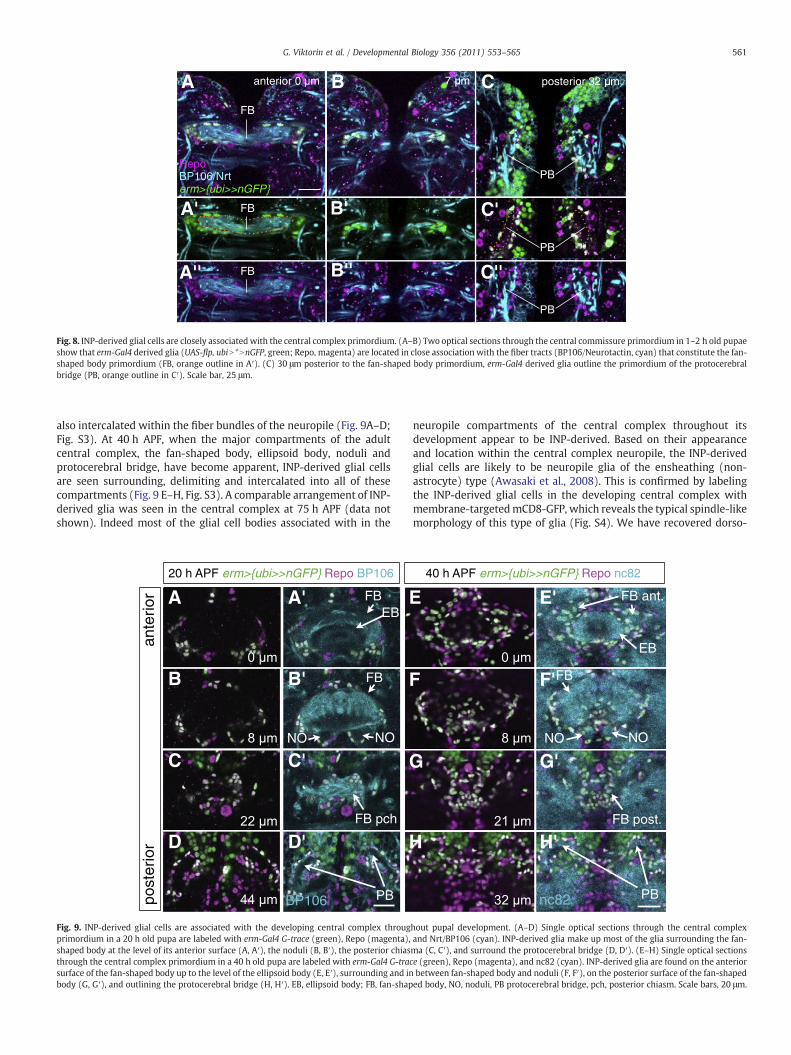
By the end of larval development, the neurotactin-positive axons ofseveral lineages including the six dorsomedial type II lineages form aplexus of commissural fibers that represents the central complexprimordium. To determine if INP-derived glial cells are associated withthis central complex primordium, we combined erm-Gal4 G-Tracelineage labeling with anti-neurotactin (BP106) and anti-Repo immu-nolabeling. Fig. 8 shows the results of this type of experiment in the earlypupal central brain (1–2 h APF, After Puparium Formation) in threeoptical sections taken through the central commissure primordium. Inall cases, Repo-positive glial cells that derive lineally from INPs areclosely associated with the neuropile primordia of the fan-shaped body(Fig. 8A–B) and the protocerebral bridge (Fig. 8C). They are seenaggregated at the two lateral entry zones to the fan-shaped bodyprimordium, surrounding almost the entire midline domain of theprimordium, and in some cases are intercalated in its fiber tracts. Inaddition, INP-derived glial nuclei clearly delineate the protocerebralbridgeprimordium.Althougha fewglial cells surrounding theprimordiaare not INP-derived, the clear majority of the glial cells associated withthis central complex structure are INP lineal descendants.
The close association of INP-derived glial cells with the developingcentral complex is also observed during subsequent pupal develop-ment in comparable experiments performed at 20 and 40 h APF. At20 h APF, when the central complex has grown significantly in sizeand shows initial internal compartmentalization, its neuropile issurrounded by numerous INP-derived glial cells, and glial cells are
C''A'' B''
B' C'
B CA
A'
anterior 0 m 7 m posterior 32 m
PB
PB
PB
FB
FB
FB
Repo BP106/Nrterm>{ubi>>nGFP}
Fig. 8. INP-derived glial cells are closely associatedwith the central complex primordium. (A–B) Two optical sections through the central commissure primordium in 1–2 h old pupaeshow that erm-Gal4 derived glia (UAS-flp, ubiN *NnGFP, green; Repo, magenta) are located in close association with the fiber tracts (BP106/Neurotactin, cyan) that constitute the fan-shaped body primordium (FB, orange outline in A′). (C) 30 μm posterior to the fan-shaped body primordium, erm-Gal4 derived glia outline the primordium of the protocerebralbridge (PB, orange outline in C′). Scale bar, 25 μm.
561G. Viktorin et al. / Developmental Biology 356 (2011) 553–565
also intercalated within the fiber bundles of the neuropile (Fig. 9A–D;Fig. S3). At 40 h APF, when the major compartments of the adultcentral complex, the fan-shaped body, ellipsoid body, noduli andprotocerebral bridge, have become apparent, INP-derived glial cellsare seen surrounding, delimiting and intercalated into all of thesecompartments (Fig. 9 E–H, Fig. S3). A comparable arrangement of INP-derived glia was seen in the central complex at 75 h APF (data notshown). Indeed most of the glial cell bodies associated with in the
post
erio
r
20 h APF erm>{ubi>>nGFP} Repo BP106
BP106
A A'
B B'
C C'
D D'
E
F
G
H
FB
FB
NO NO
FB pch
PB
ante
rior
EB
Fig. 9. INP-derived glial cells are associated with the developing central complex througprimordium in a 20 h old pupa are labeled with erm-Gal4 G-trace (green), Repo (magenta),shaped body at the level of its anterior surface (A, A′), the noduli (B, B′), the posterior chiasthrough the central complex primordium in a 40 h old pupa are labeled with erm-Gal4 G-tracsurface of the fan-shaped body up to the level of the ellipsoid body (E, E′), surrounding and inbody (G, G′), and outlining the protocerebral bridge (H, H′). EB, ellipsoid body; FB, fan-shap
neuropile compartments of the central complex throughout itsdevelopment appear to be INP-derived. Based on their appearanceand location within the central complex neuropile, the INP-derivedglial cells are likely to be neuropile glia of the ensheathing (non-astrocyte) type (Awasaki et al., 2008). This is confirmed by labelingthe INP-derived glial cells in the developing central complex withmembrane-targetedmCD8-GFP, which reveals the typical spindle-likemorphology of this type of glia (Fig. S4). We have recovered dorso-
nc82
E'
F'
G'
H'
EB
PB
FB post.
NO NO
FB
FB ant.
40 h APF erm>{ubi>>nGFP} Repo nc82
hout pupal development. (A–D) Single optical sections through the central complexand Nrt/BP106 (cyan). INP-derived glia make up most of the glia surrounding the fan-ma (C, C′), and surround the protocerebral bridge (D, D′). (E–H) Single optical sectionse (green), Repo (magenta), and nc82 (cyan). INP-derived glia are found on the anteriorbetween fan-shaped body and noduli (F, F′), on the posterior surface of the fan-shapeded body, NO, noduli, PB protocerebral bridge, pch, posterior chiasm. Scale bars, 20 μm.
562 G. Viktorin et al. / Developmental Biology 356 (2011) 553–565
medially located astrocyte-like glia as well as interhemispheric ringglia (Simon et al., 1998) in our MARCM experiments (both have muchlarger nuclei than DM-derived glia and express high levels of Prosperoand glial markers), but not in association with neuroblast or INPlineages (data not shown).
Taken together, these findings indicate that INP-derived glial cellsof the type II neuroblast lineages differentiate into ensheathingneuropile glia of the central complex. Thus, the type II neuroblasts viatheir INPs not only give rise to neuronal progeny that contributes tothe central complex neuropile but they also generate the glial progenythat ensheaths this neuropile.
INP-derived glial cells proliferate locally in the developingcentral complex
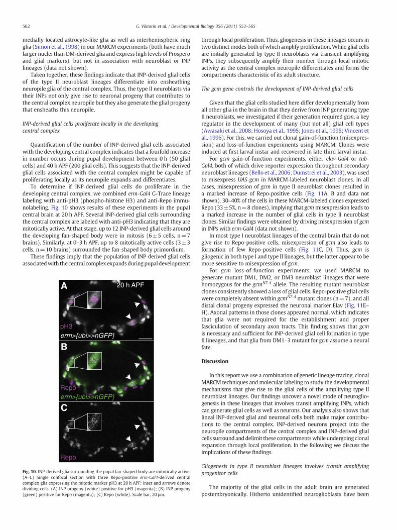
Quantification of the number of INP-derived glial cells associatedwith the developing central complex indicates that a fourfold increasein number occurs during pupal development between 0 h (50 glialcells) and 40 h APF (200 glial cells). This suggests that the INP-derivedglial cells associated with the central complex might be capable ofproliferating locally as its neuropile expands and differentiates.
To determine if INP-derived glial cells do proliferate in thedeveloping central complex, we combined erm-Gal4 G-Trace lineagelabeling with anti-pH3 (phospho-histone H3) and anti-Repo immu-nolabeling. Fig. 10 shows results of these experiments in the pupalcentral brain at 20 h APF. Several INP-derived glial cells surroundingthe central complex are labeled with anti-pH3 indicating that they aremitotically active. At that stage, up to 12 INP-derived glial cells aroundthe developing fan-shaped body were in mitosis (6±5 cells, n=7brains). Similarly, at 0–3 h APF, up to 8 mitotically active cells (3±3cells, n=10 brains) surrounded the fan-shaped body primordium.
These findings imply that the population of INP-derived glial cellsassociatedwith the central complex expands duringpupal development
Repo
Repoerm>{ubi>>nGFP}
pH3erm>{ubi>>nGFP}
A
B
C
20 h APF
Fig. 10. INP-derived glia surrounding the pupal fan-shaped body are mitotically active.(A–C) Single confocal section with three Repo-positive erm-Gal4-derived centralcomplex glia expressing the mitotic marker pH3 at 20 h APF; inset and arrows denotedividing cells. (A) INP progeny (white) positive for pH3 (magenta); (B) INP progeny(green) positive for Repo (magenta); (C) Repo (white). Scale bar, 20 μm.
through local proliferation. Thus, gliogenesis in these lineages occurs intwo distinct modes both ofwhich amplify proliferation.While glial cellsare initially generated by type II neuroblasts via transient amplifyingINPs, they subsequently amplify their number through local mitoticactivity as the central complex neuropile differentiates and forms thecompartments characteristic of its adult structure.
The gcm gene controls the development of INP-derived glial cells
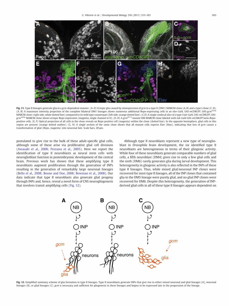
Given that the glial cells studied here differ developmentally fromall other glia in the brain in that they derive from INP generating typeII neuroblasts, we investigated if their generation required gcm, a keyregulator in the development of many (but not all) glial cell types(Awasaki et al., 2008; Hosoya et al., 1995; Jones et al., 1995; Vincent etal., 1996). For this, we carried out clonal gain-of-function (misexpres-sion) and loss-of-function experiments using MARCM. Clones wereinduced at first larval instar and recovered in late third larval instar.
For gcm gain-of-function experiments, either elav-Gal4 or tub-Gal4, both of which drive reporter expression throughout secondaryneuroblast lineages (Bello et al., 2006; Dumstrei et al., 2003), was usedto misexpress UAS-gcm in MARCM-labeled neuroblast clones. In allcases, misexpression of gcm in type II neuroblast clones resulted ina marked increase of Repo-positive cells (Fig. 11A, B and data notshown). 30–40% of the cells in these MARCM-labeled clones expressedRepo (33±5%, n=8 clones), implying that gcmmisexpression leads toa marked increase in the number of glial cells in type II neuroblastclones. Similar findings were obtained by driving misexpression of gcmin INPs with erm-Gal4 (data not shown).
In most type I neuroblast lineages of the central brain that do notgive rise to Repo-positive cells, misexpression of gcm also leads toformation of few Repo-positive cells (Fig. 11C, D). Thus, gcm isgliogenic in both type I and type II lineages, but the latter appear to bemore sensitive to misexpression of gcm.
For gcm loss-of-function experiments, we used MARCM togenerate mutant DM1, DM2, or DM3 neuroblast lineages that werehomozygous for the gcmN7-4 allele. The resulting mutant neuroblastclones consistently showed a loss of glial cells. Repo-positive glial cellswere completely absent within gcmN7-4 mutant clones (n=7), and alldistal clonal progeny expressed the neuronal marker Elav (Fig. 11E–H). Axonal patterns in those clones appeared normal, which indicatesthat glia were not required for the establishment and properfasciculation of secondary axon tracts. This finding shows that gcmis necessary and sufficient for INP-derived glial cell formation in typeII lineages, and that glia from DM1–3 mutant for gcm assume a neuralfate.
Discussion
In this report we use a combination of genetic lineage tracing, clonalMARCM techniques andmolecular labeling to study the developmentalmechanisms that give rise to the glial cells of the amplifying type IIneuroblast lineages. Our findings uncover a novel mode of neuroglio-genesis in these lineages that involves transit amplifying INPs, whichcan generate glial cells as well as neurons. Our analysis also shows thatlineal INP-derived glial and neuronal cells both make major contribu-tions to the central complex. INP-derived neurons project into theneuropile compartments of the central complex and INP-derived glialcells surroundanddelimit these compartmentswhile undergoing clonalexpansion through local proliferation. In the following we discuss theimplications of these findings.
Gliogenesis in type II neuroblast lineages involves transit amplifyingprogenitor cells
The majority of the glial cells in the adult brain are generatedpostembryonically. Hitherto unidentified neuroglioblasts have been
Elav
BP106elav>gcm, >mCD8GFPRepo
G HFE
A C
Dtub>gcm
Repo
B
gcmN7-4
tub>mCD8GFP RepogcmN7-4
tub>mCD8GFP
Fig. 11. Type II lineages generate glia in a gcm-dependent manner. (A–D) Ectopic glia caused by misexpression of gcm in a type II (DM1)MARCM clone (A, B) and a type I clone (C, D).(A, B) A maximum intensity projection of the complete bilateral DM1 lineages shows numerous additional Repo-expressing cells in an elav-Gal4, UAS-mCD8GFP, UAS-gcmm24a
MARCM clone (right side, white dotted line) compared to its wild type counterpart (left side, orange dotted line). (C,D) A single confocal slice of a type I tub-Gal4, UAS-mCD8GFP, UAS-gcmm24a MARCM clone shows ectopic Repo expression (magenta, single channel in D). (E–H) A gcmN7-4 mutant DM MARCM clone labeled with tub-Gal4 UAS-mCD8GFP lacks Repo-positive cells. (E, F) Optical projection of all cells in the clone reveals no Repo positive cell (magenta) within the clone (dotted line). In the opposite hemisphere, glial cells in thisregion are present (orange dotted outline). (G, H) A single section of the same clone shows that all mutant cells express Elav (blue), indicating that loss of gcm causes atransformation of glial (Repo, magenta) into neuronal fate. Scale bars, 20 μm.
563G. Viktorin et al. / Developmental Biology 356 (2011) 553–565
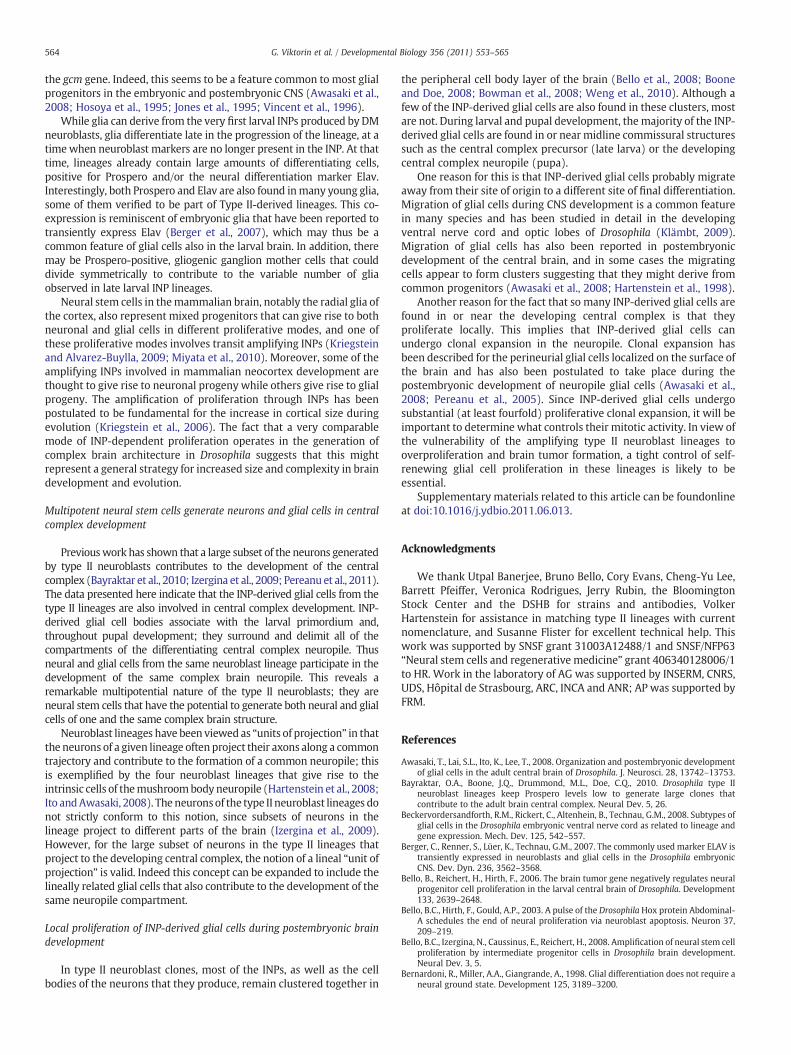
postulated to give rise to the bulk of these adult-specific glial cells,although some of these arise via proliferative glial cell divisions(Awasaki et al., 2008; Pereanu et al., 2005). Here we report theidentification of type II neuroblasts as neural stem cells withneuroglioblast function in postembryonic development of the centralbrain. Previous work has shown that these amplifying type IIneuroblasts augment proliferation through the generation of INPsresulting in the generation of remarkably large neuronal lineages(Bello et al., 2008; Boone and Doe, 2008; Bowman et al., 2008). Ourdata indicate that type II neuroblasts also generate glial progenythrough INPs and, hence, reveal a novel form of CNS neurogliogenesisthat involves transit amplifying cells (Fig. 12).
NB
INP
NB
IN
neurons
glia
n
A B
gcm
Fig. 12. Simplified summary scheme of glia formation in type II lineages. Type II neuroblastlineages (B), or glial lineages (C). gcm is necessary and sufficient for gliogenesis in these lin
Although type II neuroblasts represent a new type of neuroglio-blast in Drosophila brain development, the six identified type IIneuroblasts are heterogeneous in terms of their gliogenic activity.While four of these neuroblasts generate comparable numbers of glialcells, a fifth neuroblast (DM4) gives rise to only a few glial cells andthe sixth (DM6) rarely generates glia during larval development. Thisheterogeneity in gliogenic activity is also reflected in the INPs of thesetype II lineages. Thus, while mixed glial/neuronal INP clones wererecovered for most type II lineages, all of the INP clones that containedglia in the DM5 lineage were purely glial, and no glial INP clones wererecovered for DM6. Despite this heterogeneity, the generation of INP-derived glial cells in all of these type II lineages appears dependent on
P
NB
INP
euronsglia
C
gcm
s generate INPs that give rise to either mixed neuronal and glial lineages (A), neuronaleages and begins to be expressed late in the progression of the lineage.
564 G. Viktorin et al. / Developmental Biology 356 (2011) 553–565
the gcm gene. Indeed, this seems to be a feature common to most glialprogenitors in the embryonic and postembryonic CNS (Awasaki et al.,2008; Hosoya et al., 1995; Jones et al., 1995; Vincent et al., 1996).
While glia can derive from the very first larval INPs produced by DMneuroblasts, glia differentiate late in the progression of the lineage, at atimewhen neuroblast markers are no longer present in the INP. At thattime, lineages already contain large amounts of differentiating cells,positive for Prospero and/or the neural differentiation marker Elav.Interestingly, both Prospero and Elav are also found inmany young glia,some of them verified to be part of Type II-derived lineages. This co-expression is reminiscent of embryonic glia that have been reported totransiently express Elav (Berger et al., 2007), which may thus be acommon feature of glial cells also in the larval brain. In addition, theremay be Prospero-positive, gliogenic ganglion mother cells that coulddivide symmetrically to contribute to the variable number of gliaobserved in late larval INP lineages.
Neural stem cells in themammalian brain, notably the radial glia ofthe cortex, also represent mixed progenitors that can give rise to bothneuronal and glial cells in different proliferative modes, and one ofthese proliferative modes involves transit amplifying INPs (Kriegsteinand Alvarez-Buylla, 2009; Miyata et al., 2010). Moreover, some of theamplifying INPs involved in mammalian neocortex development arethought to give rise to neuronal progeny while others give rise to glialprogeny. The amplification of proliferation through INPs has beenpostulated to be fundamental for the increase in cortical size duringevolution (Kriegstein et al., 2006). The fact that a very comparablemode of INP-dependent proliferation operates in the generation ofcomplex brain architecture in Drosophila suggests that this mightrepresent a general strategy for increased size and complexity in braindevelopment and evolution.
Multipotent neural stem cells generate neurons and glial cells in centralcomplex development
Previouswork has shown that a large subset of the neurons generatedby type II neuroblasts contributes to the development of the centralcomplex (Bayraktar et al., 2010; Izergina et al., 2009; Pereanu et al., 2011).The data presented here indicate that the INP-derived glial cells from thetype II lineages are also involved in central complex development. INP-derived glial cell bodies associate with the larval primordium and,throughout pupal development; they surround and delimit all of thecompartments of the differentiating central complex neuropile. Thusneural and glial cells from the same neuroblast lineage participate in thedevelopment of the same complex brain neuropile. This reveals aremarkable multipotential nature of the type II neuroblasts; they areneural stem cells that have the potential to generate both neural and glialcells of one and the same complex brain structure.
Neuroblast lineages have been viewed as “units of projection” in thatthe neurons of a given lineage often project their axons along a commontrajectory and contribute to the formation of a common neuropile; thisis exemplified by the four neuroblast lineages that give rise to theintrinsic cells of themushroombody neuropile (Hartenstein et al., 2008;Ito andAwasaki, 2008). The neuronsof the type II neuroblast lineages donot strictly conform to this notion, since subsets of neurons in thelineage project to different parts of the brain (Izergina et al., 2009).However, for the large subset of neurons in the type II lineages thatproject to the developing central complex, the notion of a lineal “unit ofprojection” is valid. Indeed this concept can be expanded to include thelineally related glial cells that also contribute to the development of thesame neuropile compartment.
Local proliferation of INP-derived glial cells during postembryonic braindevelopment
In type II neuroblast clones, most of the INPs, as well as the cellbodies of the neurons that they produce, remain clustered together in
the peripheral cell body layer of the brain (Bello et al., 2008; Booneand Doe, 2008; Bowman et al., 2008; Weng et al., 2010). Although afew of the INP-derived glial cells are also found in these clusters, mostare not. During larval and pupal development, the majority of the INP-derived glial cells are found in or near midline commissural structuressuch as the central complex precursor (late larva) or the developingcentral complex neuropile (pupa).
One reason for this is that INP-derived glial cells probably migrateaway from their site of origin to a different site of final differentiation.Migration of glial cells during CNS development is a common featurein many species and has been studied in detail in the developingventral nerve cord and optic lobes of Drosophila (Klämbt, 2009).Migration of glial cells has also been reported in postembryonicdevelopment of the central brain, and in some cases the migratingcells appear to form clusters suggesting that they might derive fromcommon progenitors (Awasaki et al., 2008; Hartenstein et al., 1998).
Another reason for the fact that so many INP-derived glial cells arefound in or near the developing central complex is that theyproliferate locally. This implies that INP-derived glial cells canundergo clonal expansion in the neuropile. Clonal expansion hasbeen described for the perineurial glial cells localized on the surface ofthe brain and has also been postulated to take place during thepostembryonic development of neuropile glial cells (Awasaki et al.,2008; Pereanu et al., 2005). Since INP-derived glial cells undergosubstantial (at least fourfold) proliferative clonal expansion, it will beimportant to determine what controls their mitotic activity. In view ofthe vulnerability of the amplifying type II neuroblast lineages tooverproliferation and brain tumor formation, a tight control of self-renewing glial cell proliferation in these lineages is likely to beessential.
Supplementary materials related to this article can be foundonlineat doi:10.1016/j.ydbio.2011.06.013.
Acknowledgments
We thank Utpal Banerjee, Bruno Bello, Cory Evans, Cheng-Yu Lee,Barrett Pfeiffer, Veronica Rodrigues, Jerry Rubin, the BloomingtonStock Center and the DSHB for strains and antibodies, VolkerHartenstein for assistance in matching type II lineages with currentnomenclature, and Susanne Flister for excellent technical help. Thiswork was supported by SNSF grant 31003A12488/1 and SNSF/NFP63“Neural stem cells and regenerative medicine” grant 406340128006/1to HR. Work in the laboratory of AG was supported by INSERM, CNRS,UDS, Hôpital de Strasbourg, ARC, INCA and ANR; AP was supported byFRM.
References
Awasaki, T., Lai, S.L., Ito, K., Lee, T., 2008. Organization and postembryonic developmentof glial cells in the adult central brain of Drosophila. J. Neurosci. 28, 13742–13753.
Bayraktar, O.A., Boone, J.Q., Drummond, M.L., Doe, C.Q., 2010. Drosophila type IIneuroblast lineages keep Prospero levels low to generate large clones thatcontribute to the adult brain central complex. Neural Dev. 5, 26.
Beckervordersandforth, R.M., Rickert, C., Altenhein, B., Technau, G.M., 2008. Subtypes ofglial cells in the Drosophila embryonic ventral nerve cord as related to lineage andgene expression. Mech. Dev. 125, 542–557.
Berger, C., Renner, S., Lüer, K., Technau, G.M., 2007. The commonly used marker ELAV istransiently expressed in neuroblasts and glial cells in the Drosophila embryonicCNS. Dev. Dyn. 236, 3562–3568.
Bello, B., Reichert, H., Hirth, F., 2006. The brain tumor gene negatively regulates neuralprogenitor cell proliferation in the larval central brain of Drosophila. Development133, 2639–2648.
Bello, B.C., Hirth, F., Gould, A.P., 2003. A pulse of the Drosophila Hox protein Abdominal-A schedules the end of neural proliferation via neuroblast apoptosis. Neuron 37,209–219.
Bello, B.C., Izergina, N., Caussinus, E., Reichert, H., 2008. Amplification of neural stem cellproliferation by intermediate progenitor cells in Drosophila brain development.Neural Dev. 3, 5.
Bernardoni, R., Miller, A.A., Giangrande, A., 1998. Glial differentiation does not require aneural ground state. Development 125, 3189–3200.

565G. Viktorin et al. / Developmental Biology 356 (2011) 553–565
Boone, J.Q., Doe, C.Q., 2008. Identification of Drosophila type II neuroblast lineagescontaining transit amplifying ganglion mother cells. Dev. Neurobiol. 68,1185–1195.
Bossing, T., Udolph, G., Doe, C.Q., Technau, G.M., 1996. The embryonic central nervoussystem lineages of Drosophila melanogaster. I. Neuroblast lineages derived from theventral half of the neuroectoderm. Dev. Biol. 179, 41–64.
Bowman, S.K., Rolland, V., Betschinger, J., Kinsey, K.A., Emery, G., Knoblich, J.A., 2008.The tumor suppressors Brat and Numb regulate transit-amplifying neuroblastlineages in Drosophila. Dev. Cell 14, 535–546.
Broadus, J., Skeath, J.B., Spana, E.P., Bossing, T., Technau, G., Doe, C.Q., 1995. Newneuroblast markers and the origin of the aCC/pCC neurons in the Drosophila centralnervous system. Mech. Dev. 53, 393–402.
Doe, C.Q., 2008. Neural stem cells: balancing self-renewal with differentiation.Development 135, 1575–1587.
Dumstrei, K., Wang, F., Hartenstein, V., 2003. Role of DE-cadherin in neuroblastproliferation, neural morphogenesis, and axon tract formation in Drosophila larvalbrain development. J. Neurosci. 23, 3325–3335.
Evans, C.J., Olson, J.M., Ngo, K.T., Kim, E., Lee, N.E., Kuoy, E., Patananan, A.N., Sitz, D., Tran, P.,Do, M.T., Yackle, K., Cespedes, A., Hartenstein, V., Call, G.B., Banerjee, U., 2009. G-TRACE: rapid Gal4-based cell lineage analysis inDrosophila. Nat. Methods 6, 603–605.
Götz, M., Huttner, W.B., 2005. The cell biology of neurogenesis. Nat. Rev. Mol. Cell Biol.6, 777–788.
Hartenstein, V., Nassif, C., Lekven, A., 1998. Embryonic development of the Drosophilabrain. II. Pattern of glial cells. J. Comp. Neurol. 402, 32–47.
Hartenstein, V., Spindler, S., Pereanu, W., Fung, S., 2008. The development of theDrosophila larval brain. Adv. Exp. Med. Biol. 628, 1–31.
Haubensak, W., Attardo, A., Denk, W., Huttner, W.B., 2004. Neurons arise in the basalneuroepithelium of the early mammalian telencephalon: a major site ofneurogenesis. Proc. Natl. Acad. Sci. U.S.A. 101, 3196–3201.
Hosoya, T., Takizawa, K., Nitta, K., Hotta, Y., 1995. Glial cells missing: a binary switchbetween neuronal and glial determination in Drosophila. Cell 82, 1025–1036.
Ito, K., Awasaki, T., 2008. Clonal unit architecture of the adult fly brain. Adv. Exp. Med.Biol. 628, 137–158.
Izergina, N., Balmer, J., Bello, B., Reichert, H., 2009. Postembryonic development oftransit amplifying neuroblast lineages in the Drosophila brain. Neural Dev. 4, 44.
Jones, B.W., Fetter, R.D., Tear, G., Goodman, C.S., 1995. Glial cells missing: a geneticswitch that controls glial versus neuronal fate. Cell 82, 1013–1023.
Klämbt, C., 2009. Modes and regulation of glial migration in vertebrates andinvertebrates. Nat. Rev. Neurosci. 10, 769–779.
Knoblich, J.A., 2008. Mechanisms of asymmetric stem cell division. Cell 132, 583–597.Kriegstein, A., Alvarez-Buylla, A., 2009. The glial nature of embryonic and adult neural
stem cells. Annu. Rev. Neurosci. 32, 149–184.Kriegstein, A., Noctor, S., Martinez-Cerdeno, V., 2006. Patterns of neural stem and
progenitor cell division may underlie evolutionary cortical expansion. Nat. Rev.Neurosci. 7, 883–890.
Lee, T., Luo, L., 1999. Mosaic analysis with a repressible cell marker for studies of genefunction in neuronal morphogenesis. Neuron 22, 451–461.
Miller, F.D., Gauthier, A.S., 2007. Timing is everything: making neurons versus glia inthe developing cortex. Neuron 54, 357–369.
Miyata, T., Kawaguchi, A., Saito, K., Kawano, M., Muto, T., Ogawa, M., 2004. Asymmetricproduction of surface-dividing and non-surface-dividing cortical progenitor cells.Development 131, 3133–3145.
Miyata, T., Kawaguchi, D., Kawaguchi, A., Gotoh, Y., 2010. Mechanisms that regulate thenumber of neurons duringmouse neocortical development. Curr. Opin. Neurobiol. 20,22–28.
Noctor, S.C., Martinez-Cerdeno, V., Ivic, L., Kriegstein, A.R., 2004. Cortical neurons arisein symmetric and asymmetric division zones and migrate through specific phases.Nat. Neurosci. 7, 136–144.
Noctor, S.C., Martinez-Cerdeno, V., Kriegstein, A.R., 2007. Contribution of intermediateprogenitor cells to cortical histogenesis. Arch. Neurol. 64, 639–642.
Pereanu, W., Hartenstein, V., 2006. Neural lineages of the Drosophila brain: a three-dimensional digital atlas of the pattern of lineage location and projection at the latelarval stage. J. Neurosci. 26, 5534–5553.
Pereanu, W., Shy, D., Hartenstein, V., 2005. Morphogenesis and proliferation of thelarval brain glia in Drosophila. Dev. Biol. 283, 191–203.
Pereanu, W., Younossi-Hartenstein, A., Lovick, J., Spindler, S., Hartenstein, V., 2011. Alineage-based analysis of the development of the central complex of the Drosophilabrain. J. Comp. Neurol. 519, 661–689.
Pignoni, F., Zipursky, L., 1997. Induction of Drosophila eye development byDecapentaplegic. Development 124, 271–287.
Pfeiffer, B.D., Jenett, A., Hammonds, A.S., Ngo, T.T., Misra, S., Murphy, C., Scully, A.,Carlson, J.W.,Wan, K.H., Laverty, T.R.,Mungall, C., Svirskas, R., Kadonaga, J.T., Doe, C.Q.,Eisen, M.B., Celniker, S.E., Rubin, G.M., 2008. Tools for neuroanatomy andneurogenetics in Drosophila. Proc. Natl. Acad. Sci. U.S.A. 105, 9715–9720.
Rasband, W.S., 1997–2008. ImageJ, U. S. National Institutes of Health, Bethesda,Maryland, USA. http://rsb.info.nih.gov/ij/.
Schmidt, H., Rickert, C., Bossing, T., Vef, O., Urban, J., Technau, G.M., 1997. Theembryonic central nervous system lineages of Drosophila melanogaster. II.Neuroblast lineages derived from the dorsal part of the neuroectoderm. Dev. Biol.189, 186–204.
Schindelin, J., 2008. Fiji is Just ImageJ — Batteries Included. ImageJ User and DeveloperConference.
Skeath, J.B., Thor, S., 2003. Genetic control of Drosophila nerve cord development. Curr.Opin. Neurobiol. 13, 8–15.
Simon, A., Boquet, I., Synguélakis, M., Préat, T., 1998. The Drosophila putative kinaseLinotte (Derailed) prevents central brain axons from converging on a newlydescribed interhemispheric ring. Mech. Dev. 76, 45–55.
Soustelle, L., Trousse, F., Jacques, C., Ceron, J., Cochard, P., Soula, C., Giangrande, A., 2007.Neurogenic role of Gcm transcription factors is conserved in chicken spinal cord.Development 134, 625–634.
Technau, G.M., Berger, C., Urbach, R., 2006. Generation of cell diversity and segmentalpattern in the embryonic central nervous system of Drosophila. Dev. Dyn. 235,861–869.
Van De Bor, V., Giangrande, A., 2002. glide/gcm: at the crossroads between neurons andglia. Curr. Opin. Gen. Dev. 12, 465–472.
Vincent, S., Vonesch, J.L., Giangrande, A., 1996. Glide directs glial fate commitment andcell fate switch between neurones and glia. Development 122, 131–139.
Weng, M., Golden, K.L., Lee, C.Y., 2010. dFezf/Earmuff maintains the restricteddevelopmental potential of intermediate neural progenitors in Drosophila. Dev.Cell 18, 126–135.
Weng, M., Lee, C.Y., 2010. Keeping neural progenitor cells on a short leash duringDrosophila neurogenesis. Curr. Opin. Neurobiol. 21, 1–7.
Young, J.M., Armstrong, J.D., 2010. Building the central complex inDrosophila: the generationand development of distinct neural subsets. J. Comp. Neurol. 518, 1525–1541.


![Clinical-Grade Multipotent Adult Progenitor Cells Durably ... · PDF fileClinical-Grade Multipotent Adult Progenitor Cells Durably ... epidermal growth factor [R&D Systems], dexamethasone](https://img.pdfslide.us/doc/110x75/5ab94a877f8b9a28468de5e2/clinical-grade-multipotent-adult-progenitor-cells-durably-multipotent-adult.jpg)