Embed Size (px)
Citation preview
Science in medicine
788 The Journal of Clinical Investigation http://www.jci.org Volume 113 Number 6 March 2004
Nonstandard abbreviations used: acute disseminated encephalomyelitis (ADEM);altered peptide ligand (APL); experimental autoimmune encephalomyelitis (EAE); glatiramer acetate (GA); myelin basic protein peptide p85–99 (MBP p85–99); primaryprogressive multiple sclerosis (PPMS); relapsing-remitting multiple sclerosis (RRMS);single nucleotide polymorphism (SNP); secondary progressive multiple sclerosis(SPMS); T cell receptor (TCR).
Conflict of interest: The author has declared that no conflict of interest exists.
Citation for this article: J. Clin. Invest. 113:788–794 (2004). doi:10.1172/JCI200421357.
Multiple sclerosisDavid A. Hafler
Laboratory of Molecular Immunology, Center for Neurologic Diseases, Brigham and Women’s Hospital and Harvard Medical School, Boston, Massachusetts, USA.The Broad Institute, Massachusetts Institute of Technology and Harvard University, Cambridge, Massachusetts, USA.
Multiple sclerosis is a complex genetic disease associated with inflammation in the CNS white matterthought to be mediated by autoreactive T cells. Clonal expansion of B cells, their antibody products,and T cells, hallmarks of inflammation in the CNS, are found in MS. This review discusses new meth-ods to define the molecular pathology of human disease with high-throughput examination of germlineDNA haplotypes, RNA expression, and protein structures that will allow the generation of a new seriesof hypotheses that can be tested to develop better understanding of and therapies for this disease.
Historical perspectiveA French neurologist at the Salpetrière in Paris, Jean Martin Char-cot, first described multiple sclerosis (MS) in 1868, noting the accu-mulation of inflammatory cells in a perivascular distribution with-in the brain and spinal cord white matter of patients with intermittentepisodes of neurologic dysfunction (1–3). This led to the term scléroseen plaques disseminées, or multiple sclerosis. The more recent obser-vation in 1948 by Elvin Kabat of increases in oligoclonalimmunoglobulin in the cerebrospinal fluid of patients with MS pro-vided further evidence of an inflammatory nature to the disease (4,5). In the past half-century, several large population–based MS twinstudies demonstrated a strong genetic basis to this clinical-patho-logic entity (6–13). Lastly, the demonstration of an autoimmune, attimes demyelinating, disease in mammals with immunization ofCNS myelin (experimental autoimmune encephalomyelitis, orEAE), first made by Thomas Rivers at the Rockefeller Institute in1933 with the repeated injection of rabbit brain and spinal cordinto primates (14), has led to the generally accepted hypothesis thatMS is secondary to an autoimmune response to self-antigens in agenetically susceptible host (see box, What we know about MS). Itshould be pointed out that although the inflammation found inthe CNS of patients with MS is thought to represent an autoim-mune response, this is based on negative experiments where inves-tigators have not been able to consistently isolate a microbial agentfrom the tissue of diseased patients. Nevertheless, primary viralinfections in the CNS may induce an autoimmune response (15),and the recurring lesson from the EAE model is that the minimalrequirement for inducing inflammatory, autoimmune CNSdemyelinating disease is the activation of myelin-reactive T cells inthe peripheral immune system (16, 17).
Advances in immunology have provided clinicians with powerfultools to better understand the underlying causes of MS, leading tonew therapeutic advances. The future calls for extending the origi-nal observations of Charcot and Kabat by defining the molecularpathology of MS at the level of DNA haplotype structure, CNS andperipheral mRNA and protein expression, leading to the generationof a new series of disease-related hypotheses.
PathologyGross examination of brain tissue of individuals with MS revealsmultiple sharply demarcated plaques in the CNS white matter witha predilection to the optic nerves and white matter tracts of theperiventricular regions, brain stem, and spinal cord. As was recog-nized early on and so elegantly investigated in more recent studies,substantial axonal injury with axonal transections is abundantthroughout active MS lesions (18).
The inflammatory cell profile of active lesions is characterized byperivascular infiltration of oligoclonal T cells (19) consisting ofCD4+/CD8 α/β (20, 21) and γ/δ (22) T cells and monocytes withoccasional B cells and infrequent plasma cells (23). Lymphocytesmay be found in normal-appearing white matter beyond the mar-gin of active demyelination (24). Macrophages are most prominentin the center of the plaques and are seen to contain myelin debris,while oligodendrocyte counts are reduced. In chronic-active lesions,the inflammatory cell infiltrate is less prominent and may be large-ly restricted to the rim of the plaque, suggesting the presence ofongoing inflammatory activity along the lesion edge. Recently fourpathologic categories of the disease were defined on the basis ofmyelin protein loss, the geography and extension of plaques, thepatterns of oligodendrocyte destruction, and the immunopatho-logical evidence of complement activation. Two patterns (I and II)showed close similarities to T cell–mediated or T cell plus anti-body–mediated autoimmune encephalomyelitis, respectively. Theother patterns (III and IV) were highly suggestive of a vasculopathyor primary oligodendrocyte dystrophy, reminiscent of virus- ortoxin-induced demyelination rather than autoimmunity (25). It wasof interest that the pattern of pathology tended to be the same inmultiple lesions from any single individual with MS.
Natural historyMS, like other presumed autoimmune diseases, is more common infemales and often first manifests clinical symptoms during youngadulthood. At its onset, MS can be clinically categorized as eitherrelapsing-remitting MS (RRMS, observed in 85–90% of patients) orprimary progressive MS (PPMS). Relapses or “attacks” typically pre-sent subacutely, with symptoms developing over hours to severaldays, persisting for several days or weeks, and then gradually dissi-pating. The attacks are likely caused by the traffic of activated,myelin-reactive T cells into the CNS, causing acute inflammationwith associated edema. The ability of high dose steroids to so quick-ly abrogate MS symptoms suggests that the acute edema and itssubsequent resolution underlie the clinical relapse and remission,respectively. Studies in acute disseminated encephalomyelitis
science in medicine
The Journal of Clinical Investigation http://www.jci.org Volume 113 Number 6 March 2004 789
(ADEM) in humans (26) and EAE in rodents (27) suggest thatimmunologically, acute attacks are self-limited by regulatory T cells.
The outcome in patients with RRMS is variable; untreated, approx-imately 50% of all MS patients require the use of a walking aid withinten years after clinical onset (28), although the consequences on prog-nosis of newer treatment regiments are not as yet clear. Increasedattack frequency and poor recovery from attacks in the first years ofclinical disease predict a more rapid deterioration. Multiple MRIlesions, particularly those that gadolinium enhance on the first MRIscan, also predict a more severe subsequent course. Early on in the dis-ease, there are frequent gadolinium-enhanced MRI lesions, consistentwith acute fluxes of activated, autoreactive T cells into the CNS caus-ing a breakdown of the blood-brain barrier which may be associatedwith clinical events. However, with time, the extent of recovery fromattacks is often decreased, and baseline neurological disability accrues.Ultimately, approximately 40% of relapsing-remitting patients stophaving attacks and develop what may be a progressive neurodegener-ative secondary disorder related to the chronic CNS inflammation,known as secondary progressive multiple sclerosis (SPMS) (29). Theevolution to this secondary progressive form of the disease is associ-ated with significantly fewer gadolinium-enhanced lesions and adecrease in brain parenchymal volume (30, 31). Similarly, while earli-er RRMS is sensitive to immunosuppression (32), as times goes on,responsiveness to immunotherapy decreases and may in fact disap-pear in later forms of SPMS. Thus, rather than conceiving MS as firsta relapsing-remitting and then a secondary progressive disease, itcould be hypothesized that MS is a continuum where there are acuteinflammatory events early on with secondary induction of a neu-rodegenerative process refractory to immunologic intervention. Thishypothesis awaits experimental verification where early immunother-apy prevents the onset of secondary progressive disease. Such criticalinvestigations require new models of investigation using natural his-tory studies that can be performed over decades.
The primary progressive form of MS is characterized from theonset by the absence of acute attacks and instead involves a gradualclinical decline. Clinically, this form of the disease is associated witha lack of response to any form of immunotherapy (32). This leads tothe notion that PPMS may in fact be a very different disease as com-pared to RRMS. A recent commentary points out the similaritiesbetween PPMS and human T lymphotropic virus type I–associatedmyelopathy (15), where there is a progressive decline in neurologicfunction from the disease onset.
DiagnosisIn the absence of a specific immune-based assay, the diagnosis of MScontinues to be predicated on the clinical history and neurological
exam; that is, finding multiple lesions in time and space in the CNS.The use of MRI has had a major impact on allowing the early andmore precise diagnosis of the disease (33). In a recent prospectivestudy, patients experiencing their first episode suggestive of CNSdemyelination and having MRI evidence of at least three typicallesions were followed for an average of 42 months. Within that peri-od, a significant proportion of patients developed an additionalrelapse, thus qualifying for the diagnosis of clinically definite MS. Ifthere were no MRI lesions, the probability of developing MS was sub-stantially less. More than half of those developing MS experiencedthe additional relapse within one year of their first episode (34). Thus,it seems reasonable to label the first attack of what appears to be MSas “singular sclerosis” and to explain to patients that there is a highlikelihood of developing MS. This indicates to the patient that youhave an understanding of the underlying problem but that the prog-nosis is not as yet clear, allowing patients who never have anotherattack to be saved from carrying a diagnosis of MS.
ADEM is a monophasic demyelinating illness that can presentwith clinical, imaging, and laboratory manifestations indistin-guishable from an acute MS attack (35). However, typical ADEM isseen in pediatric populations and has more of an explosive courseassociated with alterations in mental status, and a post-viral or post-vaccination history is often elicited. This disease is associated withsignificant responses to myelin proteins, indicated both by T cell andantibody measurements (26, 36). It is usually not difficult to distin-guish this disease entity from “singular sclerosis,” and the laborato-ry measurement of circulating high affinity antimyelin basic proteinantibodies in ADEM but not MS may aid in the diagnosis. It shouldbe noted that there are many reports demonstrating low affinityantimyelin autoantibodies by ELISA in sera and CSF of patients withMS, though their role either in the disease’s pathogenesis or in pre-dicting outcome is still not well defined. However, high affinityantimyelin basic protein or myelin oligodendrocyte antibodiesappear to be more difficult to detect in the serum or CSF of patientswith MS while antimyelin oligodendrocyte antibodies can be foundin MS CNS plaque tissue (37).
MRI is the optimal imaging modality for MS. From a diagnosticstandpoint, it is important to keep in mind that the typical appear-ance of multiple lesions on MRIs is not specific for MS. In the appro-priate clinical setting, however, this appearance provides an impor-tant ancillary diagnostic tool that may establish the multifocality ofCNS involvement. MRI has also been used to assess MS disease activ-ity, disease burden, and the dynamic evolution in these parametersover time (38). MRI is 4–10 times more sensitive than the clinicalevaluation in capturing CNS lesions (39), and serial studies haveunequivocally demonstrated that clinically apparent changes reflect
What we know about MS
Inflammation in CNS white matter Different pathological subtypes. Early γδ T cell infiltration. Later CD4 and CD8 T cellswith loss of myelin and axons.
Increase in IgG in CSF CSF Ig is oligoclonal. B and T cells in CNS and CSF are also oligoclonal.Complex genetic disease λs of 20–40. High degree of inheritability. So far, only MHC region on chromosome 6 clearly
associated with MS.Antibody autoreactivity Variable results among different studies. Agreement on presence of autoantibodies in MS plaque tissue.
No high affinity autoantibodies in serum.T cell autoreactivity Autoreactivity to self-myelin antigens in patients with MS and normal subjects. In patients with MS,
modest increase in frequency of myelin-reactive, activated T cells.
science in medicine
790 The Journal of Clinical Investigation http://www.jci.org Volume 113 Number 6 March 2004
only a minor component of disease activity. Lesions in the cerebrumare much more likely to be clinically silent, as compared to lesionsin the brainstem or spinal cord.
TherapyTherapies for MS have emerged over the last two decades with thedemonstration of efficacy of three classes of immunomodulatingtherapies that impact the course of early MS: immunosuppressivedrugs such as mitoxantrone and cyclophosphamide; β-IFNs; and anMHC-binding protein that engages the T cell receptor (TCR), glati-ramer acetate (GA). The underlying pathology of MS as an inflam-matory CNS disease was instrumental in leading to the drug treat-ments presently used. While these drug therapies were notprospectively designed based on a detailed understanding of the dis-ease’s pathophysiology, examination of these drug’s mechanisms ofaction has provided insight into the etiology of MS. Newer therapiesin clinical trials are based on a more rational understanding of thedisease, and these will be discussed in more detail.
Disease mechanismsImmunopathophysiology of MS. It is often asked whether EAE, brieflydiscussed above, is really MS. Our laboratory investigates thepathophysiology of MS by directly studying patients with the dis-ease and does not directly investigate mouse models. Some of ourcolleagues will even note in good humor that MS is a superbmodel of EAE! (V. Kuchroo and S. Miller, personal communica-tion.) The truth is, we are and we will always be appropriately lim-ited with human experimentation. Experimental models, be theyNewtonian physics or rodent models of autoimmunity, are justthat — models. They do not represent truth (try to investigate thevelocity of a sub-atomic particle approaching the speed of lightusing Newtonian physics), and are only as useful as the questionone asks of the model. For example, it was shown almost a decadeago that the α4β7 integrin, VLA-4, was critical for T cell trafficinto the CNS of mice with EAE (40). This resulted in a highly suc-cessful phase II trial of anti–VLA-4 in patients with RRMS (41),which is now in phase III investigations. This is an excellent exam-
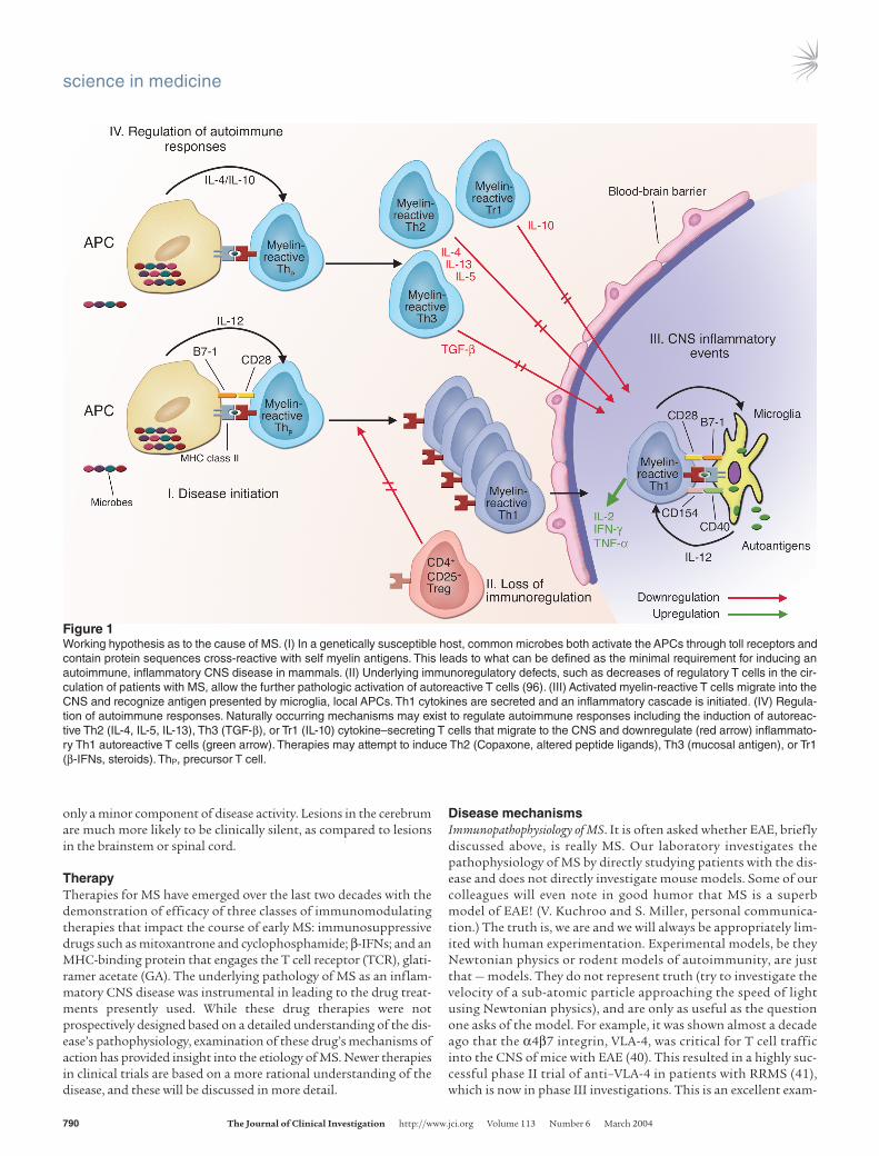
Figure 1Working hypothesis as to the cause of MS. (I) In a genetically susceptible host, common microbes both activate the APCs through toll receptors andcontain protein sequences cross-reactive with self myelin antigens. This leads to what can be defined as the minimal requirement for inducing anautoimmune, inflammatory CNS disease in mammals. (II) Underlying immunoregulatory defects, such as decreases of regulatory T cells in the cir-culation of patients with MS, allow the further pathologic activation of autoreactive T cells (96). (III) Activated myelin-reactive T cells migrate into theCNS and recognize antigen presented by microglia, local APCs. Th1 cytokines are secreted and an inflammatory cascade is initiated. (IV) Regula-tion of autoimmune responses. Naturally occurring mechanisms may exist to regulate autoimmune responses including the induction of autoreac-tive Th2 (IL-4, IL-5, IL-13), Th3 (TGF-β), or Tr1 (IL-10) cytokine–secreting T cells that migrate to the CNS and downregulate (red arrow) inflammato-ry Th1 autoreactive T cells (green arrow). Therapies may attempt to induce Th2 (Copaxone, altered peptide ligands), Th3 (mucosal antigen), or Tr1(β-IFNs, steroids). ThP, precursor T cell.

science in medicine
The Journal of Clinical Investigation http://www.jci.org Volume 113 Number 6 March 2004 791
ple of how the EAE model, if used to ask the correct question,might be highly useful in developing therapies for MS.
A second critical lesson from the EAE model is that of epitope spread-ing, first observed by Eli Sercarz (42). With the injection of a singlemyelin protein epitope into mice with subsequent development ofEAE, it was observed that T cells became activated against other epi-topes of the same protein; this was followed by T cell activation inresponse to other myelin proteins that become capable of adoptivelytransferring the disease to naive mice. The epitope spreading requirescostimulation with B7/CD28, suggesting that with tissue damage inthe CNS an adjuvant is created in the CNS with the expression of highamounts of B7.1 costimulatory molecules associated with antigenrelease (43). Moreover, we have recently observed that a transgenicmouse expressing DR2 (DRB1*1501) and a TCR (Ob1A12) clonedfrom the blood of a patient with MS recognizing an immunodominantmyelin basic protein peptide p85–99 (MBP p85–99) spontaneouslydeveloped EAE with epitope spreading to a number of antigens impli-cated in MS including α-β crystalline, and proteolipid apoprotein (D.Altman, V. Kuchroo, and D. Hafler, unpublished observations). As wehave observed high expression of B7.1 costimulatory molecules in theCNS white matter of patients with MS (44), and as most patients exhib-it T cell reactivity to a number of myelin antigens (45), it is likely thatby the time a patient develops clinical MS there has been epitopespreading with reactivity to multiple myelin epitopes. However, thepresence of clonally expanded T cells in the CSF and brain tissue ofpatients with the disease raises the issue that there may be clonal reac-tivity to just a few myelin antigens. Single cell cloning of T cells fromthe inflamed CNS tissue screened against combinatorial and proteinlibraries may allow new insights into the pathophysiology of MS.
Data from a number of laboratories combined with experimentaldata from the EAE model where myelin antigen is injected with adju-vant into mammals indicate that there are autoreactive T cells recog-nizing myelin antigens in the circulation of mammals. It appears thatthe activation of these T cells is the critical event in inducing autoim-mune disease (Figure 1). We and others first demonstrated over adecade ago that T cell clones isolated from the blood of patients withMS frequently exhibit exquisite specificity for the immunodominantp85–99 epitope of myelin basic protein (45–47). However, while theTCR appears to be highly specific in recognizing this peptide, alteringthe peptide ligand can change the TCR conformation to yield a high-er degree of T cell cross-reactivity (48). Using combinatorial chemistry,even a greater degree of cross-reactivity could be demonstrated, and anumber of viral epitopes were identified that could trigger autoreac-tive T cell clones in a manner that would not be predicted by simplealgorithms (49). Indeed, one MBP-reactive T cell clone recognized anepitope of an entirely different self-protein, the myelin oligodendro-cyte glycoprotein. Hence, a significant degree of functional degenera-cy exists in the recognition of antigens by T cells. This is consistentwith the hypothesis that MS is triggered by autoreactive T cells acti-vated by microbial antigens cross-reactive with myelin (50). The highfrequency of activated, myelin-reactive T cells in the circulation andCSF of patients with MS (51, 52) is consistent with the hypothesis thatthe disease is initiated by a microbial infection.
Novel therapeuticsPeptides bound to MHC as therapeutic options. It was recognized almost adecade ago that the strength of signal delivered through the TCRdetermines which cytokines are secreted by the T cell (53). The cellapparently measures affinity in part by timing the engagementbetween the TCR and the peptide/MHC complex. With longer
engagement, a qualitatively different TCR complex has time to form,and the extent of ζ chain phosphorylation increases corresponding-ly (54). Altered peptide ligands (APLs), which bind with low affinityto the TCR, weaken this signal. The ability of APLs to change thecytokine program of a T cell from a Th1 to a Th2 response wasexploited first by Kuchroo and coworkers as a therapy for autoim-mune disease (55). Using the EAE model of MS, these authorsshowed that APLs can activate IL-4 secretion by both encephalito-genic T cells and naive T cell clones that cross-react with self-antigens.
Injection of APLs is of clear therapeutic value in treating differentmodels of EAE (56, 57), and autoreactive human T cell clones canalso be induced to secrete the anti-inflammatory cytokines IL-4 andTGF-β after TCR engagement by APLs (58, 59). However, it wasnoted that while APLs can induce Th2 cytokine secretion of MBP-reactive T cells isolated from the peripheral blood T cell of patientswith MS, they can also induce a heteroclitic response in somepatients, activating these MBP-reactive T cells against the patient’sown tissues (60). These data provide a strong rationale for the ther-apeutic use of APLs in patients with autoimmune disease. However,they also raise the issue that in some instances, highly degenerateTCRs can recognize APLs as self-antigens.
A recently published phase II clinical trial testing an altered MBPp85–99 peptide confirms both of these conclusions. At the higherpeptide dosage tested, two of seven MS patients developed remark-ably high frequencies of myelin basic protein–reactive T cells, andthese responses were likely associated with significant increases inMRI-detectable lesions (61) and perhaps even disease exacerbations.In contrast, patients treated with lower doses of the APL showed nosuch disease flare-ups and may have indeed exhibited some degree ofimmune deviation towards increases in IL-4 secretion of MBP-reac-tive T cells (61, 62). Thus, APLs represent a classic double-edgedsword. In our outbred population, given the high degree of degener-acy in the immune system, it is unclear whether it is possible to findAPLs of self-peptides that pose no risk of cross-reactivity with self.
An alternative approach to the use of a single APL is the administra-tion of peptide mixtures that contain many different antigen speci-ficities. Random copolymers that contain amino acids commonly usedas MHC anchors and TCR contact residues have been proposed as pos-sible “universal APLs.” GA (Copaxone) is a random sequence polypep-tide consisting of four amino acids (alanine (A), lysine (K), glutamate(E), and tyrosine (Y) at a final molar ratio of A:K:E:Y of 4.5:3.6:1.5:1)with an average length of 40–100 amino acids (63). Directly labeled GAbinds efficiently to different murine H-2 I-A molecules, as well as totheir human counterparts, the MHC class II DR molecules, but doesnot bind MHC class II DQ or MHC class I molecules in vitro (64). Inphase III clinical trials, GA subcutaneously administered to patientswith RRMS decreases the rate of exacerbations and prevents theappearance of new lesions detectable by MRI (65, 66). This representsperhaps the first successful use of an agent that ameliorates autoim-mune disease by altering signals through the TCR.
A “universal antigen” containing multiple epitopes would beexpected to induce proliferation of naive T cells isolated from the cir-culation, due to its expected high degree of cross-reactivity withother peptide antigens. Indeed, GA induces strong MHC class II DR-restricted proliferative responses in T cells isolated from MS patientsor from healthy controls (64). In most patients, daily injection withGA causes a striking loss of responsiveness to this random polypep-tide antigen, accompanied by greater secretion of IL-5 and IL-13 byCD4+ T cells, indicating a shift toward a Th2 response (67–70). Inaddition, the surviving GA-reactive T cells exhibit a high degree of
science in medicine
792 The Journal of Clinical Investigation http://www.jci.org Volume 113 Number 6 March 2004
degeneracy, as measured by their ability to cross-react with a largevariety of peptides represented in a combinatorial library (68).
Thus, in vivo administration of GA induces highly cross-reactiveCD4+ T cells that are immune-deviated to secrete Th2 cytokines. Wehave proposed that GA-induced migration of highly cross-reactiveTh2 (and perhaps Th3) cells to sites of inflammation allows theirhighly degenerate TCRs to contact self-antigens, which they recog-nize as weak agonists, much like APLs. These T cells then apparent-ly secrete suppressive, Th2/Th3 cytokines, thus restricting localinflammation. Thus, knowledge of the strong genetic associationfor MHC in patients with MS has indirectly led to a number of ther-apeutic trials and new insights into the disease.
Cytokines and costimulatory signals. β-IFN has similarly had a majorimpact on the treatment of RRMS, though whether it can preventthe transition to SPMS is still not as yet known. The mechanism ofaction of β-IFN is also not as yet clear, and likely involves alterationsof a number of different pathways including induction of IL-10 andinhibition of T cell traffic by blocking metalloproteinases (71). Clin-ical trials that block the common IL-12 and IL-23 p40 chain areabout to begin, as are efforts to block costimulatory signals provid-ed by B7-CD28 interactions with CTLA-4 Ig.
What remains unknown: the genetic basis of MSIn summary of over a century of research on MS, the scientific com-munity has demonstrated that MS is a complex genetic inflammato-ry disease of the CNS white matter accompanied by T cell, B cell, andmacrophage infiltration; the antigenic target of these immune cells isnot certain, but are likely to be common myelin antigens shown to beencephalitogenic in the EAE model. To date, the MHC gene region isthe only area of the human genome clearly associated with the disease,though the precise genes in that region responsible for MS are not asyet known. In the same way that Charcot and then others defined thekey features of MS by simply examining brain pathology and observ-ing inflammation, it is critical to redefine the molecular pathology ofinflammatory human disease in terms of germline DNA sequencebased on the haplotype map, transcription products by RNA microar-rays (72), and translation products by tandem mass spectrometry. Thecombination of such approaches will likely generate a new series ofhypotheses that can be examined by both animal and in vitro modelsof human disease. As MS is a complex genetic disease, understandingwhich combinations of genes provide the multitude of perhaps rela-tively minor risk factors which in the population as a whole provideprotection from microbial disease but together, in unfortunate ran-dom combinations, result in human autoimmune disease is a centralgoal of present research efforts.
New approaches to understanding the genetic basis of MSApproximately 15–20% of MS patients have a family history of MS,but large extended pedigrees are uncommon, with most MS fami-lies having no more than two or three affected individuals. Stud-ies in twins (6–10, 12, 13) and conjugal pairs (73) indicate thatmuch of this familial clustering is the result of shared genetic riskfactors, while studies of migrants (74) and apparent epidemics (75)indicate a clear role for environmental factors. Detailed popula-tion-based studies of familial recurrence risk (76–78) have provid-ed estimates for familial clustering with λs, the ratio of the risk ofdisease in the siblings of an affected individual compared with thegeneral population equal to approximately 20–40 (79, 80). It hasbecome clear that this represents a complex genetic disease withno clear mode of inheritance.
Genetic diseases may fundamentally be divided into two types. Firstare the “gene disruptions,” where there is a gene mutation or deletion,which exhibits high penetrance, and where there is the emergence ofa clear clinical phenotype. Sickle cell anemia and muscular dystrophyare two such examples with mutations of the hemoglobin and dys-trophin genes, respectively. In these diseases, linkage studies, i.e., link-ing rather large segments of the human genome identified by so-calledmicrosatellite markers among family members with the disease, fol-lowed by positional cloning of the disease gene, have been a powerfultool in human genetics. Such studies in families with multiple sibpairs with MS have been less successful. Specifically, to date, the onlyconfirmed genetic feature to emerge from these efforts is the associa-tion and linkage of the disease with alleles and haplotypes from theMHC on chromosome 6p21 (81–86). In the mid 1990s, whole genomescreens for linkage (87–89) were published. While these investigationshave continued to accumulate whole genome linkage data and almostall of these screens have found more regions of potential linkage thanwould be expected by chance alone, no other clearly statistically sig-nificant region has emerged by linkage investigations.
The other types of genetic diseases are more complex; an alternativehypothesis emerging from the linkage studies is that MS, as a commondisease, is caused by common allelic variants each with only subtle butimportant variations in function. Put another way, crude theoreticalmodeling of human population history suggested that variants whichhave a high population frequency as a whole, and are likely to beresponsible for complex traits (the common disease–common varianthypothesis), will generally be very old and therefore accompanied byrather little linkage disequilibrium (90). Quantitatively, this may trans-late to dozens of gene regions each with risk factors of less than×1.1–×1.4 but which in concert lead to major risk for disease develop-ment. It may be postulated that as populations emerged out of Africa30,000 to 50,000 years ago, exposure to new microbes resulted in whatare thought to be major population bottlenecks, with survival of indi-viduals with allelic variants allowing for resistance to the novel infec-tious event. These combinations of different genes providing resistanceto the population, when randomly coming together, result in a hyper-responsive immune system, with subsequent autoimmune diseases theprice an individual may pay for protection of the general population.Organ specificity may have emerged because each infectious agentevolved with a population bottleneck would select for a single “MHCrestricting” element and subsequent antigen specificity.
Identifying the common allelic variants that may underlie such com-mon diseases requires a different approach from linkage studies. Onemethod might be to actually sequence the whole genome among a groupof 5,000 patients with MS as compared to an equal number of healthycontrols. While this would be the most sensitive approach, as all variantswould be identified, at this stage of technology it would be impossible toeven consider. It could be argued that as there appear to be only about 10million variant, single nucleotide polymorphisms (SNPs) in the popula-tion, we could just examine those in the patients with disease comparedto control subjects. This would also be far beyond present technologies.The possible emerging solution is both elegant and simple, and is basedon a recent observation that was in fact suggested by studies of the MHCregion over a decade ago. The discovery is that genetic variants tend tooccur together in what are called “haplotype blocks.” That is, recent inves-tigations (91) have shown that recombination is not uniformly dis-tributed along chromosomes, as previously assumed, but rather is con-centrated in hot spots that are on average some 20 to 40kb apart(haplotype blocks). It has also been shown that in Europeans and Amer-icans of European descent there is very little haplotype diversity within
science in medicine
The Journal of Clinical Investigation http://www.jci.org Volume 113 Number 6 March 2004 793
these genomic haplotype blocks (92). Again, this extensive linkage dise-quilibrium is most probably the consequence of a severe population bot-tleneck affecting Europeans some 30,000 to 50,000 years ago (93). TheEuropean population is thus ideal for screening for association of allel-ic variants with disease, since very few SNP markers from each of theselinkage disequilibrium blocks will be required to screen the entiregenome (94). It is expected that there will be approximately 100,000 suchhaplotype blocks. Assuming that three SNPs are required to interrogatefully the haplotype diversity associated with each block, the wholegenome could be screened using approximately 300,000 SNPs (∼10% ofall SNPs). This approach has been used by Rioux, Daly, Lander, andcoworkers to identify the IBD5 locus in a previously identified linkagepeak in patients with inflammatory bowel disease (95).
A whole genome association scan, while attractive, is only begin-ning to be feasible as the cost of genotyping continues to decrease. Itis also possible that such an approach may fail because MS may bethe result of more than the one genetic syndrome that it is generallybelieved to be or that hundreds or even thousands of genes, each rep-resenting only a fractional risk factor, are associated with the occur-rence of MS. Epistatic effects of genes will also complicate the analy-sis. Nevertheless, large, properly powered experiments will definitivelyanswer the question as to issues of disease heterogeneity and relativerisk factors, and will prevent the wasting of resources on underpow-ered investigations that may provide no definitive answers.
The formation of international consortiums, which allow significantcollections of patients, combined with high-throughput genotypingwill be critical in performing whole genome scans based on the haplo-type map. These collaborative efforts, although using many resources,will be necessary in providing a true road map for rational drug dis-covery. In this regard, the International MS Genetic Consortium was
created two years ago by institutions around the globe including theUniversity of Cambridge, the University of California at San Francisco,Duke University, Vanderbilt University, Harvard Medical School, theMassachusetts Institute of Technology, and the Brigham and Women’sHospital. These new partnerships in medical science requiring collab-orations across scientific disciplines and medical institutions will chal-lenge the fabric of funding, authorships, and scientific credit that havetraditionally defined academic success. Finally, unlike “gene knockoutdiseases” which require gene therapy that has been difficult to achieveclinically, elucidation of specific pathways will likely require only minormodification of allelic gene functions. Studies in the EAE model haveindicated that modification of only a few gene loci are required to elim-inate disease risk. Thus, pharmacologic targeting of relatively few path-ways (with proper safeguards for privacy) in populations screened fordisease risk may be the ultimate treatment for both the inflammatoryand degenerative components of MS.
AcknowledgmentsThe author wishes to thank all of the present and past laboratorymembers and colleagues for so many of the ideas and concepts usedin this article. In particular, I would like to acknowledge Amit Bar-Or, Kevin O’Connor, John Rioux, and Phil De Jager who providedspecific assistance, and my long-term partnerships with HowardWeiner, Samia Khoury, and Vijay Kuchroo.
Address correspondence to: David A. Hafler, Jack, Sadie and DavidBreakstone Professor of Neurology (Neuroscience), NRB, 77 AvenueLouis Pasteur, Harvard Medical School, Boston, Massachusetts02115, USA. Phone: (617) 525-5330; Fax: (617) 525-5333; E-mail:[email protected].
1. Charcot, J. 1868. Comptes rendus des séances et mémoireslus à la société de Biologie.
2. Charcot, J. 1868. Histologie de la sclérose en plaque.Gazette des Hôpitaux. 41:554–566.
3. Charcot, J. 1877. Lectures on the diseases of the nervoussystem. The New Sydenham Society. London, UnitedKingdom. 157–222.
4. Kabat, E.A., Glusman, M., and Knaub, V. 1948.Quantitative estimation of the albumin and gammaglobulin in normal and pathologic cerebrospinalfluid by immunochemical methods. Am. J. Med.4:653-662.
5. Kabat, E.A., Freedman, D.A., Murray, J.P., andKnaub, V. 1950. A study of the crystalline albumin,gamma globulin and the total protein in the cere-brospinal fluid of one hundred cases of multiplesclerosis and other diseases. Am. J. Med. Sci.219:55–64.
6. Mackay, R.P., and Myrianthopoulos, N.C. 1966. Mul-tiple sclerosis in twins and their relatives. Arch. Neu-rol. 15:449–462.
7. Williams, A., et al. 1980. Multiple sclerosis in twins.Neurology. 30:1139–1147.
8. Ebers, G.C., et al. 1986. A population-based study ofmultiple sclerosis in twins. N. Engl. J. Med.315:1638–1642.
9. Heltberg, A., and Holm, N. 1982. Concordance intwins and recurrence in sibships in MS. Lancet.1:1068.
10. Kinnunen, E., Koskenvuo, M., Kaprio, J., and Aho, K.1987. Multiple sclerosis in a nationwide series oftwins. Neurology. 37:1627–1629.
11. Utz, U., et al. 1993. Skewed T-cell receptor repertoirein genetically identical twins correlates with multi-ple sclerosis. Nature. 364:243–247.
12. Mumford, C., et al. 1992. The UK study of MS intwins. J. Neurol. 239:62.
13. French Research Group on Multiple Sclerosis. 1992.
MS in 54 twinships: concordance rate is independentof zygosity. Ann. Neurol. 32:724–727.
14. Rivers, T.M., Sprunt, D.H., and Berry, G.P. 1933.Observations on attempts to produce acute dissem-inated encephalomyelitis in monkeys. J. Exp. Med.58:39–53.
15. Hafler, D.A. 1999. The distinction blurs between anautoimmune versus microbial hypothesis in multi-ple sclerosis. J. Clin. Invest. 104:527–529.
16. Ben-Nun, A., Wekerle, H., and Cohen, I.R. 1981. Therapid isolation of clonable antigen-specific T lym-phocyte lines capable of mediating autoimmuneencephalomyelitis. Eur. J. Immunol. 11:195–199.
17. Goverman, J., et al. 1993. Transgenic mice thatexpress a myelin basic protein-specific T cell receptordevelop spontaneous autoimmunity. Cell.72:551–560.
18. Trapp, B.D., et al. 1998. Axonal transection in thelesions of multiple sclerosis. N. Engl. J. Med.338:278–285.
19. Wucherpfennig, K.W., et al. 1992. T cell receptor Valpha-V beta repertoire and cytokine gene expressionin active multiple sclerosis lesions. J. Exp. Med.175:993–1002.
20. Traugott, U., Reinherz, E.L., and Raine, C.S. 1983.Multiple sclerosis: distribution of T cell subsets with-in active chronic lesions. Science. 219:308–310.
21. Hauser, S.L., et al. 1986. Immunohistochemical anal-ysis of the cellular infiltrate in multiple sclerosislesions. Ann. Neurol. 19:578–587.
22. Wucherpfennig, K.W., et al. 1992. Gamma delta T-cell receptor repertoire in acute multiple sclerosislesions. Proc. Natl. Acad. Sci. U. S. A. 89:4588–4592.
23. Prineas, J.W., and Wright, R.G. 1978. Macrophages,lymphocytes, and plasma cells in the perivascularcompartment in chronic multiple sclerosis. Lab.Invest. 38:409–421.
24. Prineas, J. 1975. Pathology of the early lesion in mul-
tiple sclerosis. Hum. Pathol. 6:531–554.25. Lucchinetti, C.F., Bruck, W., Rodriguez, M., and Lass-
mann, H. 1996. Distinct patterns of multiple sclero-sis pathology indicates heterogeneity on pathogene-sis. Brain Pathol. 6:259–274.
26. Pohl-Koppe, A., Burchett, S.K., Thiele, E.A., andHafler, D.A. 1998. Myelin basic protein reactive Th2T cells are found in acute disseminatedencephalomyelitis. J. Neuroimmunol. 91:19–27.
27. Khoury, S.J., Hancock, W.W., and Weiner, H.L.1992. Oral tolerance to myelin basic protein andnatural recovery from experimental autoimmuneencephalomyelitis are associated with downregula-tion of inflammatory cytokines and differentialupregulation of transforming growth factor beta,interleukin 4, and prostaglandin E expression inthe brain. J. Exp. Med. 176:1355–1364.
28. Weinshenker, B.G. 1994. Natural history of multiplesclerosis. Ann. Neurol. 36(Suppl):S6–S11.
29. Confavreux, C., Vukusic, S., Moreau, T., andAdeleine, P. 2000. Relapses and progression of dis-ability in multiple sclerosis. N. Engl. J. Med.343:1430–1438.
30. Khoury, S.J., et al. 1994. Longitudinal MRI imagingin multiple sclerosis: correlation between disabilityand lesion burden. Neurology. 44:2120–2124.
31. Filippi, M., et al. 1995. Correlations between changesin disability and T2-weighted brain MRI activity inmultiple sclerosis: a follow-up study. Neurology.45:255–260.
32. Hohol, M.J., et al. 1999. Treatment of progressivemultiple sclerosis with pulse cyclophosphamide/methylprednisolone: response to therapy is linked tothe duration of progressive disease. Mult. Scler.5:403–409.
33. McDonald, W.I., et al. 2001. Recommended diag-nostic criteria for multiple sclerosis: guidelines fromthe International Panel on the Diagnosis of Multiple
science in medicine
794 The Journal of Clinical Investigation http://www.jci.org Volume 113 Number 6 March 2004
Sclerosis. Ann. Neurol. 50:121–127.34. Achiron, A., and Barak, Y. 2000. Multiple sclerosis —
from probable to definite diagnosis: a 7-yearprospective study. Arch Neurol. 57:974–979.
35. Griffin, D.E. 1990. Monophasic autoimmuneinflammatory diseases of the CNS and PNS. Res.Publ. Assoc. Res. Nerv. Ment. Dis. 68:91–104.
36. O’Connor, K.C., et al. 2003. Myelin basic protein-reactive autoantibodies in the serum and cere-brospinal fluid of multiple sclerosis patients arecharacterized by low-affinity interactions. J. Neu-roimmunol. 136:140–148.
37. Genain, C.P., Cannella, B., Hauser, S.L., and Raine,C.S. 1999. Identification of autoantibodies associat-ed with myelin damage in multiple sclerosis. Nat.Med. 5:170–175.
38. Bourdette, D., Antel, J., McFarland, H., and Mont-gomery, E. 1999. Monitoring relapsing remitting MSpatients. J. Neuroimmunol. 98:16–21.
39. Barkhof, F., et al. 1992. Relapsing-remitting multiplesclerosis: sequential enhanced MR imaging vs clini-cal findings in determining disease activity. AJR Am.J. Roentgenol. 159:1041–1047.
40. Yednock, T.A., et al. 1992. Prevention of experimen-tal autoimmune encephalomyelitis by antibodiesagainst alpha 4 beta 1 integrin. Nature. 356:63–66.
41. Miller, D.H., et al. 2003. A controlled trial of natal-izumab for relapsing multiple sclerosis. N. Engl. J.Med. 348:15–23.
42. Lehmann, P.V., Forsthuber, T., Miller, A., and Sercarz,E.E. 1992. Spreading of T-cell autoimmunity to cryp-tic determinants of an autoantigen. Nature.358:155–157.
43. Miller, S.D., et al. 1995. Blockade of CD28/B7-1interaction prevents epitope spreading and clinicalrelapses of murine EAE. Immunity. 3:739–745.
44. Windhagen, A., et al. 1995. Expression of costimula-tory molecules B7-1 (CD80), B7-2 (CD86), and inter-leukin 12 cytokine in multiple sclerosis lesions. J. Exp. Med. 182:1985–1996.
45. Ota, K., et al. 1990. T-cell recognition of an immun-odominant myelin basic protein epitope in multiplesclerosis. Nature. 346:183–187.
46. Pette, M., et al. 1990. Myelin basic protein-specific Tlymphocyte lines from MS patients and healthy indi-viduals. Neurology. 40:1770–1776.
47. Martin, R., et al. 1990. Fine specificity and HLArestriction of myelin basic protein-specific cytotoxicT cell lines from multiple sclerosis patients andhealthy individuals. J. Immunol. 145:540–548.
48. Ausubel, L.J., Kwan, C.K., Sette, A., Kuchroo, V., andHafler, D.A. 1996. Complementary mutations in anantigenic peptide allow for crossreactivity of autore-active T-cell clones. Proc. Natl. Acad. Sci. U. S. A.93:15317–15322.
49. Hemmer, B., et al. 1997. Identification of high poten-cy microbial and self ligands for a human autoreac-tive class II-restricted T cell clone. J. Exp. Med.185:1651–1659.
50. Wucherpfennig, K.W., and Strominger, J.L. 1995.Molecular mimicry in T cell-mediated autoimmuni-ty: viral peptides activate human T cell clones specif-ic for myelin basic protein. Cell. 80:695–705.
51. Scholz, C., Patton, K.T., Anderson, D.E., Freeman,G.J., and Hafler, D.A. 1998. Expansion of autore-active T cells in multiple sclerosis is independentof exogenous B7 costimulation. J. Immunol.160:1532–1538.
52. Lovett-Racke, A.E., et al. 1998. Decreased depen-dence of myelin basic protein-reactive T cells onCD28-mediated costimulation in multiple sclerosispatients. A marker of activated/memory T cells. J. Clin. Invest. 101:725–730.
53. Evavold, B.D., and Allen, P.M. 1991. Separation of IL-4 production from Th cell proliferation by analtered T cell receptor ligand. Science. 252:1308–1310.
54. Sloan-Lancaster, J., Shaw, A.S., Rothbard, J.B., and
Allen, P.M. 1994. Partial T cell signaling: alteredphospho-zeta and lack of zap70 recruitment in APL-induced T cell anergy. Cell. 79:913–922.
55. Nicholson, L.B., Murtaza, A., Hafler, B.P., Sette, A.,and Kuchroo, V.K. 1997. A T cell receptor antagonistpeptide induces T cells that mediate bystander sup-pression and prevent autoimmune encephalomyeli-tis induced with multiple myelin antigens. Proc. Natl.Acad. Sci. U. S. A. 94:9279–9284.
56. Nicholson, L.B., Greer, J.M., Sobel, R.A., Lees, M.B.,and Kuchroo, V.K. 1995. An altered peptide ligandmediates immune deviation and prevents autoim-mune encephalomyelitis. Immunity. 3:397–405.
57. Brocke, S., et al. 1996. Treatment of experimentalencephalomyelitis with a peptide analogue of myelinbasic protein. Nature. 379:343–346.
58. Windhagen, A., et al. 1995. Modulation of cytokinepatterns of human autoreactive T cell clones by a sin-gle amino acid substitution of their peptide ligand.Immunity. 2:373–380.
59. Ausubel, L.J., Krieger, J.I., and Hafler, D.A. 1997.Changes in cytokine secretion induced by altered pep-tide ligands of myelin basic protein peptide 85-99. J. Immunol. 159:2502–2512.
60. Ausubel, L.J., Bieganowska, K.D., and Hafler, D.A.1999. Cross-reactivity of T-cell clones specific foraltered peptide ligands of myelin basic protein. Cell.Immunol. 193:99–107.
61. Bielekova, B., et al. 2000. Encephalitogenic potentialof the myelin basic protein peptide (amino acids 83-99) in multiple sclerosis: results of a phase II clinicaltrial with an altered peptide ligand. Nat. Med.6:1167–1175.
62. Kappos, L., et al. 2000. Induction of a non-encephal-itogenic type 2 T helper-cell autoimmune responsein multiple sclerosis after administration of analtered peptide ligand in a placebo-controlled, ran-domized phase II trial. The Altered Peptide Ligand inRelapsing MS Study Group. Nat. Med. 6:1176–1182.
63. Bornstein, M.B., et al. 1987. A pilot trial of Cop 1 inexacerbating-remitting multiple sclerosis. N. Engl. J.Med. 41:533–539.
64. Fridkis-Hareli, M., and Strominger, J.L. 1998.Promiscuous binding of synthetic copolymer 1 to purified HLA-DR molecules. J. Immunol.160:4386–4397.
65. Johnson, K.P., et al. 1998. Extended use of glatirameracetate (Copaxone) is well tolerated and maintainsits clinical effect on multiple sclerosis relapse rateand degree of disability. Copolymer 1 Multiple Scle-rosis Study Group. Neurology. 50:701–708.
66. Filippi, M., et al. 2001. Glatiramer acetate reduces theproportion of new MS lesions evolving into “blackholes.” Neurology. 57:731–733.
67. Duda, P.W., Krieger, J.I., Schmied, M.C., Balentine, C.,and Hafler, D.A. 2000. Human and murine CD4 Tcell reactivity to a complex antigen: recognition ofthe synthetic random polypeptide glatiramer acetate.J. Immunol. 165:7300–7307.
68. Duda, P.W., Schmied, M.C., Cook, S.L., Krieger, J.I.,and Hafler, D.A. 2000. Glatiramer acetate (Copax-one) induces degenerate, Th2-polarized immuneresponses in patients with multiple sclerosis. J. Clin.Invest. 105:967–976.
69. Qin, Y., Zhang, D.Q., Prat, A., Pouly, S., and Antel, J.2000. Characterization of T cell lines derived fromglatiramer-acetate–treated multiple sclerosispatients. J. Neuroimmunol. 108:201–206.
70. Dabbert, D., et al. 2000. Glatiramer acetate (copoly-mer-1)-specific, human T cell lines: cytokine profileand suppression of T cell lines reactive againstmyelin basic protein. Neurosci. Lett. 289:205–208.
71. Stuve, O., et al. 1996. Interferon beta-1b decreasesthe migration of T lymphocytes in vitro: effects onmatrix metalloproteinase-9. Ann. Neurol. 40:853–863.
72. Dyment, D.A., and Ebers, G.C. 2002. An array of sun-shine in multiple sclerosis. N. Engl. J. Med.
347:1445–1447.73. Robertson, N.P., et al. 1997. Offspring recurrence
rates and clinical characteristics of conjugal multiplesclerosis. Lancet. 349:1587–1590.
74. Dean, G., McLoughlin, H., Brady, R., Adelstein, A.M.,and Tallett-Williams, J. 1976. Multiple sclerosisamong immigrants in greater London. Br. Med. J.1:861–864.
75. Kurtzke, J.F., Gudmundsson, K.R., and Bergmann, S.1982. MS in Iceland. 1. Evidence of a post-war epi-demic. Neurology. 32:143–150.
76. Carton, H., et al. 1997. Risks of multiple sclerosis inrelatives of patients in Flanders, Belgium. J. Neurol.Neurosurg. Psychiatr. 62:329–333.
77. Robertson, N.P., Clayton, D., Fraser, M., Deans, J.,and Compston, D.A. 1996. Clinical concordance insibling pairs with multiple sclerosis. Neurology.47:347–352.
78. Sadovnick, A., Baird, P., and Ward, R. 1988. Multiplesclerosis: updated risks for relatives. Am. J. Med. Genet.39:533–541.
79. Risch, N. 1990. Linkage strategies for geneticallycomplex traits. II. The power of affected relativepairs. Am. J. Hum. Genet. 46:229–241.
80. Sawcer, S., and Goodfellow, P.N. 1998. Inheritance ofsusceptibility to multiple sclerosis. Curr. Opin.Immunol. 10:697–703.
81. Allen, M., et al. 1994. Association of susceptibility tomultiple sclerosis in Sweden with HLA class II DRB1and DQB1 alleles. Hum. Immunol. 39:41–48.
82. Coraddu, F., et al. 1998. HLA associations with mul-tiple sclerosis in the Canary Islands. J. Neuroimmunol.87:130–135.
83. Haegert, D.G., et al. 1993. HLA-DQA1 and -DQB1associations with multiple sclerosis in Sardinia andFrench Canada: evidence for immunogenetically dis-tinct patient groups. Neurology. 43:548–552.
84. Hauser, S.L., et al. 1989. Extended major histocom-patibility complex haplotypes in patients with mul-tiple sclerosis. Neurology. 39:275–277.
85. Kellar-Wood, H.F., et al. 1995. Multiple sclerosis andthe HLA-D region: linkage and association studies.J. Neuroimmunol. 58:183–190.
86. Olerup, O., Hillert, J., and Fredrikson, S. 1990. TheHLA-D region-associated MS-susceptibility genesmay be located telomeric to the HLA-DP subregion.Tissue Antigens. 36:37–39.
87. Haines, J.L., et al. 1996. A complete genomic screenfor multiple sclerosis underscores a role for themajor histocompatability complex. The MultipleSclerosis Genetics Group. Nat. Genet. 13:469–471.
88. Sawcer, S., et al. 1996. A genome screen in multiplesclerosis reveals susceptibility loci on chromosome6p21 and 17q22. Nat. Genet. 13:464–468.
89. Ebers, G.C., et al. 1996. A full genome search in mul-tiple sclerosis. Nat. Genet. 13:472–476.
90. Kruglyak, L. 1999. Prospects for whole-genome link-age disequilibrium mapping of common diseasegenes. Nat. Genet. 22:139–144.
91. Daly, M.J., Rioux, J.D., Schaffner, S.F., Hudson, T.J.,and Lander, E.S. 2001. High-resolution haplotypestructure in the human genome. Nat. Genet.29:229–232.
92. Gabriel, S.B., et al. 2002. The structure of haplotypeblocks in the human genome. Science. 296:2225–2229.
93. Reich, D.E., et al. 2001. Linkage disequilibrium in thehuman genome. Nature. 411:199–204.
94. Johnson, G.C., et al. 2001. Haplotype tagging for theidentification of common disease genes. Nat. Genet.29:233–237.
95. Rioux, J.D., et al. 2001. Genetic variation in the 5q31cytokine gene cluster confers susceptibility to Crohndisease. Nat. Genet. 29:223–228.
96. Viglietta, V., Baecher-Allan, C., and Hafler, D. 2004.Loss of functional suppression by CD4+CD25+ reg-ulatory T cells in patients with multiple sclerosis. J. Exp. Med. In press.