Embed Size (px)
Citation preview
Multiple Myeloma
MM

Plasma Cell DyscrasiasAre characterized by clonal proliferation of immunoglobulin-secreting differentiated B
lymphocytes and plasma cells .*Multiple myeloma is the most common malignant
plasma cell dyscrasia .Other common plasma cell dyscrasias include : *monoclonal gammopathy of undetermined significance (MGUS)
*immunoglobulin light-chain (AL) amyloidosis *Waldenström macroglobulinemia

Introduction* Myeloma (multiple myeloma, myelomatosis)
is due to the unregulated proliferation of monoclonal plasma cells in the bone marrowTheir accumulation leads to:
- Anemia and Marrow Failure - Bone Resorption resulting in Lytic Lesions, generalized
osteoporosis, and pathological fractures - The cell of origin has not been conclusively
identified but may be a memory B-lymphocyte - The cause is unknown .

MM - DefinitionAccumulation of neoplastic plasma cells in the bones
and bone marrow .These plasma cells generally produce a homogeneous immunoglobulin protein that can be detected in the serum (Paraprotein or M-component) or urine (as the immunoglobulin light-chain component or Bence-Jones protein).

Epidemiology *Multiple myeloma occurs more commonly in
men than in women *occurs rarely before the age of 40 years (median
age at presentation 60 years) *Annual incidence: 12,500 new cases occurring
yearly in the Untied States.

Etiology
-The cause is unknown.

Clinical Manifestations
*Nonspecific symptoms as bone pain (particularly in the back or the chest) ,
*Fatigue, infections, fractures, and weight loss.The most common physical finding is pallor .
*Neurologic findings may also be present .Large plasmacytomas can cause spinal cord or nerve-root compression, and neuropathy can develop because of the neuropathic effect of the paraprotein.

Clinical features:Marrow infiltration
Malignant plasma cells accumulate in the red marrow of the axial skeleton and flat bonesAnemia is common, and frequently present at diagnosis. It results from the combination of:
Renal impairmentBone marrow suppression if the plasma cell burden is highOvert bone marrow failure is more commonly a feature of end-stage disease.

Clinical features:Bone resorption
There is abnormal bone remodeling with: 1 -)Increased osteoclastic bone resorption secondary to:
a)Increased levels of: * RANKL produced by myeloma & marrow stromal
cells * Interleukin-6
* Macrophage Inflammatory Protein 1α (MIP-1α) b)Suppression of soluble osteoprotegerin (OPG) :
the receptor for RANKL 2-)Inhibition of osteoblastic bone formation

Clinical features:Bone resorption
It Leads to: 1 -)Lytic destruction of the skeleton
2 -)Hypercalcaemia 3 -)Normal alkaline phosphatase

Clinical features:Bone resorption
Bone pain is the most common presenting complaint, especially severe back painThere may be fractures of proximal long bones, ribs, sternum, and vertebral crush fracturesHypercalcaemia and associated symptoms of thirst, polyuria, nausea, constipation, drowsiness, and even comaPlain X-ray examination typically reveals osteoporosis and typical lytic lesions that are often visualized on skull films
(punched out lesions not surrounded by sclerosis.)

Clinical features:Secretion of paraprotein
Accumulation of M- protein in the plasma may result in hyperviscosity with lethargy and confusionThere is a characteristic retinopathy in hyperviscosity syndrome, with distension of retinal veins and irregular vessel constrictions; hemorrhages and papilledema may be presentIgA and IgM paraproteins are especially likely to induce hyperviscosity though IgG also if in high level

Clinical features:Secretion of paraprotein
Bence–Jones protein is deposited in the renal tubules and leads to renal failure (cast nephropathy) .
Other factors contributing to renal failure are:Hypercalcemia and dehydration;Amyloid deposition;Infection.
Paraproteinemia is typically accompanied by
immune paresis, which contributes to the infection risk

Clinical features:Other features
Plasmacytomas may be palpable and also cause pressure effectsSpinal cord compression is most frequent and constitutes a medical emergency with the need for urgent assessment and local radiotherapy and/or decompressive surgeryAmyloidosis may present as macroglossia, renal failure, peripheral neuropathy, and cardiac failureVery occasionally the sclerotic bone lesions appear, and this variant of the disease is often accompanied by severe progressive peripheral neuropathy
Clinical features:Other features
Polyneuropathy: sensorimotorOrganomegaly (principally hepatomegaly);
Endocrinopathy (diabetes mellitus, amenorrhea, gynecomastia);M- protein;Skin changes (predominantly pigmentation) .
POEMS
Lab InvxHigh concentrations of paraprotein, including positively charged M protein, which can lead to expansion of plasma volume and decreased anion gap, pseudohyponatremia, or
pseudohypoglycemia .Hypercalcemia is present in 15% of patients at diagnosis and is mediated by cytokines (osteoclast-activating factor, IL-6, tumor necrosis factor β, and IL-1β) and immobilization caused by
inactivity .Hematologic manifestations include rouleaux formation on peripheral blood smear, anemia, thrombocytopenia, and the coating of platelets by M protein, which causes platelet
dysfunction .Abnormal serum creatinine, low potassium levels, and low serum bicarbonate levels could also occur in patients with renal
manifestations of myeloma .Suppression of normal immunoglobulin synthesis results in
recurrent infections in patients with multiple myeloma.
Anemia may be caused by bone marrow infiltration, deficiency of erythropoietin levels,
and increased inflammatory cytokine levels .Nephropathy may be caused by renal tubular dysfunction due to tubular light-chain deposition; proximal tubular dysfunction leading to Fanconi syndrome, hypercalcemia-induced distal tubular dysfunction; amyloid fibril deposition in glomeruli, resulting in the nephrotic syndrome; urinary tract infections; contrast agents; and nephrotoxic antibiotics
and drugs .


Diagnosis(1)CBC.ESR/CRP.
Urea, creatinine, electrolytes and serum calcium and albumin.
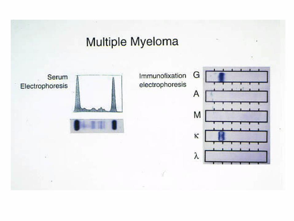
Electrophoresis and immunofixation of serum and urine proteins.
Quantification of intact paraprotein or free light chain in serum.β2 microglobulin quantification in the serum.Bone marrow aspirates and trephine biopsy.Skeletal survey.
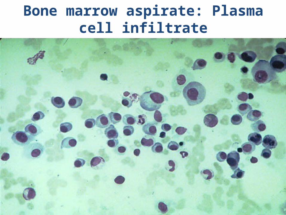
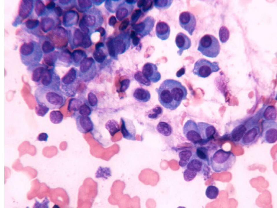
Bone marrow aspirate: Plasma cell infiltrate
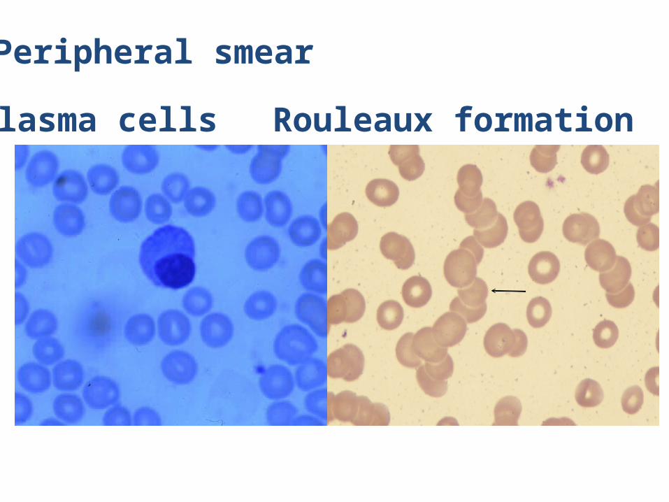
Peripheral smear
Plasma cells Rouleaux formation

Imaging
Plain radiography and MRI can detect the lytic bone lesions in multiple myeloma, whereas bone scan primarily detects osteoblastic activity

StagingThe International Myeloma Working Group has agreed on three simplified criteria for the diagnosis of symptomatic multiple myeloma. The diagnostic criteria proposed by the World Health Organization rely on major and minor diagnostic criteria . The International Myeloma Working Group criteria distinguish asymptomatic myeloma from symptomatic myeloma on the basis of whether end-organ damage has occurred. Therapy is not required in asymptomatic patients; however, they should undergo follow-up monitoring every 3 to 4 months. Laboratory testing should include serum protein electrophoresis, complete blood count, and serum creatinine and calcium measurement. Bone survey should be considered annually to evaluate
for asymptomatic bone lesions .Multiple myeloma used to be staged according to the levels of monoclonal protein production, anemia, extent of bone disease, presence of hypercalcemia, and renal failure. A simpler staging scheme uses measurement of β2 microglobulin concentration, a surrogate marker for myeloma tumor mass and renal function, and serum albumin to identify patients with good, intermediate, and poor
prognosis

Standards for Diagnosis Old Criteria
1 major and 2 minor criteria or
3 minor criteria including 1 and 2 in a patient displayingsymptoms of myeloma.
•Major criteria: 1 )biopsy-proven
plasmacytoma. 2 )bone marrow sample
showing >30% plasma cells. 3 )Elevated >30g/l
Monoclonal Ig levels in the blood
Minor criteria:
1)A bone marrow sample showing 10%-30% plasma cells.
2 )>30g/l Monoclonal Ig levels in blood
3)Imaging studies revealing lytic bone lesions
4)Low levels of normal Ig in serum
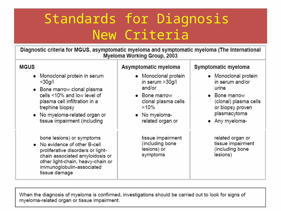
Standards for Diagnosis New Criteria

Standards for Diagnosis New Criteria
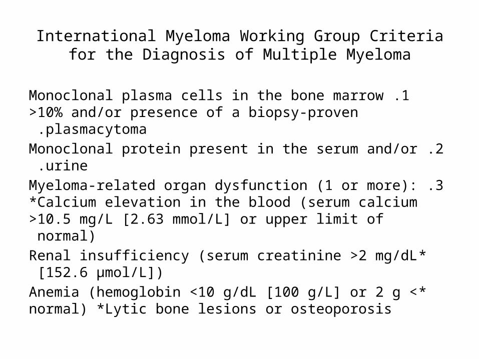
International Myeloma Working Group Criteria for the Diagnosis of Multiple Myeloma
1 .Monoclonal plasma cells in the bone marrow >10% and/or presence of a biopsy-proven plasmacytoma .
2 .Monoclonal protein present in the serum and/or urine .3 .Myeloma-related organ dysfunction (1 or more):
*Calcium elevation in the blood (serum calcium >10.5 mg/L [2.63 mmol/L] or upper limit of normal)
*Renal insufficiency (serum creatinine >2 mg/dL [152.6 µmol/L])
*Anemia (hemoglobin <10 g/dL [100 g/L] or 2 g < normal) *Lytic bone lesions or osteoporosis
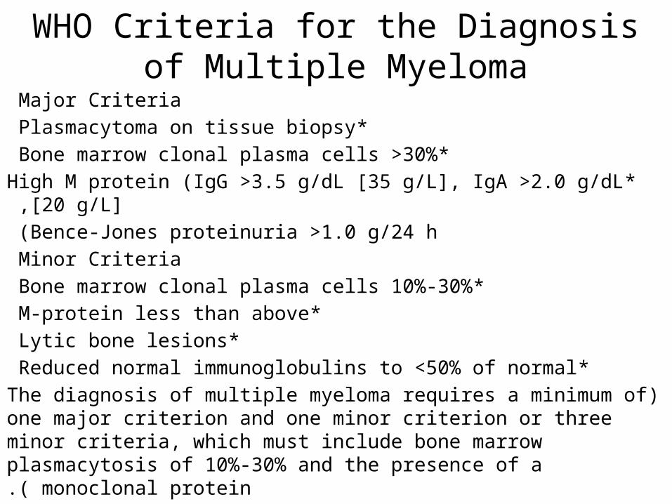
WHO Criteria for the Diagnosis of Multiple Myeloma
Major Criteria *Plasmacytoma on tissue biopsy
*Bone marrow clonal plasma cells >30% *High M protein (IgG >3.5 g/dL [35 g/L], IgA >2.0 g/dL [20 g/L] ,
Bence-Jones proteinuria >1.0 g/24 h )Minor Criteria
*Bone marrow clonal plasma cells 10%-30% *M-protein less than above
*Lytic bone lesions *Reduced normal immunoglobulins to <50% of normal
(The diagnosis of multiple myeloma requires a minimum of one major criterion and one minor criterion or three minor criteria, which must include bone marrow plasmacytosis of 10%-30% and the presence of
a monoclonal protein.)
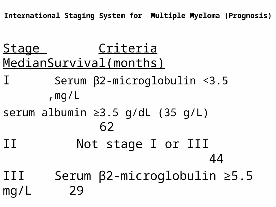
International Staging System for Multiple Myeloma (Prognosis)
Stage Criteria MedianSurvival(months) I Serum β2-microglobulin <3.5 mg/L , serum albumin ≥3.5 g/dL (35 g/L) 62
II Not stage I or III 44 III Serum β2-microglobulin ≥5.5 mg/L 29
Treatment
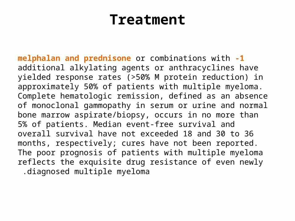
1 -melphalan and prednisone or combinations with additional alkylating agents or anthracyclines have yielded response rates (>50% M protein reduction) in approximately 50% of patients with multiple myeloma. Complete hematologic remission, defined as an absence of monoclonal gammopathy in serum or urine and normal bone marrow aspirate/biopsy, occurs in no more than 5% of patients. Median event-free survival and overall survival have not exceeded 18 and 30 to 36 months, respectively; cures have not been reported. The poor prognosis of patients with multiple myeloma reflects the exquisite drug
resistance of even newly diagnosed multiple myeloma .

2 -High-dose dexamethasone pulsing alone or in combination with continuous infusions of vincristine and doxorubicin (VAD) was regarded an effective regimen for patients whose disease has progressed while receiving
melphalan and prednisone .As initial therapy, vincristine and doxorubicin induce a more marked and rapid tumor cytoreduction than standard melphalan and prednisone or similar regimens. Overall
survival, however, is not improved .Not given now (abandoned)

3 -Interferon has been evaluated as part of remission induction and maintenance therapy as well as part of salvage therapy. Although interferon results in prolonged event-free survival, its impact on overall survival has
been negligible .Randomized and historically controlled trials have recently demonstrated the superiority of high-dose chemotherapy and autologous stem cell transplantation over standard treatment in achieving remission rates of up to 40% to 50% and extending median event-free and overall survival to 3 years and more than 5
years, respectively .

Supportive TherapyErythropoietin improves multiple myeloma–associated anemia in about 75% of patients, even in the absence of kidney disease, and is especially useful when anemia persists
because of irreversible renal failure .An important adjunct in treatment, especially for bone lesions, is parenteral bisphosphonate therapy, such as pamidronate or zoledronic acid, which delays the onset of skeletal-related events and seems to extend survival. Renal dysfunction and osteonecrosis of the jaw after dental procedures are potential adverse effects of long-term parenteral
bisphosphonate therapy .Hypercalcemia in patients with multiple myeloma is managed with saline hydration, corticosteroids, and bisphosphonates. Prophylactic immunizations for pneumonia and influenza are mandatory. Patients with recurrent bacterial infections may benefit from prophylactic monthly immune globulin infusion .

Novel Therapies
Thalidomide is a novel treatment in multiple myeloma that does not appear to act through a cytotoxic effect. Instead, it may inhibit the growth and survival of tumor cells, bone marrow stromal cells, or both; alter the profile of adhesion molecules and interactions between tumor cells and bone marrow stromal cells; modulate the cytokine milieu; inhibit angiogenesis; or increase the number of CD8 cells through its immunomodulatory effects. Thalidomide-based regimens have been evaluated in several trials and are being used in relapsed, refractory, and front-line settings. Therapy is generally well tolerated, and toxicities include fetal malformations, constipation, sedation, skin rash, peripheral neuropathy, fatigue, and thromboembolism. Discontinuation
of thalidomide is not always required to manage toxicities .

Lenalidomide, a potent analogue of thalidomide, offers great promise in myeloma. The toxicity profile is different from that of thalidomide, with less neuropathy, fatigue, and somnolence, but with increased risk for neutropenia and thrombocytopenia. Lenalidomide also can lead to
thromboembolism .Bortezomib, a proteasome inhibitor, inhibits cell growth, induces apoptosis, and allows myeloma cells in vitro to overcome drug resistance, and it has shown significant activity in myeloma. This agent was U.S. Food and Drug Administration approved as second-line therapy for myeloma in 2004. Major toxicities are fatigue, asthenia,
peripheral neuropathy, and thrombocytopenia .Novel therapies, alone and in combination regimens, have replaced oral melphalan and prednisone as well as vincristine and doxorubicin and can be used for initial
treatment and in relapsed or refractory disease .

VMP vs. VTP VT vs. VP in Elderly Newly Diagnosed Myeloma Patients (Phase III)
Primary endpoints: ORR in induction phase, CR in maintenance phase.Secondary endpoints: PFS, OS, efficacy in patients with high-risk cytogenetics.
Mateos MV, et al. ASH 2009. Abstract 3.
Patients olderthan 65 yrs of
age with newlydiagnosed
multiplemyeloma
(N = 260)
1st randomization 2nd randomization Up to3 yrsWk 31
*Thalidomide dosed at 50 mg/day on Days 1-15 of first 6-wk cycle.
VMPBortezomib 1.3 mg/m2 on Days 1, 4, 8, 11, 22, 25,29, 32 for one 6-wk cycle, then on Days 1, 8, 15, 22for five 5-wk cycles +Melphalan 9 mg/m2 QD on Days 1-4 each cycle +Prednisone 60 mg/m2 QD on Days 1-4 each cycle(n = 130)
VTPBortezomib 1.3 mg/m2 on Days 1, 4, 8, 11, 22, 25,29, 32 for one 6-wk cycle, then on Days 1, 8, 15, 22for five 5-wk cycles +Thalidomide* 100 mg/day +Prednisone 60 mg/m2 QD on Days 1-4 each cycle(n = 130)
VTBortezomib 1.3 mg/m2
on Days 1, 4, 8, 11 of90-day cycle +Thalidomide 50 mg/day(n = 91)
VPBortezomib 1.3 mg/m2
on Days 1, 4, 8, 11 of90-day cycle +Prednisone 50 mg every48 hrs(n = 87)

VMP → VT vs. VTP → VP in Elderly Newly Diagnosed Myeloma Patients: Outcomes
PFS significantly longer with VMP VT vs. VTP VP at both first and second randomizations (HR: 1.6; P = .008)
Mateos MV, et al. ASH 2009. Abstract 3.
0 15 25 35 450
40
60
80
100
20
10 20 30 40
VTPVP: 26.5VTPVT: NR
VMPVP: 32VMPVT: NR
VTPVPVTPVTVMPVPVMPVT
Treatment Group
PFS
0 15 25 35 450
40
60
80
100
20
10 20 30 40
VTPVP: 81VTPVT: 84VMPVP: 88VMPVT: 88
OS
Median PFS, Mos
2-Yr OS, %
Mos Mos
Patie
nts
With
out P
rogr
essi
on (%
)
Patie
nts
Surv
ivin
g (%
)
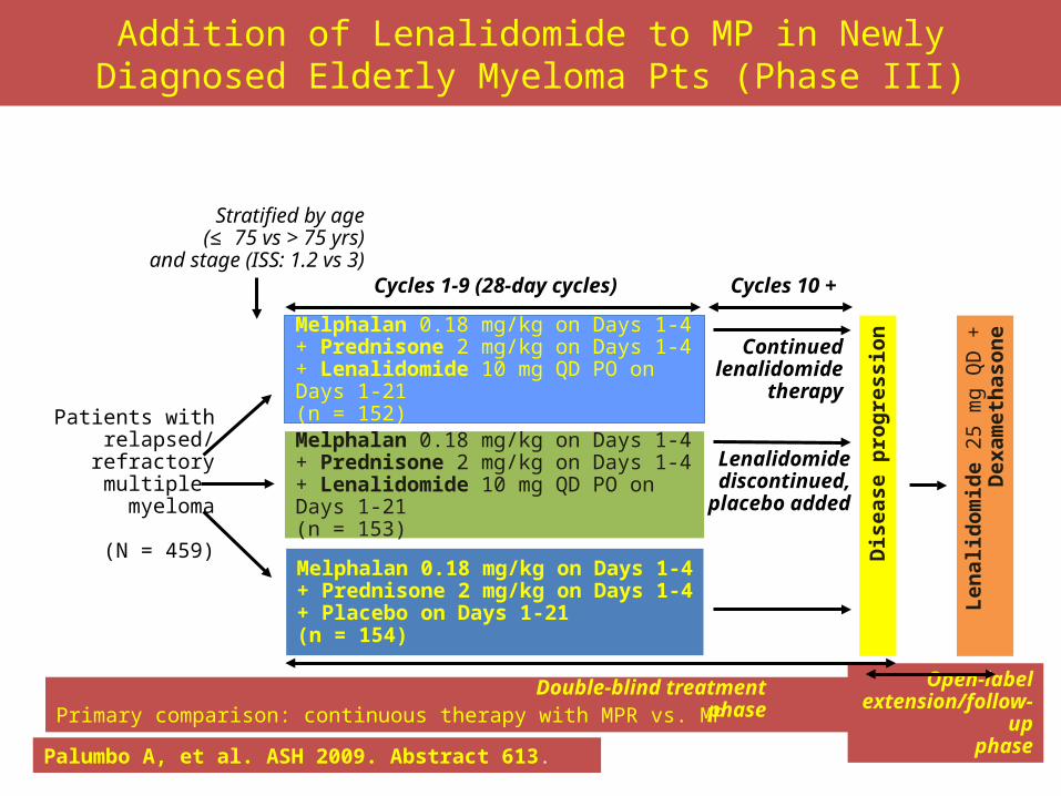
Addition of Lenalidomide to MP in Newly Diagnosed Elderly Myeloma Pts (Phase III)
Palumbo A, et al. ASH 2009. Abstract 613.
Primary comparison: continuous therapy with MPR vs. MP
Open-labelextension/follow-up
phase
Patients withrelapsed/refractorymultiple myeloma
(N = 459)Melphalan 0.18 mg/kg on Days 1-4 + Prednisone 2 mg/kg on Days 1-4 + Placebo on Days 1-21 (n = 154)
Len
alid
om
ide
25 m
g Q
D +
Dex
ame
thas
on
e
Dis
eas
e p
rog
ress
ion
Double-blind treatment phase
Melphalan 0.18 mg/kg on Days 1-4 + Prednisone 2 mg/kg on Days 1-4 + Lenalidomide 10 mg QD PO on Days 1-21
(n = 153)
Lenalidomidediscontinued,
placebo added
Melphalan 0.18 mg/kg on Days 1-4 + Prednisone 2 mg/kg on Days 1-4 + Lenalidomide 10 mg QD PO on Days 1-21
(n = 152)
Continuedlenalidomide
therapy
Cycles 1-9 (28-day cycles) Cycles 10 +
Stratified by age(≤ 75 vs > 75 yrs)
and stage (ISS: 1.2 vs 3)
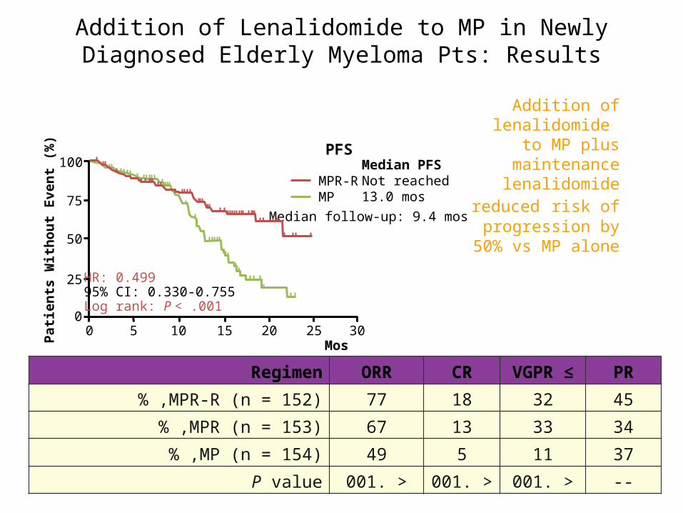
Addition of Lenalidomide to MP in Newly Diagnosed Elderly Myeloma Pts: Results
Addition of lenalidomide
to MP plus maintenance lenalidomide
reduced risk of progression by
50% vs MP alone
Palumbo A, et al. ASH 2009. Abstract 613.
PFS
Mos0 5 10 15 20 25 30
0
25
50
75
100
Patie
nts
With
out E
vent
(%)
HR: 0.49995% CI: 0.330-0.755Log rank: P < .001
Median follow-up: 9.4 mos
Median PFSNot reached13.0 mos
MPR-RMP
Regimen ORR CR ≥VGPR PR
MPR-R (n = 152)% , 77 18 32 45
MPR (n = 153)% , 67 13 33 34
MP (n = 154)% , 49 5 11 37
P value . >001 . >001 . >001 --
Monoclonal Gammopathy of Undetermined Significance
Monoclonal gammopathy of undetermined significance (MGUS) is found in approximately 1% to 2% of adults. The incidence is higher in patients older than 70 years and in blacks. MGUS is defined by the criteria listed and is characterized by a tendency towards development of multiple myeloma or a related malignancy at the rate of 1% to 1.5% per year. Whether these associations are pathogenetically related or are merely coincidental is not clear. MGUS is always asymptomatic and is usually discovered through incidental laboratory findings, most often, hyperproteinemia. A monoclonal spike is identified on subsequent protein electrophoresis.
Laboratory evaluation should include a complete blood count and measurement of serum calcium, creatinine, and electrolytes as well as immunoglobulin quantitation, serum and urine protein electrophoresis, serum and urine immunofixation electrophoresis, and a bone survey. Examination of the bone marrow is not always required, but, if done, should show less than 10% plasma cells.

IgG or IgA MGUS may progress to multiple myeloma, AL amyloidosis, or a related plasma cell disorder, whereas IgM MGUS may progress to a lymphoproliferative disorder (non-Hodgkin lymphoma, chronic lymphocytic leukemia, or Waldenström macroglobulinemia). A risk-stratification model to predict the risk of progression of MGUS uses three adverse risk factors: (1) a serum M protein level greater than or equal to 1.5 g/dL, (2) non-IgG MGUS, and (3) an abnormal serum free light-chain ratio. Using this model, the risk of disease progression over 20 years for patients with various combinations of
risk factors is as follows :three risk factors (high-risk MGUS) — 58%two risk factors (high–intermediate-risk MGUS) — 37%one risk factor (low–intermediate-risk MGUS) — 21%no risk factors (low-risk MGUS) — 5%

MGUS may rarely be associated with nonmalignant disorders, such as skin diseases (scleroderma, pyoderma gangrenosum), liver diseases (cirrhosis, primary biliary cirrhosis, hepatitis), rheumatologic diseases (rheumatoid arthritis, polymyositis,
polymyalgia rheumatica), and HIV infection .Patients with MGUS should be evaluated every 3 to 6 months for symptoms suggestive of myeloma and receive repeat laboratory tests every 6 to 12 months. Repeat bone marrow evaluation should be done only if other laboratory features suggest progression to multiple myeloma. Although there is no treatment to prevent progression of MGUS to multiple myeloma, monitoring for plasma cell dyscrasias is important because early recognition and treatment favorably
affect outcome .

Monoclonal Gammopathy of Undetermined Significance
The presence of a serum monoclonal protein (M-protein, whether IgA, IgG, or IgM), at a concentration ≤3 g/dL (30 g/L)
Bone marrow plasma cells <10% The absence of lytic bone lesions, anemia, hypercalcemia, and renal insufficiency related to the plasma cell proliferative process or related B-cell lymphoproliferative disorder

Waldenström Macroglobulinemia
Waldenström macroglobulinemia results from the proliferation of B lymphocytes that show maturation to plasma cells and is characterized by a lymphoplasmacytic infiltrate of the bone marrow, lymphadenopathy, anemia, neuropathy, organomegaly, IgM monoclonal gammopathy, and hyperviscosity syndrome. Hyperviscosity syndrome, which is related to the physicochemical properties of IgM, is identified by blurred vision; fatigue; mucosal bleeding caused by impaired platelet
function; heart failure; headache; and altered mentation .Funduscopic examination in these patients may show engorged retinal
veins .Plasmapheresis temporarily relieves acute symptoms and should be combined with specific treatment that may include chlorambucil, rituximab, and fludarabine or cladribine.