Embed Size (px)
Citation preview
MULTIPLE LARGE DNA MOLECULES OF Azospirillum
BY
ALVIN G. WOOD
A DISSERTATION PRESENTED TO THE GRADUATE COUNCILOF THE UNIVERSITY OF FLORIDA IN
PARTIAL FULFILLMENT OF THE REQUIREMENTSFOR THE DEGREE OF DOCTOR OF PHILOSOPHY
UNIVERSITY OF FLORIDA
1982
ACKNOWLEDGEMENTS
The author would like to thank his committee members,
Drs. Dennis E. Duggan, Francis C. Davis, Jr., Philip J.
Laipis, William W. Hauswirth and L.O. Ingram for their
valuable suggestions and criticisms.
He would also like to extend special thanks to
Dr. William B. Gurley for help with the hybridization
experiments and for the use of his laboratory equipment
and supplies.
TABLE OF CONTENTS
PAGE
ACKNOWLEDGEMENTS ii
ABSTRACT iv
CHAPTER I OCCURRENCE OF MULTIPLE LARGE DNA MOLECULESIN Azospirillum AND A METHOD FOR ISOLAT-ING THEM ON AGAROSE GELS 1
Introduction 1
Materials and Methods 3
Results 6
Discussion 11
CHAPTER II ACRIDINE ORANGE-INDUCED MUTATIONSIN Azospirillum 31
Introduction 31Materials and Methods 32Results 33Discussion 37
CHAPTER III Nif GENE HYBRIDIZATION STUDIESOF Azospirillum DNA MOLECULES. ... 47
Introduction 47Materials and Methods 4 8
Results and Discussion 51
APPENDICES
A NORMALIZATION OF GEL MOBILITY DATA . . 58
B REGRESSION LINE CALCULATIONS .... 59
LITERATURE CITED 60
BIOGRAPHICAL SKETCH 67
Abstract of Dissertation Presented to the Graduate Councilof the University of Florida in Partial Fulfillment of the
Requirements for the Degree of Doctor of Philosophy
MULTIPLE LARGE DNA MOLECULES OF Azospirillum
byAlvin G. Wood
May 19 82
Chairman: Dennis E. DugganMajor Department: Microbiology and Cell Science
Six strains of Azospirillum brasilense and two of
A. lipoferum were found to harbor as many as eight different-
sized circular DNA molecules ranging from 45 to 1500 mega-
daltons. Identification and separation of these very large
molecules were achieved by gently lysing bacteria in the wells
of vertical agarose gels, subjecting the lysate to electro-
phoresis at 2 mA for 6 h, and then continuing electrophoresis
at 15-30 mA for an additional 12-48 h. Optimal recovery
required lysis at 4°C in the presence of ribonuclease. The
technique has been used to isolate large DNAs from other
bacteria, including the chromosomes of Escherichia coli
and Agrobacterium tumefaciens .
Several types of mutants were isolated from acridine
orange-treated cultures of A. l ipoferum and A. brasilense .
Mutants displaying increased sensitivity to cadmium and
unable to grow on carbon-free media or on ethanol were all
found to have lost a specific plasmid. One of these strains
was shown to have suffered deletions in most of its remaining
DNA molecules. A mutant unable to grow on N2
or reduce
acetylene was isolated from the multiply-deleted strain, but
its DNA molecules showed the same electrophoretic mobilities
as those of its parent strain. Methionine-requiring auxo-
trophs, isolated at a high frequency from A. lipoferum cul-
tures, also possessed DNA molecules with unaltered mobilities.
Attempts were made to determine which Azospirillum DNA
molecule includes the genes controlling nitrogen fixation by
hybridizing a labeled recombinant probe to Southern blots
of wild type and mutant DNA molecules. The limited success
acheived with this technique indicates that the structural
genes for nitrogenase are carried on the largest Azospirillum
DNA molecule.
CHAPTER I
OCCURRENCE OF MULTIPLE LARGE DNA MOLECULESIN Azospirillum
AND A METHOD FOR ISOLATING THEM ONAGAROSE GELS
Introduction.
The genus Azospirillum comprises Gram-negative, free-living,
nitrogen-fixing bacteria found in association with roots of
cereal crops and tropical forage grasses (19). Field experiments
conducted at the University of Florida showed higher yields
of dry matter in Azospirillum-inoculated pearl millet and
guinea grass than in uninoculated controls (64). More
recently, Azospirillum inoculation has been reported to
enhance corn yields in Israel (46).
The potential agronomic value of this association has
prompted studies of carbon and nitrogen metabolism in Azo-
spirillum (2, 21, 27, 28, 38, 43, 44, 45, 48, 49, 51), and
the scores of strains isolated from various parts of the
world have been grouped, on the basis of DNA homology and
biochemical characteristics, into two species, A. lipoferum
and A. brasilense (67). Very little is known, however,
concerning the genetics of Azospirillum (26, 41, 55). In
particular, no system of genetic transfer exists which would
permit location of the genes controlling nitrogen fixation
and facilitate studies of their expression.
1
Our interest in developing a system of genetic transfer
for Azospirillum led us to examine several wild type strains
for the presence of plasmids. Our initial attempt to identify
plasmid DNA in Azospirillum (9) involved dye bouyant density
ultracentrifugation of alkaline denatured lysates (61) and
direct visualization of plasmids in the satellite bands by
electron microscopy (18). Open circular (OC) DNAs of various
contour lengths were seen, but the apparent multiplicity of
molecules in each strain and their large sizes relative to
the plasmid chosen as a size standard (ColE. ) made it difficult
to accurately assess the numbers and sizes of plasmids in
any given strain. However, plasmids with molecular weights
in excess of 300 megadaltons (Mdal) did appear to be present
in several Azospirillum strains.
This observation prompted us to try two electrophoretic
techniques specifically designed for the isolation of large
plasmids (10, 29), but neither of these permitted the isolation
of more than three plasmids from any Azospirillum strain.
We felt there was a strong possibility that very large
plasmids were present in these strains but that they were
being sheared during the mechanical manipulations, however
gentle, inherent in these procedures.
Therefore we adopted a method which is theoretically
the most gentle of all, that described by Eckhardt (22).
This technique differs fundamentally from other electrophoretic
techniques in that plasmid DNA is not extracted from cells
prior to electrophoresis. Rather, the bacteria are lysed
directly in the wells of the gel apparatus, resulting in
minimal nicking or breakage of covalently closed circular
(CCC) DNA. Using a modified version of this technique, we
have discovered the widespread occurrence of a multiplicity
of very large DNA molecules in strains of Azospirillum .
The present communication describes the electrophoretic
conditions necessary for the successful isolation of these
Azospirillum DNA molecules. Estimates of the sizes of the
molecules harbored by one Azospirillum strain are provided,
based on a comparison of their mobilities with those of
plasmids of known molecular weight. The very low mobilities
of some of the Azospirillum molecules suggest that they
represent small chromosomes rather than large plasmids.
Indeed, we have been able to isolate slowly-migrating DNA
bands from strains of Escherichia coli and Agrobacterium
tumefaciens , including two strains which do not harbor
plasmids. Evidence is provided indicating that the slowly-
migrating DNA bands isolated from Azospirillum represent CCC
DNA uncomplexed with protein.
Materials and Methods
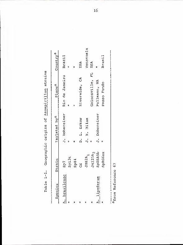
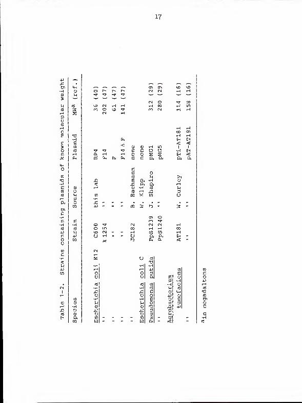
Bacterial strains . Table 1-1 lists the Azospirillum
strains examined for plasmid content. Table 1-2 lists other
bacterial strains harboring plasmids of known molecular
weights used for construction of the standard size curve.
Growth conditions . All bacteria were grown to early
stationary phase prior to harvesting for electrophoresis.
Azospirillum and A. tumefaciens strains were grown in a
succinate/mineral salts medium (50) supplemented with 0.01%
yeast extract. Pseudomonas putida and E. coli strains were
grown in nutrient broth (Difco) supplemented with 0.01%
yeast extract. Growth temperatures were 35°C for Azospirillum
and E. coli and 28°C for A. tumefaciens and P. putida .
Plasmid isolation and agarose gel electrophoresis . We
used an electrophoretic method based on that described by
Eckhardt (22). The protocol outlined here includes modifications
found to be necessary for optimal, reproducible visualization
of the electrophoretic bands representing the largest DNA
molecules.
Vertical gels were cast with 0.6% or 0.7% molten agarose
(BioRad Standard Low -m ) in Tris/borate/EDTA electrophoresis
(E) buffer (40). The agarose was tempered at 42°C for 20
min prior to casting the gel in order to minimize contracture
during solidification. The plastic comb used to form wells
had 16 teeth (13x9x1.5 mm). The gel was submerged in E
buffer and allowed to age at least 4 h at 4°C prior to
removal of the comb. Spraying the comb lightly with PAM
(3oyle-Midway ) prevented agarose from sticking to it during
removal.
Except where otherwise noted, wells were loaded in the fol-
lowing manner. One milliliter of cell culture was centrifuged
for one minute in a microcentrifuge (Fisher Model 235). The
supernatant solution was pipetted off using a vacuum aspirator
and the pellet was resuspended in 20-100 pi of 20% ficoll in
E buffer. Ten microliter aliquots of the cell suspensions were
added to wells preloaded with 15 y 1 of a solution containing
20% ficoll, 10 yg/ml lysozyme (Sigma), 100 pg/ml ribonuclease
A (Sigma), and 0.05% xylene cyanol FF (Kodak) in E buffer.
No attempt was made to mix the cell suspension with the
lysozyme solution inside the well. The cells were allowed
to interact with the lysozyme mixture at 4°C for a minimum
of 30 min, and then 30 u 1 of 10% ficoll, 1% SDS in E buffer
was added, followed by 50 y 1 of 5% ficoll, 1% SDS in E buffer.
A current of 2 mA was applied for a minimum of 6 h, and
then the current was raised to 15-30 mA (50-100 V at 4°C).
Electrophoresis was continued for 12-48 h depending on the
voltage used and the degree of molecular separation required.
The large capacity (2.5 1) of the buffer reservoirs of our
electrophoresis apparatus made recirculation of buffer un-
necessary.
Photography . Gels were stained for a minimum of 30 min
in 0.5 ug/ml ethidium bromide and visualized with either a
254 nm hand-held UV light (UV Products) or a 300 nm trans-
illuminator (Fotodyne). Photographs were taken through #4
and #29 Wratten filters, using Type 57 film (Polaroid).
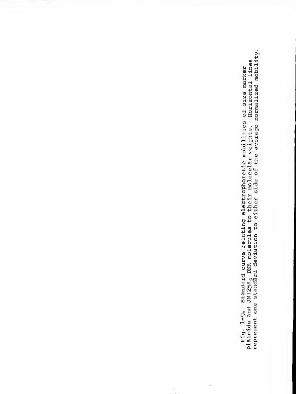
Standard curve construction . Mobility data from 19 gels
were normalized to the mobility data of the gel illustrated
in Fig. 1-6 according to the method described by Hansen and
Olsen (29, Appendix A) except that absolute mobilities
(distances of plasmid migration from origin) rather than
relative mobilities (absolute mobilities divided by gel
lengths) were used in the calculations (see Appendix A).
The number of DNA bands in common between the normalized and
standard gels ranged from 7 to 15 with an average of 12.
The logarithm of the average normalized mobility of a given
standard plasmid was plotted against the logarithm of the
molecular weight of that plasmid, and a least squares regres-
sion line was calculated (see Appendix B). This regression
line was then used to estimate the molecular weights of the
Azospirillum molecules as well as the presumed chromosomes
of E. coli and A. tumefaciens .
Plasmid nomenclature . The molecule having the lowest
mobility in each Azospirillum strain has been designated
pAZl; molecules are then numbered in order of increasing
mobility. All the molecules of a given strain are suffixed
with that strain designation. For example, the smallest
plasmid of Spl3t is pAZ6-Spl3t.
Results
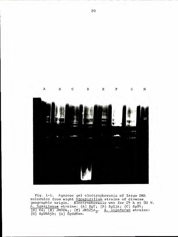
Mult iplicity of DNA molecules found in Azospirillum
strains . Figures 1-1 and 1-2 illustrate the electrophoretic
banding patterns obtained from eight geographically diverse
isolates of A. brasilense and A. lipoferum lysed in the
wells of vertical agarose gels. Each strain has a character-
istic array of DNA molecules of various mobilities, an
observation which can be used for purposes of identification.
Every strain harbors two molecules whose extremely low
7
mobilities suggest that they represent small chromosomes
rather than plasmids. Under optimal conditions, the recovery
of these DMAs is highly reproducible except for the small
plasmid which bands in the region of linear DNA in some gels
(Fig. 1-1, lane D; Fig. 1-2, lanes C and D)
.
A comparison of Figs. 1-1 and 1-2 indicates that very
long periods of electrophoresis are necessary in order to
achieve separation of all the DNA bands, in accordance with
what would be expected for very large DNAs. The increase in
resolution achieved by increasing electrophoresis time is,
unfortunately, accompanied by a tendency for the most slowly-
migrating bands to become faint or disappear altogether
(data not shown). This suggests, however, that the material
is those bands is fragile, presumably because of its high
molecular weight.
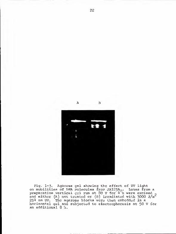
Effect of ultraviolet light on mobilities of JM125A2
molecules . Figure 1-3 compares the mobilities of UV-irradiated
and unirradiated molecules of JM125A2
. The JM125A2molecules
were isolated in the usual manner except that electrophoresis
was terminated after 4 h at 80 V. Blocks of agar were cut
from lanes of the gel, extending from the well to the position
of the tracking dye. One of the unstained agar blocks
2(lane B) was subjected to a dose of approximately 3000 J/m
of 25 4 nm UV, while the other (lane A) was untreated. This
dose should have been sufficient to introduce at least one
chain break into every CCC DNA molecule in the irradiated
gel (6, 7, 33, 56). Both treated and untreated agarose
blocks were then imbedded in a horizontal agarose gel and
subjected to electrophoresis for 8 h at 50 V. As indicated
in Fig. 1-3, UV irradiation converted the DNA molecules from
forms capable of movement through an agarose gel into forms
incapable of such movement. Presumably, this represents the
conversion of CCC DNA into OC DNA.
Effects of enzymatic treatments on DNA recovery . In
order to acquire information concerning the physical relation-
ships of the DNA molecules to other cellular components, the
roles of lysozyme, RNase, and protease (Sigma Type VI) in
optimal DNA recovery were assessed. Figure 1-4 shows that
neither the addition of protease to the standard cell
mixture nor the elimination of lysozyme had an appreciable
effect on DNA recovery or mobility. The elimination of
RNase, however, resulted in failure to recover pAZl and pAZ2
as well as poor recovery of pAZ3.
Effect of cell mass on DNA isolation . Figure 1-5 illus-
trates the result of an experiment in which cell suspensions
of JM125A- and AT181 were serially diluted prior to loading
the wells. The smearing of the bands in lanes A and E
7appears to be due to overloading. The use of only 10 cells
(lanes D and H) allowed visualization of all the DNA bands
in this experiment. However, in other experiments (data not
7shown) using 10 cells resulted in very faint bands, particu-
larly for the smaller molecules. Optimal recovery was
pusually achieved with 10 cells.
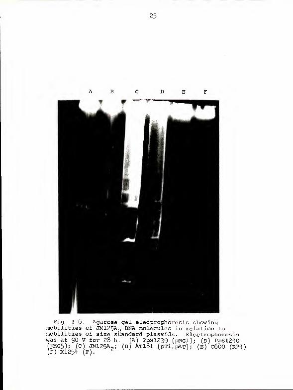
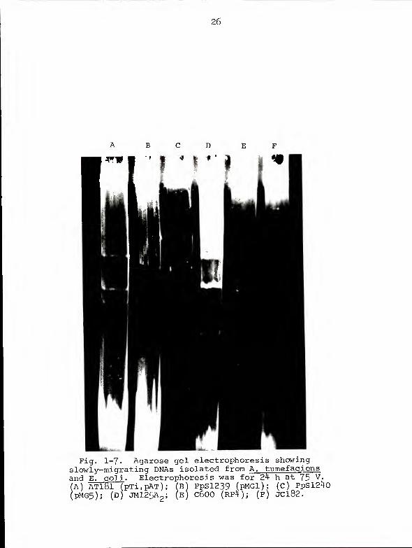
Size estimates of DNA molecules . The mobilities of the
DNA molecules of JM125A?
in relation to plasmids of known
molecular weight and slowly-migrating DNAs of other bacteria
are illustrated in Figs. 1-6, 1-7, and 1-8. Based on Fig.
1-6, JM125&2 appears to harbor five molecules larger than
the largest standard molecule (pMGl, 312 Mdal ) . Recovery of
the larger JM125A- molecules was poor in Figs. 1-6 and 1-7;
these are included primarily to show the slowly-migrating
DNAs isolated from E. coli (Fig. 1-7, lane F ; Fig 1-3, lanes
E, G, and H) and A. tumefaciens (Fig. 1-7, lane A). The two
slowly-migrating DNAs recovered from A. tumefaciens have
been given the designations pXXl-AT181 and pXX2-ATl81. The
isolation of these slowly-migrating DNAs is not completely
reproducible (Fig. 1-6, lanes E and F ; Fig 1-7, lane E;
Fig. 1-8, lanes A and D). Indeed, the difficulty of isola-
ting the molecules appears, in our experience, to be inver-
sely related to mobility. Thus, our rate of success in
isolating slowly-migrating DNAs from Azospirillum is 90% or
better, but our success rate with the slowly-migrating
E. coli DNA has never exceeded 5 0%. We have never isolated
a slowly-migrating DNA band from either of the two Pseudo-
monas strains used in the present study. This failure may
be related to an observed tendency for these strains to lyse
prematurely.
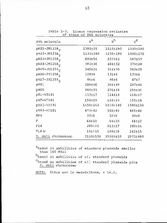
Mobility data from 20 agarose gels were normalized and
used to construct a standard curve relating electrophoretic
mobility and molecular weight (Fig. 1-9). Since reports in
the literature had suggested that CCC DNAs larger than 80
10
Mdal (72) or 140 Mdal (29) migrate faster than predicted
from linear extrapolations of standard curves based on
smaller CCC DNAs, we initially calculated a regression line
not including pMGl and pMG5. When this line was used to
estimate the sizes of the Azospirillum molecules, however,
it seemed impossible that the values obtained could be
underestimates. We therefore recalculated regression data
with the large Pseudomonas plasmids included, and again with
the slowly-migrating band from E. coli included and assigned
a molecular weight of 2800 Mdal (8,14,28). The three sets
of regression line estimates are summarized in Table 1-3.
Figure 1-9 is a graph of regression line B, chosen because
it includes only those molecules measured by electron micro-
scopic contour length.
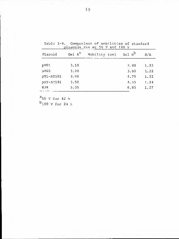
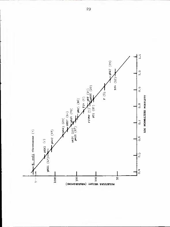
Effect of voltage gradient on regression estimates . If
the larger molecules examined in the present study were
really migrating faster than predicted, this effect should
be more pronounced at higher than at lower voltages (24).
We were particularly interested in this possibility since,
in our attempts to optimize electrophoresis conditions, we
had used voltages ranging from 50 V to 100 V and we wished
to include data from as many gels as possible in our regression
line calculations. Figure 1-10 compares the mobilities of
five standard plasmids run at 50 V for 42 h with mobilities
of the same plasmids run at 100 V for 24 h. The two curves
are nearly parallel and neither displays a convincing change
11
of slope above 140 Mdal. Table 1-4 indicates that, for each
standard plasmid, the ratio of mobility at 100 V to mobility
at 50V is a constant value.
Discussion
Eckhardt first described in situ lysis of bacteria in
agarose gels as a rapid method for plasmid isolation (22).
We have sacrificed the rapidity of the technique but exploited
its gentleness in order to isolate very high molecular
weight CCC DNAs from Azospirillum and other bacteria.
Our initial excitement in isolating slowly-migrating
DNAs on agarose gels was tempered with concern that the low
mobilities might reflect an open circular nature or some
protein interaction rather than large size. The electro-
phoretic behavior of the JM125A„ molecules subsequent to UV
irradiation, however, strongly indicates that they are
covalently closed and supercoiled. A DNA/protein interaction,
while not strictly ruled out by the failure of protease to
alter the electrophoretic mobilities of the molecules, seems
unlikely in view of this result. Furthermore, such an
association would have to be resistant to dissociation by
the SDS which quickly migrates from the upper ficoll layers
down through the DNA-containing region of the gel during
electrophoresis. Thus, the slowly-migrating bands do not
appear to represent relaxation complexes of the type isolated
from plasmid-bearing strains of E. coli , which dissociate
yielding OC DNA when exposed to SDS or protease (13).
12
A comparison of the DNA molecules isolated from
A. brasilense strains Sp7 and Spl3t provides further evidence
that the slowly-migrating DNAs are not simply isomeric forms
of smaller plasmids. Since these two strains were isolated
from the same region of Brazil and display nearly identical
electrophoretic banding patters, there is a good possibility
that they are isogenic except for the occurrence of pAZ6-Sp7
in one strain. If so, none of the slowly-migrating DNAs of
either strain could represent an isomeric form of this
relatively small molecule.
The requirement of RNase treatment for isolation of the
larger Azospirillum DNAs suggests that, in their native
forms, these molecules are attached via RNA to some cellular
component in a manner precluding entry into the gel matrix.
It is also possible that the larger Azospirillum molecules
are attached to one another via RNA. Assuming the likelihood
that essential genes are carried on the largest two or three
molecules, some mechanism to ensure cosegregation of newly-
replicated molecules into daughter cells would appear to be
necessary. Molecules as large as pAZl and pAZ2 might further
be expected to exist inside the cell in condensed, folded
states. These considerations lead us to postulate that pAZl
and pAZ2 (and perhaps pAZ3) are arranged in a chromosomal
structure closely resembling that believed to occur in
E. coli (54, 73, 74). The only difference between the two
"nucleoid" structures would be that in E. coli the RNA-
stabilized domains comprise a single, continuous DNA molecule,
13
whereas in Azospirillum these domains are divided into two
or three continuous DNA molecules. The remaining Azospi-
rillum molecules might form nonintegrative associations with
the Azospirillum nucleoid analogous to those described
between other large, stringently controlled plasmids and
their host chromosomes (34, 35).
We have attempted to estimate the sizes of the DNA
molecules of one Azospirillum strain (JM125A2* b^ comparing
their mobilities to those of plasmids whose sizes have been
calculated from electron microscopic contour length measure-
ments. A problem with this effort arose in that five of the
JM12 5Ao molecules migrated more slowly than pMGl, the lar-
gest standard plasmid available. Their sizes, therefore,
had to be estimated from a linear extrapolation of our
standard curve (Fig. 1-9) and so must be considered only
approximate.
Some investigators have cautioned against standard
curve extrapolations on the grounds that CCC DNAs larger
than 80 Mdal (72) or 140 Mdal (29) migrate faster than
predicted. For several reasons, this appears not to be the
case under our electrophoresis conditions. First, it is
difficult to believe that the calculated values for the
larger Azospirillum molecules could be underestimates.
Second, these estimates change only modestly when the Pseu-
domonas plasmids are disallowed or when the chromosome of
E. coli is included in the regression line calculation
(Table 1-4). Third, the hypothetical nonlinearity of the
standard curve should have been greater for a gel run at 100
14
V than for a gel run at 50V, but Table 1-4 indicates that
the relative mobilities of the standard plasmids were nearly
identical at the two voltages. Finally, a theoretical
justification for nonlinearity of standard curves for CCC
DNAs in the high molecular weight range has not been advanced.
The explanation offered for the fast mobilities of high
molecular weight linear DNAs, i.e., "end-on" migration (1,
25), would seemingly not apply to high molecular weight CCC
DNA.
From the estimated sizes of the Azospirillum molecules
and assuming one copy of each per cell, the full genetic
complement of DNA for these bacteria appears to be approximately
94.3x10 daltons, some 50% greater than the corresponding
value for E. coli (8, 14, 28). At present, we can only
speculate as to the reason for this discrepancy. The large
complement of DNA may simply reflect the metabolic diversity
of these bacteria; Azospirillum species are capable of
carrying out most of the known nitrogen transformations (19,
44, 45), can grow heterotrophically (49) or autotrophically
(60), and tolerate the full range of oxygen tensions from
fully aerobic (49) to anaerobic with nitrate as terminal
electron acceptor (45). Alternatively, some of the Azospirillum
DNA may be redundant. This redundancy, if it does occur,
could provide a basis for recombination among Azospirillum
DNA molecules, underlying a potential mechanism for the
evolution of new strains.
The application of the modified Eckhardt technique to
bacteria harboring size standard plasmids led to the discovery
15
that slowly-migrating DNAs could be isolated from species
other than Azospirillum . Molecules with apparent molecular
weights of 500 and 1800 Mdal were isolated from A. tumefaciens
AT181 along with the two previously described plasmids.
These four molecules may well represent the full genetic
complement of this A. tumefaciens strain since the sum of
Qtheir estimated sizes is 2.6x10 daltons. Hence, pXXl-AT181
may, in fact, represent the Agrobacterium chromosome. The
slowly-migrating DNA isolated from E. coli strains appears
to represent the E. coli chromosome since it displayed an
appropriate mobility and could be recovered from both plasmid-
harboring and plasmidless strains. Isolation of intact
E. coli chromosomes by ultracentrifugation of gently lysed
cells through neutral sucrose gradients has been described
by others (66, 73)
.
In summary, we have demonstrated that strains of
Azospirillum harbor unique arrays of large DNA molecules.
The probability that these molecules comprise the full
genetic complement of their host bacteria suggests that
Azospirillum should be considered a multichromosomal
prokaryote. Arrangement of genetic material in this fashion
contrasts sharply with the situation in E. coli , in which
more than 90% of the DNA is carried on a single large molecule.
The common assumption that the DNA of most prokaryotes is
arranged as it is in E. coli may reflect, to a certain
extent, the previous lack of a suitable protocol for isolating
CCC DNAs larger than 500 Mdal.
16
17
4J
13
Table 1-3. Linear regression estimatesof sizes of DNA molecules
DNA molecule B
pAZl-JM125A
pAZ2-JM125A
pAZ3-JM125A
pAZ4-JM125A
pAZ5-JM12 5A„
pAZ6-JMl25A
pAZ7-JM125A2
pMGl
pMG5
pTi-AT181
pAT-AT181
pXXl-AT181
PXX2-AT181
RP4
F
F14
F14 AF
E. coli chromosome
1380+25
1130±180
600+56
385±30
345±26
128±6
46±6
289±46
260±35
113+17
154±19
1650+160
473+42
33 + 6
64±10
205+16
141+15
3150±530
1510±290
1230±190
637±62
404±32
362±30
131 + 6
46±6
302±49
270±38
114±18
158+19
18101180
500±45
32±6
64±10
212±17
144±16
3500±610
1330+240
1090±170
587+53
379±28
342±25
130±6
47±7
287+46
259+35
114±17
155±13
1580+150
465+40
34±6
66±10
205116
142115
29701490
based on mobilities of standard plasmids smallerthan 160 Mdal
based on mobilities of all standard plasmids
'based on mobilities of all standard plasmids plusE. coli chromosome
NOTE: Sizes are in megadaltons, + ls.d.
19
Table 1-4. Comparison of mobilities of standardplasmids run at 50 V and 100 V
Plasmid Gel Aa Mobility (cm) Gel B° B/A
pMGl
20
Fig. 1-1. Agarose gel electrophoresis of large DNAmolecules from eight Azospirillum strains of diversegeographic origin. Electrophoresis was for 24 h at 80 V.A . brasilense strains: (A) Sp7; (b) Spl3t; (c) Sp84;D) Cd; (E) JM82A n ; (f) JM125A . A. lipoferum strains:
SpUSA5bE) JM82A,; (F) JM125A
2.
5b; (H) SpRG6xx.''
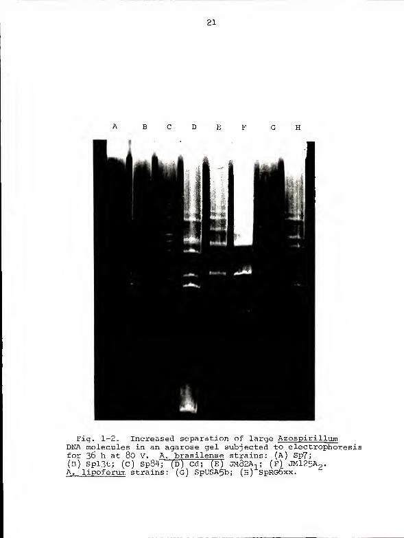
21
Fig. 1-2* Increased separation of large AzospirillumDNA molecules in an agarose gel subjected to electrophoresisfor 36 h at 80 V. A. brasilense strains: (A) Sp7;(B) Spl3t; (C) Sp84; (D) Cd; (E) JM82A
1 ; (f) JM125A2
.
A. lipoferum strains: (g) SpUSA5bj (h) SpRGbxx.
22
Fig. 1-3- Agarose gel showing the effect of UV lighton mobilities of DNA molecules from JMl25Ap. Lanes from apreparative vertical gel run at 80 V for 4 h were excised
?and either (A) not treated or (B) irradiated with 3000 J/m25^ nm UV. The agarose blocks were then embedded in ahorizontal gel and subjected to electrophoresis at 50 V foran additional 8 h.
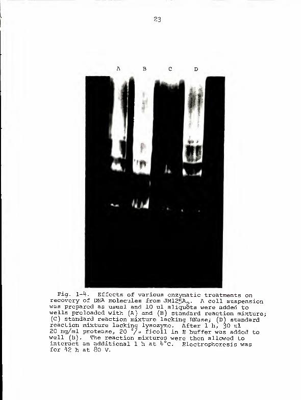
23
I
24
A B CD EFGH
Fig. 1-5 . Effect of cell number on isolation of DNAmolecules from (A-D) JM125A
2and (E-H) ATl8l. Electro-
phoresis was at 80 V for 24 h. (A) and (e)q
ft
3x10 cells; (B) and (F) 10 cells; (c) and (G) 3x10
cells; (D) and (h) 10^ cells.
7
25
Fig. 1-6. Agarose gel electrophoresis showingmobilities of JM125A,mobilities of size s\
was at 90 V for 28 h.(pMG5); (C) JM125AP ;
(F) X125^ (F). d
DNA molecules in relation toandard plasmids. Electrophoresis
(A) PPS1239 (pMGl); (B) PpSl2^0(D) AT181 (pTi,pAT); (E) C600 (RP4)
26
Fig. 1-7. Agarose gel electrophoresis showingslowly-migrating DNAs isolated from A. tumefaciensand E. coli . Electrophoresis was for 2)\ h at 75 v -
(A) AT181 (pTi^pAT); (B) PpSl239 (pMGl)j (c) PpS1240(pMG5); (D) JM125A
2 ; (E) C600 (RP 1!); (F) JCl82.
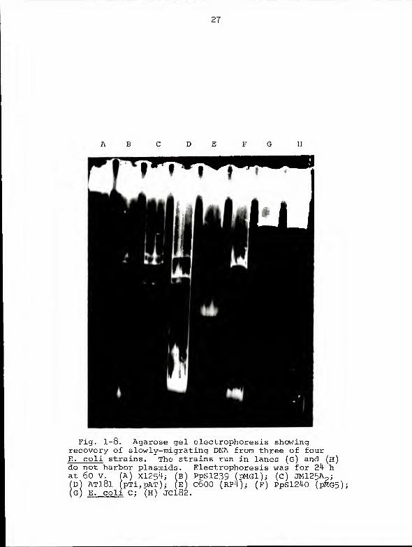
27
Fig. 1-8. Agarose gel electrophoresis showingrecovery of slowly-migrating DNA from three of fourE. coli strains. The strains run in lanes (g) and (h)do not harbor plasmids. Electrophoresis was for 24 hat 60 V. (A) X1254; (b) PpSl239 (pMGl); (c) JM125A
(S)D) AT181 (pTi,pAT); (E) C600 (RP4); (f) PpSl240 (pMG5);G) E. coli C; (H) JC182.
29
(SNoxivavoaw) xhoism avinoaiow
30
- 500
pMGl
- 100
50
J. 1 10.5 0.6 ,7
LOG MOBILITY (cm)
0.8 o.q
Fig. 1-10. Comparison of mobilities of standardplasmids in gels run at different voltages. The 50 Vgel was run for 42 h and the 100 V gel for 24 h.
CHAPTER IIACRIDINE ORANGE-INDUCED MUTATIONS IN Azospirillum
Introduction
Genetic material in the genus Azospirillum appears to
be arranged in a manner unusual for bacteria. Rather than
possessing a single large chromosome with or without acces-
sory DNA in the form of plasmids, Azospirillum strains
harbor unique arrays of up to eight different-sized co-
valently closed circular (CCC) DNA molecules, the largest
estimated to be 1500 megadaltons (Mdal) (see Chapter I).
The occurrence of so many molecules intermediate in size
between what is normally considered to be very large for a
plasmid (300 Mdal) and what would be considered a small
chromosome (1500 Mdal) makes the usual plasmid/chromosome
dichotomy less than obvious in these bacteria.
Plasmid DNA is generally defined as encoding functions
not essential for cell growth under usual conditions.
Growth of plasmid-harboring bacteria in the presence of
acridine orange (AO) often permits the isolation, at an in-
creased frequency, of strains which have been "cured" of the
plasmid (30). We therefore grew a representative strain of
A. lipoferum and one of A. brasilense in AO-containing
medium and tested individual colonies for a variety of
nonessential phenotypic traits. Since the capacity for
31
32
nitrogen fixation can be considered nonessential, the potentially
mutant colonies were tested for growth on nitrogen-free
medium. In addition, because of the blurring of the distinction
between plasmid and chromosome in these bacteria, all colonies
were screened for auxotrophy. Mutant colonies identified in
this manner were analyzed for DNA content by agarose gel
electrophoresis.
The present communication describes the successful cor-
relation of three phenotypic properties with the presence of
a specific plasmid in both Azospirillum strains examined.
Furthermore, we report the isolation of a mutant having
suffered multiple deletions in its DNA molecules and the
isolation of both Nif" and auxotrophic strains for which no
obvious loss of DNA could be demonstrated.
Materials and Methods
Bacteria . A. brasilense JM125A2
and A. lipoferum
SpUSA5b were obtained from N. R. Krieg. Mutant strains
derived from them are listed in Table 2-1.
Media. The succinate/mineral salts medium described by
Okon et al . (50) was used for nitrogen-free growth. The
same medium supplemented with 0.5% (NH.KSO, was used as
minimal agar (MA). Carbon-free agar (CFA) was MA lacking
succinate. Carbon source utilization tests were done on MA
with the appropriate carbon source replacing succinate at a
concentration of 1%. Sensitivities of bacteria to inhibitors
were determined on nutrient agar (Difco) supplemented with
0.01% yeast extract and filter-sterilized antibiotics or
separately-autoclaved heavy metal salts.
33
Curing . Bacteria were treated with AO as described by
Hirota (30), except that the pH was 8.0. The maximum concen-
tration of AO which consistently permitted growth was 2.5 p g/ral
Detection of DNA molecules . The electrophoretic method
used to detect the various DNA molecules present in wild
type and mutant strains has been extensively described
elsewhere (see Chapter I).
Results
Table 2-1 lists the types of mutants isolated from cul-
tures of A. brasilense JM125A- and A. lipoferum SpUSA5b
grown in 2.5 P g/ral AO at pH 8.0 and the approximate fre-
quencies with which they were found to occur in such cul-
tures. No spontaneous mutants (from untreated cultures)
were found.
Isolat ion of JM125A mutants with the T phenotype .
Originally, we screened JM125A„ colonies derived from AO-
treated cultures for a number of properties: 1) growth on
minimal agar plates containing arabinose, galactose, ethanol,
or butanol as sole carbon source; 2) sensitivity to tri-
methoprim (since wild type Azospirillum strains are resistant
(3)); and 3) growth on succinate/mineral salts media with and
without (NH4
)2S0
4. No auxotrophic, Nif~, Ara~, Gal", or
trimethoprim-sensitive mutants were found. However, mutants
unable to grow on ethanol or butanol were isolated at a high
frequency (Table 2-1). Further testing showed that acetate
or acetaldehyde would support growth of the mutants.
34
Somewhat surprisingly, wild type colonies grew on the
medium (intended as a control) to which no carbon source had
been added. Growth on the carbon-free agar was slower and
less luxurious than growth on any of the media containing
added carbon. The mutant colonies failed to grow appreciably
on the carbon-free medium. Although it appeared from these
observations that Azospirillum was capable of autotrophic
growth and that the mutants had lost this ability, both wild
type and mutant strains produced a visible band of growth
when a small inoculum (100 cells) was introduced into the
carbon-free soft agar (0.02%) under a predominantly H^/CO^
atmosphere. Others have recently described autotrophic
growth of Azospirillum (60).
Since heavy metal ion resistance is a characteristic
often associated with plasmids in other bacteria (12, 42,
63, 71), we assessed the sensitivities of the mutant and
wild type strains to the salts of seven heavy metals. No
differential sensitivity to Ag , Cu , Co , Pb , Hg , or
Ni was observed; however, 3x10 M Cd permitted growth
of the wild type but not the mutant strains. Further analysis
showed the mutants to be 3 0-fold more sensitive than the
wild type to Cd+
(Table 2-2). We have designated the "T"
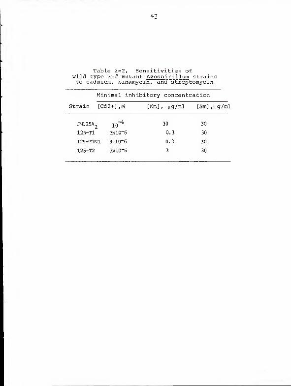
phenotype to represent the triad of cadmium sensitivity,
inability to metabolize alcohols, and failure to grow on
carbon-free agar under ambient atmosphere.
35
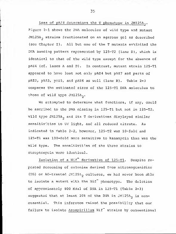
Loss of pAZ4 determines the T phenotype in JM125A?
.
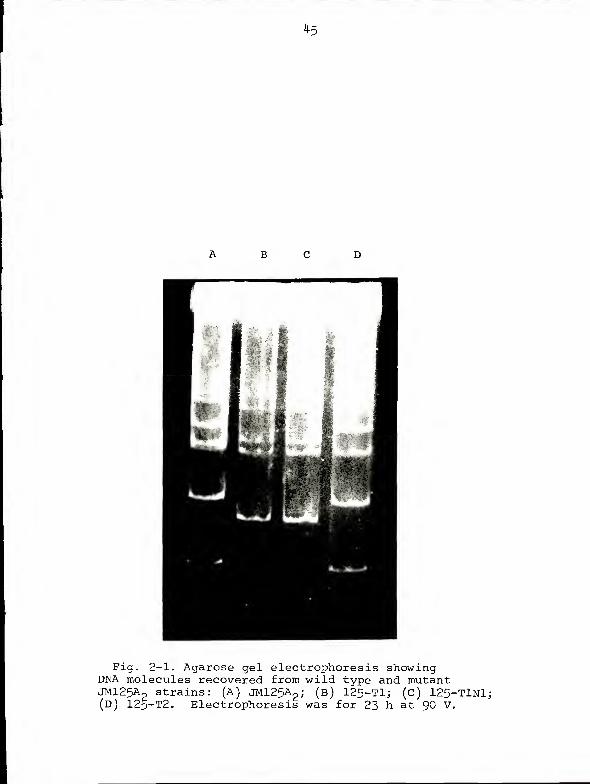
Figure 2-1 shows the DNA molecules of wild type and mutant
JM125A„ strains fractionated on an agarose gel as described
(see Chapter I). All but one of the T mutants exhibited the
DNA banding pattern represented by 125-T2 (lane D), which is
identical to that of the wild type except for the absence of
pAZ4 (cf. lanes A and D). In contrast, mutant strain 125-T1
appeared to have lost not only pAZ4 but pAZ7 and parts of
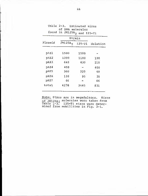
pAZ2, pAZ3, pAZ5, and pAZ6 as well (lane B). Table 2-3
compares the estimated sizes of the 125-Tl DNA molecules to
those of wild type JM125A„.
We attempted to determine what functions, if any, could
be ascribed to the DNA missing in 125-Tl but not in 125-T2.
Wild type JM125A and its T derivatives displayed similar
sensitivities to UV light, and all reduced nitrate. As
indicated in Table 2-2, however, 125-T2 was 10-fold and
125-Tl was 100-fold more sensitive to kanamycin than was the
wild type. The sensitivities of the three strains to
streptomycin were identical.
Isolation of a Nif derivative of 125-Tl . Despite re-
peated screening of colonies derived from nitrosoguanidine
(NG) or AO-treated JM125A- cultures, we had never been able
to isolate a mutant with the Nif phenotype. The deletion
of approximately 800 Mdal of DNA in 125-Tl (Table 2-3)
suggested that at least 20% of the DNA in JM125A- is non-
essential. This inference raised the possibility that our
failure to isolate Azospirillum Nif strains by conventional
36
mutagenesis might be due to occurrence of the nif genes in
more than one copy. Therefore, we screened 2000 colonies
derived from AO-treated 125-Tl cultures for growth on
succinate/mineral salts medium with and without (NH.)„SO..
One mutant was isolated which grew on the minimal medium
only when nitrogen was supplied. This strain (125-T1N1)
also failed to reduce acetylene even in the presence of low
concentrations of (NH.)2SO. or yeast extract. The electro-
phoretic mobilities of its DNA molecules, however, could not
be distinguished from those of its parent strain (Fig. 2-1,
lanes B and C )
.
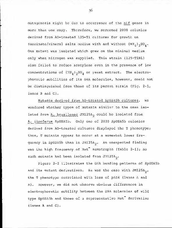
Mutants derived from AO-treated SpUSA5b cultures . We
wondered whether types of mutants similar to the ones iso-
lated from A. brasilense JM125A?could be isolated from
A. lipoferum SpUSA5b. Only one of 2000 SpUSA5b colonies
derived from AO-treated cultures displayed the T phenotype;
thus, T mutants appear to occur at a somewhat lower fre-
quency in SpUSA5b than in JM125A-. An unexpected finding
was the high frequency of Met-
auxotrophs (Table 2-1); no
such mutants had been isolated from JM125A2
.
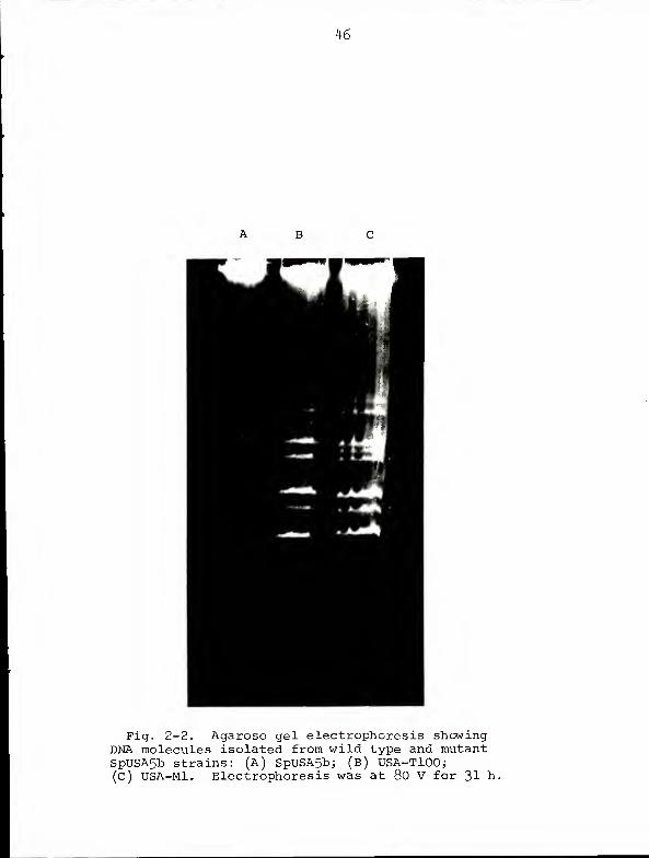
Figure 2-2 illustrates the DNA banding patterns of SpUSA5b
and its mutant derivatives. As was the case with JM125A-,
the T phenotype correlated with loss of pAZ4 (lanes A and
B). However, we did not observe obvious differences in
electrophoretic mobility between the DNA molecules of wild
type SpUSA5b and those of a representative Met derivative
( lanes A and C)
.
37
125-T1N1 and USA-MI are nonreverting . To investigate
the possibility that 125-T1N1 and USA-MI arose from AO-induced
frameshift mutations, we attempted to isolate revertants of
these strains from untreated, AO-treated, and NG- treated
cultures. No true revertants were found. Slowly-growing
— 8colonies did appear at a frequency of approximately 10 when
untreated 125-T1N1 was plated on nitrogen-free agar. However,
when restreaked on fresh nitrogen-free plates they failed to
flourish, and they were unable to reduce acetylene. Further-
more, their isolation frequency was enhanced to a much
greater extent by NG than by AO.
A similar situation occurred with potential Met revertants
of USA-MI. Colonies arising on minimal agar grew when
restreaked on fresh minimal plates, but growth on all types
of media was slower than wild type growth. Like the pseudo-
revertants of 125-T1N1, their isolation frequency was in-
creased significantly by NG but only marginally by AO.
These Met strains also displayed a deep reddish pigmentation,
whereas colonies of our other SpUSA5b strains are peach
colored.
Discus s ion
The results show that pAZ4-JM125A2
and pAZ4-SpUSA5b
R +determine, for their respective hosts, the Cad , Adh , and
Cfa phenotypes. The two plasmids are not entirely homo-
logous, however, since the electrophoretic mobility of
pAZ4-SpUSA5b is slightly lower than that of pAZ4-JMl25A2
(see Chapter I). Loss of pAZ4-JM125A2
at a high frequency
33
in AO-treated JM125A- cultures is consistent with the known
curing effects of acridine dyes (30). On the other hand,
high-frequency AO-induced methionine auxotrophy and
AO-induced multiple deletions in DNA have not been
described.
The sensitivities of wild type and mutant Azospirillum
strains to cadmium are, interestingly enough, comparable to
R Sthe sensitivities exhibited by Cad and Cad Staphylococcus
aureus strains (11). Recently, cadmium resistance in
S. aureus has been shown to depend on an energy-dependent,
plasmid-encoded efflux system (53, 69). A similar system
may operate to confer cadmium resistance in Azospirillum .
Alternatively, the configuration of membrane proteins in
Azospirillum T mutants may result in a lesser degree of
thiol group shielding than occurs in the wild type (58).
Growth of the T mutants on acetate and acetaldehyde but
not on ethanol implies the existance of an alcohol dehydro-
genase encoded by pAZ4-JM125A2
and pAZ4-SpUSA5b. The basis
for the inability of the T mutants to flourish on carbon-free
agar under ambient atmosphere is less obvious. The defect
does not appear to be in the capacity for autotrophy per se ,
as both mutant and wild type strains grow in carbon-free
soft agar under an autotrophic atmosphere. Biosynthesis of
ribulose diphosphate carboxylase in other bacteria has been
shown to be repressed under conditions of high oxygen tension
(37). Therefore, a single cell plated on carbon-free agar
under ambient atmosphere cannot immediately begin to grow
39
autotrophically ; it must instead utilize some intracellular
carbon reserve to grow and divide several times, so that an
aerobic layer of cells is formed under which a micro-
aerophilic environment is created. Azospirillum strains
do, in fact, accumulate large amounts of poly-B-hydroxy-
butyrate (PHB), particularly (but not exclusively) when
grown in nitrogen-free media (49). Although the T mutants
as well as the wild type can be seen microscopically to
contain PHB granules, it is possible that they have
difficulty utilizing this storage polymer for growth.
The discovery that one of the T mutants (125-T1) had
suffered multiple deletions in its DNA molecules is an
observation which may be relevant to the proposed mode of
action of AO in plasmid elimination. Some investigators
have suggested that plasmids, owing to their relatively
small size, are more accessible to AO than is the bacterial
chromosome (52) or that AO selectively inhibits plasmid
replication (31, 32). In contrast, others have maintained
that AO causes nonspecific loss of DNA via inhibition of
polymerase I (4, 5); since only cells having lost nonessential
DNA survive, this nonspecific mode of action translates into
an apparent specificity for plasmid (i.e. nonessential) DNA.
Assuming that the generation of 125-T1 really was AO-mediated
and not merely a spontaneous event which happened to occur
in an AO-treated culture, our results tend to support the
idea of a nonspecific interaction of AO with DNA. If AO
specifically interacted with or inhibited replication of
40
plasmid DNA, one would not expect to observe partially
deleted plasmids unless those plasmids were capable of
dissociation into self-replicating component molecules. The
possibility of four DNA molecules undergoing dissociation of
this kind within the same cell seems remote.
Wild and mutant JM125A- strains displayed three levels
of sensitivity to kanamycin, correlating with degree of DNA
loss. This tends to implicate two or more proteins or one
protein encoded by two or more loci (on at least two DNA
molecules) in the determination of kanamycin sensitivity
levels in Azospiril lum . Furthermore, these proteins must
act specifically on kanamycin and not on aminoglycosides in
general, since the sensitivities of the various mutants to
streptomycin were identical. Several types of aminoglycoside
modifying enzymes have been described in other bacteria, but
the mechanism by which the modified antibiotic confers
resistance is not known (17). The mechanism of aminoglycoside
uptake by sensitive cells is also obscure (17).
The Met" derivatives of SpUSA5b and the Nif derivative
of 125-T1 possess DNA molecules whose electrophoretic mobilities
cannot readily be distinguished from those of their parent
strains. These phenomena may be accounted for in one of
three ways. First, the mutants might have arisen via the
action of AO as a frameshift mutagen (15, 52). This possibil-
ity seems unlikely in view of the observed inability of AO
to promote reversions in the mutant strains. Furthermore,
the frameshift action of acridine dyes has been described
41
primarily in bacteriophage (15, 52); frameshift mutagenesis
of bacteria at the frequencies reported here has not been
described. A second possibility is that the mutants are
deleted for all or part of a molecule not identified by the
electrophoretic method used. This explanation also seems
rather unlikely since we have used the method to identify
molecules as large as the E. coli chromosome (see Chapter I).
The third, and most likely, possibility is that the mutants
carry deletions not large enough to lead to obvious differences
in mobility for the affected molecules. Thus, 125-T1N1
might have arisen by deletion of the entire nif cluster from
pAZl-JM125A2
(1500 Mdal); the wild type and mutant pAZl-
JM125A_ molecules, differing in molecular weight by only 1%,
would not be resolved by electrophoresis. We are currently
attempting to determine whether this is the case by hybridizing
a labeled nif probe to Southern blots (65) of fragmented
mutant and wild type molecules.
42
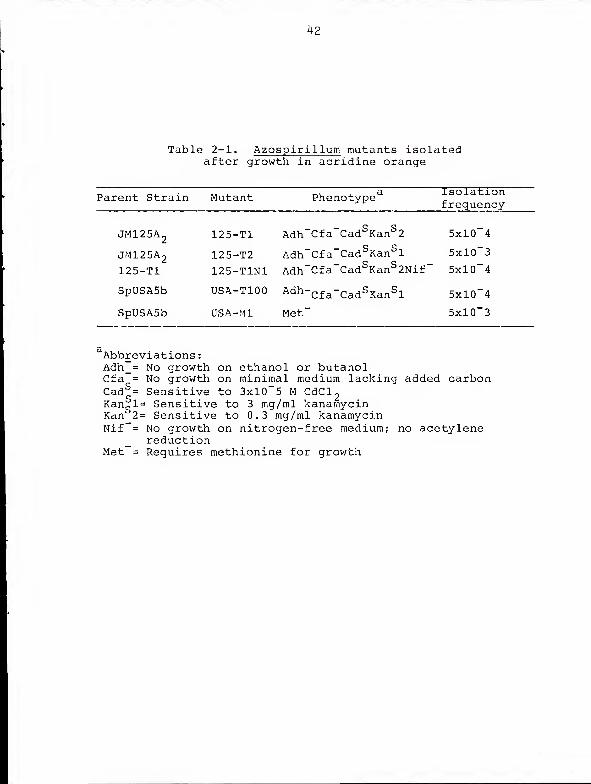
Table 2-1. Azospirillum mutants isolatedafter growth in acridine orange
™ j. r.j_ ^«j-j. t-.i *. a IsolationParent Strain Mutant Pnenotype_ frequency
JM125A2
125-T1 Adh~Cfa~Cad SKanS 2 5xl0~4
JM125A2
125-T2 Adh~Cfa"CadSKanS l 5xl0~3
125-T1 125-T1N1 Adh"Cfa~CadS KanS 2Nif" 5xl0~4
SpUSASb USA-T100 Adh-Cf
a
-Cad SKanS l 5xl0"4
SpUSA5b USA-MI Met" 5xl0"3
Abbreviations
:
Adh_= No growth on ethanol or butanolCfa = No growth on minimal medium lacking added carbonCad^= Sensitive to 3xl0~5 M CdCl
2Kan
ql= Sensitive to 3 mg/ml kanarnycin
Kan 2= Sensitive to 0.3 rng/ml kanarnycinNif~= No growth on nitrogen-free medium; no acetylene
reductionMet = Requires methionine for growth
4 3
Table 2-2. Sensitivities ofwild type and mutant Azospirillum strainsto cadmium, kanamycin^ and streptomycin
Minimal inhibitory concentration
Strain [Cd2+] ,M [Km], yg/ral [Sm],yg/ml
jmi25a2
44
Table 2-3. Estimated sizesof DNA molecules
found in JM125A2 an(j 125-T1
45
B C
Fig. 2-1. Agarose gel electrophoresis showingDNA molecules recovered from wild type and mutantJMl25Ap strains: (A) JM125A
2 ; (b) 125-Tl; (c) 125-TlNl;(D) 125-T2. Electrophoresis was for 23 h at 90 V.
K6
Fig. 2-2. Agarose gel electrophoresis showingDNA molecules isolated from wild type and mutantSpUSA5b strains: (A) SpUSA5b; (b) USA-TlOO;(C) USA-Mi. Electrophoresis was at 80 V for 31 h.
CHAPTER IIINif GENE HYBRIDIZATION STUDIESOF Azospirillum DNA MOLECULES
Introduction
In previous chapters we showed that strains of
Azospirillum brasilense and A. lipoferum harbor unique
arrays of large circular DNA molecules and that phenotypically
altered strains, some exhibiting a change in plasmid array,
could be isolated at a high frequency from cultures treated
with acridine orange. We now address the question of which
of these molecules determines the phenotype of greatest
general interest, i.e. nitrogen fixation. We had hoped that
the Nif" derivative of JM12 5A 2would show a change in the
mobility of one or more of its DNA molecules, but such was
not the case. We therefore decided to try a different
approach to the problem, based on molecular hybridization.
The structural genes for nitrogenase are thought to
have been either introduced recently in evolutionary history
into the various nitrogen-fixing bacteria or conserved to a
greater extent than other translated prokaryotic genes (59).
The basis for this view is the observation that Klebsiella
pneumoniae nif structural genes can hybridize to DNA from
all types of nitrogen-fixing prokaryotes, including both
Gram-negative and Gram-positive bacteria, Actinomycetes, and
Cyanobacteria (59). This interspecies homology has been
exploited to study the organization of nif genes in
47
48
blue-green algae (39). Although Azospirillum DNA has not
been examined for homology to Klebsiella nif DUA, there is
no reason to think that Azospirillum would behave differently
from other nitrogen-fixing prokaryotes in this regard,
especially since the individual Azospirillum nitrogenase
subunit proteins can form active complexes in vitro with
complementary proteins from other nitrogen-fixing bacteria
(23).
We obtained, from W. Klipp (36), an Escherichia coli
strain harboring pWK27, a recombinant plasmid which includes
an EcoRI fragment carrying the nif K, nifD, and nifH genes
from K. pneumoniae . This molecule was isolated in large
quantity, labeled to high specific activity by nick trans-
lation (57), and hybridized to Southern blots (65) of gels
containing DNA molecules from wild type and mutant Azospirillum
strains. The very limited success we have achieved with
this technique indicates that pAZl, the largest of the
Azospirillum molecules, carries the nif structural genes in
Azospirillum .
Materials and Methods
Bacterial strains . HB101(pWK27) was supplied by W. Klipp
(36). Origins of Azospirillum strains are described in
Table 1-1 (Chapter I) and Table 2-1 (Chapter II).
Isolation of pWK27 . To one liter of cell culture grown
at 37°C to Klett 90 was added 170 mg chloramphenicol. The
cells were incubated at 37°C for 20 h, harvested, washed
with 10 mM NaCl, and resuspended in 6 ml 0.02 M EDTA, 0.025
4 9
M Tris, 0.9% glucose (pH 8.0). Lysozyme (12 mg) was added
and the cells were chilled on ice for 30 min. Next, 12 ml
0.08% NaOH, 0.8% SDS was added and the lysate was gently
swirled for 5 min. Following addition, with gentle mixing,
of 9 ml KAc (pH 4.8), the lysate was incubated on ice for 2
h. The lysate was then centrifuged at 15,000 rpm for 30 min
and the supernatant solution was transferred to a fresh
tube. The addition of PEG 6000 to a final concentration of
10% followed by incubation on ice for 2 h caused the plasmid
to precipitate and it was pelleted at 2,500 rpm for 5 min.
The pellet was resuspended in 2 ml 50mM Tris, ImM EDTA
(pH 8.0). Ribonuclease was added to a concentration of
5 \i g/ml and the solution was incubated at 37 °C for 30 min.
Following adjustment of the volume to 10 ml with 50 mM Tris,
ImM EDTA (pH 8.0), the solution was extracted twice each
with phenol /chloroform, chloroform, and ether. Residual
ether was blown off with air. The plasmid was then ethanol
precipitated, washed in 70% ethanol, and further purified by
dye bouyant density ultracentrifugation. Plasmid bands,
visualized with UV light, were removed from the centrifuge
tubes with a plastic syringe. Ethidium bromide was removed
by extraction with isopropanol and CsCl by dialysis against
three changes (2 1 each) of 2 5 mM Tris, 1 mM EDTA.
Blotting . Agarose gels containing separated Azospirillum
DNA. molecules were prepared as described in Chapter I. DNA
from the gels was blotted onto strips of nitrocellulose
according to the method of Wahl et al. (70). This is a
50
slight modification of Southern's original technique (65).
The gels are treated with 0.25 M HC1 as an initial step in
order to partially depurinate the DNA and fragment it for
more efficient transfer.
Nick translation of pWK2 7 . The probe was labeled with
32P according to the method described by Rigby et al
.
(57).
32One hundred microCuries Pa-dCTP was dried under vacuum in a
1.5 ml microfuge tube. To this was added 5 pi 0.5 M Tris, 0.1 M
3-mercaptoethanol, 0.05 M MgCl2
; 24 p 1 of a 1:1:1 dNTP mix (200
mM each); IP g pWK27 DNA; and water to make 48 p 1 total
volume. After 5 min at 30°C, 1 pi diluted, activated DNase
I was added. (DNase was activated by diluting a 1 mg/ml
stock in 10 mM HC1 1:2000 into 10 mM Tris, 5 mM MgCl2
, 1
mg/ml BSA (pH 7.6)). After 2 min at 30°C, 0.5 pi DNA
polymerase I (5 U/ml ) was added and the reaction was held
at 15°C for 1 h, whereupon it was terminated by the addition
of 5 Pi 0.25 M EDTA. The reaction mixture was extracted
with phenol and the labeled plasmid was separated from the
unincorporated nucleotides by passage through a Sepharose 4B
or Sephadex G-100 column or by electrophoresis through a
0.7% low melting point agarose gel.
Hybridi zation . All hybridizations and prehybridizations
were carried out in Sears Seal-n-Save plastic bags. Filters
were prehybridized for a minimum of 12 h at 65°C in a solu-
tion consisting of 0.5% SDS, 20 p g/ml denatured salmon sperm
DNA, lOx Denhardt's solution (20), 2 . 5x SSC, and 0.05 M
Na^HPO. (pH 8.0). Hybridizations were carried out under the

51
same conditions except that lx Denhardt's solution was used
and denatured, labeled pWK27 was included. The washing
protocol of Thomashow et al. v/as followed (68). This consists
of one 30 min wash with 2.5x SSC at room temperature followed
by four washes (30 min each) with 2 . 5x SSC at 65°C and one
wash at 65°C with O.lx SSC.
Autoradiography . The dried filters were exposed to
X-ray film at -70°C in the presence of an intensifying
screen. Exposure times were 2 h for the control hybridi-
zation of pWK27 to its restriction fragments and a minimum
of 1 week for the Azospirillum hybridizations.
Results and Discussion
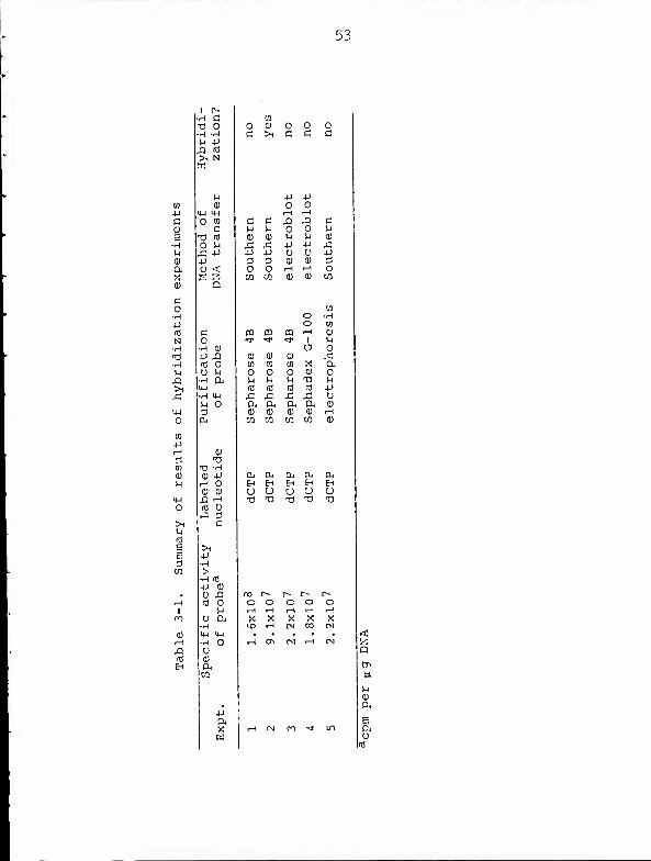
Table 3-1 summarizes the variations in protocol and
outcomes of five hybridization experiments. Both Fig. 3-2
and Fig 3-4 were taken from Experiment 2. Despite the
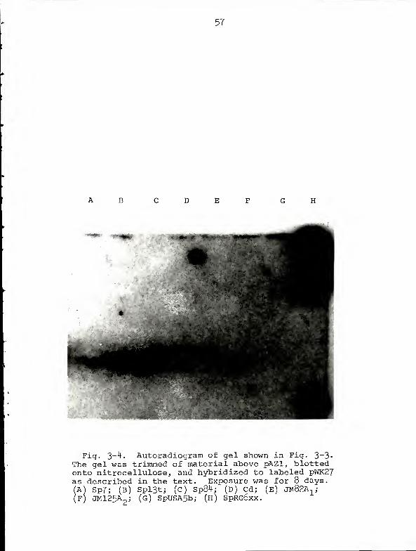
somewhat disappointing results, we can conclude from Fig.
3-4 that the Azospirillum nif structural genes are carried
on pAZl, the molecule which probably represents the chromo-
some of these bacteria.
Since the control hybridization (Fig. 3-2) gave such a
strong signal it is doubtful that our difficulty in detect-
ing Azospiril lum nif sequences reflects a serious flaw in
experimental procedure. Rather, the problem seems to be one
of sensitivity; the autoradiogram illustrated in Fig. 3-4
required 8 days exposure and shows only faint bands of
hybridization. There would appear to be two explanations
for this low level of hybridization. First, assuming that
52
the Azospirillum nif genes are carried on pAZl, less than
0.5% of the DNA in the band representing pAZl is capable of
hybridizing to the probe. Second, since there is some
degree of divergence in the DNA sequences of nif structural
genes in various bacteria (59), the probe DNA is not entirely
homologous to that portion of pAZl which is capable of
hybridization. DNA preparations from different strains of
Rhizobium hybridize with differing intensities to Klebsiella
nif DNA (59). We do not think that the low level of hybrid-
ization exhibited by Azospirillum DNA reflects poor transfer
from gel to nitrocellulose. Gels were always restained and
examined for DNA after transfer; none was ever found.
53
+jc<u
e•HM0)
OUX0)
CoHPrr!
NH^•iH
u&
>1p
EE
m
nj OH --H
P -P
>i N
Pa)
O Wc
o PX -P
-P
Q
CO•h <jj
-P X!(t! Oo u•H Onu-l
•H IM
p O
T3TS -HO PH Oo aj
m oPI 3
c
>1p•H
>H f0
4-> a>
(C op
O DjrH14-t IHrHOoa,CO
4-)
a.
0)00
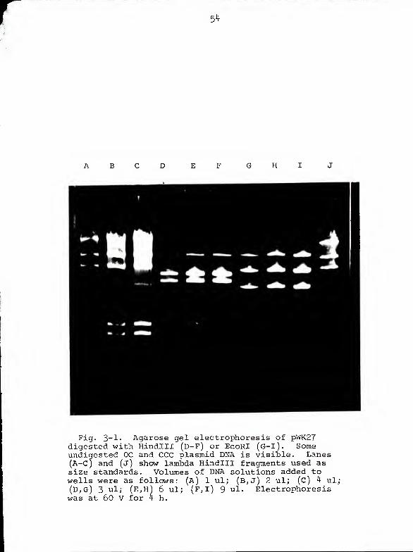
r 54
A B
Fig. 3_ 1- Agarose gel electrophoresis of pWK27digested with Hindi I I (D-F) or EcoRI (G-l). Someundigested OC and CCC plasmid DNA is visible. Lanes(A-C) and (j) show lambda Hindlll fragments used assize standards. Volumes of DNA solutions added towells were as follows: (A) 1 ul; (B,J) 2 ul; (C) 4 ul;(D,G) 3 ul; (E,H) 6 ul; (F,l) 9 ul. Electrophoresiswas at 60 V for
I) 6 ul;4 h.
55
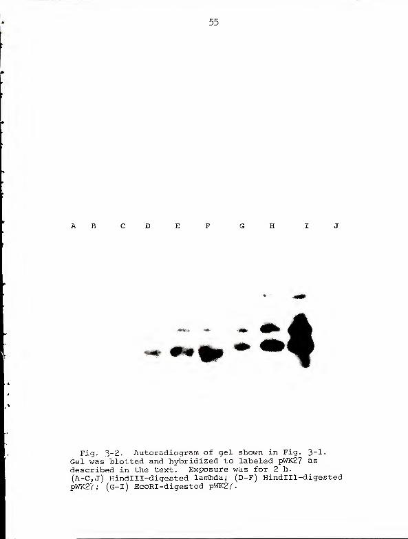
Fig. 3-2. Autoradiogram of gel shown in Fig. 3-1
•
Gel was blotted and hybridized to labeled pWK27 asdescribed in the text. Exposure was for 2 h.
(A-C,J) Hindlll-digested lambda; (D-F) Hindlll-digestedpWK27; (G-I) EcoRI-digested pWK27
.
56
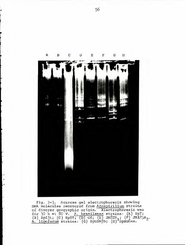
Fig. 3- 3> Agarose gel electrophoresis showingDNA molecules recovered from Azospirillum strainsof diverse geographic origin. Electrophoresis wasfor 30 h at 80 V. A. brasilense strains: (A) Sp7;(B) Spl3t; (C) Sp84; (D) Cd; (E) JM82A,; (F) JMl25Ap,A. lipoferum strains: (g) SpUSA5b; (h) SpRGDxx.
57
D E
Fig. 3~^' Autoradiogram of gel shown in Fig. 3-3«The gel was trimmed of material above pAZl, blottedonto nitrocellulose, and hybridized to labeled pWK27as described in the text. Exposure was for 8 days.A) Sp7; (B) Spl3t; (C) Sp84; (D) Cd; (E) JM82A.,;
F) JM125A2
j (G) SpUSA5bj (H) SpRG6xx.
APPENDIX ANORMALIZATION OF GEL MOBILITY DATA
In order to calculate a regression line relating electro-
phoretic mobility to molecular weight, it was necessary
to pool mobility data from 20 gels run for various lengths
of time under slightly differing conditions. Therefore,
the mobility data from 19 of the gels had to be normalized
to the data from one gel chosen as a standard. The gel
illustrated in Fig. 1-6 was chosen as the standard because
it includes the greatest number (15) of DNA bands.
The standard gel was designated B and the mobilities
of its DNA bands were B L , B 2 , , Bn » The mobilities
of the bands in gel A (which had in common with gel B n different
plasmid bands numbered l,2,...,n) were designated
Al' A 2' • • • ' An* ™*le constant Ka was calculated for
which the expression
ZI
KaAi-B i l/(KaAi +B i )
where i=l, 2, . . . ,n
was at a minimum.
Then the mobilities of the plasmids in gel A were multi-
plied by K a different K = was calculated for each gela. a.
to be normalized. 58
APPENDIX B
REGRESSION LINE CALCULATIONS
If the logarithms of the average normalized mobilities
of the standard plasmids were designated as X^, X2,...Xn and
the logarithms of their molecular weights (in megadaltons)
were designated Ylf Y2 ,...Ynthen the logarithm of the
molecular weight (Yfc
) of the unknown molecule could be
calculated from the logarithm of its mobility (Xt ) according
to the formula
Yt= A+BX
t
where A=T-BX
EX2
- I (EX.)2
n
X = EX./n
Y = EY./n
59
LITERATURE CITED
1. Aaij , C, and P. Borst. 1972. The gel electrophoresisof DNA. Biochim. Biophys. Acta 269 ;192-200.
2. Ahmad, M. H. 1978. Influence of nitrogen on growth,free amino acids and nitrogenase activity in Spirillumlipoferum . J. Gen. Appl. Microbiol. 24 : 271-278
.
3. Albrecht, S. L. , and Y. Okon. 1980. Cultures ofAzospirillum , p. 746. In A. San Pietro (ed.), Methodsin enzymology, vol. 69. Academic Press, New York.
4. Barker, G. R. 1978. Genetic expression and its controlin naturally occurring bacterial plasmids, pp. 25-29.
In P. W. Kent (ed.), New approaches to genetics:Developments in molecular genetics. Oriel Press, Boston.
5. Barker, G. R. , and N. Hardman. 1978. The effects ofacridine orange on deoxyribonucleic acid in Escherichiacoli . Biochern. J. 171 :567-573.
6. Brent, T. P. 1972. Repair enzyme suggested by mammalianendonuclease activity specific for ultraviolet-irradiated DNA. Nature (London), New Biol. 239 :172-173.
7. Bujard, H. 1970. Electron microscopy of single-strandedDNA. J. Mol. Biol. 49:125-137.
8. Cairns, J. 196 3. The chromosome of Escherichia coli .
Cold Spring Harbor Symp. Quant. Biol. 28: 43-45.
9. Carr, T. C. 1978. Cryptic plasmids in Azospirillum .
Master's Thesis, University of Florida, Gainesville.
10. Casse, F., C. Boucher, J. S. Julliot, M. Michel, andJ. Denarie. 1979. Identification and characterizationof large plasmids in Rhizobium meliloti using agarosegel electrophoresis. J. Gen. Microbiol. 113 : 229-242.
11. Chopra, I. 1975. Mechanism of plasmid-mediated resist-ance to cadmium in Staphylococcus aureus . Antimicrob.Agents Chemother. 7_:8-14.
12. Clark, D. , A. A. Weiss, and S. Silver. 1977. Mercuryand organomercurial resistances determined by plasmidsin Pseudomonas. J. Bacteriol. 132:186-196.
60
61
13. Clewell, D. B. , and D. R. Helenski. 1969. Supercoiledcircular DNA-protein complex in Escherichia coli :
Purification induced conversion to an open circularDNA form. Proc. Natl. Acad. Sci. U.S.A. 62 ; 1159-1166.
14. Cooper, S., and C. E. Helmstetter. 1968. Chromosomereplication and the division cycle of Escherichiacoli. B/r. J. Mol. Biol. 3JL:519_54 °-
15. Crick, F. H. C, L. Barnett, S. Brenner, and R. J.
Watts-Tobin. 1961. General nature of the genetic codefor proteins. Nature (London) 192 ; 1227-1232.
16. Currier, T. C, and E. W. Nester. 1976. Evidence fordiverse types of large plasmids in tumor-inducingstrains of Agrobacterium . J. Bacteriol. 126 : 157-165.
17. Davis, J., and D. I. Smith. 1978. Plasmid-determinedresistance to antimicrobial agents. Ann. Rev.Microbiol. 32:469-518.
18. Davis, R. W. , M. Simon, and N. Davidson. 1971. Electronmicroscope heteroduplex methods for mapping regions of
base sequence homology in nucleic acids. In L. Grossmanand K. Moldave (ed.), Methods in enzymology, vol. 21.
Academic Press. New York.
19. Day, J. M. , and J. Dobereiner. 1976. Physiologicalaspects of N -fixation by a Spirillum from Digitariaroots. Soil Biol. Biochem. £: 45-50.
20. Denhardt, D. 1966. A membrane filter technique forthe detection of complementary DNA. Biochem. Biophys.Res. Commun. 23: 641-646.
21. Dobereiner, J., I. E. Marriel, and M. Nery. 1976.Ecological distribution of Spirillum lipoferumBeijerinck. Can. J. Microbiol. 22: 1464-1473.
22. Eckhardt, T. 1978. A rapid method for the identificationof plasmid desoxyribonucleic acid in bacteria.Plasmid JL:534-583.
23. Emerich, D. W. , and R. H. Burris. 1978. Complementaryfunctioning of the component proteins of nitrogenasefrom several bacteria. J. Bacteriol. 134 :936-943.
24. Fangman, W. L. 1978. Separation of very large DNAmolecules by gel electrophoresis. Nucleic AcidsRes. _5:653-665.
25. Fisher, M. P., and C. W. Dingman. 1971. Role of mole-lecular conformation in determining the electrophoreticproperties of polynucleotides in agarose-acrylamidecomposite gels. Biochemistry 10 : 1895-1899.
62
26. Franche, C, and C. Elmerich. 1981. Physiologicalproperties and plasmid content of several strainsof Azospirillum brasilense and A. lipoferum . Ann.
Microbiol. (Inst. Pasteur) 132A: 3-18.
27. Gauthier, D. , and C. Elemrich. 1977. Relationshipbetween glutamine synthetase and nitrogenase inSpirillum lipoferum . FEMS Microbiol. Letters 2_:101-104.
28. Gillis, M., J. De Ley, and M. De Cleene. 1970. Thedetermination of molecular weight of bacterial genomeDNA from renaturation rates. Eur. J. Biochem. 12:143-153,
29. Hansen, J. B. , and R. H. Olsen. 1978. Isolation of largebacterial plasmids and characterization of the P2
incompatibility group plasmids pMGl and pMG5. J.
Bacteriol. 135 :227-238.
30. Hirota, Y. 1960. The effect of acridine dyes on matingtype factors in Escherichia coli . Genetics 46 : 57-64.
31. Hohn, B., and D. Korn. 1969. Cosegregation of a sexfactor with the Escherichia coli chromosome duringcuring by acridine orange. J. Mol. Biol. 45 : 385-395.
32. Jacob, F., S. Brenner, and F. Curzin. 1963. On theregulation of DNA replication in bacteria. Cold SpringHarbor Symp. Quant. Biol. 28:329-348.
33. Kato, A. C., and M. J. Fraser. 1973. Action of a single-strand specific Neurospora crassa endonuclease onultraviolet light-irradiated native DNA. Biochem.Biophys. Acta 312 :645-655.
34. Kline, B. C., and J. R. Miller. 1975. Detection of non-integrated plasmid deoxyribonucleic acid in the foldedchromosome of Escherichia coli : Physicochemical approachto studying the unit of segregation. J. Bacteriol. 121 :
165-172".
35. Kline, B. C., J. R. Miller, D. E. Cress, M. Wlodarczyk,J. J. Manis, and M. R. Otten. 1976. Nonintegratedplasmid-chromosome complexes in Escherichia coli .
J. Bacteriol. 127 :881-889." ""
36. Klipp, W. 1980. Personal communication.
37. Kuehn, G. D. , and B. A. McFadden. 1968. Factorsaffecting the synthesis and degradation of ribulose-1, 5-diphosphate carboxylase in Hydrogenomonas facilisand Hydrogenomonas eutropha . J. Bacteriol. 95:937-946.
63
38. Magalhaes, L. M. S., C. A. Neyra, and J. Dobereiner.1978. Nitrate and nitrite reductase negative mutantsof N„-fixing Azospirillum spp. Arch. Microbiol.117:247-252.
39. Mazur, B. J., D. Rice, and R. Haselkorn. 1980. Inter-species homology of nitrogenase genes. Proc. Natl.Acad. Sci. USA 1J_: 191-195.
40. Meyers, J. A., D. Sanchez, L. P. Elwell, and S. Falkow.1976. Simple agarose gel electrophoretic method forthe identification and characterization of plasmiddeoxyribonucleic acid. J. Bacteriol. 127 :1529-1537.(Erratum, J. Bacteriol. 129 :1171, 1977.)
41. Mishra, A. K. , P. Roy, and S. Bhattacharya. 1980.Deoxyribonucleic acid-mediated transformation ofSpirillum lipoferum . J. Bacteriol. 137 :1425-1427.
42. Nakahara, H. , T. Ishikawa, S. Sarai, I. Kondo, and S.
Mitshuhashi. 1977. Frequency of heavy metal resistancein bacteria from inpatients in Japan, Nature 266 : 165-167,
43. Nelson, L. M. , and R. Knowles. 1978. Effect of oxygenand nitrate on nitrogen fixation and denitrif icationby Azospirillum brasilense grown in continuous cultureCan. J. Microbiol. 24:1395-1403.
44. Neyra, C. A., J. Dobereiner, R. Lalande, and R. Knowles.1977. Denitrification by N
2~fixing Spirillum lipoferum .
Can. J. Microbiol. 2^3:300-305.
45. Neyra, C. A., and P. Van Berkum. 1977. Nitrate re-duction and nitrogenase activity in Spirillum lipoferum .
Can. J. Micbriol. 2_3: 306-310.
46. Nur, I., Y. Okon, and Y. Henis. 1980. An increase innitrogen content of Setaria Italica and Zea maysinoculated with Azospirillum . Can. J. Microbiol._26:432-485.
47. Ohtsubo, E., R. C. Deonier, H. J. Lee, and N. Davidson.Electron microscope heteroduplex studies of sequencerelations among plasmids of Escherichia coli . IV. Thesequence in F14. J. Mol. Biol. 89: 565-584.
43. Okon, Y., S. L. Albrecht, and R. H. Burris. 1976.Factors affecting growth and nitrogen fixation ofSpirillum lipoferum . J. Bacteriol. 127:1248-1254.
49. Okon, Y., S. L. Albrecht, and R. H. Burris. 1976.Carbon and ammonia metabolism of Spirillum lipoferum .
J. Bacteriol. 128: 592-597.
64
50. Okon, Y. , S. L. Albrecht, and R. H. Burris. 1977. Methodsfor growing Spirillum lipoferum and for counting itin pure culture and in association with plants. Appl.Env. Microbiol. 33.:85-88.
51. Okon, Y., J. P. Houchins, S. L. Albrecht, and R. H.Burris. 1977. Growth of Spirillum lipoferum at constantpartial pressures of oxygen, and the properties of itsnitrogenase in cell-free extracts. J. Gen. Microbiol.9^:87-93.
52. Orgel, A., and S. Brenner. 1961. Mutagenesis of bacterio-phage T4 by acridines. J. Mol. Biol.
_3_ :762-768.
53. Perry, R. D. , and S. Silver. 1981. Transport studieson cadmium resistance in whole cells and subcellularmembranes of Staphylococcus aureus , p. 159. In Abstractsof the annual meeting of the American Society forMicrobiology, 1981. ASM Publications, Washington, D.C.
54. Pettijohn, D. E. , and R. Hecht. 1973. RNA moleculesbound to the folded bacterial genome stabilize DNAfolds and segregate domains of supercoiling. ColdSpring Harbor Symp. Quant. Biol. 38: 31-41.
55. Polsinelli, M. , E. Baldanzi, M. Bazzicalupo, and E.Gallori. 1980. Transfer of plasmid pRDl from Escherichiacoli to Azosp irillum brasilense . Mol. Gen. Genet.178:709-711.
56. Rainbow, A. J., and S. Mak. 1973. DNA damage and bio-logical function of human adenovirus after U.V.-irradiation. Int. J. Radiat. Biol. 24 : 59-72.
57. Rigby, P. W. J., M. Dieckmann, C. Rhodes, and P. Berg.1977. Labeling deoxyribonucleic acid to high specificactivity in vitro by nick translation with DNA poly-merase. J. Mol. Biol. 113 :237-251.
58. Rothstein, A. 1959. Cell membrane as site of action ofheavy metals. Fed. Proc. 18:1026-1035.
59. Ruvkun, G. B. , and F. M. Ausubel. 1980. Interspecieshomology of nitrogenase genes. Proc. Natl. Acad. Sci.USA 72:191-195.
60. Sampaio, J. A. M. , E. M. R. daSilva, J. Dobereiner,M. G. Yates, and F. 0. Pedrosa. 1981. Autotrophy andmethylotrophy in Derxia gummosa , Azospirillum brasilense ,
and A. lipoferum , p. 444. In A. H. Gibson and W. E.~~
Newton (ed.), Current perspectives in nitrogen fixation.Australian Academy of Science, Canberra City.
6 5
61. Sharp, P. A., M Hsu, E. Ohtsubo, and N. Davidson.197 2. Electron microscope heteroduplex studies of se-quence relations among plasmids of Escherichia coli .
I. Structure of F-prime factors. J. Mol. Biol. 71: 471-487.
62. Silver, S. , E. Levine, and P. M. Spielman. 1968.Acridine binding by Escherichia coli : pH dependencyand strain differences. J. Bacteriol. 95: 333-339.
63. Smith, D. H. 1967. R factors mediate resistance to mercury,nickel, and cobalt. Science 156 : 1114-1116.
64. Smith, R. L. , J. H. Bouton, S. C. Schank, K. H.
Quesenberry, M. E. Tyler, J. R. Milam, M. H. Gaskins,and R. C. Littell. 1976. Nitrogen fixation in grassesinoculated with Spirillum lipoferum . Science 193 :1003-1005.
65. Southern, E. M. 1975. Detection of specific sequencesamong DNA fragments separated by gel electrophoresis.J. Mol. Biol. 98:503-517.
66. Stonington, 0. G. , and D. E. Pettijohn. 1971. The foldedgenome of Escherichia coli isolated in a protein-DNA-RNAcomplex. Proc. Natl. Acad. Sci. USA 68:6-9.
67. Tarrand, J. J., N. R. Krieg, and J. Dobereiner. 1978.A taxonomic study of the Spirillum lipoferum group,with descriptions of a new genus, Azospirillum gen. nov.and two species, Azospirillum lipoferum (Beijerinck)comb. nov. and Azospirillum brasilense sp. nov. Can. J.
Microbiol. _2_4: 967-980.
68. Thomashow, M. F., R. Nutter, A. L. Montoya, M. P. Gordon,and E. W. Nester. 1980. Integration and organizationof Ti plasmid sequences in crown gall tumors. Cell 19 :
729-739.
69. Tynecka, Z., Z. Gos, and J. Zajac. 1981. Plasmid-determined Cd2 resistance in Staphylococcus aureus :
Accelerated efflux and reduced net uptake, p. 159.
In Abstracts of the annual meeting of the AmericanSociety for Microbiology, 1981. ASM Publications, Washing-ton, D.C.
70. Wahl, G. M., M. Stern, and G. R. Stark. 1979. Efficienttransfer of large DNA fragments from agarose gels todiazobenzylozymethlyl-paper and rapid hybridization byusing dextran sulfate. Proc. Natl. Acad. Sci. USA 76:
3633-3687.
71. Weiss, A. A., S. D. Murphy, and S. Silver. 1977. Mercuryand organomercurial resistances determined by plasmidsin Staphylococcus aureus . J. Bacteriol. 132 : 197-208.
66
72. Willshaw, G. A., H. R. Smith, and E. S. Anderson. 1979,Application of agarose gel electrophoresis to thecharacterization of plasmid DNA in drug-resistantEnterobacteria. J. Gen. Microbiol. 114 : 15-25.
73. Worcel, A., and E. Burgi. 1972. On the structure ofthe folded chromosome of Escherichia coli . J. Mol.Biol. TL-.121-1M .
74. Worcel, A., E. Burgi, J. Robinton, and C. L. Carlson.1973. Studies on the folded chromosome of Escherichiacoli. Cold Spring Harbor Symp. Quant. Biol. 38:43-51.
BIOGRAPHICAL SKETCH
The author was born Alvin Gleave Wood on October 5,
1951, in St. Petersburg, Florida. He is the only son of
Mary Gleave Harris and the late Rowland Emery Wood. Follow-
ing graduation from Northeast High School in St. Petersburg,
he attended the University of Chicago and received a B.A. in
biology in June 1973. He received his M.S. degree from the
Department of Microbiology and Cell Science, University of
Florida, 1978. He is currently a candidate for the degree
of Doctor of Philosophy, also in the Department of Microbiology
and Cell Science.
67
I certify that I have read this study and that in myopinion it conforms to acceptable standards of scholarlypresentation and is fully adequate, in icope and quality,as a dissertation for the degree of Doctor- of Philosophy.
/,
Dennis. E. Duggan, ChairmanAssociate Professor ofMicrobiology and Cell Science
I certify that I have read this study and that in myopinion it conforms to acceptable standards of scholarlypresentation and is fully adequate, in scope and quality,as a dissertation for the deqree of Doctor of Philosophy.)— .- <:", I
-t . , ^" 7 '
.
• VFrancis C. Davis, Jr. /Associate Professor ofMicrobiology and Cell Science
I certify that I have read this study and that in myopinion it conforms to acceptable standards of scholarlypresentation and is fully adequate, in scope and quality,as a dissertation for the degree of Doctor of Philosophy.
Lonnie 0. IngramAssociate Professor ofMicrobiology and Cell Science
I certify that I have read this study and that in myopinion it conforms to acceptable standards of scholarlypresentation and is fully adequate, in scope and quality,as a dissertation for the degree of Doctor of Philosophy. ^
«S*'
William W. HauswirthAssociate Professor of Immunology
and Medical Microbiology
This dissertation was submitted to the Graduate Faculty ofthe College of Agriculture and to the Graduate Council, andwas accepted as partial fulfillment of %he requirements forthe degree of Doctor of Philosophy. ( )
May 1982 XJ <A fr - JVWDean, College of Agriculture
1/
Dean for Graduate Studies andResearch

UNIVERSITY OF FLORIDA
llllllllli3 1262 08553 2124