Embed Size (px)
Citation preview

HAL Id: hal-01521993https://hal.univ-lorraine.fr/hal-01521993
Submitted on 12 May 2017
HAL is a multi-disciplinary open accessarchive for the deposit and dissemination of sci-entific research documents, whether they are pub-lished or not. The documents may come fromteaching and research institutions in France orabroad, or from public or private research centers.
L’archive ouverte pluridisciplinaire HAL, estdestinée au dépôt et à la diffusion de documentsscientifiques de niveau recherche, publiés ou non,émanant des établissements d’enseignement et derecherche français ou étrangers, des laboratoirespublics ou privés.
Multiple and Variable NHEJ-Like Genes Are Involved inResistance to DNA Damage in Streptomyces
ambofaciensGrégory Hoff, Claire Bertrand, Lingli Zhang, Emilie Piotrowski, Ludovic
Chipot, Cyril Bontemps, Fabrice Confalonieri, Stephen Mcgovern, FrançoisLecointe, Annabelle Thibessard, et al.
To cite this version:Grégory Hoff, Claire Bertrand, Lingli Zhang, Emilie Piotrowski, Ludovic Chipot, et al.. Multipleand Variable NHEJ-Like Genes Are Involved in Resistance to DNA Damage in Streptomyces ambo-faciens. Frontiers in Microbiology, Frontiers Media, 2016, 7, pp.1901. �10.3389/fmicb.2016.01901�.�hal-01521993�

fmicb-07-01901 November 24, 2016 Time: 16:29 # 1
ORIGINAL RESEARCHpublished: 28 November 2016
doi: 10.3389/fmicb.2016.01901
Edited by:Frank T. Robb,
University of Maryland, Baltimore,USA
Reviewed by:Igor Kovalchuk,
University of Lethbridge, CanadaAndrzej Stasiak,
University of Lausanne, Switzerland
*Correspondence:Pierre Leblond
Specialty section:This article was submitted to
Evolutionary and GenomicMicrobiology,
a section of the journalFrontiers in Microbiology
Received: 07 October 2016Accepted: 14 November 2016Published: 28 November 2016
Citation:Hoff G, Bertrand C, Zhang L,
Piotrowski E, Chipot L, Bontemps C,Confalonieri F, McGovern S,
Lecointe F, Thibessard A andLeblond P (2016) Multiple
and Variable NHEJ-Like Genes AreInvolved in Resistance
to DNA Damage in Streptomycesambofaciens.
Front. Microbiol. 7:1901.doi: 10.3389/fmicb.2016.01901
Multiple and Variable NHEJ-LikeGenes Are Involved in Resistance toDNA Damage in StreptomycesambofaciensGrégory Hoff1,2, Claire Bertrand1,2, Lingli Zhang1,2, Emilie Piotrowski1,2, Ludovic Chipot1,2,Cyril Bontemps1,2, Fabrice Confalonieri3, Stephen McGovern4, François Lecointe4,Annabelle Thibessard1,2 and Pierre Leblond1,2*
1 UMR 1128, Dynamique des Génomes et Adaptation Microbienne, Université de Lorraine, Vandœuvre-lès-Nancy, France,2 UMR 1128, Institut National de la Recherche Agronomique, Dynamique des Génomes et Adaptation Microbienne,Vandœuvre-lès-Nancy, France, 3 Institute for Integrative Biology of the Cell (I2BC), CEA, Centre National de la RechercheScientifique, Université Paris-Sud, Orsay, France, 4 Institut Micalis, INRA, AgroParisTech, Université Paris-Saclay,Jouy-en-Josas, France
Non-homologous end-joining (NHEJ) is a double strand break (DSB) repair pathwaywhich does not require any homologous template and can ligate two DNA endstogether. The basic bacterial NHEJ machinery involves two partners: the Ku protein,a DNA end binding protein for DSB recognition and the multifunctional LigD proteincomposed a ligase, a nuclease and a polymerase domain, for end processing andligation of the broken ends. In silico analyses performed in the 38 sequenced genomesof Streptomyces species revealed the existence of a large panel of NHEJ-like genes.Indeed, ku genes or ligD domain homologues are scattered throughout the genome inmultiple copies and can be distinguished in two categories: the “core” NHEJ gene setconstituted of conserved loci and the “variable” NHEJ gene set constituted of NHEJ-likegenes present in only a part of the species. In Streptomyces ambofaciens ATCC23877,not only the deletion of “core” genes but also that of “variable” genes led to an increasedsensitivity to DNA damage induced by electron beam irradiation. Multiple mutants of ku,ligase or polymerase encoding genes showed an aggravated phenotype compared tosingle mutants. Biochemical assays revealed the ability of Ku-like proteins to protect andto stimulate ligation of DNA ends. RT-qPCR and GFP fusion experiments suggested thatku-like genes show a growth phase dependent expression profile consistent with theirinvolvement in DNA repair during spores formation and/or germination.
Keywords: Streptomyces, non-homologous end joining, DNA repair, double strand breaks (DSBs), ligases, Kuprotein, DNA damage
INTRODUCTION
Double strand breaks (DSB) are the most detrimental genomic lesions and their repair constitutesa major challenge for cell viability. In eukaryotes and prokaryotes, two main repair machinerieshave evolved to repair these DSBs, namely homologous recombination (HR) and non-homologousend joining (NHEJ) (Alonso et al., 2013; Deriano and Roth, 2013; Glickman, 2014). In contrast
Frontiers in Microbiology | www.frontiersin.org 1 November 2016 | Volume 7 | Article 1901
fmicb-07-01901 November 24, 2016 Time: 16:29 # 2
Hoff et al. NHEJ-Like Genes in S. ambofaciens
to HR which needs an intact template to ensure repair of thebreak, NHEJ can bind DNA ends together without any templateafter a facultative DNA ends remodeling step. In multicellulareukaryotes, the NHEJ mechanism represents the predominantDSB repair pathway and requires the intervention of the endtethering heterodimer Ku70/80 as well as diverse end processingproteins (for review: Deriano and Roth, 2013). In prokaryotes,NHEJ is not ubiquitous and the minimal NHEJ machinery firstdescribed in Mycobacterium consists of two actors: a Ku homologand an ATP dependent ligase LigD (Weller et al., 2002; Dellaet al., 2004; Gong et al., 2005). The latter one is a multifunctionalprotein that carries three distinct enzymatic domains conferringnuclease, polymerase and ligase activities (Pitcher et al., 2005).Unlike DSB repair through HR, DSB repair carried out byNHEJ is mutagenic. Nucleotide addition and/or deletion are scarsfrequently left after DNA ends ligation (Stephanou et al., 2007;Aniukwu et al., 2008; Gupta et al., 2011). The initial modelinvolving two unique actors is now renewed by the discovery ofalternative actors. When the ligase domain of LigD (LigDom) isdefective, a back-up ligation activity is provided by LigC, anotherATP dependent ligase (Della et al., 2004; Bhattarai et al., 2014). Inthe same way, Mycobacterium smegmatis possesses two additionalpolymerases which share the same in vitro activity than thepolymerase domain of LigD (PolDom) (Zhu et al., 2012).
Streptomyces are filamentous bacteria mainly found in soils,sediments and seawater as well as symbionts with plants,fungi and animals (Seipke et al., 2012). Soil is a complexand changing environment where Streptomyces have to adaptto the heterogeneousness of the niche (moisture, nutrients,temperature. . .) and to cope with biotic competition. Their richand varied secondary metabolism such as signaling moleculesor chemical weapons is assumed to mediate their relationshipswith other organisms (Davies and Davies, 2010; Abrudanet al., 2015). Under limited nutritive conditions, their hyphaedifferentiate into long chains of exospores providing an efficientway for resistance and dissemination until suitable conditionsfor another growth cycle are met (Chater, 1993). While hyphaeare large multi-genomic compartments, spores are known toinclude a single genome (Hopwood, 2006). In contrast tothe vast majority of bacteria, Streptomyces possess a linearchromosome whose size is ranging from 5.96 to 11.94 Mb1 witha central replication origin and terminally inverted repeats thatare covalently bound to specific telomere proteins (Lin et al.,1993). The Streptomyces genome presents a high plasticity andtheir chromosome is highly compartmented, with a conservedcentral region including mostly housekeeping genes whereas thedispensable genes are predominantly located in the chromosomalarms (Bentley et al., 2002; Ikeda et al., 2003; Kim et al., 2015).The loss of synteny at the ends of the chromosome resultsfrom horizontally acquired DNA insertion or deletion events(Choulet et al., 2006; Thibessard and Leblond, 2014). Thisterminal variability reveals the strong recombination activitytaking place at the ends of the genome which is reminiscentof the DNA rearrangements reported several decades ago(Leblond et al., 1990; Volff and Altenbuchner, 1998). Hence,
1http://www.ncbi.nlm.nih.gov/assembly/?term=Streptomyces
Streptomyces are also subject to a high level of genetic instabilitywhich is directly linked to major genome rearrangements.Characterization of some of these rearrangements revealed largedeletions, chromosome circularization, (Fischer et al., 1997;Inoue et al., 2003), arm replacement (Fischer et al., 1998;Uchida et al., 2003), chromosome end-to-end fusion (Wenneret al., 2003) or large DNA amplifications (Yanai et al., 2006).Molecular analyses showed that chromosome circularizationand end-to-end fusion events especially rely on illegitimaterecombination whereas chromosome arm replacement orDNA amplifications is dependent on HR. This terminalrecombination pattern may result from higher recombinationfrequencies triggered by the preferential formation of DSBsin these regions. On the other hand, genome rearrangementscould also be more tolerated if formed in these dispensableregions.
Nevertheless, little is known about DSB repair in Streptomyces.Like the other bacteria, Streptomyces possess a RecA homologueinvolved in HR (Aigle et al., 1997; Muth et al., 1997; Vierling et al.,2001; Huang and Chen, 2006). However, recent studies that weperformed suggest that Streptomyces has an atypical HR pathway.In fact, in contrast to bacterial models like Escherichia coli,Bacillus subtilis or Mycobacterium, deletion of genes encodinga potential helicase-nuclease complex supposed to be involvedin the initial end resection step during HR is not viable inS. ambofaciens (Zhang et al., 2014). On the other hand, deletionof ruvABC and recG, two loci encoding factors that have aredundant role in migration and resolution of Holliday junctionduring late steps of HR, has only a mild effect on recombinationefficiency (Hoff et al., 2016). Early bioinformatics studies showedthat the two model species, S. coelicolor and S. avermitilis, bothpossess a ku ortholog suggesting also the existence of a NHEJpathway in Streptomyces (Rocha et al., 2005). Here, we performedin silico analyses in the fully sequenced Streptomyces genomesleading to the identification of a large panel of conserved andvariable genes suspected to be involved in a NHEJ pathway.Construction of single and multiple NHEJ-like genes mutantstrains in S. ambofaciens allowed us to study the impact of thesedifferent genes in DNA damage repair. All the results we obtainedsuggest the existence of a complex and atypical bacterial NHEJpathway in Streptomyces.
MATERIALS AND METHODS
Phylogenetic AnalysesA total of 38 sequenced Streptomyces genomes, including thatof S. ambofaciens ATCC23877 was scanned to identify genesputatively encoding NHEJ Ku-like or LigD-like proteins basedon BLAST similarity search against the non-redundant database.A preliminary BLASTP search (e-value cut-off 10−30
; identitythreshold 30%) using M. tuberculosis Rv0937c and Rv0938 (Kuand LigD respectively) protein sequences as a query allow toidentify and numerate the candidates. Secondly, BLASTP wereperformed using S. ambofaciens ATCC23877 NHEJ-like genesand reciprocal best hits allowed to organize them into groups oforthologs.
Frontiers in Microbiology | www.frontiersin.org 2 November 2016 | Volume 7 | Article 1901

fmicb-07-01901 November 24, 2016 Time: 16:29 # 3
Hoff et al. NHEJ-Like Genes in S. ambofaciens
For the phylogeny of Ku-like proteins, the tree was builtusing a maximum-likelihood method based on a GTR + G + Imodel. Phylogenetic trees were built and edited with Mega6(Tamura et al., 2013). Support of the tree branches was estimatedwith 100 bootstrap replicates and all positions with <80% sitecoverage were eliminated. A total of 295 and 309 positions wererespectively used for the Ku-like and the LigC/LigD proteinphylogenetic trees.
Bacterial Strains and PlasmidsAll strains used in this study are listed in Table 1. TheDH5α E. coli strain is used as conservation host for BACsand plasmids. For biochemical analyses, plasmid constructionswere done in E.coli MC1061 (Casadaban and Cohen, 1980).ER2566 (NEB) was used for kuB gene expression and Rosetta(DE3) PLysS (Stratagene) for kuA and kuC gene expression.E.coli strains were grown in Luria-Bertani (LB) mediumat 37◦C except for the BW25113 pKD20 thermosensitivestrain used for PCR targeting, which was grown at 30◦C.S. ambofaciens strains were grown at 30◦C on Soya flourmannitol (SFM) plates except for phenotypical analyses andfor liquid cultures which were performed on solid HickeyTresner (HT) medium. All Streptomyces mutants derive from ourreference strain S. ambofaciens ATCC23877 (Pinnert-Sindico,1954). Mutant strains were constructed by PCR targeting asdescribed by Gust et al. (2003). To summarize, a recombinantBAC containing the locus of interest was transformed in thehighly recombinogenic E. coli BW25113/pKD20 strain in order toreplace the target gene by an oriT- aac(3)IV resistance disruptioncassette (Raynal et al., 2006). The ET12567 non-methylatingstrain containing the mobilizing pUZ8002 plasmid was usedas donor for intergeneric conjugation of the modified BACbetween E. coli and S. ambofaciens Double-crossover (CO) events
TABLE 1 | Streptomyces ambofaciens strains used in this study.
Strains Characteristics Sources
ATCC23877 Used as a WT strain Pinnert-Sindico, 1954
1kuA 1SAM23877_5082 This study
1kuB 1SAM23877_6929 This study
1kuC 1SAM23877_6942 This study
1kuABC 1SAM23877_50821SAM23877_69291SAM23877_6942
This study
1ligC 1SAM23877_6361 This study
1ligD 1SAM23877_0862 This study
1ligCD 1SAM23877_63611SAM23877_0862
This study
1polR 1SAM23877_6362 This study
1polK 1SAM23877_5081 This study
1polRK 1SAM23877_63621SAM23877_5081
This study
1recA 1SAM23877_5473 This study
1kuABC ArecA 1SAM23877_50821SAM23877_69291SAM23877_69421SAM23877_5473
This study
leading to the replacement of the target gene by the cassettewere selected by the sensitivity to kanamycin (loss of the BACvector part) along with the resistance to apramycin (insertionof the resistance cassette). The disruption cassette flanked byattL and attR sites was excised in the newly obtained mutantstrains thanks to the introduction of pOSV508 a plasmid allowingthe expression of int and xis genes from pSAM2 (Raynal et al.,2006). Strains expressing translational EGFP fusions were alsoconstructed by in vivo recombination. The egfp gene with thetranscription terminator from phage λ was cloned from plasmidpIJ8660 (Sun et al., 1999) into an oriT aac(3)IV disruptioncassette (Raynal et al., 2006). This cassette was then used as thesource to make kuA-egfp, kuB-egfp and kuC-egfp fusions. PCRwas performed using egfp-oriT-aac(3)IV as the template withspecific oligonucleotides which anneal to the linker sequence atthe 5′ end of egfp and downstream of the 3′ end of aac(3)IV andintroduce 40 bases of homology to the 3′ ends of kuA, kuB, orkuC, as well as removing the stop codons. PCR products wereintroduced into E. coli BW25113/pKD20 containing the cognateBACs with either kuA, kuB, or kuC, and the following steps of theprocedure are described above. In these recombinant strains, thefusion genes (kuA-egfp, kuB-egfp or kuC-egfp, respectively) arethe only copies of kuA, kuB, or kuC.
Electron Beam Irradiation (EBI)An amount of 109 spores of different Streptomyces strains wasdiluted in sterile water in 96 well microplates and exposed to 0,0.1, 0.3, 0.5, 0.7, and 1 kGy of radiation doses. Processing wascarried out in a Van de Graaf R© electron accelerator (VIVIRAD,Handschuheim, France) in the technological resource centerAérial (Illkirch-Graffenstaden, France). Energy delivered by theelectron accelerator reach 2.2 MeV with an intensity of 10 µA.Dose rate equals approximately 60 kGy/h. After irradiation,several spore dilutions were plated with the easySpiral R© apparatus(Interscience, Saint-Nom-la-Bretèche, France) on SFM Petridishes and the number of colonies was counted after 3 daysincubation at 30◦C. For each genetic context, at least twoindependent mutant strains were exposed in two independentexperiences.
Fluorescent MicroscopyStrains for microscopic observations were inoculated in theacute-angle junction of standard-sized microscope coverslipsinserted with an angle of 45◦ in SFM agar, and grown for 48 h.The coverslips were then carefully pulled from the medium anddirectly mounted in a drop of sterile water on a microscopeslide. Fluorescent microscopy observations were carried outusing a Axiovert 200M MOT device (Carl Zeiss Micro imaging)equipped with a mercury vapor lamp and a FITC filter set (470/40excitation, 540/50 emission). Both contrast phase and fluorescentobservations were made with an X100 objective.
DNA PreparationTo verify the mutant strains, Streptomyces genomic DNA wasextracted by low binding sulfate salt method as described byKieser et al. (2000). Liquid HT medium was used for growth ofmycelium before DNA extraction. For biochemical analyses, the
Frontiers in Microbiology | www.frontiersin.org 3 November 2016 | Volume 7 | Article 1901

fmicb-07-01901 November 24, 2016 Time: 16:29 # 4
Hoff et al. NHEJ-Like Genes in S. ambofaciens
gene coding for KuA was amplified from the BAC 16ZB5. KuBand KuC genes were amplified from the BAC 27ZF1. Primersused for amplification were designed to flank the open readingframes with the coding sequence of a His6-tag followed by aNdeI site on their 5′ side, and by a XhoI site on their 3′ side.The resulting ORFs were cloned under the T7 promoter ofthe pJ411 vector (DNA2.0) giving the plasmids pSMG258, 259,260 allowing expression of the proteins 6His-KuA, 6His-KuB,6His-KuC, respectively (named hereafter KuA, KuB, and KuC).Sequences of the primers used in this study are available uponrequest.
RNA Extraction and RT-qPCR AnalysesFor RNA extraction from cultures on solid medium, streakswere harvested after growth (from 24 to 60 h depending ofthe experiment) at 30◦C on HT solid medium covered withcellophanes membranes. For RNA extraction after a genotoxicstress, pre-germinated spores of Streptomyces were grown until0.2 OD600 was reached and exposed to mitomycin C (MMC,1.5 µg.mL−1, i.e., CMI/4) for 30 min. RNA isolation wasperformed using the Aurum Total RNA mini-kit (Bio-Rad)according to the manufacturer’s instructions. To optimize celllysis, an additional sonication step (3 × 30 s) using a Bioruptorapparatus was added. The RNA quality was assessed aftermigration on 1% agarose gel. In order to eliminate genomicDNA contamination, the RNA samples were treated beforereverse transcription with DnaseI (Ambion) in the presence of10 U of RNase inhibitor per µg of RNA. DnaseI was theninactivated at 75◦C during 10 min in the presence of EDTA.Reverse transcription was performed on 2 µg RNA with aniScript Advanced cDNA synthesis kit (Bio-Rad) according tothe manufacturer’s instructions. The absence of contaminationwith genomic DNA was validated by a 30 cycle-PCR on acontrol aliquot removed before reverse transcription using hrdB-F and hrdB-R primers. Quantitative PCR was undertaken ina CFX96 (Bio-Rad) apparatus in a 10 µL reaction mixturecontaining respectively 5 µL of SYBR supermix (Bio-Rad),0.2 µM of each primer and 4 µL of a 10-fold dilution ofcDNA. The reaction conditions were as follow: a 3 min initialdenaturation step at 95◦C and 40 cycles of 10 s at 95◦C and30 s at 60◦C. In order to check the absence of secondaryproducts, melting curves were realized from 65 to 95◦C withan increase rate of 0.5◦C/5 s. The transcription level of interestgenes was analyzed using the mathematical model proposedby Pfaffl (2001). The hrdB gene, encoding the principal sigmafactor in Streptomyces, was used as the reference gene forthe normalization of the expression level as it was the casein former studies (Galet et al., 2015; Martínez-Burgo et al.,2015).
Proteins PurificationPurification of KuAEscherichia coli Rosetta PlysS cells transformed with pSMG258were grown at 30◦C in 2 l of LB medium supplemented with25 mg/mL thymine, 30 µg/mL kanamycin and 25 µg/mLchloramphenicol to OD600 = 0.7. Production of protein wasinduced with 0.5 mM IPTG for 15 h at 20◦C. Cells were harvested
by centrifugation and the pellet was resuspended in 40 mL lysisbuffer A (50 mM Tris-HCl pH 8, 0.25 M NaCl). Cells were brokenby a freeze/thaw step in liquid N2/37◦C and then centrifuged1 h at 100 000 g at 4◦C. The pellet was resuspended with40 mL of a 20 mM Tris pH 7.5, 2.5 mM MgCl2, 0.5 mM CaCl2,
1 mg/mL DNaseI buffer. After 1 h at 20◦C, the mixture wascentrifuged 20 min at 100 000 g at 4◦C. All subsequent stepswere carried at 4◦C. The pellet was washed with 40 ml bufferP1 (50 mM Tris-HCl, pH 8, 1 M NaCl), and then solubilizedwith 40 mL of buffer P2 (50 mM Tris pH 8, 8 M urea). After1 h the mixture was centrifuged 20 min at 100 000 g and 3 mLof Ni2+ affinity column (Ni-NTA agarose, Qiagen) was added tothe supernatant which was transferred onto an Econo-Column(Biorad). The flow through was discarded and the resin waswashed with series of 25 mL of buffer P3 (50 mM Tris pH 8,1% sucrose, 0.5 M NaCl) with urea concentration decreasingfrom 8 M to 0 M by steps of 0.5 M. The Ni-NTA column waswashed with buffer P3, then with buffer W (50 mM Tris pH8.0, 0.5 M NaCl) supplemented with 20 mM imidazole. KuAprotein was eluted in buffer W supplemented with 250 mMimidazole and then dialysed against buffer D (50 mM Tris-HClpH 8, 0.4 M NaCl, 50% glycerol, 1 mM DTT) prior to storage at−20◦C.
Purification of KuCThe expression of KuC gene was induced in E. coli Rosetta PLysScells transformed with pSMG260 as described for KuA. Cellswere harvested by centrifugation and the pellet was resuspendedin 40 mL lysis buffer B (50 mM Tris-HCl pH 8, 0.25 M NaCl,0.2 mg/mL lysozyme) supplemented with Complete Antiprotease(Roche) and incubated 1 h at 20◦C. The mixture was thencentrifuged and the pellet was treated with DnaseI, as describedfor KuA purification. Purification procedure of KuC at this stepis the same as the one described for KuA.
Purification of KuBER2566 cells transformed with pSMG259 were grown at 30◦Cin 2 l of LB medium supplemented with 25 mg/mL thymineand 30 µg/mL kanamycin and kuB gene expression wasinduced as for kuA and kuC genes. Cells were harvested bycentrifugation, the pellet was resuspended in 40 mL of lysisbuffer B and incubated 1 h at 20◦C. All subsequent steps werecarried at 4◦C. Cell lysis mixture was supplemented with 0.5M NaCl, 0.1% Triton and 0.1% Non-idet P40 and incubated1 h. Two mL of polyethyleneimine (PEI) 10%, pH 8 wasadded to precipitate nucleic acids; the mixture was stirredfor 1 h and then centrifuged 1 h at 100 000 g. The solublephase was next precipitated by raising the concentration ofammonium sulfate (AS) firstly to 50% of saturation, KuB wasmainly found in the supernatant at this step, and secondlyto 80% of saturation to precipitate KuB. The correspondingpellet was resuspended in 25 mL of buffer P1 and 3 mL ofNi-NTA agarose, pre-equilibrated in buffer P1, were added.The Ni-NTA agarose was washed with buffer P1, then withbuffer W supplemented with 20 mM imidazole and KuB waseluted in buffer W supplemented with 250 mM imidazole.
Frontiers in Microbiology | www.frontiersin.org 4 November 2016 | Volume 7 | Article 1901

fmicb-07-01901 November 24, 2016 Time: 16:29 # 5
Hoff et al. NHEJ-Like Genes in S. ambofaciens
The eluted fraction was dialyzed against buffer D and storedat−20◦C.
Final purification step analyzed by SDS PAGE showedthat purified KuA, KuB and KuC were obtained at 90%purity and were a mix of full length and truncated forms(Supplementary Figure S1). Their concentration was determinedby UV absorption at λ = 280 nm on a NanoDrop 1000Spectrophotometer (Thermo Scientific).
Purification of LigDBsubLigD from Bacillus subtilis fused in C-terminal to a His6 tag waspurified as previously described McGovern et al. (2016).
T5 Protection AssayFifty nanograms (∼75 fmol) of a 1001 bp linear DNA substratewith 5′-phosphorylated blunt-ends were preincubated withraising concentrations of KuA, KuB, or KuC in 19 µL of buffer T5(50 mM Tris-HCl pH 8, 1 mM DTT, 5 mM MgCl2, 40 mM NaCland 5% glycerol) for 30 min at 37◦C. One µL of T5 exonuclease(0.5 U/µL, Biolabs) was added to the reaction mixture andreactions were incubated for 30 min at 37◦C. Reactions werestopped by adding 4 µL of stop buffer (5 mg/mL proteinaseK, 2% SDS and 0.1 M EDTA) and incubated at 37◦C for30 min. Four microliter of DNA loading buffer (33% glyceroland 0.25% xylene cyanol) were added, and the reactions wereloaded on a 0.7% agarose gel in TBE Buffer. After electrophoresis,DNA was stained by the SYBR Gold reagent (Invitrogen) andthe gel was photographed using a Chemidoc apparatus (Bio-Rad).
Ligation AssayIn a final reaction volume of 20 µL in the T4 DNA ligase ligationbuffer (NEB, 50 mM Tris-HCl pH 7.5, 10 mM MgCl2, 10 mMDTT, 1 mM ATP), 50 nanograms (∼75 fmol) of the 1001 bp linearDNA substrate were preincubated with 0.25 or 1 µM of KuA,KuB, or KuC for 30 min at 30◦C. Then, raising concentrationsof LigDBsub were added to reactions. Ligation reactions werestopped after 2 h at 37◦C by adding 4 µL of stop buffer andanalyzed as described above.
RESULTS
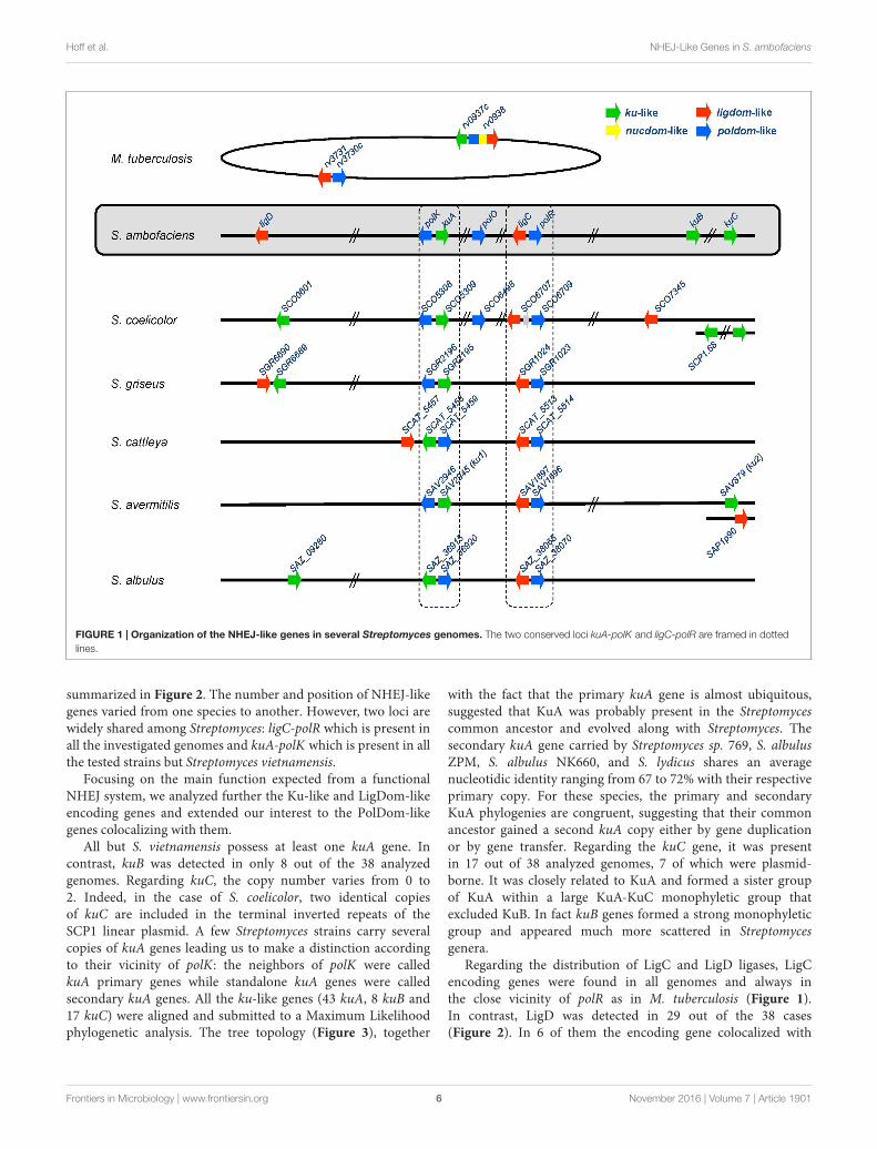
A “Core” and a “Variable” NHEJ GeneSet in StreptomycesGenes homologous to the bacterial NHEJ factors ku and ligDwere sought in the genome of our model strain Streptomycesambofaciens ATCC23877 (Thibessard et al., 2015). To do so,the M. tuberculosis Rv0937c and Rv0938 (KuMtub and LigDMtubrespectively) protein sequences were used as query in a BLASTPsearch. The first observation was that the LigDMtub proteinhad no full length homologue in S. ambofaciens, but that eachcandidate protein matched only with one single domain at a time.Therefore, the 759 amino acid sequence of LigDMtub was splitinto 3 slightly overlapping parts (1–300, 271–459, and 451–759)corresponding to the polymerase (PolDom), the nuclease(NucDom) and the ligase (LigDom) domains, respectively. In
this way, 3 Ku-like encoding genes (sharing 36, 39, and 40%of protein identity with KuMtub), 3 LigDom-like encoding genes(sharing 30, 32, and 41% of protein identity with the LigDomof LigDMtub) and 3 PolDom-like encoding genes (sharing 29,34, and 41% of protein identity with the PolDom of LigDMtub)were identified in S. ambofaciens ATCC23877 genome (Figure 1).No NucDom-like encoding gene was detected. The 3 ku-likegenes (SAM23877_5082, SAM23877_6929 and SAM23877_6942)were named kuA, kuB, and kuC respectively. Two of the 3LigDom-like genes (SAM23877_6361 and SAM23877_0862) werenamed ligC and ligD, respectively. Indeed, SAM23877_6361appeared to share more amino-acid identity to M. tuberculosisLigC (LigCMtub), i.e., 60% than to the LigDom of LigDMtubi.e., 30% and was therefore considered as LigCMtub ortholog.Similarly, SAM23877_0862 appeared to be the closest relative ofthe LigDom of LigDMtub. The third ATP-dependent DNA ligaseencoding gene detected (SAM23877_1283) would encode a LigBprotein known to exhibit a strong nick sealing activity in vitro andwhose defect would not alter mycobacterial NHEJ (Gong et al.,2004). Therefore, this third ligase was not included in our furtherinvestigations. The 3 PolDom-like genes (SAM23877_5081,SAM23877_6202, and SAM23877_6362) were named polK, polOand polR, respectively. It was noticeable that 2 of the 3 PolDomlike genes (polK and polR) colocalized with kuA and ligC.
KuA, KuB, and KuC proteins are composed of 365, 383,and 302 amino-acids, respectively. All these proteins harborthe conserved Ku core domain at their N-terminal side, theconserved minimal C-ter domain (red in Supplementary FigureS2), that has been shown to be required for the interaction withLigD in B. subtilis (McGovern et al., 2016), and a basic extendedC-terminal domain, which is of 114, 128, and 47 aa, respectively.The pIs of these extended domains (11.64, 10.97, and 10.65)are consistent with those described for the majority of bacterialKu proteins. This suggests that their roles in DNA bindingas well as in the modulation of the ability to thread inwardthe DNA molecule are conserved in Streptomyces (Kushwahaand Grove, 2013; McGovern et al., 2016). Interestingly, a SAPdomain, previously predicted in the S. coelicolor KuB homologue(SCO0601) by Aravind and Koonin (2001) is also detectedin S. ambofaciens KuB (at position 337–370, SupplementaryFigure S2). This SAP domain is assumed to be also involved innucleic acid binding (Aravind et al., 2000) and could modifythe DNA binding abilities of KuB compare to KuA and KuC.Further experiments are required to highlight the roles and thedifferences of the extended C-terminal domains of these three Kuproteins.
Once this list of genes potentially involved in a StreptomycesNHEJ pathway established, the question of their conservationin the Streptomyces genus was addressed by seeking theirhomologues in 37 other Streptomyces complete genomesequences available at http://www.ncbi.nlm.nih.gov/genome.Therefore, S. ambofaciens KuA, KuB, KuC, LigC, LigD, PolK,PolO and PolR were used as query in BLASTP searches.A wide variety of situations were encountered and a few ofthem are illustrated in Figure 1. The homologues identifiedin 38 Streptomyces genomes are listed by their locus tag inSupplementary Table S1 and their presence or absence is
Frontiers in Microbiology | www.frontiersin.org 5 November 2016 | Volume 7 | Article 1901
fmicb-07-01901 November 24, 2016 Time: 16:29 # 6
Hoff et al. NHEJ-Like Genes in S. ambofaciens
FIGURE 1 | Organization of the NHEJ-like genes in several Streptomyces genomes. The two conserved loci kuA-polK and ligC-polR are framed in dottedlines.
summarized in Figure 2. The number and position of NHEJ-likegenes varied from one species to another. However, two loci arewidely shared among Streptomyces: ligC-polR which is present inall the investigated genomes and kuA-polK which is present in allthe tested strains but Streptomyces vietnamensis.
Focusing on the main function expected from a functionalNHEJ system, we analyzed further the Ku-like and LigDom-likeencoding genes and extended our interest to the PolDom-likegenes colocalizing with them.
All but S. vietnamensis possess at least one kuA gene. Incontrast, kuB was detected in only 8 out of the 38 analyzedgenomes. Regarding kuC, the copy number varies from 0 to2. Indeed, in the case of S. coelicolor, two identical copiesof kuC are included in the terminal inverted repeats of theSCP1 linear plasmid. A few Streptomyces strains carry severalcopies of kuA genes leading us to make a distinction accordingto their vicinity of polK: the neighbors of polK were calledkuA primary genes while standalone kuA genes were calledsecondary kuA genes. All the ku-like genes (43 kuA, 8 kuB and17 kuC) were aligned and submitted to a Maximum Likelihoodphylogenetic analysis. The tree topology (Figure 3), together
with the fact that the primary kuA gene is almost ubiquitous,suggested that KuA was probably present in the Streptomycescommon ancestor and evolved along with Streptomyces. Thesecondary kuA gene carried by Streptomyces sp. 769, S. albulusZPM, S. albulus NK660, and S. lydicus shares an averagenucleotidic identity ranging from 67 to 72% with their respectiveprimary copy. For these species, the primary and secondaryKuA phylogenies are congruent, suggesting that their commonancestor gained a second kuA copy either by gene duplicationor by gene transfer. Regarding the kuC gene, it was presentin 17 out of 38 analyzed genomes, 7 of which were plasmid-borne. It was closely related to KuA and formed a sister groupof KuA within a large KuA-KuC monophyletic group thatexcluded KuB. In fact kuB genes formed a strong monophyleticgroup and appeared much more scattered in Streptomycesgenera.
Regarding the distribution of LigC and LigD ligases, LigCencoding genes were found in all genomes and always inthe close vicinity of polR as in M. tuberculosis (Figure 1).In contrast, LigD was detected in 29 out of the 38 cases(Figure 2). In 6 of them the encoding gene colocalized with
Frontiers in Microbiology | www.frontiersin.org 6 November 2016 | Volume 7 | Article 1901

fmicb-07-01901 November 24, 2016 Time: 16:29 # 7
Hoff et al. NHEJ-Like Genes in S. ambofaciens
FIGURE 2 | Distribution of NHEJ-like genes in 38 complete genomes of Streptomyces. The presence of a homolog is mentioned by a black box and theabsence of a homolog by an empty box. The number inside the boxes indicates when several homologs were identified; ∗ plasmidic; ∗∗ closed to kuC; ∗∗∗ partial(cover of 35%).
Frontiers in Microbiology | www.frontiersin.org 7 November 2016 | Volume 7 | Article 1901

fmicb-07-01901 November 24, 2016 Time: 16:29 # 8
Hoff et al. NHEJ-Like Genes in S. ambofaciens
FIGURE 3 | Phylogeny of Ku-like proteins identified in 38 Streptomyces strains. Streptomyces Ku-like proteins were identified in 38 Streptomyces strainsand compared with the closest homologues of M. tuberculosis, M. smegmatis, B. subtilis, B cereus, B halodurans, and P. aeruginosa. The tree was built using amaximum-likelihood method with a 295 aa alignment. Only bootstrap values higher than 70% are reported. Heavy lines indicate branches supported by bootstrapvalues >90% (100 replicates). The scale represents mutations per amino-acid. Primary KuA are in red, secondary KuA are in orange, KuB are in green and KuC inblue. The asterisks symbolized the proteins encoded by plasmid borne-genes. The locus-tag of each sequence is mentioned in brackets. The suspected event ofgene duplication is depicted by the two braces. B, Bacillus; M, Mycobacterium; P, Pseudomonas; S, Streptomyces. NRRL 8057 = DSM 46488; C34 = DSM42122 = NRRL B-24963; MA-4680 = NBRC 14893.
kuC, and in S. cattleya, it colocalized with kuA-polK (seeFigure 1). Phylogenetic analysis by Maximum Likelihood method(Supplementary Figure S3) showed that the LigC proteins
branched together with M. tuberculosis LigC with a topology thatseemed quite congruent with the species relations, suggesting avertical inheritance of this conserved locus. LigD proteins also
Frontiers in Microbiology | www.frontiersin.org 8 November 2016 | Volume 7 | Article 1901

fmicb-07-01901 November 24, 2016 Time: 16:29 # 9
Hoff et al. NHEJ-Like Genes in S. ambofaciens
branched together and together with LigDom of M. tuberculosisLigD.
Altogether, two NHEJ-like gene categories were delineated:(i) those that are conserved among the Streptomyces and wereconsidered as “core” putative NHEJ factors (i.e., kuA and thecolocalized polK, ligC and the colocalized polR) and (ii) those thatare sporadically represented among the Streptomyces and wereconsidered as “variable” putative NHEJ factors (i.e., kuB, kuC andligD).
NHEJ-Like Genes Are Not Essential inS. ambofaciensAfter in silico identification of the “core” and “variable” NHEJputative factors in Streptomyces, we constructed deleted mutantstrains by replacing the chromosomal locus with an excisionableantibiotic resistance cassette (Raynal et al., 2006) by the use ofPCR targeting (Gust et al., 2003). The whole selection procedurewas performed several times simultaneously in order to select atleast two independent mutant lineages for each genetic context.Table 1 summarizes the different genotypes constructed forthis study. The mutations were checked by PCR amplificationof the mutated locus. The maintenance of the chromosomallinearity of the mutants was checked by PCR amplification of thetelomeres and of some loci scattered along the chromosome (notshown).
All the mutations, simple or multiple, were obtained withclassical frequencies indicating that none of the targeted genes(“core” or “variable”) was essential. In addition, there was nosignificant difference between the wild-type reference strain andany of mutants in colonial morphology and sporulation undernormal growth conditions (not shown).
Implication of NHEJ-Like Genes in DNADamage ResponseElectronic beam irradiation (EBI) was shown to induce loss ofviability of Bacillus spores by damaging spore coat, by alteringmembrane permeability or by DNA fragmentation (Fiester et al.,2012). High level energy beam such as those used in this work(2.2 MeV) is likely to trigger DSB as the most significant lesionsin the spore embedded DNA. In order to test the involvementof the NHEJ-like factors to DNA damage response, spores ofthe wild-type S. ambofaciens reference strain as well as sporesof at least two independent mutants for each genetic contextwere irradiated by EBI from 0.1 to 1 kGy and their survivalwas assessed after growth on SFM medium. The results arerepresented in Figure 4.
Both “Core” and “Variable” ku genes Are Involved inDNA Damage ResponseDepending on the dose, deletion of kuA, which belongs to the“core” NHEJ-like gene set, conferred a 2–4 fold increase ofthe sensitivity compared to the wild-type (Figure 4A). SincekuA is present in the large majority of the sequenced genome,it appears as the major Ku actor of the putative StreptomycesNHEJ. Surprisingly, mutations of the variable ku-like genes,kuB and kuC, also conferred an increased sensitivity but at
a lower level, with a maximal twofold cell viability decreasereached after 0.5 and 0.7 kGy exposition for both contexts(Figure 4A). In addition, the sensitivity was aggravated whenkuB and kuC were both deleted in a 1kuA background togenerate a triple 1kuABC mutant, until reaching a sevenfold cellviability diminution in comparison to the WT for the mediumradiation doses (Figure 4A). This result suggests that the threeKu products participate to the response to DNA damagingagents in spores, each of them assuming their share of the DNArepair independently from the two others. Additionally, the triple1kuABC mutation assumed to abolish any putative NHEJ repairstrongly increased the sensitivity of a HR deficient context (i.e.,1recA) (Figure 4D). This last result shows that RecA and Ku-likeproteins are both important contributors to the S. ambofaciensspore resistance to EBI.
Two ATP Dependent Ligases Involved in DNADamage ResponseIn Mycobacterium, LigD is the main NHEJ ligase whereasLigC only provides a back-up activity (Della et al., 2004). Incontrast to Mycobacterium where a single ligC mutation doesnot impact survival rate to DNA damage, the defect of both theconserved ligC and the variable ligD of S. ambofaciens conferreda significant sensitivity to EBI. Surprisingly, the deletion of ligDconferred an even higher sensitivity than the mutation of ligC(Figure 4B). Furthermore, the 1ligCD double mutant showedan aggravated phenotype in the same proportions as a 1kuABCtriple mutant (Figure 4B). These data clearly show that thetwo ligase activities participate to DNA repair in response toEBI.
PolDom Genes Are Involved in DNA DamageResponseSingle deletion of polK or polR, the two conserved homologuesof Mycobacterium PolDom colocalized respectively with kuAand ligC led to a weak sensitivity to electronic beam exposure.For both single mutants, increase of sensitivity in comparisonto the wild-type does not exceed 1.5 fold for each dose(Figure 4C). On the other hand, double polRK mutant strainsare approximately twice more sensitive than the single mutants(Figure 4C). Both loci are therefore involved in DNA damageresponse.
The ku Genes Are DifferentiallyExpressed during DevelopmentIn order to investigate the spatial and temporal expression ofNHEJ-like genes during development, a time-course of RT-PCRexperiments was performed and translational fusions with EGFPwere observed by fluorescent microscopy. First of all, the resultsshowed that NHEJ-like genes are weakly expressed throughoutthe development. For instance, the expression of ligC, ligD, polKand polR was too low to be monitored by qPCR whereas theexpression of ku genes was 10 to 50 fold lower than hrdBexpression (Figure 5A). Secondly, kuA expression was clearlygrowth-phase dependent with a 10 fold increase between 24 and60 h after germination. It is important to indicate that sporulationbegan to be observable after 48 h of development in these growth
Frontiers in Microbiology | www.frontiersin.org 9 November 2016 | Volume 7 | Article 1901

fmicb-07-01901 November 24, 2016 Time: 16:29 # 10
Hoff et al. NHEJ-Like Genes in S. ambofaciens
FIGURE 4 | Electronic beam irradiation of S. ambofaciens strains deficient in NHEJ-like genes. Spores of mutants of NHEJ-like genes, namely single andmultiple ku (A), ligase (B) and polymerase (C) mutants, were exposed to 0.1, 0.3, 0.5, 0.7, and 1 kGy of electronic beam irradiation. (D) In the same way, a 1ku-like1recA mutant also affected in HR was exposed to the same irradiation doses. In this latter case, a logarithmic scale was used for survival rate representation.Survival rates were calculated as the number of clones for a given dose relative to the number of clones unexposed to radiation. For each graph, wild-type strain isrepresented as a control. Each curve corresponds to data obtained from an individual mutant strain. Error bars indicate standard error of a minimum of threereplicates.
conditions. There was no difference throughout the time-coursefor kuB and kuC expression. Both of them were less expressedthan kuA at the late stages of development. The WT strainwas also grown to OD600 of 0.2 in HT liquid medium andthen exposed to MMC (1.5 µg·ml−1) for 30 min. RNAs wereextracted and reverse-transcribed to assess by PCR the regulationof NHEJ-like genes during a genotoxic stress. No induction orrepression of NHEJ-like genes was noticeable, while the positiveinduction control (recA) was up-regulated up to 20 fold (notshown).
To observe the cellular localization of Ku proteins inphysiological conditions, egfp translational fusions wereconstructed at kuA, kuB, and kuC chromosomal loci. Fusionstrains were inoculated on SFM agar plates and grown for48 h before observations by fluorescent microscopy along witha negative control strain without any egfp gene. A positive
control strain constitutively expressing egfp gene exhibited astrong green fluorescence signal in all cells throughout thedevelopmental stages of the bacteria, proving the reportergene to be functional in S. ambofaciens (not shown). For KuAfusion, the green fluorescence signal was significant in sporesand segmenting aerial hyphae but not in vegetative mycelium(Figure 5B), showing a specific accumulation of KuA inspores. This result corroborates the phase dependent expressionpattern of kuA observed in the previous qPCR analysis. ForKuB and KuC fusions, although the signals were weaker thanfor KuA fusion, they also revealed the accumulation of KuBand KuC in spore chains. Since no significant modulationof gene expression was detected for kuB and kuC along thegrowth cycle (Figure 5A), this protein accumulation patternsuggests a translational (and/or a post-translational) regulationlevel.
Frontiers in Microbiology | www.frontiersin.org 10 November 2016 | Volume 7 | Article 1901

fmicb-07-01901 November 24, 2016 Time: 16:29 # 11
Hoff et al. NHEJ-Like Genes in S. ambofaciens
FIGURE 5 | Growth-dependent and cell-type specific expression of NHEJ-like genes. (A) Expression level of S. ambofaciens ATCC23877 ku genes relativeto hrdB after 24, 36, 48, and 60 h growth on SFM plates at 30◦C. Error bars represent standard error. Expression of kuA after 60 h growth is significantly higher thanthat at 24 h. Furthermore, kuA expression at 60 h growth is significantly different than that of kuB and kuC (t-test with Bonferroni correction, α = 0.05). (B) Strainsexpressing EGFP fusion with KuA, KuB, or KuC were inoculated in the acute-angle junction of standard-sized microscope coverslips inserted at 45◦ in SFM agar,and grown for 48 h before observations by fluorescent microscopy. The control strain does not contain any GFP gene. Representative examples of spores or aerialmycelium are shown as a contrast phase image (upper panels) and as EGFP fluorescent channel (lower panels). The weak fluorescent foci observed mainly inmycelia were readily detected in the control strain and are not likely to correspond to GFP signal. The scale is represented by a 5 µm size bar.
The Ku Proteins Protect Linear dsDNAfrom the T5 Exonuclease and Stimulatethe Ligase Activity of LigDBsubTo assess which proteins are required in a putative S. ambofaciensNHEJ, we tried to purify all the proteins putatively implicated inthis pathway. Among them, we succeeded to express and purifiedS. ambofaciens KuA, KuB and KuC proteins (39.1, 42.4, and33.6 KDa respectively). To evaluate the oligomeric state of thethree Ku proteins, we performed gel filtration experiments. KuAand KuB were eluted in fractions corresponding to a molecularweight of 160 and 100 kDa, respectively, strongly suggestingthat KuA and KuB are multimeric (Supplementary Figure S1and Supplementary Data Sheet S1). To date, characterizedbacterial Ku are dimers. However, the elongated shape of
the B. subtilis Ku (KuBsub) dimer (McGovern et al., 2016)led to the elution of this protein with apparent molecularweight corresponding to a tetramer. Then KuA and KuBcould also be dimers displaying elongated shape, as forKuBsub. KuC was clearly purified in different oligomericstates ranging from 70 to more than 600 kDa. The mainelution peak, corresponding to a protein of apparent molecularweight around 160 kDa, was similar to the one observedfor KuA.
KuA, KuB, and KuC were able to protect a linear dsDNAmolecule from the T5 exonuclease (Figure 6A), a propertyshared by the KuBsub protein (McGovern et al., 2016) and thePseudomonas aeruginosa Ku against different exonucleases (Zhuand Shuman, 2010). This result suggests that these proteins areable to bind DNA ends. Another role of Ku in the bacterial
Frontiers in Microbiology | www.frontiersin.org 11 November 2016 | Volume 7 | Article 1901

fmicb-07-01901 November 24, 2016 Time: 16:29 # 12
Hoff et al. NHEJ-Like Genes in S. ambofaciens
FIGURE 6 | KuA, KuB, and KuC protect linear dsDNA from the T5 exonuclease and stimulate the ligase activity of LigDBsub. (A) A 1001 bp linear dsDNAwith 5′-phosphorylated ends (3.8 nM) was incubated 30 min at 37◦C with increasing amounts of KuA, KuB, or KuC as indicated. T5 exonuclease (0.5 U) was thenadded in each reaction mixtures for 30 min at 37◦C and digested products were analyzed as described in the Section “Material and Methods.” Control experimentsshowing the 1001 ds DNA substrate and the nearly complete degradation of the DNA molecule by the T5 exonuclease without other proteins added are shown onthe left. (B) A 1001 bp linear dsDNA with 5′-phosphorylated ends (3.8 nM) was incubated with 0.25 or 1 µM of KuA, KuB, or KuC as indicated. Increasing amountsof LigDBsub were then added (allowing final concentrations of LigDBsub indicated above the gels) and ligation products were analyzed as described in Section“Material and Methods.” Control experiments showing the weak activity of LigDBsub alone at different concentration are shown on the left (No Ku). For comparison,KuBsub was assessed for its LigDBsub stimulation activity in the same conditions (right part).
NHEJ is to recruit the LigD at DNA ends and to stimulateits ligase activity. Remarkably, KuA, KuB, and KuC were ableto stimulate the ligation activity of the B. subtilis LigD protein(Figure 6B), even if they were less efficient than KuBsub. However,we observed a different concentration-dependent stimulationof the ligase by the S. ambofaciens Ku proteins. Raising the
concentration of KuA and KuC from 0.25 to 1 µM in theligation assay seemed to slightly increase ligation efficiency. Atthe opposite, the stimulation activity observed with 0.25 µMof KuB appeared to be reduced when its concentration wasraised to 1 µM. A similar inhibitory effect was observed withKuBsub.
Frontiers in Microbiology | www.frontiersin.org 12 November 2016 | Volume 7 | Article 1901

fmicb-07-01901 November 24, 2016 Time: 16:29 # 13
Hoff et al. NHEJ-Like Genes in S. ambofaciens
DISCUSSION
Streptomyces NHEJ-Like GeneOrganization Is AtypicalThe presence of a NHEJ pathway in prokaryotes appears to belimited to some phyla representing about 20% of the species(estimated on a sample of 2645 complete bacterial genomes)(McGovern et al., 2016). The minimal set of NHEJ gene encodesa two partner system including the homodimeric Ku protein anda ligase polypeptide composed of the ligase enzymatic domain(LigDom) to a nuclease (NucDom) and a polymerase (PolDom)domain. The latter enzymatic activities process DNA ends beforeligation. The NHEJ gene repertoire ranges from the simplest inP. aeruginosa with a single Ku and a ligase (ligase D) to themore complex with those described in Agrobacterium (Zhu andShuman, 2007) or Sinorhizobium (Kobayashi et al., 2008) andStreptomyces as described in this work. Hence, while B. subtilisand Mycobacterium possess a single Ku, a single LigD and oneor two LigC, A. tumefaciens possesses three Ku, two LigD andthree LigC (Zhu and Shuman, 2007). Sinorhizobium melilotiand other rhizobia possess up to 4 Ku, 5 LigD and one LigC(Kobayashi et al., 2008); the ligase, nuclease and polymerasedomains are present in different organizations, sometimes (i)fused together in a single polypeptide or (ii) encoded bydistinct genes. This situation is similar in archaea where theputative ligase, nuclease and polymerase domains are encodedby distinct genes, however, kept into an operonic organizationassumed to ensure the co-regulation of the activities (Bartlettet al., 2013). However, among actinobacteria, Streptomycesappears as an exception, since other main phyla (Nocardia,Frankia, Rhodococcus, and Corynebacterium) seem to possess amultidomain LigD homologue (not shown).
In Streptomyces, several levels of complexity are alsonoticeable. First, the activities putatively involved in NHEJ areencoded by standalone genes that are scattered along the genome.Consequently the repertoire is rather expanded: the number ofgenes ranging from none up to 4 for ku, from one to two LigDomencoding genes (ligC, ligD), from 2 to 3 PolDom encoding genes,from none to one NucDom encoding gene. The total numberof NHEJ-like genes ranges from 4 in S. bingchenggensis andS. venezuelae to 8 in S. ambofaciens and S. reticuli: 3 ku (kuA,kuB, kuC), one ligD and one ligC, and 3 polymerase genes (polK,polR, polO). These genes can fall into two groups. On one hand,the “core” set includes kuA, ligC, polK and polR which are presentin almost all Streptomyces species and organized in two loci ofrather conserved organization (i.e., kuA-polK, ligC-polR). Theonly exception appears to be in S. vietnamiensis which lacks thecouple kuA-polK and the other ku genes. On the other hand, therest of the genes constitute the “variable” gene set. An interestingfeature to highlight is a gene duplication event involving kuAor kuC. The duplicated copies are either located in the variableregion of the genome or present within the terminal invertedrepeats of the linear replicons. This phenomenon is interestingsince duplicated copies can evolve through gene divergence toraise new function. This mechanism is rather rare in bacteria butgene duplication seems frequent in Streptomyces, notably with
large terminal duplication probably resulting from DSB repair bya mechanism related to break-induced replication.
Involvement of the “Core” and “Variable”NHEJ-Like Gene Sets in DNA RepairThe large number of NHEJ-like genes in Streptomyces as wellas their variability questions their implication in DNA repairmechanisms. It is tempting to imply the “core” NHEJ gene setin a classical NHEJ pathway with a Ku (i.e., KuA) and an ATP-dependent DNA ligase (i.e., LigC). In addition to being theconserved ku gene, kuA is the more expressed of the 3 ku-likegenes in the late growth phases, its defect conferred the mostmarked sensitivity to EBI and finally, KuA is the most efficientof the 3 Ku-like proteins in stimulating LigDBsub activity in vitro.
It might be surprising that the NHEJ ligase could be LigCrather than LigD which is the canonical NHEJ ligase in otherbacterial species. However, it should be reminded that inStreptomyces, LigD carries the only ligase domain, as doesLigC. Both ligases are devoid of the PolDom domain knownto be involved in its recruitment by Ku. Therefore LigD andLigC may ensure similar functional roles in Streptomyces. Onecould hypothesize that a gene rearrangement split the ligasedomain from the two others (i.e., PolDom and NucDom) in aStreptomyces common ancestor. The two ligase genes would havethen provided an equivalent contribution to the bacterium, finallyresulting in the replacement of LigD by LigC in the “core” NHEJset by a founder effect.
In S. ambofaciens, in which both ligC and ligD cohabits, singlemutants displayed a sensitive phenotype to EBI and the doublemutant displayed an aggravated phenotype. In this context, atleast two hypotheses can be drawn. First, both LigC and LigDcould participate to the same repair pathway; in that case, thesensitivity of single mutants and the aggravated phenotype ofdouble mutants could be explained by a dose effect that is theneed for a certain amount of ligases to cope for the inducedDNA damage. In that case, both ligase genes would contributeto achieve the required level of ligase. Alternatively, they couldparticipate to different repair pathways required to take overvaried DNA damage (see below).
If the “core” NHEJ gene set indeed establishes a functionalNHEJ pathway, the presence of two PolDoms in the “core”NHEJ gene set constitutes another intriguing feature, especiallyconsidering that they do not seem to have redundant functions.Hence, the sensitivity of both polK and polR simple mutants wasaggravated in the double mutant. As shown in M. tuberculosisNHEJ, the role of the PolDom domain of LigD would be lessto provide a polymerase activity than to recruit LigD to Ku(Pitcher et al., 2005). Then PolK and/or PolR may play a roleof linker/adapter to recruit a ligase; the more likely being thatPolK and PolR recruit LigC, however, on different types of DNAextremities (e.g., blunt, 3′ or 5′ overhanging ends). In the case ofthe presence of LigD (as in S. ambofaciens for example), the twoPolK and PolR may recruit both LigC and LigD.
It is particularly noticeable that the “variable” genes (i.e., ligDbut also kuB, kuC) were involved in the response to EBI. Theexposure to EBI is assumed to trigger DSBs. However, different
Frontiers in Microbiology | www.frontiersin.org 13 November 2016 | Volume 7 | Article 1901

fmicb-07-01901 November 24, 2016 Time: 16:29 # 14
Hoff et al. NHEJ-Like Genes in S. ambofaciens
types of lesions can result from direct transmission of energyto the DNA structure and from secondary oxidative stress (e.g.,oxidized bases). In consequence, damage induced by EBI requiresNHEJ but may also require other DNA repair pathways. Thus,these “variable” genes would (i) support the NHEJ encoded bythe “core” gene set in an opportunistic way, (ii) constitute analternative NHEJ pathways or (iii) constitute a repair pathwaydedicated to other DNA damage single strand gaps or abasicsites). The first hypothesis would imply a dose effect of theKu as well as the ligase proteins to explain the aggravatedphenotype of the multiple mutants (i.e., 1kuABC and 1ligCD).In this first hypothesis, considering that prokaryote Ku act asdimers (McGovern et al., 2016), we can speculate that these threeKu proteins could act as homodimers as well as heterodimers.A data in favour of the second hypothesis (an alternative NHEJpathway) is the colocalization of the kuC and ligD genes in 6Streptomyces species clustering with S. griseus (SupplementaryTable S1; Figure 3). However, no clear trend toward the co-occurrence of these two “variable” genes can be noticed in the38 analysed genomes. In B. subtilis, the recent involvement ofLigDBsub and KuBsub in base excision repair along with theirmajor role in NHEJ supports the third hypothesis (de Ory et al.,2014, 2016).
In order to solve the question of the involvement of thesegenes in a NHEJ pathway, a forward-looking approach could beto specifically induce DSB formation at targeted chromosomalloci and assessed the sensitivity of the NHEJ-candidate genes toidentify those truly involved in DSB repair per se.
A Late Growth Phase Role for NHEJ inStreptomycesThe presence of a NHEJ pathway in bacteria is usually assignedto DSB repair in low rate growth phases characterizing bacterialspecies involved in pathogenic processes or exposed to harshenvironmental living conditions. When bacteria replicate such asin laboratory conditions, sister chromosomes providing an intacttemplate are preferentially used to ensure faithful DSB repair byHR. However, when replication is slowing down (i.e., stationaryphases) or stopping (e.g., in resistance forms such as spores), DSBrepair strongly relies on NHEJ. This is consistent with the factthat NHEJ genes are expressed preferentially when DSB repaircannot be ensured by a faithful process. Hence, in B. subtilisas well as in Streptomyces, the NHEJ genes were shown to beexpressed in stationary phase and to accumulate in spores. Theforespore-specific sigma factor σG in B. subtilis directly regulatesykoU (coding for LigD) and ykoV (coding for Ku), inducingtheir expression during the forespore development (Wang et al.,2006). In addition, B. subtilis Ku protein specifically localizesto the nucleoid of germinating spores while it disappears atlater stages of germination. In Streptomyces, gene transcriptionanalyses showed in this work an expression of the ku in stationaryphase with an increase of kuA expression during sporulation.Moreover, GFP fusions showed a predominant localization of theKu proteins in spores. Furthermore, mutations in ku and ligasegenes confer sensitivity to genotoxic agents to B. subtilis as wellas Streptomyces spores. Sensitivity to heat and desiccation which
are biotic parameters known to trigger DSB is also associated tothe NHEJ mutations in B. subtilis and Mycobacterium smegmatis(Moeller et al., 2007; Pitcher et al., 2007; Stephanou et al., 2007).The presence of multiple ku genes (and of other NHEJ-like genes)in Sinorhizobium meliloti is assumed to correspond to the needfor DNA repair at different steps of the cell cycle. Although noinduction of the NHEJ pathways was observed under genotoxicconditions (i.e., IR exposure), the expression patterns of thesku genes suggest that each Ku protein has a distinct roleunder different conditions (free bacterial form, bacteroids). Incontrast, NHEJ would not be required during the establishmentof symbiosis since a strain lacking all four sku genes was shownto establish functional nodules (Kobayashi et al., 2008). As inS. meliloti, it was shown that each of the S. ambofaciens kugenes is functional since their deletion conferred a phenotype ofsensitivity toward genotoxic agents. Mutant strains lacking kuA,kuB, and kuC genes showed an aggravated phenotype suggestingthat each participated to the response to DNA damage. We cantherefore suppose that like in S. meliloti, intervention of theStreptomyces ku genes is done under specific conditions.
Recently in Pseudomonas putida, the NHEJ was shown toparticipate to another late growth phase phenomenon (Pariset al., 2015). Hence, bacteria frequently encountered nutrientstarvation during late-growth phase. Populations starving displaya phenomenon called stationary-phase mutagenesis (also knownas adaptive mutations) from which emerge mutants able toovercome the shortage and initiate new vegetative cycles.The underlying mechanisms notably involve unfaithful DNApolymerases during HR events (Shee et al., 2012). Although theinvolvement of NHEJ in late mutagenesis remains unknown,deficiency of each of the NHEJ activities triggered an alteredpattern of mutations (Paris et al., 2015). The authors speculateon ability to bypass DNA lesions of the LigD polymerase domain.
Origin of the “Core” and “Variable” NHEJGene Sets in StreptomycesWhile the origin of the NHEJ “core” gene set is likely toresult from vertical inheritance from a common Streptomycesancestor, the origin of the “variable” set is questionable. Thesporadic distribution of the “variable” genes may reflect eitherthe existence of NHEJ in a bacterial common ancestor andits subsequent sporadic loss during genus/species separation.Reciprocally, it may reflect the acquisition of NHEJ genes throughhorizontal gene transfer in some phyla. Both hypotheses canalso be combined and result in the observed complexity. Themost explicit argument about their origin is their localization inthe variable regions of the chromosome or in plasmids. Whileplasmids can be mobilized and even self-transmitted, the terminalregions of the Streptomyces chromosome is known to be targetedby recombination phenomena leading to insertions of acquiredDNA and further losses (Choulet et al., 2006).
Impact of NHEJ on Genome EvolutionWhatever the evolutionary past of the NHEJ gene sets, thepresence of a conserved “core” gene set enriched by additionalfunctional “variable” genes may confer some advantage. In all
Frontiers in Microbiology | www.frontiersin.org 14 November 2016 | Volume 7 | Article 1901

fmicb-07-01901 November 24, 2016 Time: 16:29 # 15
Hoff et al. NHEJ-Like Genes in S. ambofaciens
bacteria studied so far, NHEJ is not essential (Ku/LigD) forgrowth, at least under laboratory conditions. Mutants of NHEJin Mycobacterium, Pseudomonas, or Bacillus are not affectedalthough they show sensitivity to environmental stress (Welleret al., 2002; Jacobs et al., 2003; Gong et al., 2004, 2005). Wecame to the same conclusion in S. ambofaciens although it wasparticularly interesting to note that a part of the “variable” genes(i.e., kuB, kuC and ligD) was involved in response to genotoxicagents. Hence the aggravated phenotype observed in doubleor multiple mutant strains (i.e., kuABC, ligCD) revealed thecooperative actions of the multiple alleles. The “variable” genes inStreptomyces may increase the flexibility and/or efficiency of theDSB repair mechanism and influence evolution of the genome.Lessons about the role of NHEJ in genome evolution can bedrawn from organisms lacking NHEJ. Hence, the only eukaryotelacking NHEJ is the yeast Lachancea kluyveri (Gordon et al.,2011). Compared to other species of the Saccharomycetaceaefamily, this species showed a significantly low number ofgenomic rearrangements and the lack of telomere-to-telomerefusions. The authors suggest that the loss of the NHEJ andan alternative end joining pathway [named microhomology-mediated end joining (MMEJ) or alternative end joining (A-EJ)]is responsible for the low recombination rate assessed in thisphylum. In this way, we can hypothesize that in Streptomycesthe presence/absence of NHEJ genes in addition to the “core”NHEJ (the “variable” genes participating to a unique NHEJ orother alternative pathways) may modulate the frequency of DNArearrangements and DNA acquisition through horizontal genetransfer and confer different evolution rates to Streptomycesspecies. These genes being themselves subject to transfer,the recombinant phenotype (i.e., hyperrecombinant) could bevariable within a population and favor genetic diversificationand adaptation to the environment. This situation could bereminiscent of mutator strains identified in E. coli (for review:Giraud et al., 2001) but applied to the capacity to recombine DNAincluding incoming genetic information.
Future PerspectivesA key question is the relative contribution of HR and NHEJ tothe evolution and the remarkable compartmentalization of thelinear Streptomyces chromosome. Is there any regulation betweenthe two pathways during the development of the bacteria? In
S. avermitilis, the deletion of the ku homologues (ku1 and ku2corresponding to kuA and kuC respectively) was associated toan increased efficiency of HR (Zhang et al., 2012). This mayreveal the existence of a balance resulting from the competitionbetween HR and NHEJ repair mechanisms, and that both repairpathways co-exist at least during replicating phases. Is there anyspatial regulation of the DSB repair mechanisms? DSB could behealed by different mechanisms depending of their chromosomallocation leading to either faithful repair or possible integration offoreign information.
AUTHOR CONTRIBUTIONS
Investigation: GH, CB, LZ, EP, LC, and SM; Computer analysis:AT and CyB; Methodology: CyB, FC, and FL; Conceptualization:CB, AT, FL, and PL; Writing original draft: GH, CB, AT, andPL; Writing-review and editing: FL, FC, CyB, CB, AT, and PL;Resources: FC, FL, and PL; Funding acquisition: CB, AT, and PL;Supervision: AT and PL.
FUNDING
This work was supported by the French National ResearchAgency program Streptoflux (ANR Blanc 0096_01), by ANRthrough the Laboratory of Excellence ARBRE (ANR-12-LABXARBRE-01) and by the Région Lorraine.
ACKNOWLEDGMENTS
We would like to thank Sébastien Riegler and Florian Kuntz fromthe Technological Resource center Aérial (Illkirch-Graffenstaden,France) for their technical support and their warm welcome.
SUPPLEMENTARY MATERIAL
The Supplementary Material for this article can be foundonline at: http://journal.frontiersin.org/article/10.3389/fmicb.2016.01901/full#supplementary-material
REFERENCESAbrudan, M. I., Smakman, F., Grimbergen, A. J., Westhoff, S., Miller,
E. L., van Wezel, G. P., et al. (2015). Socially mediated inductionand suppression of antibiosis during bacterial coexistence. Proc.Natl. Acad. Sci. U.S.A. 112, 11054–11059. doi: 10.1073/pnas.1504076112
Aigle, B., Holl, A., Angulo, J. F., Leblond, P., and Decaris, B. (1997).Characterization of two Streptomyces ambofaciens recA mutants: identificationof the RecA protein by immunoblotting. FEMS Microbiol. Lett. 149, 181–187.doi: 10.1111/j.1574-6968.1997.tb10326.x
Alonso, J. C., Cardenas, P. P., Sanchez, H., Hejna, J., Suzuki, Y., andTakeyasu, K. (2013). Early steps of double-strand break repair in Bacillussubtilis. DNA Repair(Amst.) 12, 162–176. doi: 10.1016/j.dnarep.2012.12.005
Aniukwu, J., Glickman, M. S., and Shuman, S. (2008). The pathways and outcomesof mycobacterial NHEJ depend on the structure of the broken DNA ends. GenesDev. 22, 512–527. doi: 10.1101/gad.1631908
Aravind, L., and Koonin, E. V. (2001). Prokaryotic homologs of the eukaryoticDNA-end-binding protein Ku, novel domains in the Ku protein and predictionof a prokaryotic double-strand break repair system. Genome Res. 11, 1365–1374.doi: 10.1101/gr.181001
Aravind, L., Makarova, K. S., and Koonin, E. V. (2000). Holliday junction resolvasesand related nucleases: identification of new families, phyletic distribution andevolutionary trajectories. Nucleic Acids Res. 28, 3417–3422. doi: 10.1093/nar/28.18.3417
Bartlett, E. J., Brissett, N. C., and Doherty, A. J. (2013). Ribonucleolyticresection is required for repair of strand displaced nonhomologous end-joiningintermediates. Proc. Natl. Acad. Sci. U.S.A. 110, E1984–E1991. doi: 10.1073/pnas.1302616110
Frontiers in Microbiology | www.frontiersin.org 15 November 2016 | Volume 7 | Article 1901

fmicb-07-01901 November 24, 2016 Time: 16:29 # 16
Hoff et al. NHEJ-Like Genes in S. ambofaciens
Bentley, S. D., Chater, K. F., Cerdeno-Tarraga, A.-M., Challis, G. L., Thomson,N. R., James, K. D., et al. (2002). Complete genome sequence of the modelactinomycete Streptomyces coelicolor A3 (2). Nature 417, 141–147. doi: 10.1038/417141a
Bhattarai, H., Gupta, R., and Glickman, M. S. (2014). DNA ligase C1mediates the LigD-independent nonhomologous end-joining pathway ofMycobacterium smegmatis. J. Bacteriol. 196, 3366–3376. doi: 10.1128/JB.01832-14
Casadaban, M. J., and Cohen, S. N. (1980). Analysis of gene control signals byDNA fusion and cloning in Escherichia coli. J. Mol. Biol. 138, 179–207. doi:10.1016/0022-2836(80)90283-1
Chater, K. F. (1993). Genetics of differentiation in Streptomyces. Annu. Rev.Microbiol. 47, 685–711. doi: 10.1146/annurev.mi.47.100193.003345
Choulet, F., Aigle, B., Gallois, A., Mangenot, S., Gerbaud, C., Truong, C.,et al. (2006). Evolution of the terminal regions of the Streptomyceslinear chromosome. Mol. Biol. Evol. 23, 2361–2369. doi: 10.1093/molbev/msl108
Davies, J., and Davies, D. (2010). Origins and evolution of antibiotic resistance.Microbiol. Mol. Biol. Rev. 74, 417–433. doi: 10.1128/MMBR.00016-10
de Ory, A., Nagler, K., Carrasco, B., Raguse, M., Zafra, O., Moeller, R., et al. (2016).Identification of a conserved 5’-dRP lyase activity in bacterial DNA repair ligaseD and its potential role in base excision repair. Nucleic Acids Res. 44, 1833–1844.doi: 10.1093/nar/gkw054
de Ory, A., Zafra, O., and de Vega, M. (2014). Efficient processing of abasic sitesby bacterial nonhomologous end-joining Ku proteins. Nucleic Acids Res. 42,13082–13095. doi: 10.1093/nar/gku1029
Della, M., Palmbos, P. L., Tseng, H., Tonkin, L. M., Daley, J. M., Topper,L. M., et al. (2004). Mycobacterial Ku and ligase proteins constitute a two-component NHEJ repair machine. Science 306, 683–685. doi: 10.1126/science.1099824
Deriano, L., and Roth, D. B. (2013). Modernizing the nonhomologous end-joiningrepertoire: alternative and classical NHEJ share the stage. Annu. Rev. Genet. 47,433–455. doi: 10.1146/annurev-genet-110711-155540
Fiester, S. E., Helfinstine, S. L., Redfearn, J. C., Uribe, R. M., and Woolverton,C. J. (2012). Electron beam irradiation dose dependently damages the Bacillusspore coat and spore membrane. Int. J. Microbiol. 2012, 1–9. doi: 10.1155/2012/579593
Fischer, G., Decaris, B., and Leblond, P. (1997). Occurrence of deletions, associatedwith genetic instability in Streptomyces ambofaciens, is independent of thelinearity of the chromosomal DNA. J. Bacteriol. 179, 4553–4558. doi: 10.1128/jb.179.14.4553-4558.1997
Fischer, G., Wenner, T., Decaris, B., and Leblond, P. (1998). Chromosomalarm replacement generates a high level of intraspecific polymorphism in theterminal inverted repeats of the linear chromosomal DNA of Streptomycesambofaciens. Proc. Natl. Acad. Sci. U.S.A. 95, 14296–14301. doi: 10.1073/pnas.95.24.14296
Galet, J., Deveau, A., Hôtel, L., Frey-Klett, P., Leblond, P., and Aigle, B.(2015). Pseudomonas fluorescens pirates both ferrioxamine and ferricoelichelinsiderophores from Streptomyces ambofaciens. Appl. Environ. Microbiol. 81,3132–3141. doi: 10.1128/AEM.03520-14
Giraud, A., Radman, M., Matic, I., and Taddei, F. (2001). The rise and fallof mutator bacteria. Curr. Opin. Microbiol. 4, 582–585. doi: 10.1016/S1369-5274(00)00254-X
Glickman, M. S. (2014). Double-strand DNA break repair in mycobacteria.Microbiol. Spectr. 2:MGM2-0024-2013. doi: 10.1128/microbiolspec.MGM2-0024-2013
Gong, C., Bongiorno, P., Martins, A., Stephanou, N. C., Zhu, H., Shuman, S., et al.(2005). Mechanism of nonhomologous end-joining in mycobacteria: a low-fidelity repair system driven by Ku, ligase D and ligase C. Nat. Struct. Mol. Biol.12, 304–312. doi: 10.1038/nsmb915
Gong, C., Martins, A., Bongiorno, P., Glickman, M., and Shuman, S. (2004).Biochemical and genetic analysis of the four DNA ligases of mycobacteria.J. Biol. Chem. 279, 20594–20606. doi: 10.1074/jbc.M401841200
Gordon, J. L., Byrne, K. P., and Wolfe, K. H. (2011). Mechanisms of chromosomenumber evolution in yeast. PLoS Genet. 7:e1002190. doi: 10.1371/journal.pgen.1002190
Gupta, R., Barkan, D., Redelman-Sidi, G., Shuman, S., and Glickman, M. S. (2011).Mycobacteria exploit three genetically distinct DNA double-strand break
repair pathways. Mol. Microbiol. 79, 316–330. doi: 10.1111/j.1365-2958.2010.07463.x
Gust, B., Challis, G. L., Fowler, K., Kieser, T., and Chater, K. F. (2003). PCR-targeted Streptomyces gene replacement identifies a protein domain needed forbiosynthesis of the sesquiterpene soil odor geosmin. Proc. Natl. Acad. Sci. U.S.A.100, 1541–1546. doi: 10.1073/pnas.0337542100
Hoff, G., Bertrand, C., Piotrowski, E., Thibessard, A., and Leblond, P.(2016). Implication of RuvABC and RecG in homologous recombination inStreptomyces ambofaciens. Res. Microbiol. doi: 10.1016/j.resmic.2016.07.003[Epub ahead of print].
Hopwood, D. A. (2006). Soil to genomics: the Streptomyces chromosome. Annu.Rev. Genet. 40, 1–23. doi: 10.1146/annurev.genet.40.110405.090639
Huang, T.-W., and Chen, C. W. (2006). A recA null mutation may be generated inStreptomyces coelicolor. J. Bacteriol. 188, 6771–6779. doi: 10.1128/JB.00951-06
Ikeda, H., Ishikawa, J., Hanamoto, A., Shinose, M., Kikuchi, H., Shiba, T., et al.(2003). Complete genome sequence and comparative analysis of the industrialmicroorganism Streptomyces avermitilis. Nat. Biotechnol. 21, 526–531. doi: 10.1038/nbt820
Inoue, S., Higashiyama, K., Uchida, T., Hiratsu, K., and Kinashi, H. (2003).Chromosomal circularization in Streptomyces griseus by nonhomologousrecombination of deletion ends. Biosci. Biotechnol. Biochem. 67, 1101–1108.doi: 10.1271/bbb.67.1101
Jacobs, M. A., Alwood, A., Thaipisuttikul, I., Spencer, D., Haugen, E., Ernst, S., et al.(2003). Comprehensive transposon mutant library of Pseudomonas aeruginosa.Proc. Natl. Acad. Sci. U.S.A. 100, 14339–14344. doi: 10.1073/pnas.2036282100
Kieser, T., Bibb, M. J., Buttner, M. J., Chater, K. F., and Hopwood, D. A. (2000).Practical Streptomyces Genetics. Norwich: John Innes Foundation.
Kim, J.-N., Kim, Y., Jeong, Y., Roe, J.-H., Kim, B.-G., and Cho, B.-K. (2015).Comparative genomics reveals the core and accessory genomes of Streptomycesspecies. J. Microbiol. Biotechnol. 25, 1599–1605. doi: 10.4014/jmb.1504.04008
Kobayashi, H., Simmons, L. A., Yuan, D. S., Broughton, W. J., and Walker, G. C.(2008). Multiple Ku orthologues mediate DNA non-homologous end-joiningin the free-living form and during chronic infection of Sinorhizobium meliloti.Mol. Microbiol. 67, 350–363. doi: 10.1111/j.1365-2958.2007.06036.x
Kushwaha, A. K., and Grove, A. (2013). C-terminal low-complexity sequencerepeats of Mycobacterium smegmatis Ku modulate DNA binding. Biosci. Rep.33:e00016. doi: 10.1042/BSR20120105
Leblond, P., Demuyter, P., Simonet, J. M., and Decaris, B. (1990). Genetic instabilityand hypervariability in Streptomyces ambofaciens: towards an understanding ofa mechanism of genome plasticity. Mol. Microbiol. 4, 707–714. doi: 10.1111/j.1365-2958.1990.tb00641.x
Lin, Y.-S., Kieser, H. M., Hopwood, D. A., and Chen, C. W. (1993). Thechromosomal DNA of Streptomyces lividans 66 is linear. Mol. Microbiol. 10,923–933. doi: 10.1111/j.1365-2958.1993.tb00964.x
Martínez-Burgo, Y., Álvarez-Álvarez, R., Rodríguez-García, A., and Liras, P.(2015). The pathway-specific regulator ClaR of Streptomyces clavuligerus hasa global effect on the expression of genes for secondary metabolism anddifferentiation. Appl. Environ. Microbiol. 81, 6637–6648. doi: 10.1128/AEM.00916-15
McGovern, S., Baconnais, S., Roblin, P., Nicolas, P., Drevet, P., Simonson, H.,et al. (2016). C-terminal region of bacterial Ku controls DNA bridging, DNAthreading and recruitment of DNA ligase D for double strand breaks repair.Nucleic Acids Res. 44, 4785–4806. doi: 10.1093/nar/gkw149
Moeller, R., Stackebrandt, E., Reitz, G., Berger, T., Rettberg, P., Doherty, A. J.,et al. (2007). Role of DNA repair by nonhomologous-end joining in Bacillussubtilis spore resistance to extreme dryness, mono- and polychromatic UV, andionizing radiation. J. Bacteriol. 189, 3306–3311. doi: 10.1128/JB.00018-07
Muth, G., Frese, D., Kleber, A., and Wohlleben, W. (1997). Mutational analysisof the Streptomyces lividans recA gene suggests that only mutants withresidual activity remain viable. Mol. Gen. Genet. 255, 420–428. doi: 10.1007/s004380050514
Paris, Ü, Mikkel, K., Tavita, K., Saumaa, S., Teras, R., and Kivisaar, M. (2015).NHEJ enzymes LigD and Ku participate in stationary-phase mutagenesis inPseudomonas putida. DNA Repair (Amst.) 31, 11–18. doi: 10.1016/j.dnarep.2015.04.005
Pfaffl, M. W. (2001). A new mathematical model for relative quantification inreal-time RT-PCR. Nucleic Acids Res. 29, 2002–2007. doi: 10.1093/nar/29.9.e45
Frontiers in Microbiology | www.frontiersin.org 16 November 2016 | Volume 7 | Article 1901

fmicb-07-01901 November 24, 2016 Time: 16:29 # 17
Hoff et al. NHEJ-Like Genes in S. ambofaciens
Pinnert-Sindico, S. (1954). Une nouvelle espèce de Streptomyces productriced’antibiotiques: Streptomyces ambofaciens n. sp. caractères culturaux. Ann. Inst.Pasteur. 87, 702–707.
Pitcher, R. S., Green, A. J., Brzostek, A., Korycka-Machala, M., Dziadek, J., andDoherty, A. J. (2007). NHEJ protects mycobacteria in stationary phase againstthe harmful effects of desiccation. DNA Repair (Amst.) 6, 1271–1276. doi: 10.1016/j.dnarep.2007.02.009
Pitcher, R. S., Tonkin, L. M., Green, A. J., and Doherty, A. J. (2005). Domainstructure of a NHEJ DNA repair ligase from Mycobacterium tuberculosis. J. Mol.Biol. 351, 531–544. doi: 10.1016/j.jmb.2005.06.038
Raynal, A., Karray, F., Tuphile, K., Darbon-Rongere, E., and Pernodet, J.-L. (2006).Excisable cassettes: new tools for functional analysis of Streptomyces genomes.Appl. Environ. Microbiol. 72, 4839–4844. doi: 10.1128/AEM.00167-06
Rocha, E. P. C., Cornet, E., and Michel, B. (2005). Comparative and evolutionaryanalysis of the bacterial homologous recombination systems. PLoS Genet. 1:e15.doi: 10.1371/journal.pgen.0010015
Seipke, R. F., Kaltenpoth, M., and Hutchings, M. I. (2012). Streptomyces assymbionts: an emerging and widespread theme? FEMS Microbiol. Rev. 36,862–876. doi: 10.1111/j.1574-6976.2011.00313.x
Shee, C., Gibson, J. L., and Rosenberg, S. M. (2012). Two mechanisms producemutation hotspots at DNA breaks in Escherichia coli. Cell Rep. 2, 714–721.doi: 10.1016/j.celrep.2012.08.033
Stephanou, N. C., Gao, F., Bongiorno, P., Ehrt, S., Schnappinger, D., Shuman, S.,et al. (2007). Mycobacterial nonhomologous end joining mediates mutagenicrepair of chromosomal double-strand DNA breaks. J. Bacteriol. 189, 5237–5246.doi: 10.1128/JB.00332-07
Sun, J., Kelemen, G. H., Fernández-Abalos, J. M., and Bibb, M. J. (1999). Greenfluorescent protein as a reporter for spatial and temporal gene expressionin Streptomyces coelicolor A3 (2). Microbiology 145, 2221–2227. doi: 10.1099/00221287-145-9-2221
Tamura, K., Stecher, G., Peterson, D., Filipski, A., and Kumar, S. (2013). MEGA6:molecular evolutionary genetics analysis version 6.0. Mol. Biol. Evol. 30, 2725–2729. doi: 10.1093/molbev/mst197
Thibessard, A., Haas, D., Gerbaud, C., Aigle, B., Lautru, S., Pernodet, J.-L., et al.(2015). Complete genome sequence of Streptomyces ambofaciens ATCC 23877,the spiramycin producer. J. Biotechnol. 214, 117–118. doi: 10.1016/j.jbiotec.2015.09.020
Thibessard, A., and Leblond, P. (2014). “Subtelomere plasticity in the bacteriumStreptomyces,” in Subtelomeres, eds E. J. Louis and M. M. Becker (Berlin:Springer), 243–258.
Uchida, T., Miyawaki, M., and Kinashi, H. (2003). Chromosomal arm replacementin Streptomyces griseus. J. Bacteriol. 185, 1120–1124. doi: 10.1128/JB.185.3.1120-1124.2003
Vierling, S., Weber, T., Wohlleben, W., and Muth, G. (2001). Evidence thatan additional mutation is required to tolerate insertional inactivation of theStreptomyces lividans recA gene. J. Bacteriol. 183, 4374–4381. doi: 10.1128/JB.183.14.4374-4381.2001
Volff, J.-N., and Altenbuchner, J. (1998). Genetic instability of the Streptomyceschromosome. Mol. Microbiol. 27, 239–246. doi: 10.1046/j.1365-2958.1998.00652.x
Wang, S. T., Setlow, B., Conlon, E. M., Lyon, J. L., Imamura, D., Sato, T., et al.(2006). The forespore line of gene expression in Bacillus subtilis. J. Mol. Biol.358, 16–37. doi: 10.1016/j.jmb.2006.01.059
Weller, G. R., Kysela, B., Roy, R., Tonkin, L. M., Scanlan, E., Della, M., et al. (2002).Identification of a DNA nonhomologous end-joining complex in bacteria.Science 297, 1686–1689. doi: 10.1126/science.1074584
Wenner, T., Roth, V., Fischer, G., Fourrier, C., Aigle, B., Decaris, B., et al. (2003).End-to-end fusion of linear deleted chromosomes initiates a cycle of genomeinstability in Streptomyces ambofaciens. Mol. Microbiol. 50, 411–425. doi: 10.1046/j.1365-2958.2003.03698.x
Yanai, K., Murakami, T., and Bibb, M. (2006). Amplification of the entirekanamycin biosynthetic gene cluster during empirical strain improvement ofStreptomyces kanamyceticus. Proc. Natl. Acad. Sci. U.S.A. 103, 9661–9666. doi:10.1073/pnas.0603251103
Zhang, L., Nguyen, H. C., Chipot, L., Piotrowski, E., Bertrand, C., Thibessard, A.,et al. (2014). The adnAB locus, encoding a putative helicase-nuclease activity,is essential in Streptomyces. J. Bacteriol. 196, 2701–2708. doi: 10.1128/JB.01513-14
Zhang, X., Chen, W., Zhang, Y., Jiang, L., Chen, Z., Wen, Y., et al. (2012). Deletionof ku homologs increases gene targeting frequency in Streptomyces avermitilis.J. Ind. Microbiol. Biotechnol. 39, 917–925. doi: 10.1007/s10295-012-1097-x
Zhu, H., Bhattarai, H., Yan, H.-G., Shuman, S., and Glickman, M. S. (2012).Characterization of Mycobacterium smegmatis PolD2 and PolD1 as RNA/DNApolymerases homologous to the POL domain of bacterial DNA ligase D.Biochemistry 51, 10147–10158. doi: 10.1021/bi301202e
Zhu, H., and Shuman, S. (2007). Characterization of Agrobacterium tumefaciensDNA ligases C and D. Nucleic Acids Res. 35, 3631–3645. doi: 10.1093/nar/gkm145
Zhu, H., and Shuman, S. (2010). Gap filling activities of Pseudomonas DNA ligaseD (LigD) polymerase and functional interactions of LigD with the DNA end-binding Ku protein. J. Biol. Chem. 285, 4815–4825. doi: 10.1074/jbc.M109.073874
Conflict of Interest Statement: The authors declare that the research wasconducted in the absence of any commercial or financial relationships that couldbe construed as a potential conflict of interest.
Copyright © 2016 Hoff, Bertrand, Zhang, Piotrowski, Chipot, Bontemps,Confalonieri, McGovern, Lecointe, Thibessard and Leblond. This is an open-accessarticle distributed under the terms of the Creative Commons Attribution License(CC BY). The use, distribution or reproduction in other forums is permitted, providedthe original author(s) or licensor are credited and that the original publication in thisjournal is cited, in accordance with accepted academic practice. No use, distributionor reproduction is permitted which does not comply with these terms.
Frontiers in Microbiology | www.frontiersin.org 17 November 2016 | Volume 7 | Article 1901