Embed Size (px)
Citation preview

Living up to Life
1
Multiphoton Microscopy
Living up to Life
2
Jablonski Diagram:Nonlinear Optical Microscopy
F.- Helmchen, W. Denk, Deep tissue two-photon microscopy, Nat. Methods 2, 932-940
Living up to Life
3
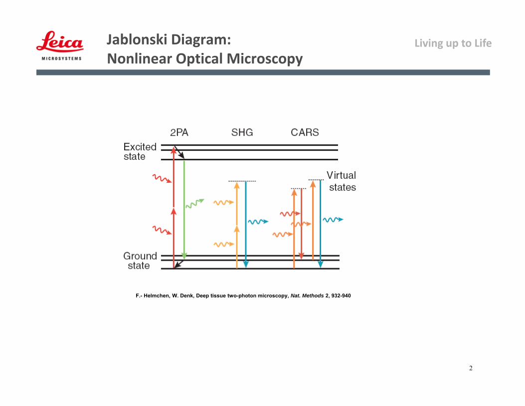
T. NevianInstitute of PhysiologyUniversity of Bern, Switzerland
• Somata 10-30 µm
• Dendrites 1-5 µm
• Spines ~0.5 µm
• Axons 1-2 µm
ls ~ 50-100 µm (@ 630 nm)
ls ~ 200 µm (@ 800 nm)
F.- Helmchen, W. DenkDeep tissue two-photon microscopy.Nat. Methods 2, 932-940
Typical Samples –Small Dimensions & Highly Scattering
Living up to Life
4
• Today main challenge:
To go deeper into samples for improved studies of cells, organs or tissues, live animals
Less photodamage, i.e. less bleaching and phototoxicity
• Why is it possible?
Due to the reduced absorption and scattering of the excitation light
Why Multiphoton microscopy?

Living up to Life
Page 5
• Achievable depth: ~ 300 – 600 µm
• Maximum imaging depth depends on:
– Available laser power
– Scattering mean-free-path
– Tissue properties
• Density properties• Microvasculature organization• Cell-body arrangement• Collagen / myelin content
– Specimen age
– Collection efficiency
The depth limit
Acute mouse brain sections containing YFP neurons,maximum projection, Z stack: 233 m
Courtesy: Dr Feng Zhang, Deisseroth laboratory, Stanford University, USA

Living up to Life
6
2-photonexcitation1-photonexcitation
exciting photon
emitted photon
What is Two‐Photon Microscopy?
Simultaneous absorption of 2 longer wavelength photons to excite a fluorochrome, emission as with 1-photon
A 3-dimensional imaging technique in which 2 photons are used to excite fluorescence emission
S0
S1
Living up to Life
7
na Pavg
2
f 2
NA2
hc
2
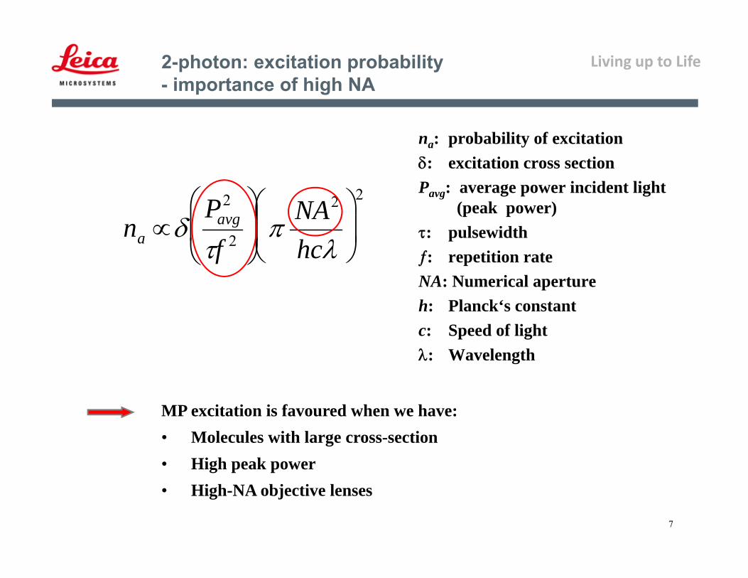
na: probability of excitation: excitation cross sectionPavg: average power incident light
(peak power): pulsewidth: repetition rateNA: Numerical apertureh: Planck‘s constantc: Speed of light: Wavelength
2-photon: excitation probability- importance of high NA
MP excitation is favoured when we have:• Molecules with large cross-section• High peak power• High-NA objective lenses
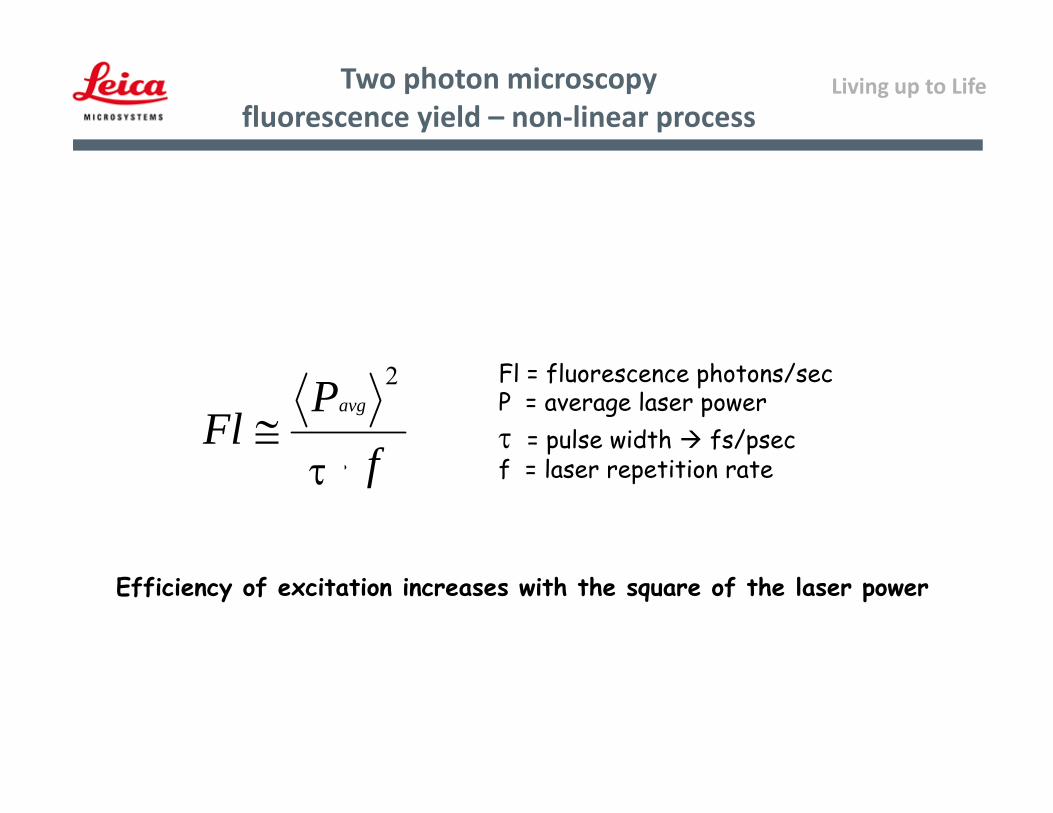
Living up to LifeTwo photon microscopyfluorescence yield – non‐linear process
Fl = fluorescence photons/secP = average laser power = pulse width fs/psecf = laser repetition rate
Efficiency of excitation increases with the square of the laser power
fTP
Flavg
2

Living up to Life
9
What is Two‐Photon Microscopy ?
1-Photon 2-Photon label is excited only at the focus of the beam where probability of 2P event is highest
No out-of-focus-fluorescence:
- No need of confocal aperture
- Dye bleaching and photo
toxicity limited to the plane
of focus
Living up to Life
10
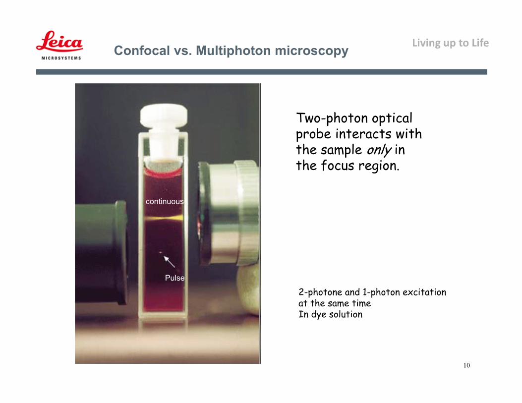
Confocal vs. Multiphoton microscopy
Pulse
continuous
Two-photon optical probe interacts with the sample only in the focus region.
2-photone and 1-photon excitationat the same timeIn dye solution
Living up to Life
11
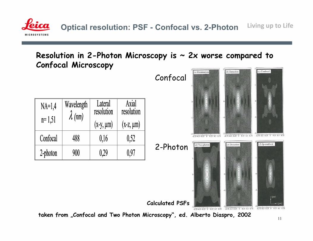
Optical resolution: PSF - Confocal vs. 2-Photon
Resolution in 2-Photon Microscopy is ~ 2x worse compared to Confocal Microscopy
Confocal
2-Photon0,970,299002-photon0,520,16488Confocal
Axial resolution(x-z, µm)
Lateralresolution(x-y, µm)
Wavelength (nm)
NA=1,4n= 1,51
0,970,299002-photon0,520,16488Confocal
Axial resolution(x-z, µm)
Lateralresolution(x-y, µm)
Wavelength (nm)
NA=1,4n= 1,51
Calculated PSFs
taken from „Confocal and Two Photon Microscopy“, ed. Alberto Diaspro, 2002
Living up to Life
Page 12
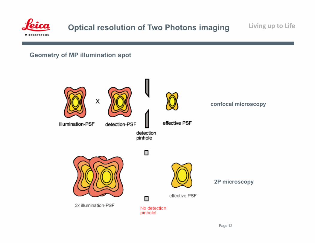
Optical resolution of Two Photons imaging
Geometry of MP illumination spot
confocal microscopy
2P microscopy
Living up to Life
Page 13
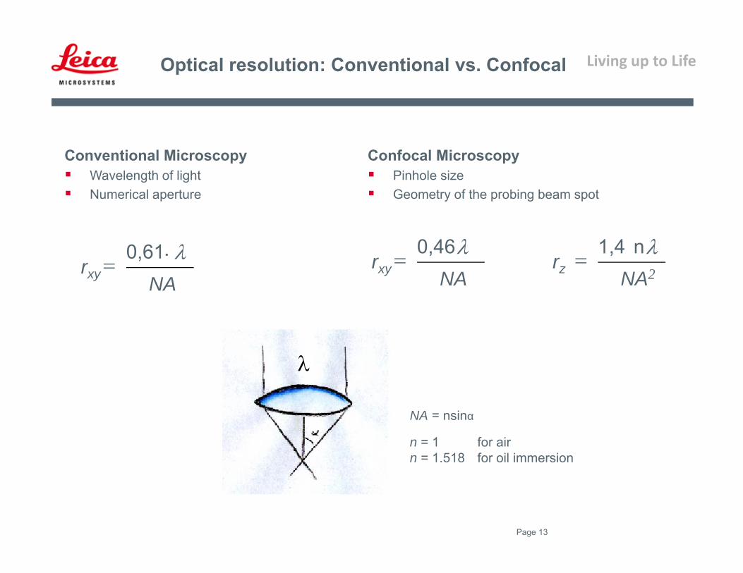
Optical resolution: Conventional vs. Confocal
Conventional Microscopy Wavelength of light Numerical aperture
NArxy
0,61
NA = nsinα
n = 1 for airn = 1.518 for oil immersion
NArxy
0,46NA2
rzn 1,4
Confocal Microscopy Pinhole size Geometry of the probing beam spot
Living up to Life
Page 14
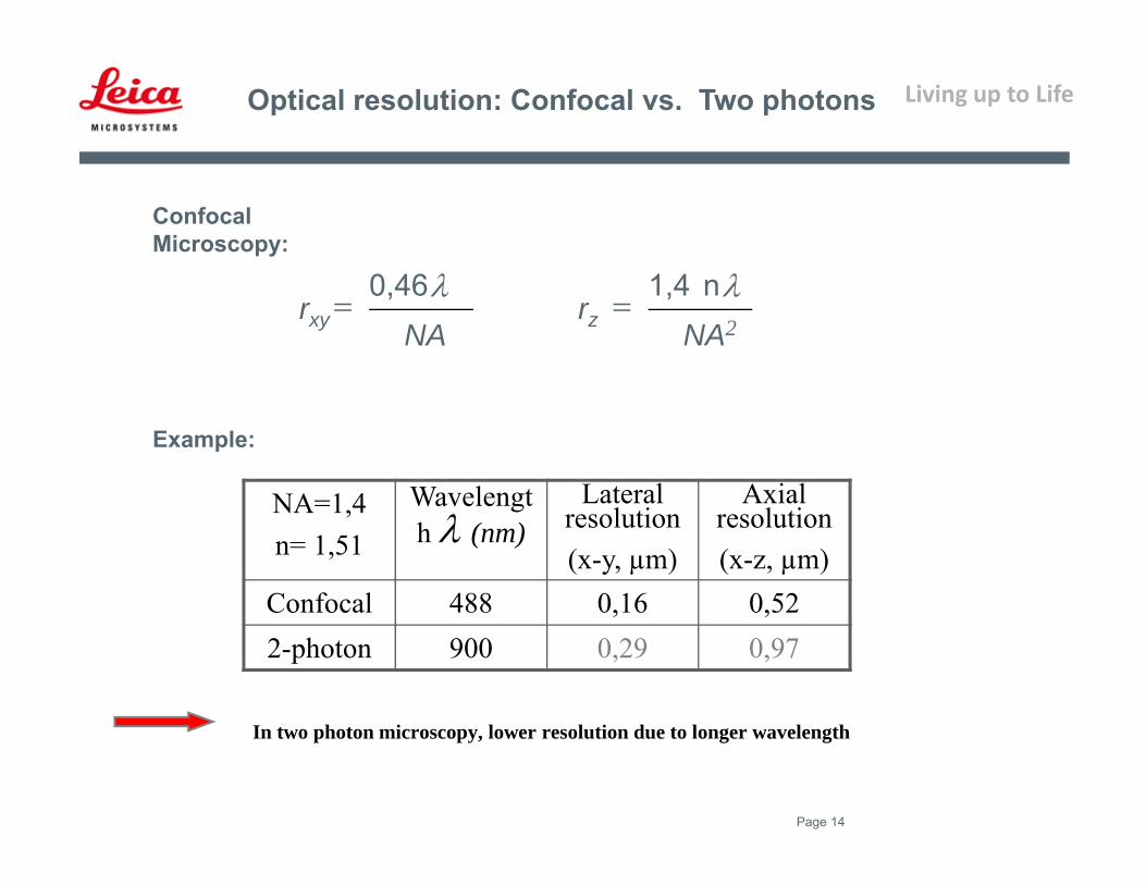
Optical resolution: Confocal vs. Two photons
NA=1,4n= 1,51
Wavelength (nm)
Lateral resolution(x-y, µm)
Axial resolution(x-z, µm)
Confocal 488 0,16 0,522-photon 900 0,29 0,97
NArxy
0,46NA2
rzn 1,4
Confocal Microscopy:
In two photon microscopy, lower resolution due to longer wavelength
Example:

Living up to Life
Page 15
Rubart , M., Two-Photon Microscopy of Cells and Tissue, Circ. Res. 2004;95;1154-1166
TPE volumes: wavelength & NA dependence

Living up to Life
16
Comparison of penetration: UV – IR (internal detectors)
Eye of zebrafish larvae (stained with DAPI)
Image size (xz): 125 m x 125 m - Objective: 63x 1.2 Water - Detection range: 400nm – 500nm
Exc.: UVPMT:
300V
Exc.: IRPMT: 360V
Living up to Life
17
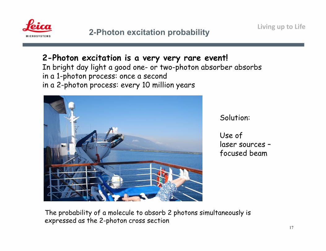
2-Photon excitation probability
2-Photon excitation is a very very rare event!In bright day light a good one- or two-photon absorber absorbsin a 1-photon process: once a secondin a 2-photon process: every 10 million years
The probability of a molecule to absorb 2 photons simultaneously is expressed as the 2-photon cross section
Solution:
Use oflaser sources –focused beam
Living up to Life
18
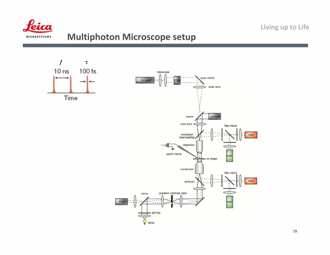
Multiphoton Microscope setup
Living up to Life
19
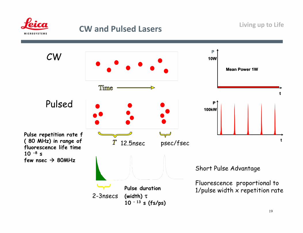
CW and Pulsed Lasers
CW
Pulsed
Short Pulse Advantage
Fluorescence proportional to1/pulse width x repetition rate
psec/fsec12.5nsec
t
P
Mean Power 1W
10W
t
P
Mean Power 1W
10W
t
P100kW
t
P100kW
2-3nsecsPulse duration(width) 10 – 13 s (fs/ps)
Pulse repetition rate f( 80 MHz) in range offluorescence life time10 -8 sfew nsec 80MHz
Living up to Life
Page 20
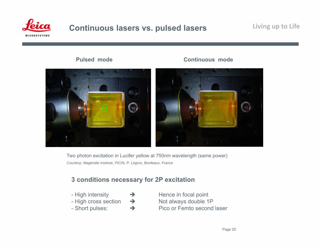
Continuous lasers vs. pulsed lasers
Pulsed mode Continuous mode
Two photon excitation in Lucifer yellow at 750nm wavelength (same power)Courtesy: Magendie institute, PICIN, P. Legros, Bordeaux, France
3 conditions necessary for 2P excitation
- High intensity Hence in focal point- High cross section Not always double 1P- Short pulses: Pico or Femto second laser

Living up to Life
21
CoherentChameleon Vision II
Typical Tuning Curve IR Laser
Spectra PhysicsMaiTai DeepSee
Living up to Life
22
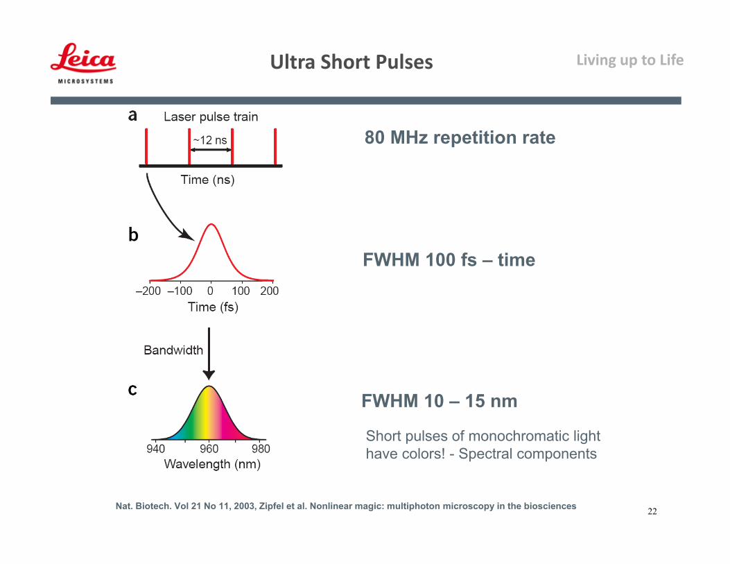
80 MHz repetition rate
FWHM 100 fs – time
FWHM 10 – 15 nm
Nat. Biotech. Vol 21 No 11, 2003, Zipfel et al. Nonlinear magic: multiphoton microscopy in the biosciences
Short pulses of monochromatic lighthave colors! - Spectral components
Ultra Short Pulses
Living up to Life
Page 23
Continuous or pulsed laser
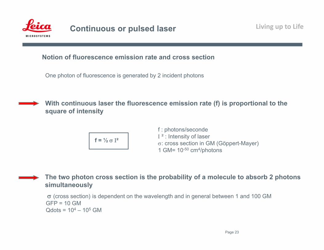
Notion of fluorescence emission rate and cross section
f : photons/seconde ² : Intensity of laser: cross section in GM (Göppert-Mayer)1 GM= 10-50 cm4/photons
(cross section) is dependent on the wavelength and in general between 1 and 100 GMGFP = 10 GMQdots = 104 – 105 GM
f = ½ ²
One photon of fluorescence is generated by 2 incident photons
With continuous laser the fluorescence emission rate (f) is proportional to the square of intensity
The two photon cross section is the probability of a molecule to absorb 2 photons simultaneously

Living up to Life
Page 24
Continuous or pulsed laser
With pulsed laser, the fluorescence emision rate is described as follows:
t : Pulse duration (s)F : Frequency (Hz)fm : Average fluorescence emission rate m² : Average intensityt. : Intensity per pulse (peak power): cross section in GM (Göppert-Mayer)
So for equal power we have 105 time more excitation of fluorophore with pulsed laser
fm = ½ (t.F)-1 m²
Examples t = 10-13 sF = 108 Hz (t.F)-1 = 105
To compare (t.F)-1 = 1 for continuous laser
Because m = t.F.
Living up to Life
25
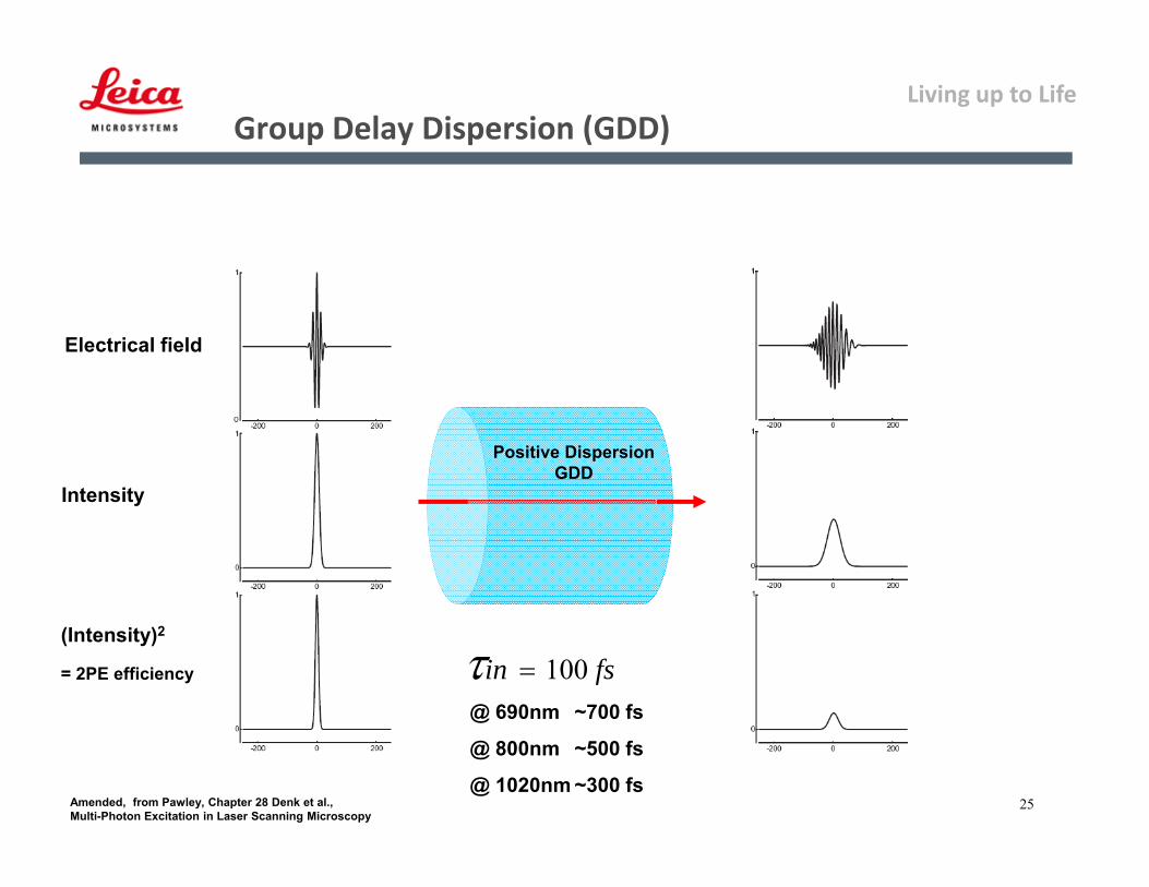
Electrical field
Intensity
(Intensity)2
= 2PE efficiency
Positive DispersionGDD
Amended, from Pawley, Chapter 28 Denk et al.,Multi-Photon Excitation in Laser Scanning Microscopy
Group Delay Dispersion (GDD)
fsin 100@ 690nm ~700 fs
@ 800nm ~500 fs
@ 1020nm ~300 fs

Living up to Life
26
Principle of Precompensation
Laser Microscope
Sample
redblue
100 fs 400 fs+GDD
bluered
Laser MircroscopePre-Chirp100 fs100 fs 400 fs +GDD-GDD
Sample
Group Delay Dispersion: GDD
Living up to Life
27
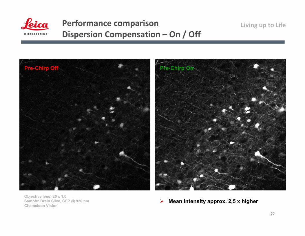
Performance comparisonDispersion Compensation – On / Off
Pre-Chirp Off Pre-Chirp On
Objective lens: 20 x 1,0Sample: Brain Slice, GFP @ 920 nmChameleon Vision
Mean intensity approx. 2,5 x higher
Living up to Life
28
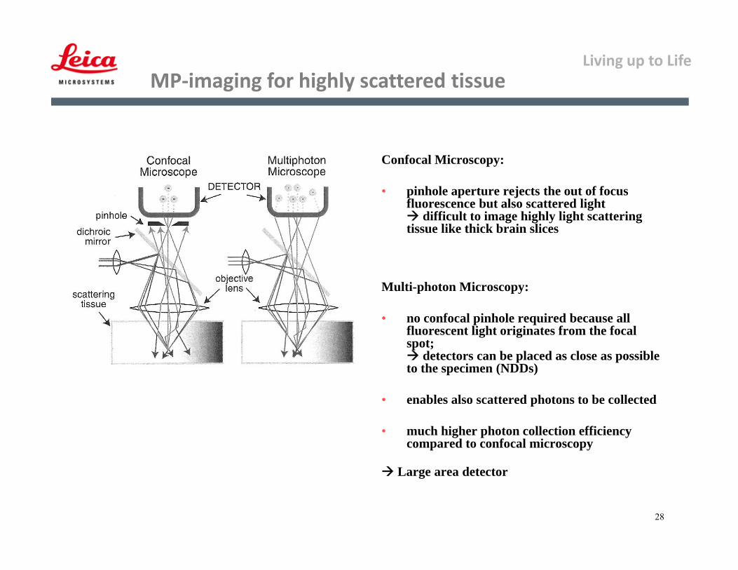
MP‐imaging for highly scattered tissue
Confocal Microscopy:
• pinhole aperture rejects the out of focus fluorescence but also scattered light difficult to image highly light scattering tissue like thick brain slices
Multi-photon Microscopy:
• no confocal pinhole required because all fluorescent light originates from the focal spot; detectors can be placed as close as possible to the specimen (NDDs)
• enables also scattered photons to be collected
• much higher photon collection efficiency compared to confocal microscopy
Large area detector

Living up to Life
29
Detection Path
• Descanned pathway can be used – but clipping at pinhole
• Strategy: collect as many photons as possible
→ i.e. if descanned detection: open pinhole completely!
• Non-Descanned-Detection:– Large-area detectors (predominantly PMTs)– epi-detection– trans-collection – high-NA Condensor, prefferably oil (!)

Living up to Life
30
Photon Collection Efficiency ‐ Internal vs. NDDs
Mouse brain slice: ~ 20 µm (center plane)Detection range: 500 – 550 nmPMT: 950 VObjective: 20 x 1.0 W
internal RLD TLD
Mean intensity: 20 52 58

Living up to Life
31
Fluorochromes: TPE ‐ Overview 1
Bestvater et el.Two-photon fluorescence absorption and emission spectra ofdyes relevant for cell imagingJournal of Microscopy, Vol. 208, Pt 2 November 2002, pp. 108–115
Living up to Life
32
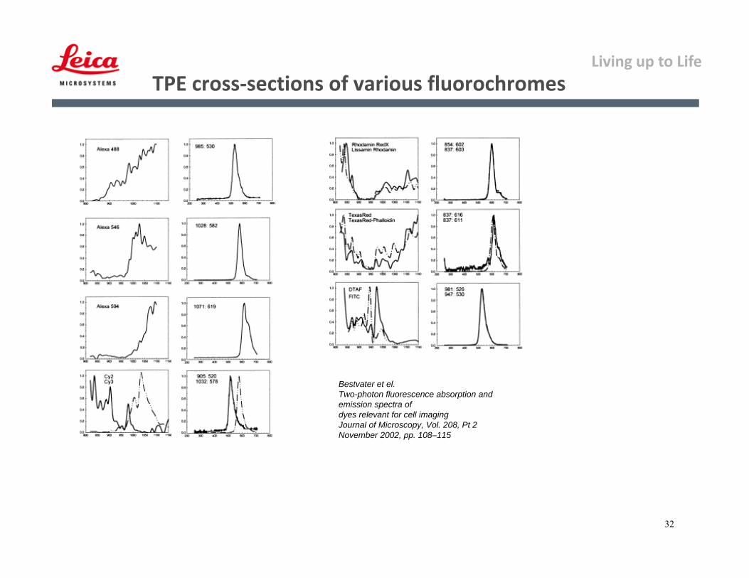
TPE cross‐sections of various fluorochromes
Bestvater et el.Two-photon fluorescence absorption and emission spectra ofdyes relevant for cell imagingJournal of Microscopy, Vol. 208, Pt 2 November 2002, pp. 108–115
Living up to Life
33
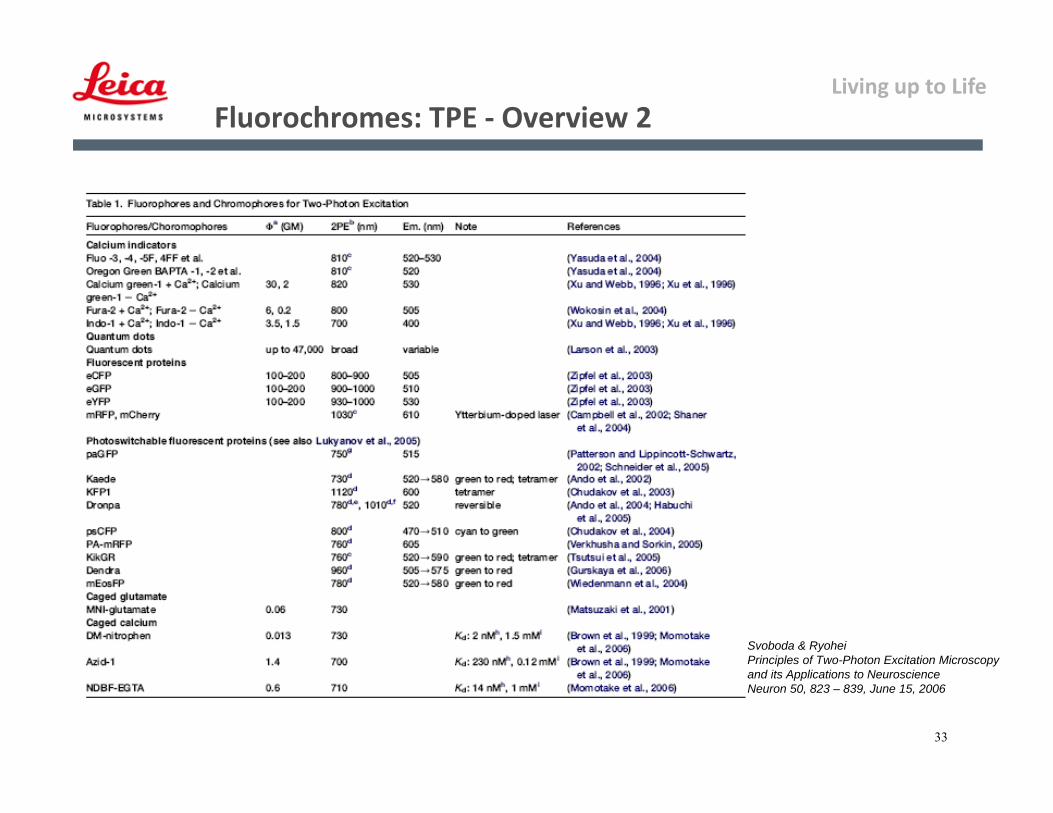
Fluorochromes: TPE ‐ Overview 2
Svoboda & RyoheiPrinciples of Two-Photon Excitation Microscopy and its Applications to NeuroscienceNeuron 50, 823 – 839, June 15, 2006
Living up to Life
34
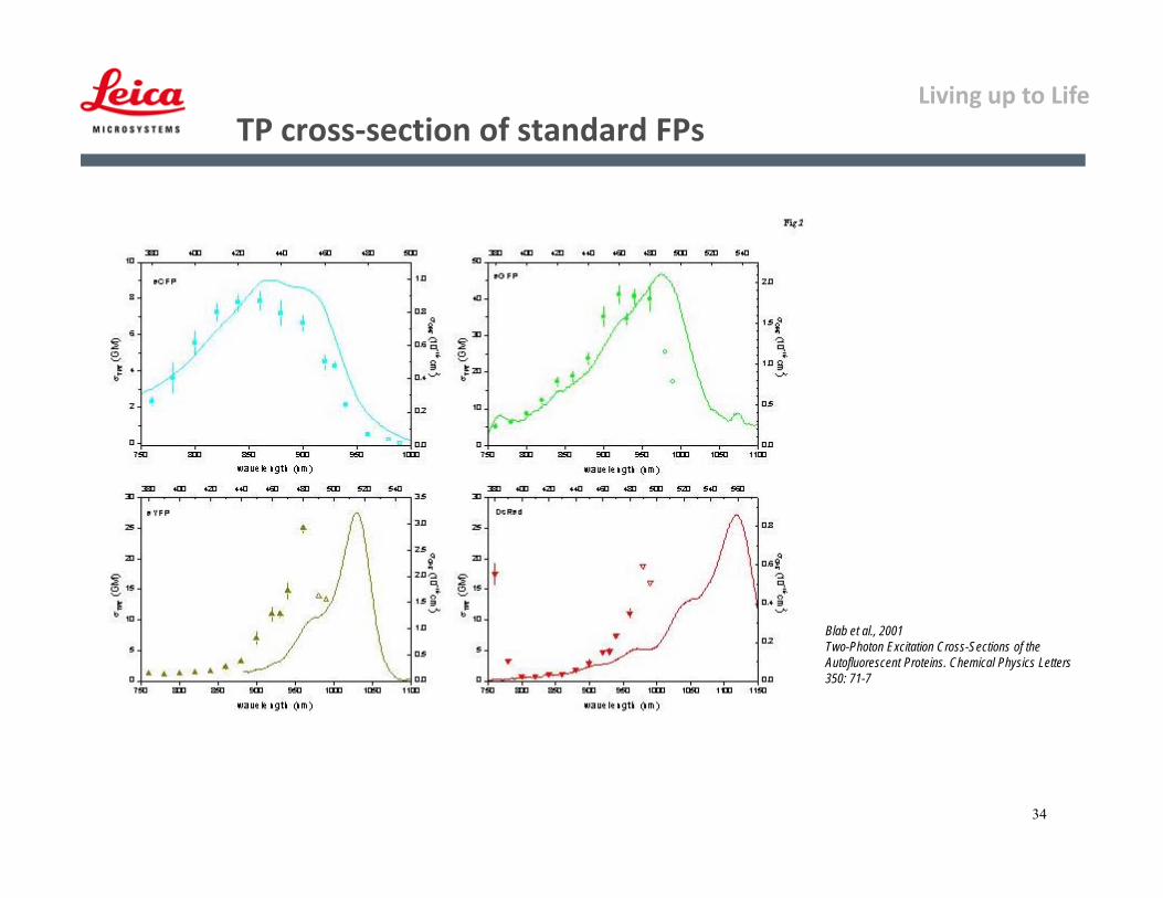
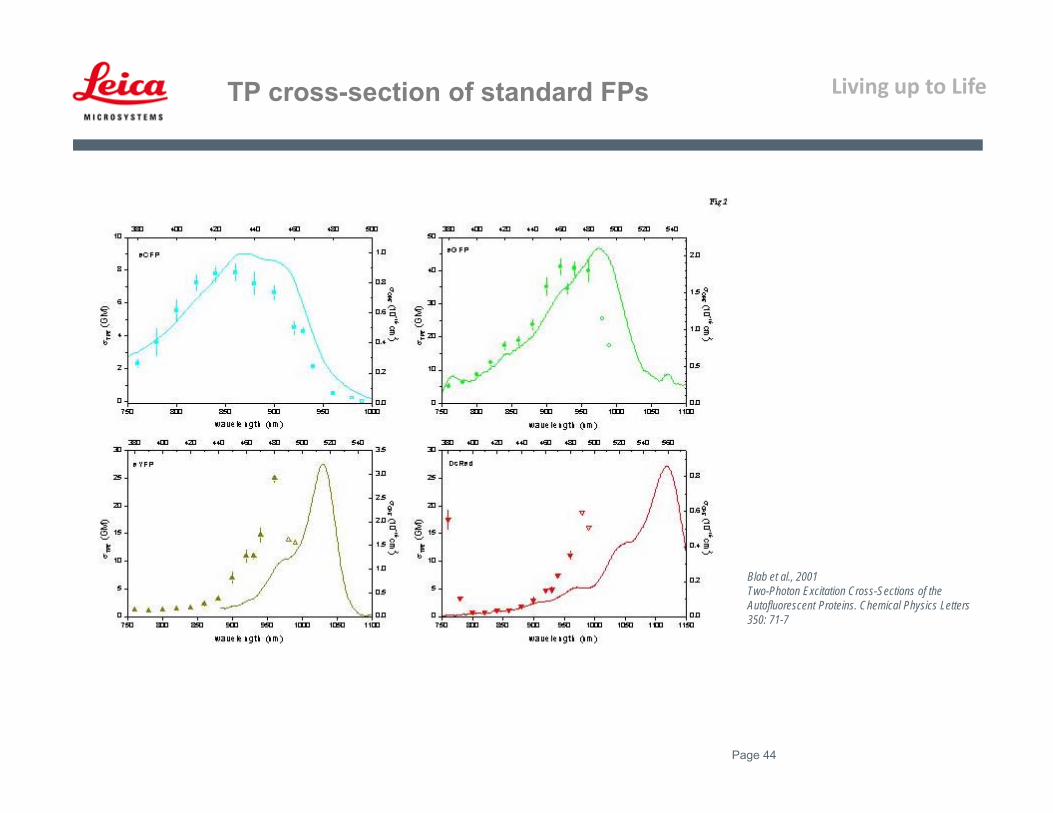
Blab et al., 2001Two-Photon Excitation Cross-Sections of the Autofluorescent Proteins. Chemical Physics Letters 350: 71-7
TP cross‐section of standard FPs

Living up to Life
35
TCS SP 5 MP: Typical Setup ‐ inverted
1) IR Laser (Mai Tai DeepSee)
2) Safety Box
3) EOM (Driver)
4) Beam Routing (direct coupling)
5) Scanhead
6) DMI 6000 Microscope Stand
7) NDD Detection Unit
8) NDD: RLD
9) NDD: TLD
1
2
3
4
5
6
78
9

Living up to Life
36
• SHG is a nonlinear scattering process that conserves energy and results in the SHG exactly half ofthe illumination ’
• involves virtual transitions in which no energy is absorbed
• 2 photons „simultaneously“ scattered, resulting in „frequency doubling“
• In contrast 2PE involves absorption (real transition) and excitation of molecules
• SHG = λincident/2
• Investigate with spectrophoto-meter-PMT or NDD
Excited state
Ground state
hν
hν
hνSHG
Virtual state
Virtual state
Second Harmonic Generation (SHG)
Living up to Life
37
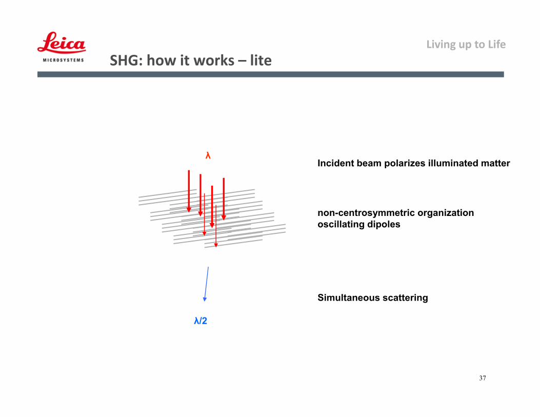
SHG: how it works – lite
Incident beam polarizes illuminated matter
non-centrosymmetric organizationoscillating dipoles
λ
λ/2
Simultaneous scattering
Living up to Life
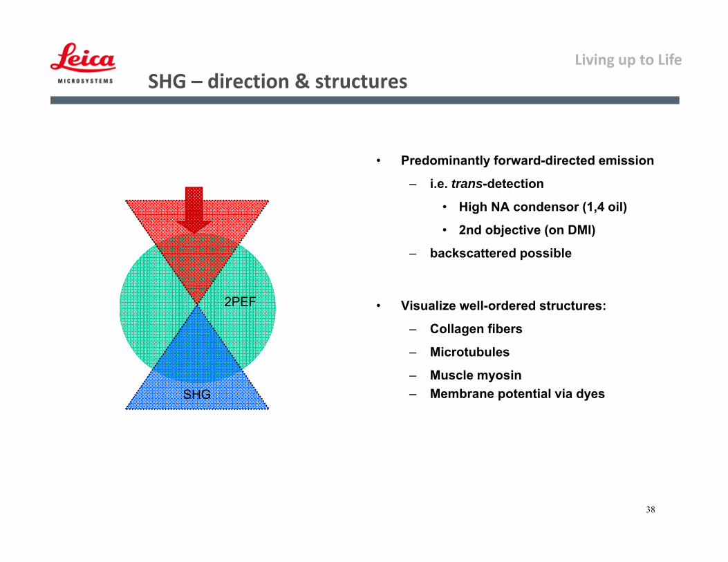
38
2PEF
SHG
• Predominantly forward-directed emission
– i.e. trans-detection
• High NA condensor (1,4 oil)
• 2nd objective (on DMI)
– backscattered possible
• Visualize well-ordered structures:
– Collagen fibers
– Microtubules
– Muscle myosin– Membrane potential via dyes
SHG – direction & structures

Living up to Life
39
1
2
Striation pattern of murine heartSHG combined with fluorescence:Collagen fibrils (SHG, grey), Macrophages(Fluorescence, green & red)
SHG – images
Living up to Life

Living up to Life
Fluorophore (cm-1 M-1)
Oregon Green®488 87,000
BODIPY FL 91,000
FAM 79,000
JOE 71,000
TAMRA 103,000
ROX 82,000
Texas Red 139,000
Fluorophore QY
Oregon Green®488
0.9
BODIPY FL 0.9
FAM 0.9
JOE 0.6
TAMRA 0.2
ROX 0.7
Texas Red 0.9
Living up to Life
Living up to Life
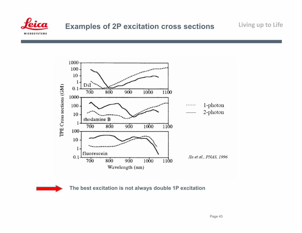
Page 43
Examples of 2P excitation cross sections
The best excitation is not always double 1P excitation
Living up to Life
Page 44
Blab et al., 2001Two-Photon Excitation Cross-Sections of the Autofluorescent Proteins. Chemical Physics Letters 350: 71-7
TP cross-section of standard FPs
Living up to Life
Page 45
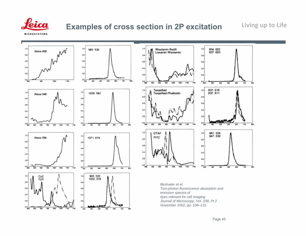
Bestvater et el.Two-photon fluorescence absorption and emission spectra ofdyes relevant for cell imagingJournal of Microscopy, Vol. 208, Pt 2 November 2002, pp. 108–115
Examples of cross section in 2P excitation
Living up to Life
Page 46
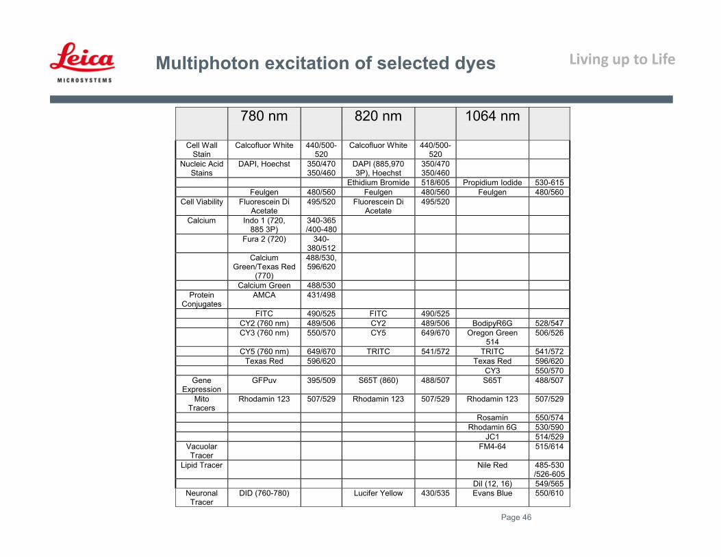
Multiphoton excitation of selected dyes
780 nm 820 nm 1064 nm
Cell WallStain
Calcofluor White 440/500-520
Calcofluor White 440/500-520
Nucleic AcidStains
DAPI, Hoechst 350/470350/460
DAPI (885,9703P), Hoechst
350/470350/460
Ethidium Bromide 518/605 Propidium Iodide 530-615Feulgen 480/560 Feulgen 480/560 Feulgen 480/560
Cell Viability Fluorescein DiAcetate
495/520 Fluorescein DiAcetate
495/520
Calcium Indo 1 (720,885 3P)
340-365/400-480
Fura 2 (720) 340-380/512
CalciumGreen/Texas Red
(770)
488/530,596/620
Calcium Green 488/530Protein
ConjugatesAMCA 431/498
FITC 490/525 FITC 490/525CY2 (760 nm) 489/506 CY2 489/506 BodipyR6G 528/547CY3 (760 nm) 550/570 CY5 649/670 Oregon Green
514506/526
CY5 (760 nm) 649/670 TRITC 541/572 TRITC 541/572Texas Red 596/620 Texas Red 596/620
CY3 550/570Gene
ExpressionGFPuv 395/509 S65T (860) 488/507 S65T 488/507
MitoTracers
Rhodamin 123 507/529 Rhodamin 123 507/529 Rhodamin 123 507/529
Rosamin 550/574Rhodamin 6G 530/590
JC1 514/529VacuolarTracer
FM4-64 515/614
Lipid Tracer Nile Red 485-530/526-605
DiI (12, 16) 549/565Neuronal
TracerDID (760-780) Lucifer Yellow 430/535 Evans Blue 550/610

Living up to Life
ExcitationMaximum (nm)
EmissionMaximum (nm)
MolarExtinctionCoefficient
QuantumYield
in vivoStructure
RelativeBrightness(% of EGFP)
GFP (wt) 395/475 509 21,000 0.77 Monomer* 48EGFP 484 507 56,000 0.6 Monomer* 100AcGFP 480 505 50,000 0.55 Monomer* 82
TurboGFP 482 502 70,000 0.53 Monomer* 110Emerald 487 509 57,500 0.68 Monomer* 116
Azami Green 492 505 55,000 0.74 Monomer 121ZsGreen 493 505 43,000 0.91 Tetramer 117
Green Fluorescent proteins
EBFP 383 445 29,000 0.31 Monomer* 27Sapphire 399 511 29,000 0.64 Monomer* 55
T-Sapphire 399 511 44,000 0.6 Monomer* 79
Blue Fluorescent proteins
ECFP 439 476 32,500 0.4 Monomer* 39mCFP 433 475 32,500 0.4 Monomer 39
Cerulean 433 475 43,000 0.62 Monomer* 79CyPet 435 477 35,000 0.51 Monomer* 53
AmCyan1 458 489 44,000 0.24 Tetramer 31Midori-Ishi Cyan 472 495 27,300 0.9 Dimer 73
mTFP1 (Teal) 462 492 64,000 0.85 Monomer 162
Cyan Fluorescent proteins
EYFP 514 527 83,400 0.61 Monomer* 151Topaz 514 527 94,500 0.6 Monomer* 169Venus 515 528 92,200 0.57 Monomer* 156
mCitrine 516 529 77,000 0.76 Monomer 174YPet 517 530 104,000 0.77 Monomer* 238
PhiYFP 525 537 124,000 0.39 Monomer* 144ZsYellow1 529 539 20,200 0.42 Tetramer 25
Yellow Fluorescent proteins
Kusabira Orange 548 559 51,600 0.6 Monomer 92mOrange 548 562 71,000 0.69 Monomer 146
dTomato 554 581 69,000 0.69 Dimer 142dTomato-Tandem 554 581 138,000 0.69 Monomer 283
DsRed 558 583 75,000 0.79 Tetramer 176DsRed2 563 582 43,800 0.55 Tetramer 72
DsRed-Express (T1) 555 584 38,000 0.51 Tetramer 58DsRed-Monomer 556 586 35,000 0.1 Monomer 10
mTangerine 568 585 38,000 0.3 Monomer 34mStrawberry 574 596 90,000 0.29 Monomer 78
AsRed2 576 592 56,200 0.05 Tetramer 8mRFP1 584 607 50,000 0.25 Monomer 37JRed 584 610 44,000 0.2 Dimer 26
mCherry 587 610 72,000 0.22 Monomer 47HcRed1 588 618 20,000 0.015 Dimer 1
mRaspberry 598 625 86,000 0.15 Monomer 38HcRed-Tandem 590 637 160,000 0.04 Monomer 19
mPlum 590 649 41,000 0.1 Monomer 12AQ143 595 655 90,000 0.04 Tetramer 11
Orange Fluorescent proteins
Fluorescent Proteins –Covering the entire visible spectra
Living up to Life
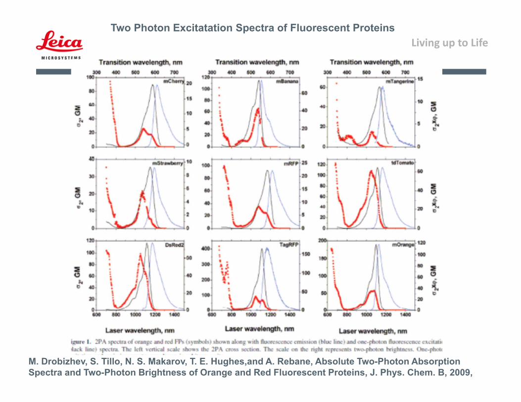
M. Drobizhev, S. Tillo, N. S. Makarov, T. E. Hughes,and A. Rebane, Absolute Two-Photon Absorption Spectra and Two-Photon Brightness of Orange and Red Fluorescent Proteins, J. Phys. Chem. B, 2009,
Two Photon Excitatation Spectra of Fluorescent Proteins
Living up to Life
Page 49
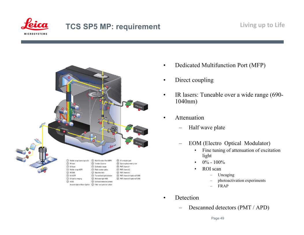
• Dedicated Multifunction Port (MFP)
• Direct coupling
• IR lasers: Tuneable over a wide range (690-1040nm)
• Attenuation– Half wave plate
– EOM (Electro Optical Modulator)• Fine tuning of attenuation of excitation
light • 0% - 100%• ROI scan
– Uncaging– photoactivation experiments – FRAP
• Detection– Descanned detectors (PMT / APD)
TCS SP5 MP: requirement
Living up to Life
Page 50
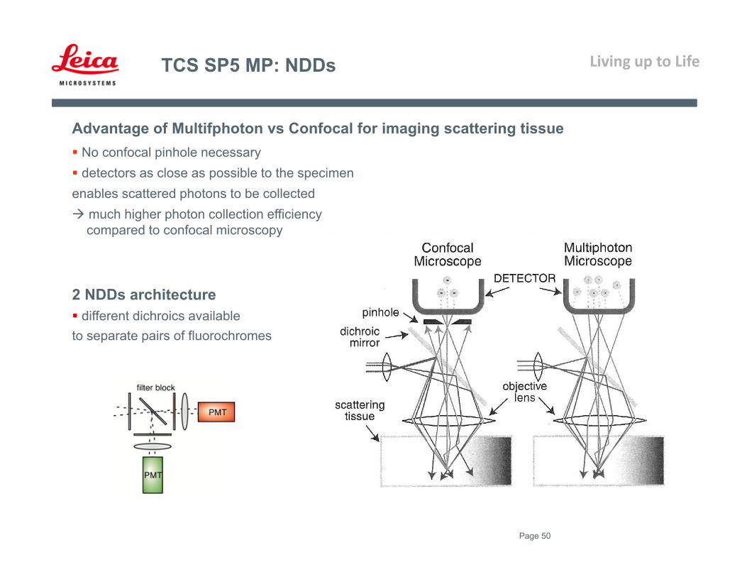
Advantage of Multifphoton vs Confocal for imaging scattering tissue No confocal pinhole necessary detectors as close as possible to the specimen enables scattered photons to be collected much higher photon collection efficiency
compared to confocal microscopy
2 NDDs architecture different dichroics availableto separate pairs of fluorochromes
TCS SP5 MP: NDDs

Living up to Life
Page 51
Highest photon collection efficiency
Detectors directly behind Objective, RLD
Detectors directly behind Condensor, TLD
Advantage:
Scattered fluorescent photons can also becollected
Special dichroic allows simultaneousacquisition of fluorescence and IR-SGC
Protected by Leica patent US 6,831,780 B2
TCS SP5 MP: NDDs
Living up to Life
Page 52
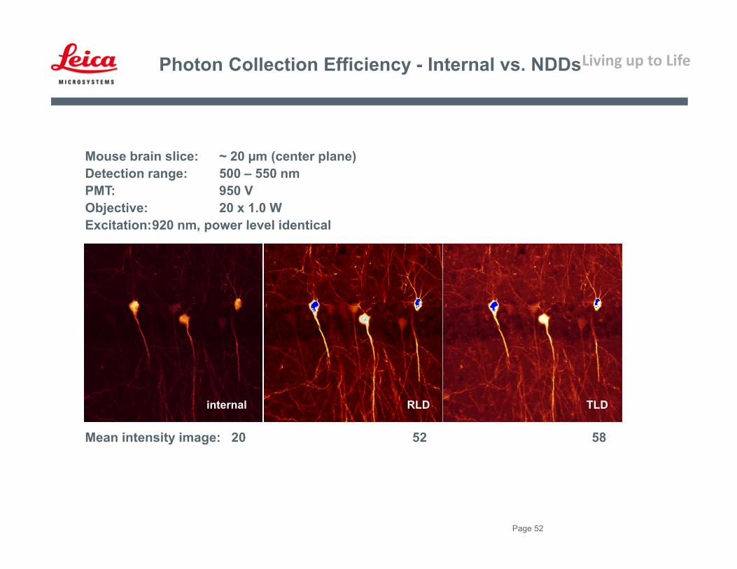
Mouse brain slice: ~ 20 µm (center plane)Detection range: 500 – 550 nmPMT: 950 VObjective: 20 x 1.0 WExcitation:920 nm, power level identical
internal RLD TLD
Mean intensity image: 20 52 58
Photon Collection Efficiency - Internal vs. NDDs

Living up to Life
Page 53
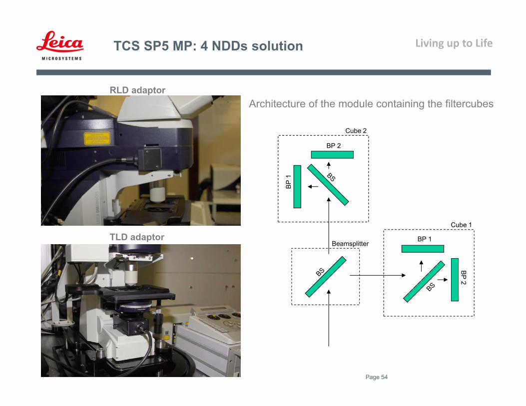
TCS SP5 MP: 4 NDDs solution
4 NDDs solution
Simultaneous acquisition of 4 colors
Solution: adaptor + liquid light guide + 4NDDs module
Photon transmission via liquid light guide
Adaptor directly behind Objective, RLD
Adaptor directly behind Condensor, TLD
Living up to Life
Page 54
TLD adaptor
RLD adaptor
BP 2
BP 1
BP 2
BP 1
Cube 2
Cube 1
Beamsplitter
Architecture of the module containing the filtercubes
TCS SP5 MP: 4 NDDs solution
Living up to Life
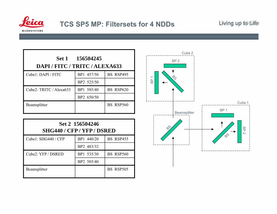
Set 1 156504245DAPI / FITC / TRITC / ALEXA633
Cube1: DAPI / FITC BP1 457/50 BS RSP495
BP2 525/50
Cube2: TRITC / Alexa633 BP1 585/40 BS RSP620
BP2 650/50
Beamsplitter BS RSP560
Set 2 156504246SHG440 / CFP / YFP / DSRED
Cube1: SHG440 / CFP BP1 440/20 BS RSP455
BP2 483/32
Cube2: YFP / DSRED BP1 535/30 BS RSP560
BP2 585/40
Beamsplitter BS RSP505
BP 2
BP 1
BP 2
BP 1
Cube 2
Cube 1
Beamsplitter
TCS SP5 MP: Filtersets for 4 NDDs Living up to Life
Living up to Life
Page 56
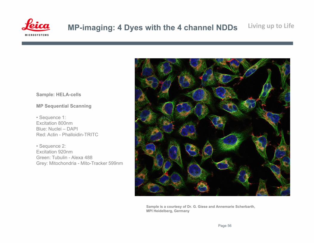
MP-imaging: 4 Dyes with the 4 channel NDDs
Sample: HELA-cells
MP Sequential Scanning
• Sequence 1: Excitation 800nmBlue: Nuclei – DAPIRed: Actin - Phalloidin-TRITC
• Sequence 2:Excitation 920nmGreen: Tubulin - Alexa 488Grey: Mitochondria - Mito-Tracker 599nm
Sample is a courtesy of Dr. G. Giese and Annemarie Scherbarth,MPI Heidelberg, Germany
Living up to Life
Page 57
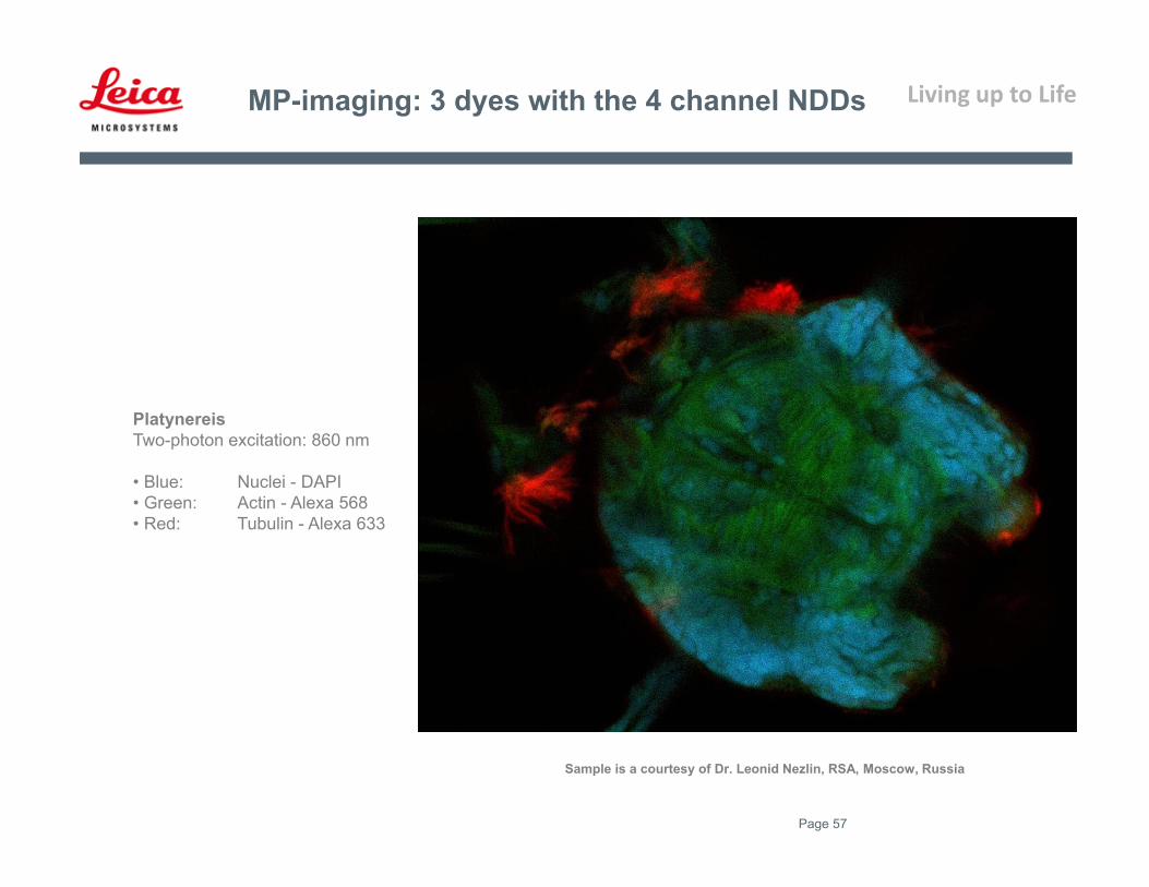
MP-imaging: 3 dyes with the 4 channel NDDs
PlatynereisTwo-photon excitation: 860 nm
• Blue: Nuclei - DAPI • Green: Actin - Alexa 568 • Red: Tubulin - Alexa 633
Sample is a courtesy of Dr. Leonid Nezlin, RSA, Moscow, Russia

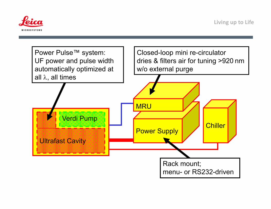
Living up to LifeCoherant laser
Living up to Life
Power Supply
MRU
ChillerVerdi Pump
Ultrafast Cavity
Closed-loop mini re-circulatordries & filters air for tuning >920 nmw/o external purge
Rack mount;menu- or RS232-driven
Power Pulse™ system:UF power and pulse width automatically optimized at all , all times