Embed Size (px)
Citation preview

Optimizing Implementation of Prostate MRI
Andrei S. Purysko, M.D.
Section of Abdominal Imaging &
Nuclear Radiology Department

• To review the basic components of a state-of-the-art
prostate MRI protocol
• To list resources available and under development to
assist in the implementation and optimization of a
prostate MRI program
Objectives


1980
0.04 Tesla Magnet
The Evolution of Prostate MRI

“At present, MR imaging is the most accurate diagnostic modality for the local staging of carcinoma of the prostate, but for optimal
results, multiple sequences and two orthogonal planes of imaging are needed.”
1980
The Evolution of Prostate MRI
0.35 Tesla Magnet

1980
The Evolution of Prostate MRI

1980 1990
The Evolution of Prostate MRI
1.5 Tesla Magnet

1980 1990
The Evolution of Prostate MRI
1980 1990
The Evolution of Prostate MRI

1980 1990 2000
The Evolution of Prostate MRI

1980 1990 2000
The Evolution of Prostate MRI

1980 1990 2000
The Evolution of Prostate MRI
2010
PI-RADS Version 1

1980 1990 2000
The Evolution of Prostate MRI
2010
Minimum acceptable technical parameters
Standardized lexicon
Revised scoring system
Standardized scheme for deriving an overall assessment category


State-of-the-art technique
• Hardware
– 1.5 or 3.0 T
– Surface coil with or without ER coil
• Pulse Sequences:
– Multiplanar T2-WI
– DWI/ADC
– DCE T1-WI
– MRSI (optional – not included in PI-RADS v2)
Multiparametric

Prostate MRI Pulse Sequences
• T2-WI
– Detailed anatomic information – staging
– Dominant parameter for TZ lesions
Base Mid Apex

• T2-WI
– Detailed anatomic information – staging
– Dominant parameter for TZ lesions
PCa BPH Nodules
Prostate MRI Pulse Sequences

• DWI /ADC map
– Tissue microarchitecture and cellularity
– Dominant parameter for PZ lesions
– ADC: Inverse correlation with Gleason score
Prostate MRI Pulse Sequences
B-0 B-500 B-1000 B-2000

Prostate MRI Pulse Sequences
Gleason 3 + 3
Gleason 5 + 5
DWI ADC
• DWI /ADC map
– Tissue microarchitecture and cellularity
– Dominant parameter for PZ lesions
– ADC: Inverse correlation with Gleason score

Prostate MRI Pulse Sequences
• DCE
– Tissue vascularity
– PCa: poorly formed vessels with ↑ permeability
– Lesion detection and characterization (limited role)
T2 ADC DWI DCE

Abnormality location
Peripheral Zone Transition Zone
DWI/ADC T2-WI Assessment Category
PI-RADS 1 – highly unlikely Score 1
Score 2
Score 3
Score 4
Score 5
Score 1
Score 2
Score 3
Score 4
DCE - DWI ≤ 4
PI-RADS 2 – unlikely
PI-RADS 3 – equivocal
PI-RADS 4 – likely
PI-RADS 5 – highly unlikely Score 5

DWI ADC T2-WI
PI-RADS 1
PI-RADS 2
PI-RADS 3
PI-RADS 4
PI-RADS 5
<1.5 cm
> 1.5 cm
< 1.5-cm and No EPE
≥ 1.5-cm and/or EPE

Predictions for PI-RADS v2.1 and beyond
Biopsy recommendations
Inclusion of quantitative assessment
Likely requires further standardization of technical parameters
Radiomics
Inclusion of imaging criteria for other applications
Staging
Criteria for active surveillance
Evaluation of recurrence

Implementation of Prostate MRI Program
Personnel involved
Referring physicians (Urologists, Rad Onc, Med Onc)
Hospital administration/leadership
IT Department
Radiologists/technologists
• “The Director of Prostate Imaging”*
*Westphalen et al. PMID 28396916

Roles of local champion
• Collaborate with urologists/referring physicians on institutional
policies for imaging utilization and in the acquisition and
deployment of related technologies
Implementation of Prostate MRI Program

Roles of local champion
• Technologist engagement
• Ensure consistent and adequate image quality
• Development of imaging protocols ensuring they meet or
exceed the parameters standardized by PI-RADS v2
*Weinreb et al. (PMID: 26427566)
Implementation of Prostate MRI Program

Roles of local champion
• Radiology engagement
• Development and use of report templates
• To assist in the improvement of consistency and accuracy
of reports
Implementation of Prostate MRI Program

Educational tools
• Prostate MRI workshop
• ACR Education Center, Reston VA
• 2-day hands-on course 100+ cases and lectures
• Faculty:
- Katarzyna Macura MD, PhD (JHU) - Jeff Weinreb, MD (Yale) - Claire Tempany, MD (BWH) - Peter Choyke, MD (NIH)
- Baris Turkbey, MD (NIH) - Andy Rosenkrantz (NYU) - Daniel Margulis, MD (Weill Cornel) - Sadhna Verma, MD (CH) - Andrei Purysko, MD (Cleveland Clinic)
Implementation of Prostate MRI Program

ACR prostate MR workshop
Implementation of Prostate MRI Program
Educational tools
• Prostate MRI workshop
• Radiological Society of North America (Nov/Dec)
• Society of Abdominal Radiology (March)
Implementation of Prostate MRI Program
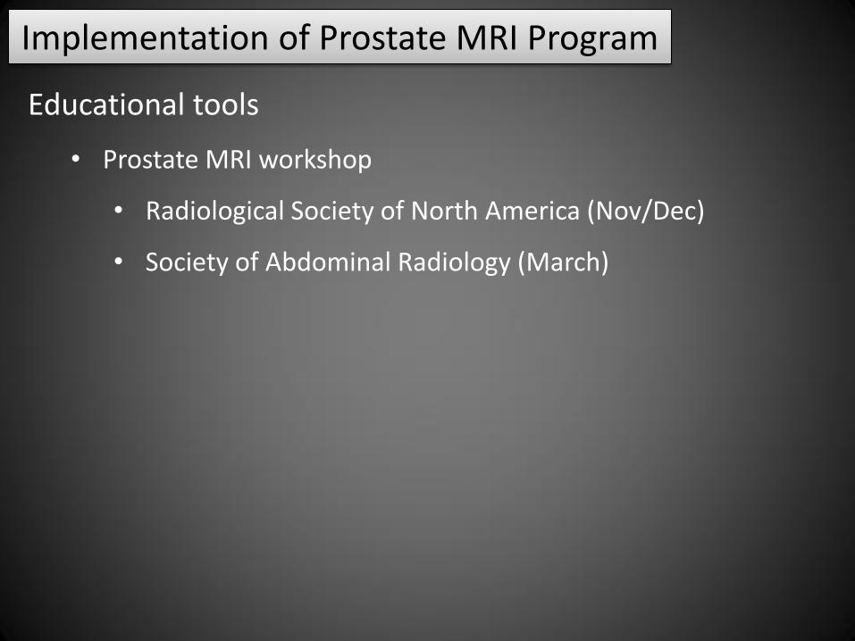
Educational tools
• PI-RADS Atlas
• ACR.org
Implementation of Prostate MRI Program

Educational tools
• “Learn Prostate MRI”
• learnprostatemri.com
Implementation of Prostate MRI Program
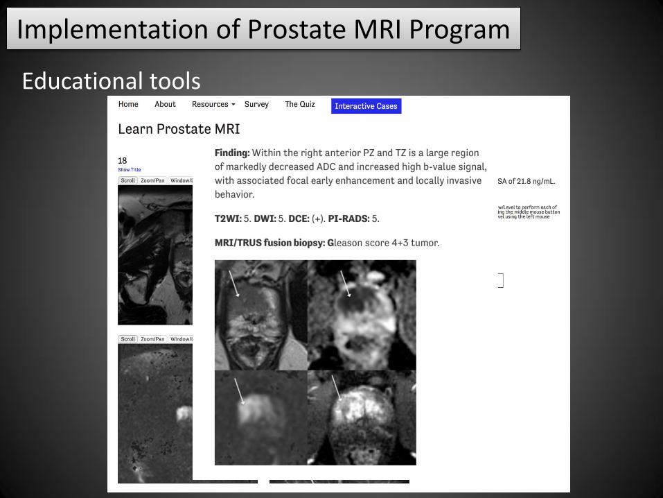
Educational tools
Implementation of Prostate MRI Program
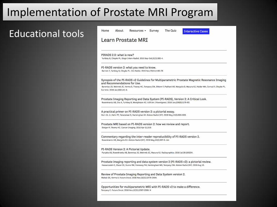
Educational tools
Implementation of Prostate MRI Program

Continued improvement
• Informal review and formal case discussions in conferences,
tumor boards and periodic radiologic–pathologic correlation
• Feedback mechanism that enables evaluation and
interpretative skills and the impact of the technology
Implementation of Prostate MRI Program
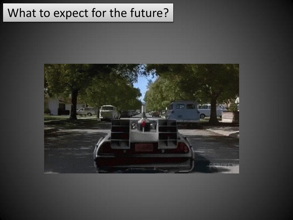
What to expect for the future?

PI-RADS v2.1 and beyond (predictions)
Biopsy recommendations
Inclusion of quantitative assessment
Likely requires further standardization of technical parameters
Radiomics
Inclusion of imaging criteria for other applications
Staging
Criteria for active surveillance
Evaluation of recurrence

ACR Prostate MR Accreditation Program
Interpreting physician qualification
• Minimum number of cases per year
• CME activities
MR technologist and physicist qualifications
Reporting
• PI-RADS and ACR practice parameters for communication of
diagnostic findings
Must have capability to perform or coordinate biopsy
planning
Policy for feedback/follow up on biopsy results
Implementation of Prostate MRI Program

• Advances in hardware and software over the last 3
decades helped establishing MRI as an accurate method
for PCa detection
• The development of technical standards for imaging
acquisition and reporting have facilitated the utilization
of prostate MRI in clinical practice
• Numerous resources are available to assist in the
implementation and optimization of prostate MRI
Summary

Thank you!


![[2016.174] PI-RADS v2 in Practice: A Pictorial Review](https://img.pdfslide.us/doc/110x75/586e09061a28ab20708b63b7/2016174-pi-rads-v2-in-practice-a-pictorial-review.jpg)