Embed Size (px)
Citation preview

Multiparametric Prostate MRI and
Fusion Biopsy- When and How
David J. Hernandez, MDAssociate Professor
Director, USF Urology Clinic South
USF Health Morsani College of Medicine
Tampa, FL

Disclosures
• None
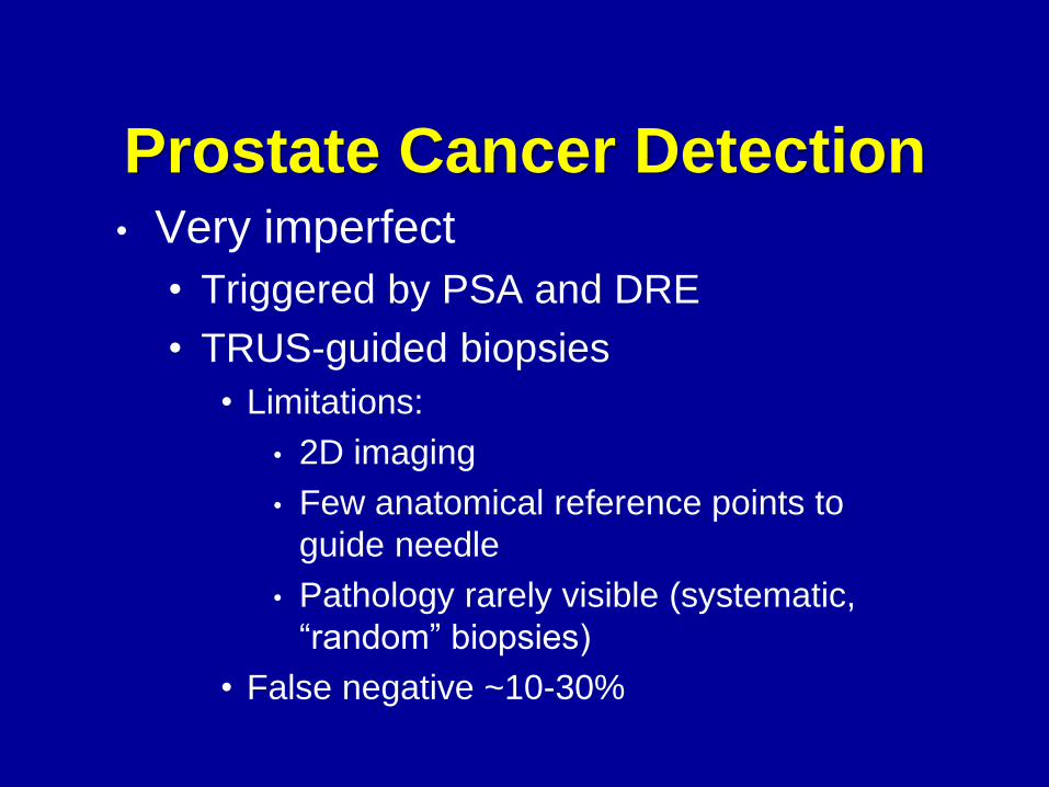
Prostate Cancer Detection• Very imperfect
• Triggered by PSA and DRE
• TRUS-guided biopsies
• Limitations:
• 2D imaging
• Few anatomical reference points to
guide needle
• Pathology rarely visible (systematic,
“random” biopsies)
• False negative ~10-30%
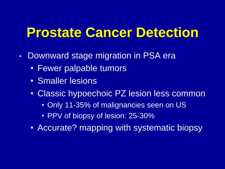
Prostate Cancer Detection
• Downward stage migration in PSA era
• Fewer palpable tumors
• Smaller lesions
• Classic hypoechoic PZ lesion less common
• Only 11-35% of malignancies seen on US
• PPV of biopsy of lesion: 25-30%
• Accurate? mapping with systematic biopsy
Prostate Cancer Detection
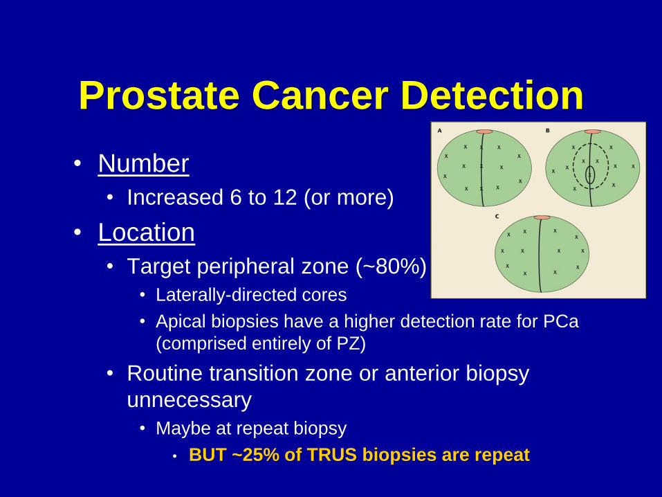
• Number
• Increased 6 to 12 (or more)
• Location
• Target peripheral zone (~80%)
• Laterally-directed cores
• Apical biopsies have a higher detection rate for PCa
(comprised entirely of PZ)
• Routine transition zone or anterior biopsy
unnecessary
• Maybe at repeat biopsy
• BUT ~25% of TRUS biopsies are repeat
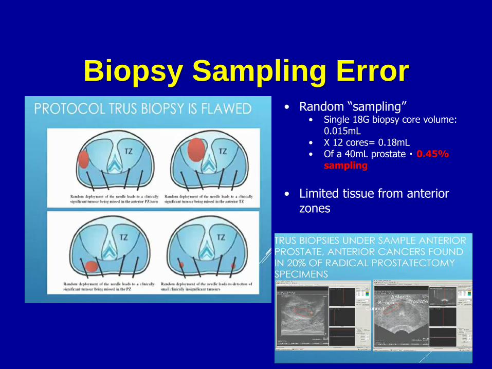
Biopsy Sampling Error• Random “sampling”
• Single 18G biopsy core volume: 0.015mL
• X 12 cores= 0.18mL• Of a 40mL prostate 0.45%
sampling
• Limited tissue from anterior zones
Biopsy Sampling Error
• Undersampling, undergrading and
nondetection
• Biggest limitation to active surveillance (or
focal therapies)
• (eg, missed anterior tumors)
• Overdetection of insignificant disease
• Image-guided (targeted) biopsy is
becoming standard of care
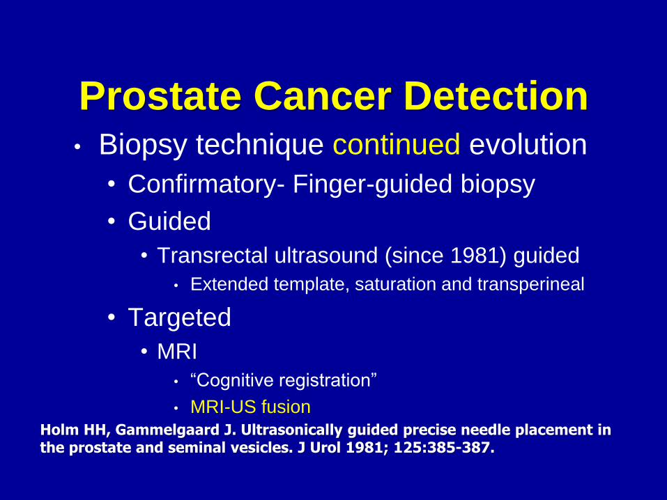
Prostate Cancer Detection• Biopsy technique continued evolution
• Confirmatory- Finger-guided biopsy
• Guided
• Transrectal ultrasound (since 1981) guided
• Extended template, saturation and transperineal
• Targeted
• MRI
• “Cognitive registration”
• MRI-US fusion
Holm HH, Gammelgaard J. Ultrasonically guided precise needle placement in the prostate and seminal vesicles. J Urol 1981; 125:385-387.
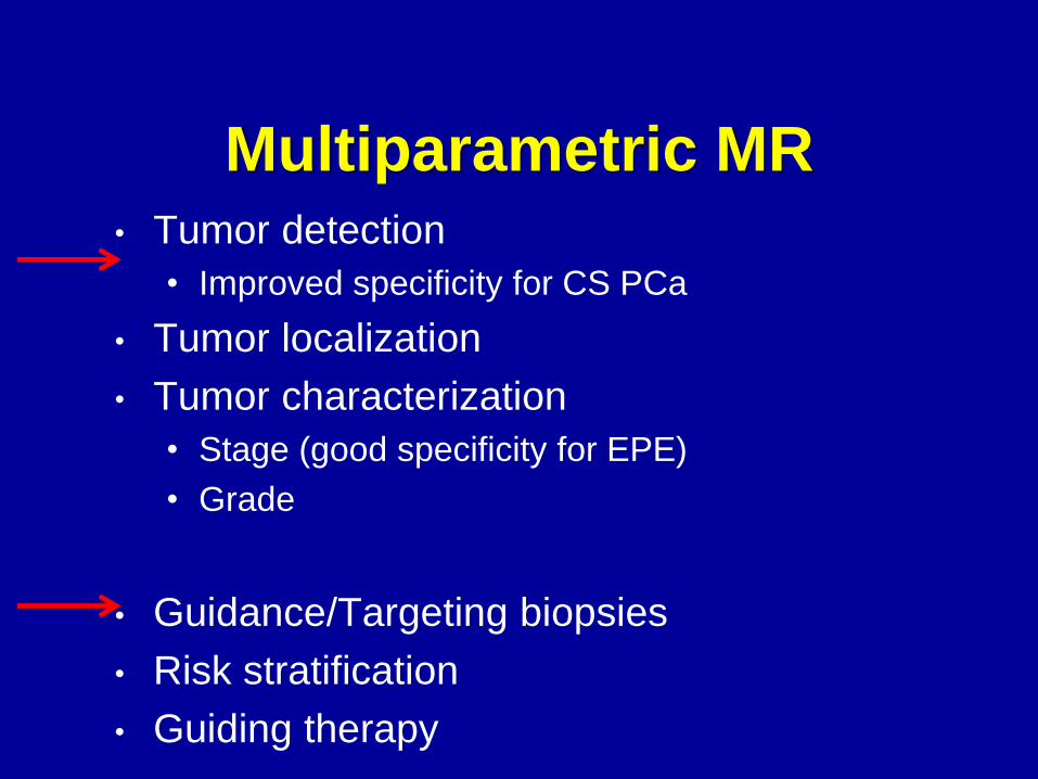
Multiparametric MR• Tumor detection
• Improved specificity for CS PCa
• Tumor localization
• Tumor characterization
• Stage (good specificity for EPE)
• Grade
• Guidance/Targeting biopsies
• Risk stratification
• Guiding therapy
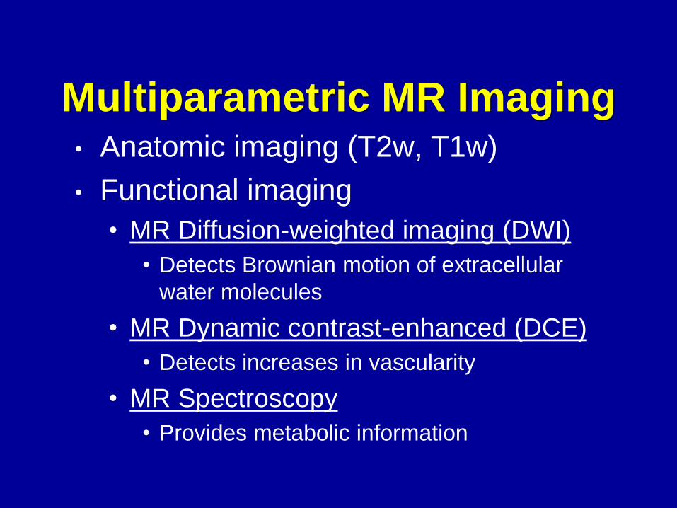
Multiparametric MR Imaging• Anatomic imaging (T2w, T1w)
• Functional imaging
• MR Diffusion-weighted imaging (DWI)
• Detects Brownian motion of extracellular
water molecules
• MR Dynamic contrast-enhanced (DCE)
• Detects increases in vascularity
• MR Spectroscopy
• Provides metabolic information
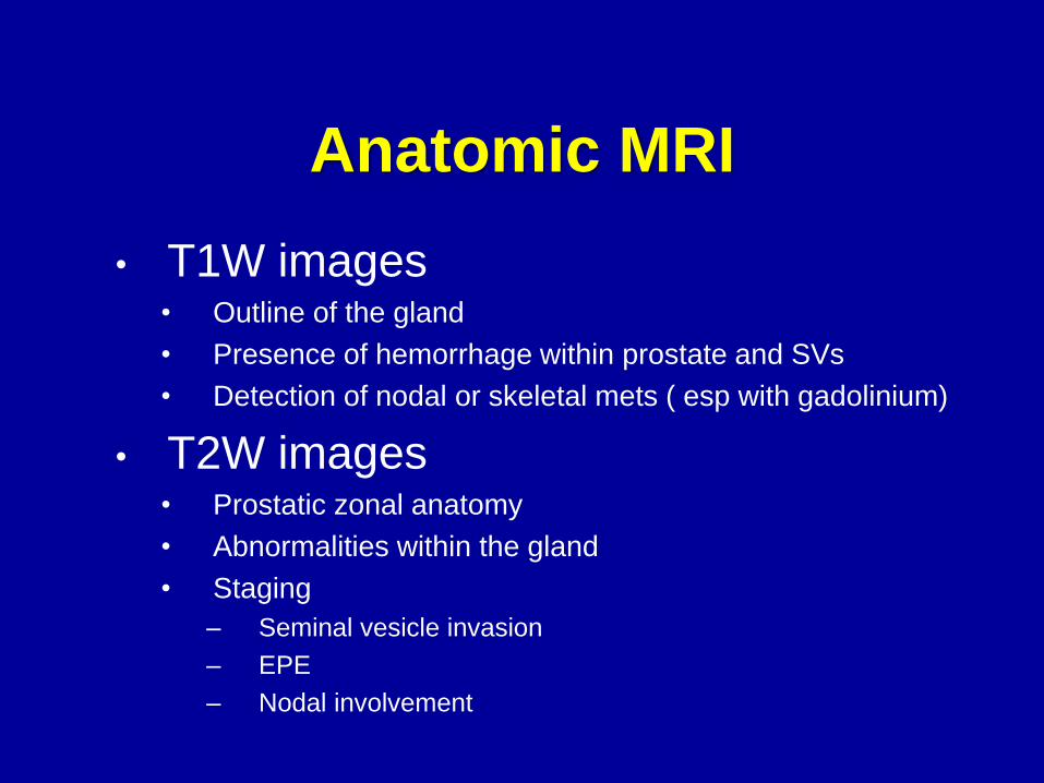
Anatomic MRI
• T1W images• Outline of the gland
• Presence of hemorrhage within prostate and SVs
• Detection of nodal or skeletal mets ( esp with gadolinium)
• T2W images• Prostatic zonal anatomy
• Abnormalities within the gland
• Staging
– Seminal vesicle invasion
– EPE
– Nodal involvement
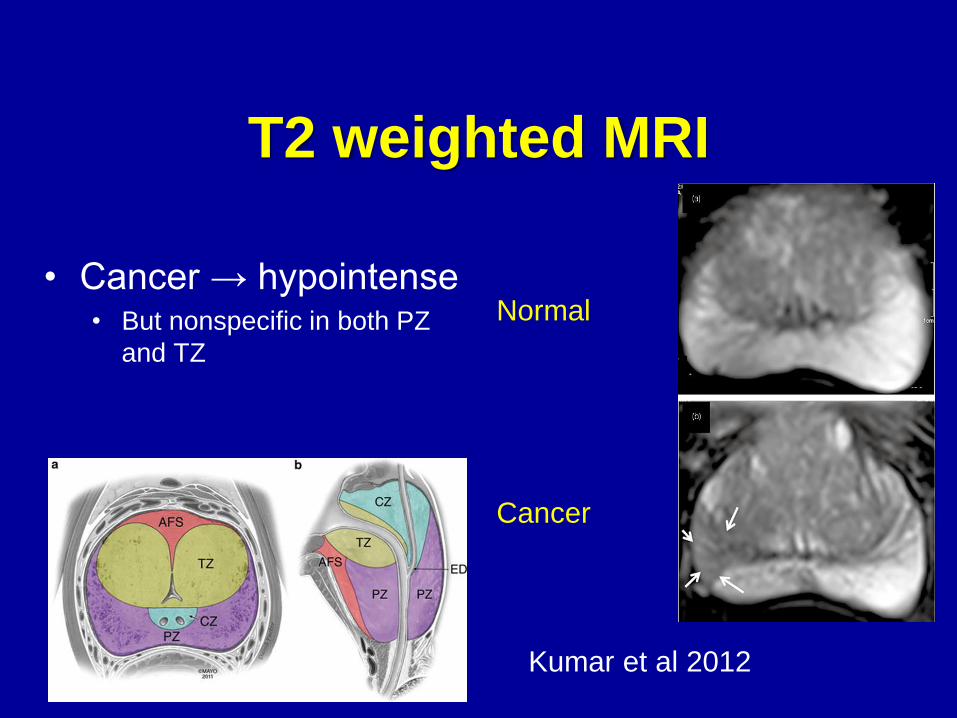
T2 weighted MRI
• Cancer → hypointense• But nonspecific in both PZ
and TZ
Normal
Cancer
Kumar et al 2012
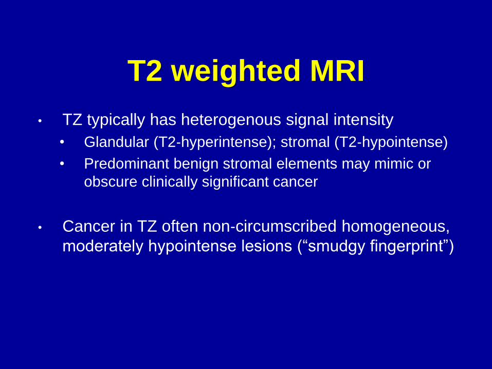
T2 weighted MRI
• TZ typically has heterogenous signal intensity
• Glandular (T2‐hyperintense); stromal (T2‐hypointense)
• Predominant benign stromal elements may mimic or
obscure clinically significant cancer
• Cancer in TZ often non‐circumscribed homogeneous,
moderately hypointense lesions (“smudgy fingerprint”)
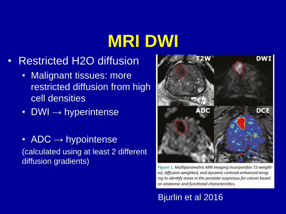
MRI DWI• Restricted H2O diffusion
• Malignant tissues: more
restricted diffusion from high
cell densities
• DWI → hyperintense
• ADC → hypointense
(calculated using at least 2 different
diffusion gradients)
Bjurlin et al 2016
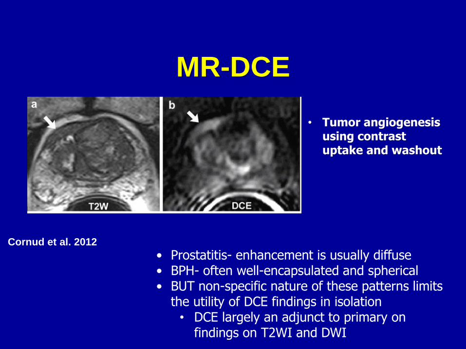
MR-DCE
Cornud et al. 2012
• Prostatitis- enhancement is usually diffuse• BPH- often well-encapsulated and spherical• BUT non-specific nature of these patterns limits
the utility of DCE findings in isolation• DCE largely an adjunct to primary on
findings on T2WI and DWI
• Tumor angiogenesis using contrast uptake and washout
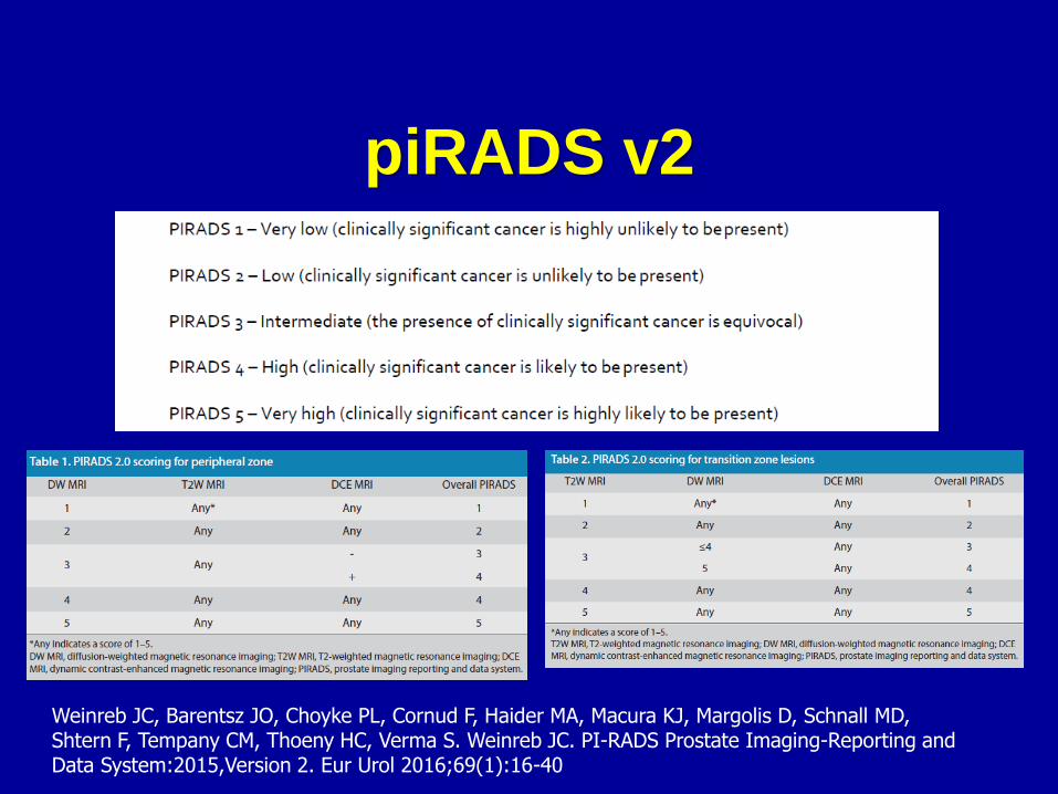
piRADS v2
• Prostate Imaging Reporting and Data
System (ESUR and ACR)
• Timing- at least 6 (8-12) wks post biopsy (takes
~6 months for hemorrhage to resolve in PZ)
• No consensus on patient preparation but BM
before important
• 3 T or 1.5 T (3T typically better but 1.5 T only
option with some implants)
• Endorectal coil not necessary (helpful in some
1.5 T for DWI and DCE)
Weinreb JC, Barentsz JO, Choyke PL, Cornud F, Haider MA, Macura KJ, Margolis D, Schnall MD, Shtern F, Tempany CM, Thoeny HC, Verma S. Weinreb JC. PI-RADS Prostate Imaging-Reporting and Data System:2015,Version 2. Eur Urol 2016;69(1):16-40
piRADS v2
Weinreb JC, Barentsz JO, Choyke PL, Cornud F, Haider MA, Macura KJ, Margolis D, Schnall MD, Shtern F, Tempany CM, Thoeny HC, Verma S. Weinreb JC. PI-RADS Prostate Imaging-Reporting and Data System:2015,Version 2. Eur Urol 2016;69(1):16-40
piRADS v2
Weinreb JC, Barentsz JO, Choyke PL, Cornud F, Haider MA, Macura KJ, Margolis D, Schnall MD, Shtern F, Tempany CM, Thoeny HC, Verma S. Weinreb JC. PI-RADS Prostate Imaging-Reporting and Data System:2015,Version 2. Eur Urol 2016;69(1):16-40
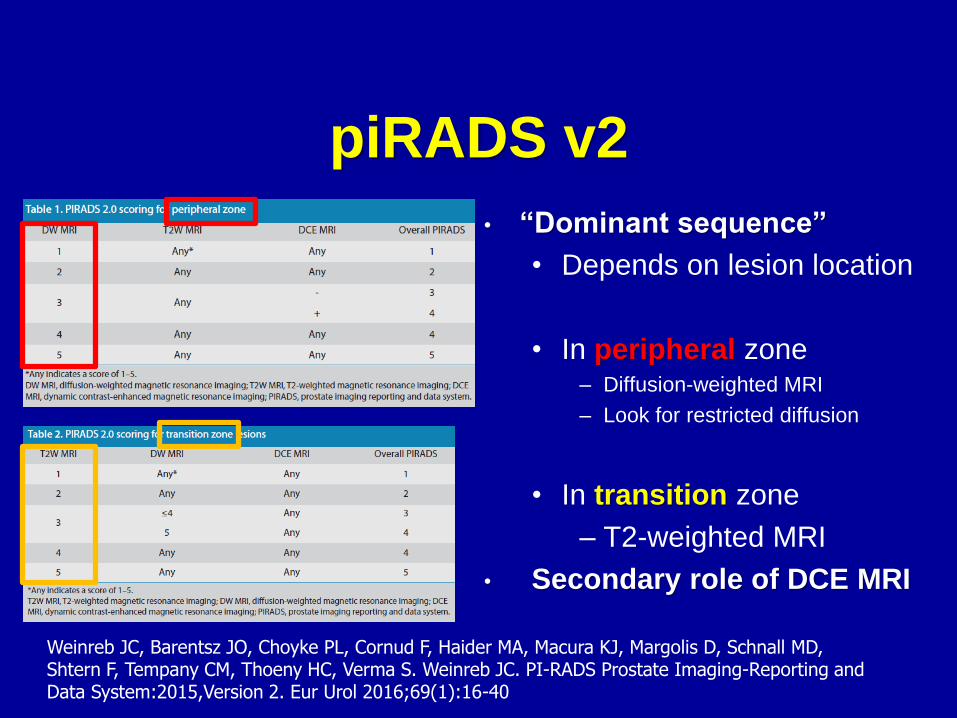
• “Dominant sequence”
• Depends on lesion location
• In peripheral zone– Diffusion-weighted MRI
– Look for restricted diffusion
• In transition zone
– T2-weighted MRI
• Secondary role of DCE MRI
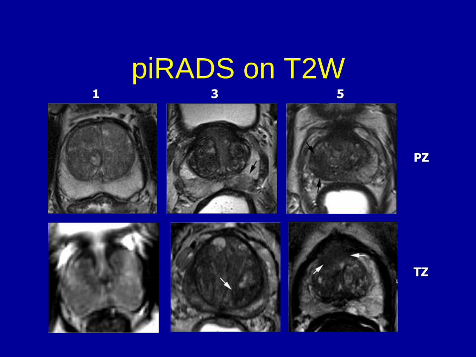
piRADS on T2W
PZ
TZ
1 3 5
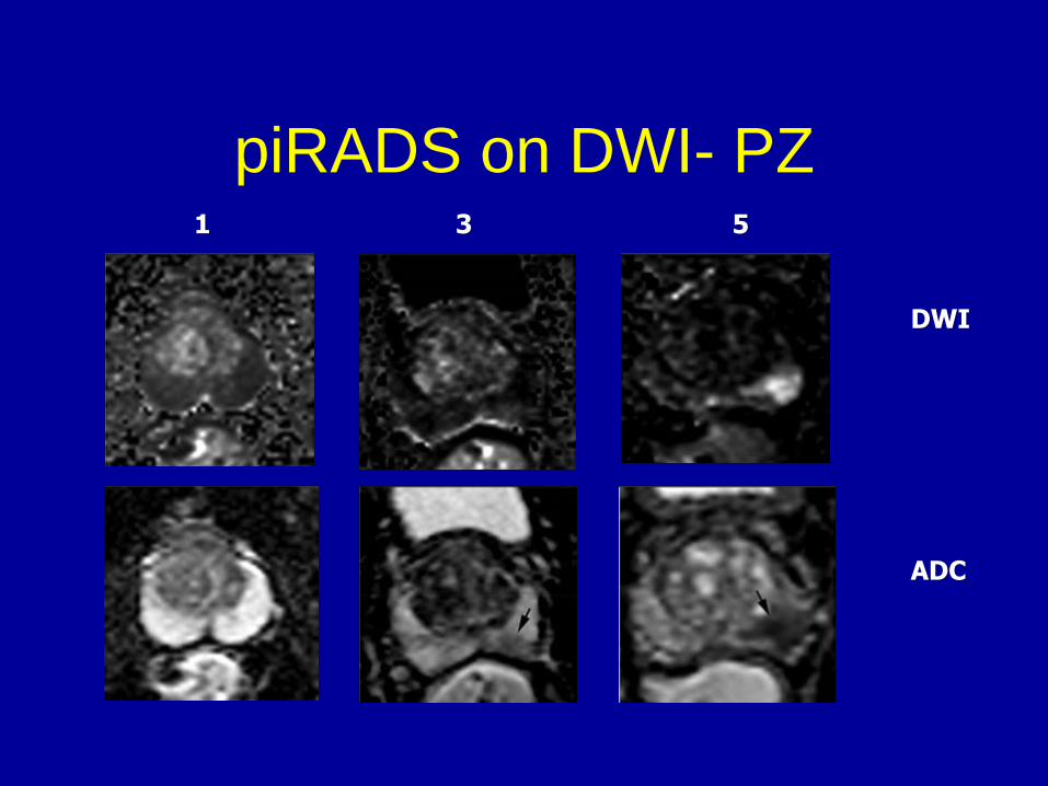
piRADS on DWI- PZ
DWI
ADC
1 3 5
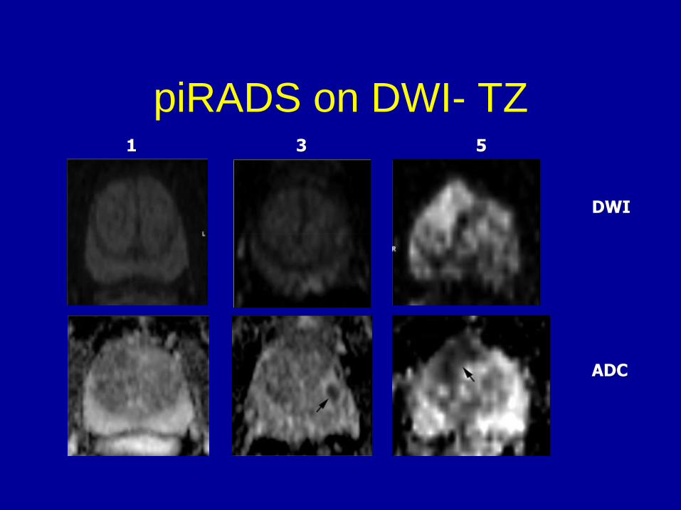
piRADS on DWI- TZ
DWI
ADC
1 3 5
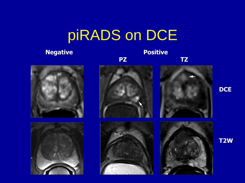
piRADS on DCENegative Positive
PZ TZ
DCE
T2W
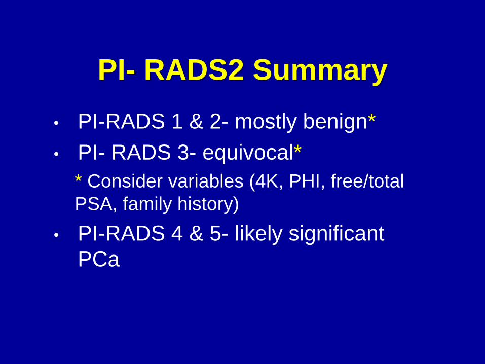
PI- RADS2 Summary
• PI-RADS 1 & 2- mostly benign*
• PI- RADS 3- equivocal*
* Consider variables (4K, PHI, free/total
PSA, family history)
• PI-RADS 4 & 5- likely significant
PCa
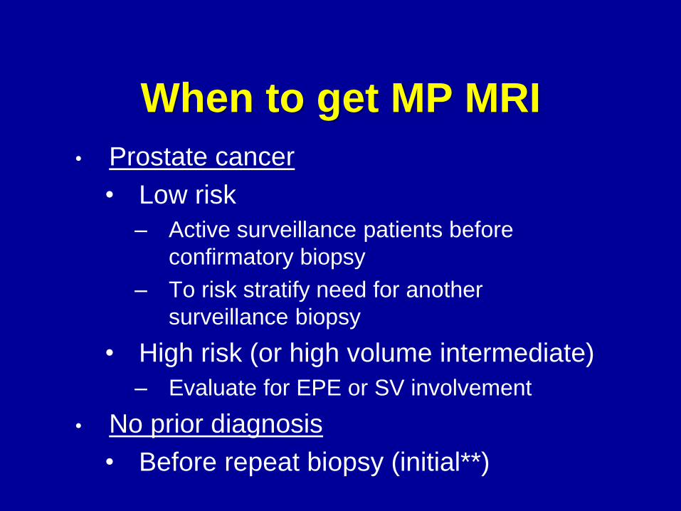
When to get MP MRI
• Prostate cancer
• Low risk
– Active surveillance patients before
confirmatory biopsy
– To risk stratify need for another
surveillance biopsy
• High risk (or high volume intermediate)
– Evaluate for EPE or SV involvement
• No prior diagnosis
• Before repeat biopsy (initial**)
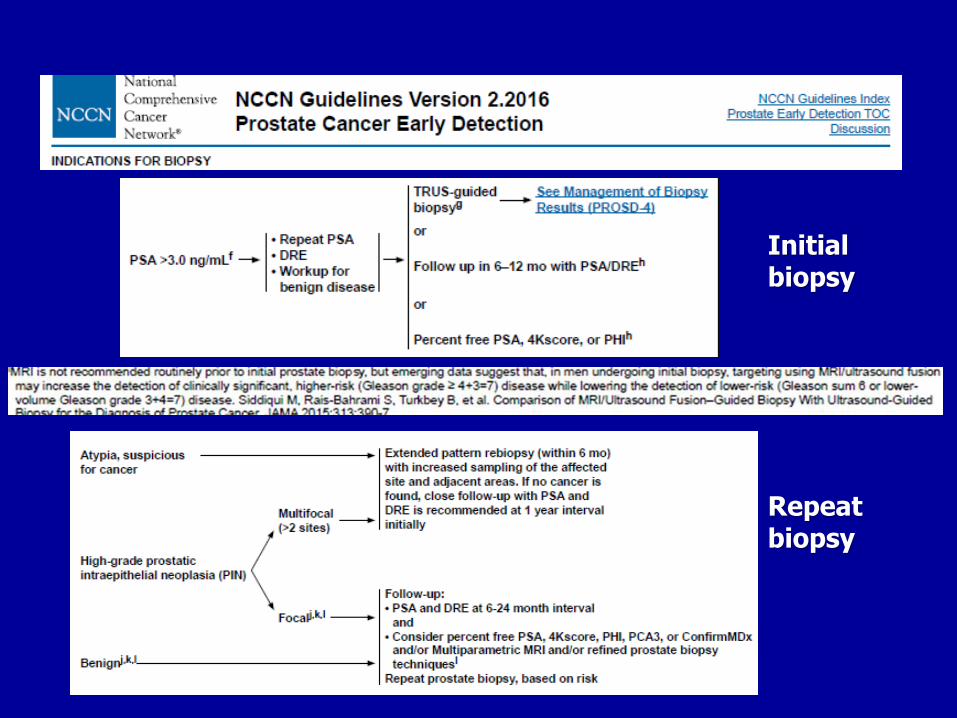
Initial biopsy
Repeat biopsy
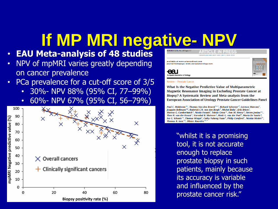
If MP MRI negative- NPV• EAU Meta-analysis of 48 studies• NPV of mpMRI varies greatly depending
on cancer prevalence• PCa prevalence for a cut-off score of 3/5
• 30%- NPV 88% (95% CI, 77–99%)• 60%- NPV 67% (95% CI, 56–79%)
“whilst it is a promising tool, it is not accurate enough to replace prostate biopsy in such patients, mainly because its accuracy is variable and influenced by the prostate cancer risk.”
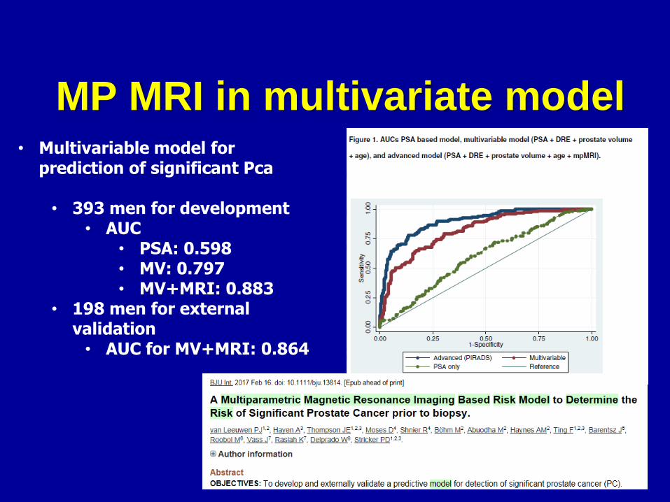
MP MRI in multivariate model• Multivariable model for
prediction of significant Pca
• 393 men for development• AUC
• PSA: 0.598• MV: 0.797• MV+MRI: 0.883
• 198 men for external validation
• AUC for MV+MRI: 0.864
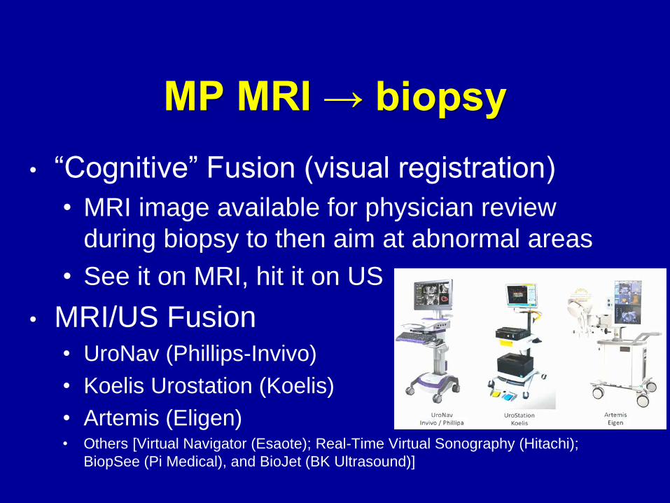
MP MRI → biopsy
• “Cognitive” Fusion (visual registration)
• MRI image available for physician review
during biopsy to then aim at abnormal areas
• See it on MRI, hit it on US
• MRI/US Fusion• UroNav (Phillips-Invivo)
• Koelis Urostation (Koelis)
• Artemis (Eligen) • Others [Virtual Navigator (Esaote); Real-Time Virtual Sonography (Hitachi);
BiopSee (Pi Medical), and BioJet (BK Ultrasound)]
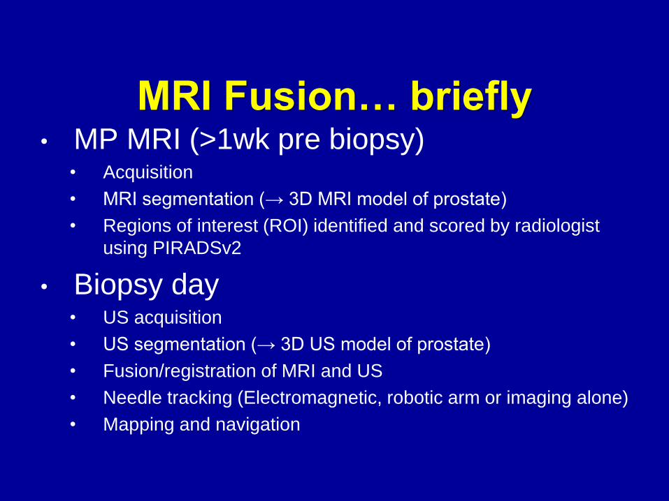
MRI Fusion… briefly• MP MRI (>1wk pre biopsy)
• Acquisition
• MRI segmentation (→ 3D MRI model of prostate)
• Regions of interest (ROI) identified and scored by radiologist
using PIRADSv2
• Biopsy day• US acquisition
• US segmentation (→ 3D US model of prostate)
• Fusion/registration of MRI and US
• Needle tracking (Electromagnetic, robotic arm or imaging alone)
• Mapping and navigation
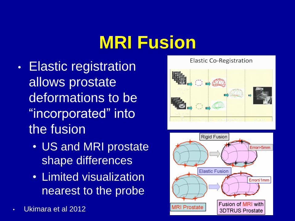
MRI Fusion
• Elastic registration
allows prostate
deformations to be
“incorporated” into
the fusion
• US and MRI prostate
shape differences
• Limited visualization
nearest to the probe
• Ukimara et al 2012
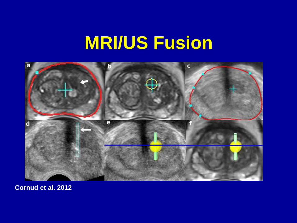
MRI/US Fusion
Cornud et al. 2012
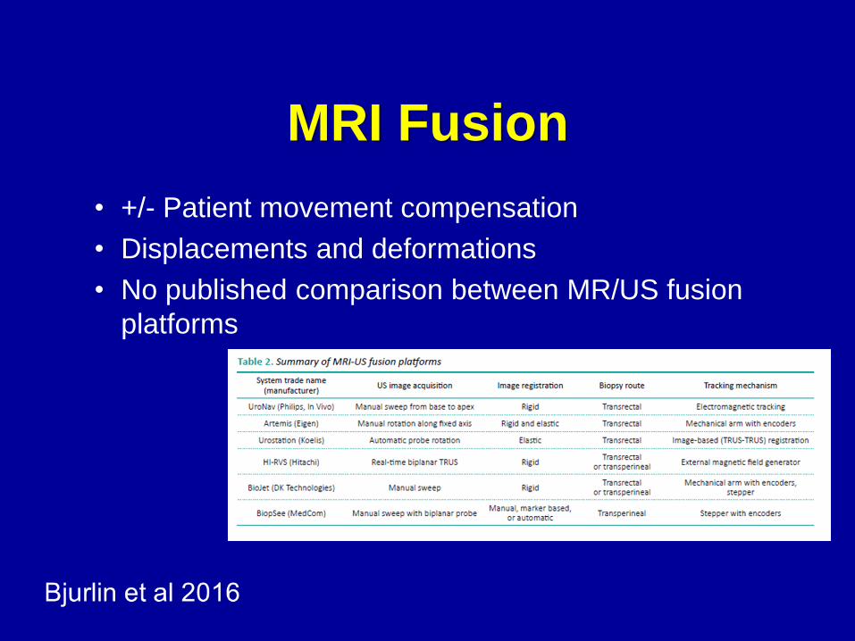
MRI Fusion
• +/- Patient movement compensation
• Displacements and deformations
• No published comparison between MR/US fusion
platforms
MRI/US Fusion
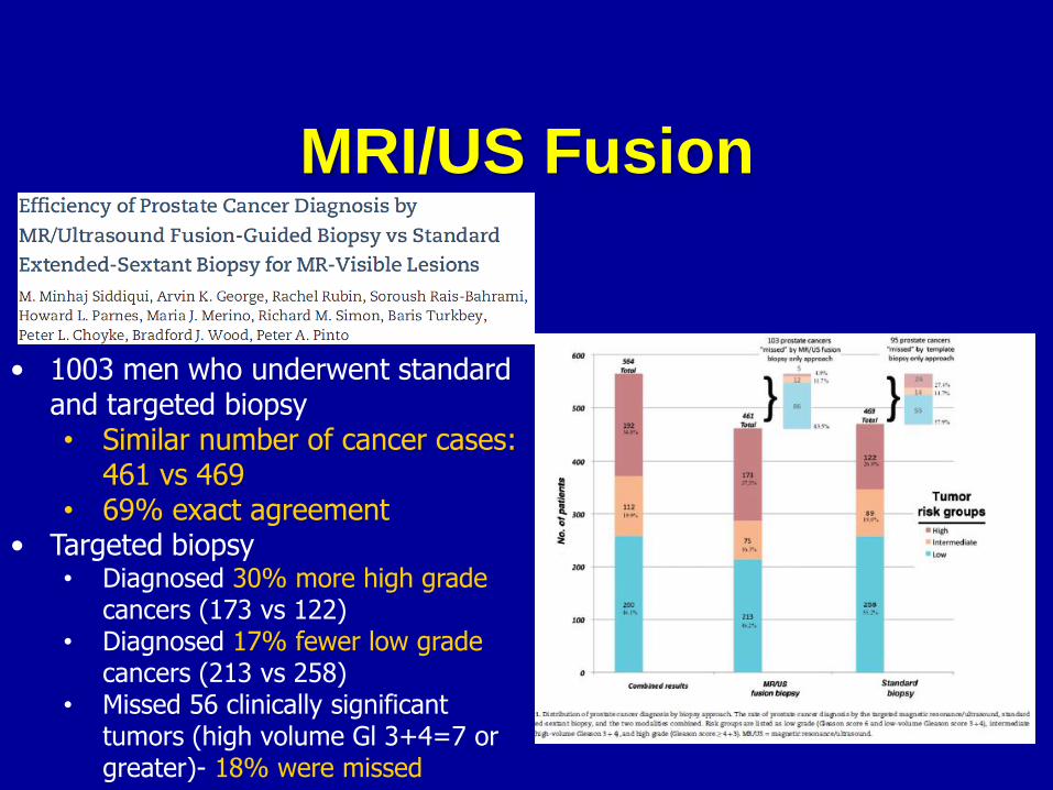
• 1003 men who underwent standard and targeted biopsy• Similar number of cancer cases:
461 vs 469• 69% exact agreement
• Targeted biopsy• Diagnosed 30% more high grade
cancers (173 vs 122)• Diagnosed 17% fewer low grade
cancers (213 vs 258)• Missed 56 clinically significant
tumors (high volume Gl 3+4=7 or greater)- 18% were missed
MRI/US Fusion
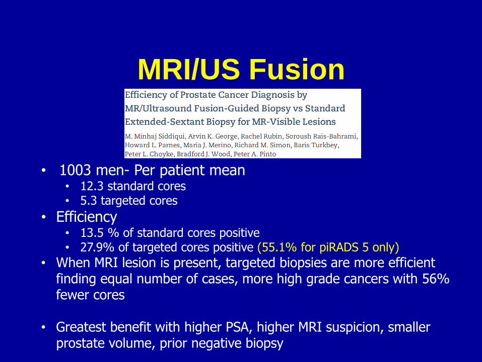
• 1003 men- Per patient mean• 12.3 standard cores• 5.3 targeted cores
• Efficiency• 13.5 % of standard cores positive• 27.9% of targeted cores positive (55.1% for piRADS 5 only)
• When MRI lesion is present, targeted biopsies are more efficient finding equal number of cases, more high grade cancers with 56% fewer cores
• Greatest benefit with higher PSA, higher MRI suspicion, smaller prostate volume, prior negative biopsy
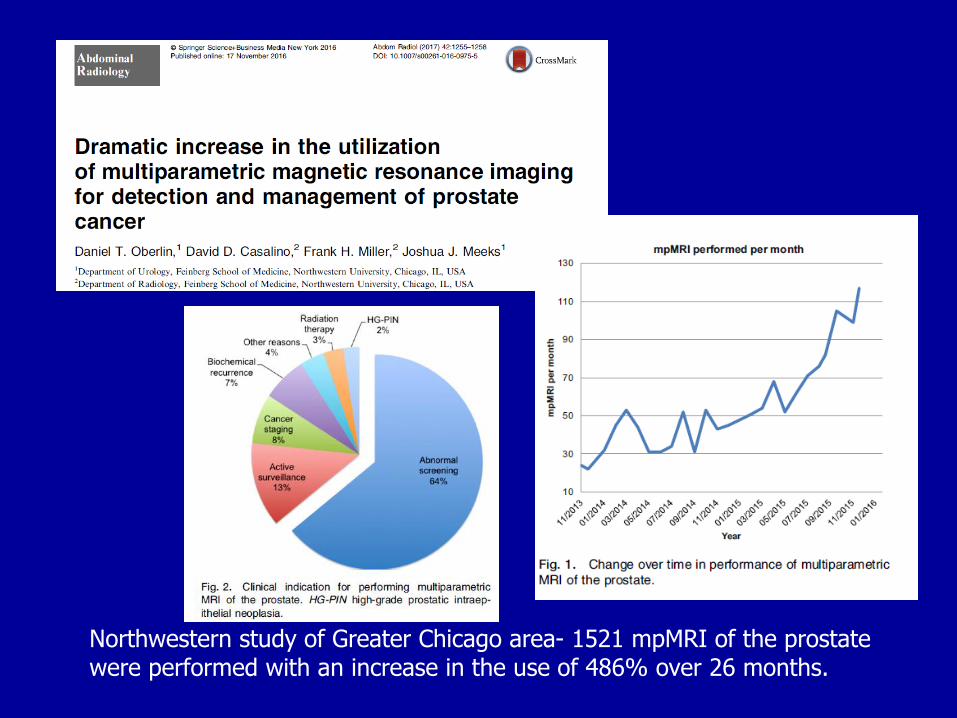
Northwestern study of Greater Chicago area- 1521 mpMRI of the prostate were performed with an increase in the use of 486% over 26 months.
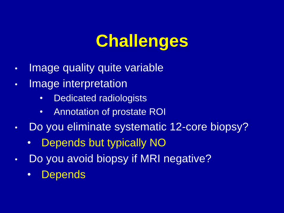
Challenges
• Image quality quite variable
• Image interpretation
• Dedicated radiologists
• Annotation of prostate ROI
• Do you eliminate systematic 12-core biopsy?
• Depends but typically NO
• Do you avoid biopsy if MRI negative?
• Depends
Challenges• Systematic biopsy remains an essential component
of a targeted biopsy• ~30% of concurrent systematic biopsies will reveal clinically
significant disease (may be higher in “bad” prostates-piRADS 4 and 5)
• Major obstacles• Learning curve, dedicated radiologist, cost of machine,
insurance coverage, time and reimbursement

Summary• Prostate biopsy is evolving
• Lesion-directed sampling → image-guided sampling → modeled systematic sampling with targeting
• Ideal PCa detection will involve combination • Targeting of image-visible suspicious lesions
• Targeting to sites most likely to contain cancer
• MPMRI allows for identification/targeting of more “visible” lesions
• MP MRI should be done before repeat biopsy with TBx of suspicious lesions AND standard biopsy
References• Moldovan PC, et al. What Is the Negative Predictive Value of Multiparametric
Magnetic Resonance Imaging in Excluding Prostate Cancer at Biopsy? A
Systematic Review and Meta-analysis from the European Association of
Urology Prostate Cancer Guidelines Panel. Eur Urol (2017).
• van Leeuwen PJ, Hayen A, Thompson JE, et al. A Multiparametric Magnetic
Resonance Imaging Based Risk Model to Determine the Risk of Significant
Prostate Cancer prior to biopsy. BJU Int. 2017 Feb 16.
• Oberlin DT, Casalino DD, Miller FH, Meeks JJ. Dramatic increase in the
utilization of multiparametric magnetic resonance imaging for detection and
management of prostate cancer. Abdom Radiol (NY). 2016 Nov 17.
• Michael Kongnyuy M, George AK, Rastinehad AR, Pinto PA. Magnetic
Resonance Imaging-Ultrasound Fusion-Guided Prostate Biopsy: Review of
Technology, Techniques, and Outcomes. Curr Urol Rep (2016) 17: 32.
• Bjurlin MA1, Mendhiratta N2, Wysock JS3, Taneja SS3. Multiparametric MRI
and targeted prostate biopsy: Improvements in cancer detection, localization,
and risk assessment. Cent European J Urol. 2016;69(1):9-18.
References• Weinreb JC, Barentsz JO, Choyke PL, Cornud F et al. PI-RADS Prostate
Imaging-Reporting and Data System:2015,Version 2. Eur Urol 2016;69(1):16-40
• Hamoen EHJ, et al. Use of the Prostate Imaging Reporting and Data System
(PI-RADS) for Prostate Cancer Detection with Multiparametric Magnetic
Resonance Imaging: A Diagnostic Meta-analysis. Eur Urol 2015; 67:1112.
• Ukimara O, Faber K, Gill I. Intraprostatic targeting. Curr Opin Urol. 2012; 22:97.
• Cornud F, Delongchamps NB, Mozer P, Beuvon F, Schull A, Muradyan N et al.
Value of multiparametric MRI in the work-up of prostate cancer. Curr Urol Rep
2012; 13:82.
• Ukimara O, Desai MM, Palmer S, Valencerina S, Gross M, Abreu AL et al. 3-
dimensional elastic registration system of prostate biopsy location by real-time
3-dimensional transrectal ultrasound guidance with magnetic
resonance/transrectal ultrasound image fusion. J Urol 2012 ; 187:1080.
• Kumar V, Jagannathan NR, Thulkar S, Kumar R. Prebiopsy magnetic
resonance spectroscopy and imaging in the diagnosis of prostate cancer. Int J
Urol 2012
References• Haffner J, Lemaitre L, Puech P, Haber GP, Leroy X, Jones JS et al. Role of
magnetic resonance imaging before initial biopsy: comparison of magnetic
resonance imaging-targeted and systematic biopsy for significant prostate
cancer detection. BJU Int 2011; 108:E171.
• Pinto PA, Chung PH, Rastinehad AR, Baccala AA, Kruecker J, Benjamin CJ
et al. Magnetic resonance imaging/ultrasound fusion guided prostate biopsy
improves cancer detection following transrectal ultrasound biopsy and
correlates with multiparametric magnetic resonance imaging. J Urol 2011;
186:1281.
• Mitterberger M, Horninger W, Aigner F, Pinggera GM, Steppan I, Rehder P
et al. Ultrasound of the prostate. Cancer Imaging 2010; 10:40.
• Trabulsi EJ, Sackett D, Gomella LG, Halpern EJ. Enhanced transrectal
ultrasound modalities in the diagnosis of prostate cancer. Urology 2010;
76:1025.
• Taneja SS. Prostate biopsy: targeting cancer for detection and therapy. Rev
Urol. 2006;8:173.
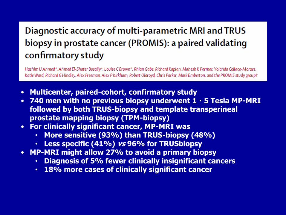
• Multicenter, paired-cohort, confirmatory study• 740 men with no previous biopsy underwent 1・5 Tesla MP-MRI
followed by both TRUS-biopsy and template transperineal prostate mapping biopsy (TPM-biopsy)
• For clinically significant cancer, MP-MRI was• More sensitive (93%) than TRUS-biopsy (48%)• Less specific (41%) vs 96% for TRUSbiopsy
• MP-MRI might allow 27% to avoid a primary biopsy• Diagnosis of 5% fewer clinically insignificant cancers• 18% more cases of clinically significant cancer