Embed Size (px)
Citation preview
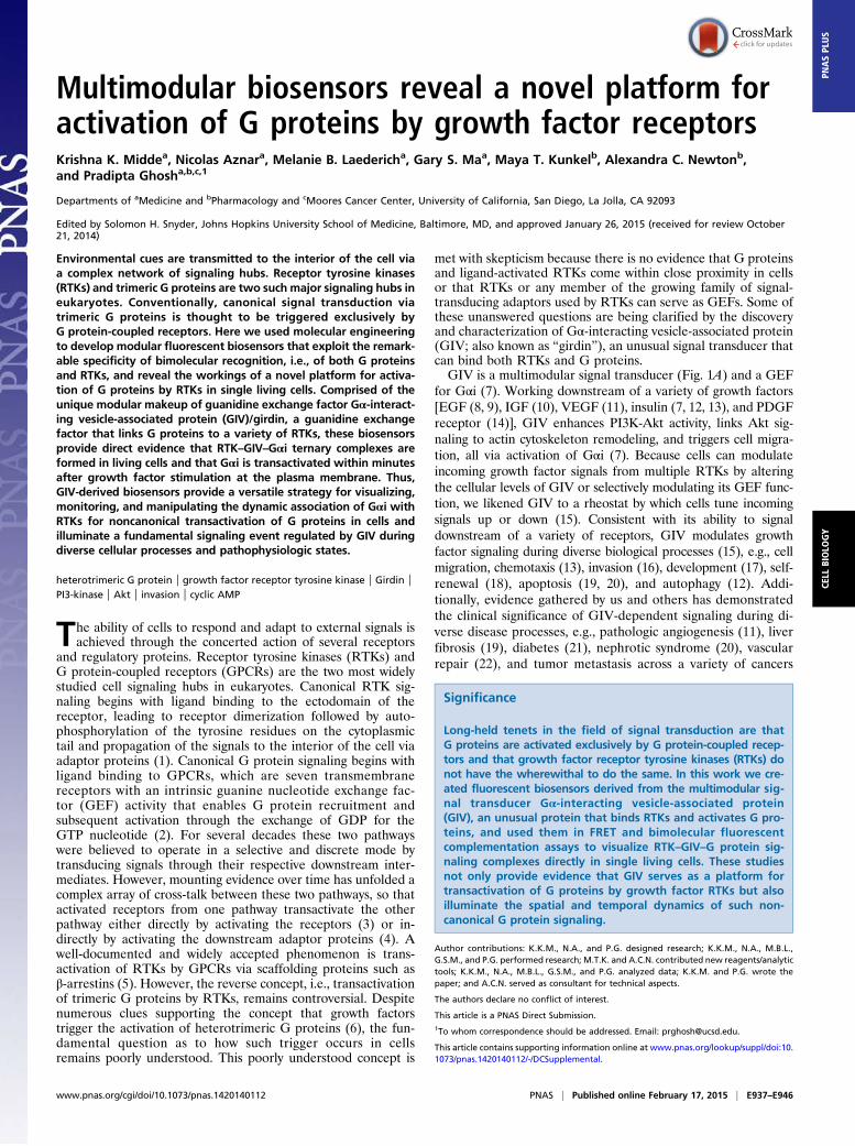
Multimodular biosensors reveal a novel platform foractivation of G proteins by growth factor receptorsKrishna K. Middea, Nicolas Aznara, Melanie B. Laedericha, Gary S. Maa, Maya T. Kunkelb, Alexandra C. Newtonb,and Pradipta Ghosha,b,c,1
Departments of aMedicine and bPharmacology and cMoores Cancer Center, University of California, San Diego, La Jolla, CA 92093
Edited by Solomon H. Snyder, Johns Hopkins University School of Medicine, Baltimore, MD, and approved January 26, 2015 (received for review October21, 2014)
Environmental cues are transmitted to the interior of the cell viaa complex network of signaling hubs. Receptor tyrosine kinases(RTKs) and trimeric G proteins are two such major signaling hubs ineukaryotes. Conventionally, canonical signal transduction viatrimeric G proteins is thought to be triggered exclusively byG protein-coupled receptors. Here we used molecular engineeringto develop modular fluorescent biosensors that exploit the remark-able specificity of bimolecular recognition, i.e., of both G proteinsand RTKs, and reveal the workings of a novel platform for activa-tion of G proteins by RTKs in single living cells. Comprised of theunique modular makeup of guanidine exchange factor Gα-interact-ing vesicle-associated protein (GIV)/girdin, a guanidine exchangefactor that links G proteins to a variety of RTKs, these biosensorsprovide direct evidence that RTK–GIV–Gαi ternary complexes areformed in living cells and that Gαi is transactivated within minutesafter growth factor stimulation at the plasma membrane. Thus,GIV-derived biosensors provide a versatile strategy for visualizing,monitoring, and manipulating the dynamic association of Gαi withRTKs for noncanonical transactivation of G proteins in cells andilluminate a fundamental signaling event regulated by GIV duringdiverse cellular processes and pathophysiologic states.
heterotrimeric G protein | growth factor receptor tyrosine kinase | Girdin |PI3-kinase | Akt | invasion | cyclic AMP
The ability of cells to respond and adapt to external signals isachieved through the concerted action of several receptors
and regulatory proteins. Receptor tyrosine kinases (RTKs) andG protein-coupled receptors (GPCRs) are the two most widelystudied cell signaling hubs in eukaryotes. Canonical RTK sig-naling begins with ligand binding to the ectodomain of thereceptor, leading to receptor dimerization followed by auto-phosphorylation of the tyrosine residues on the cytoplasmictail and propagation of the signals to the interior of the cell viaadaptor proteins (1). Canonical G protein signaling begins withligand binding to GPCRs, which are seven transmembranereceptors with an intrinsic guanine nucleotide exchange fac-tor (GEF) activity that enables G protein recruitment andsubsequent activation through the exchange of GDP for theGTP nucleotide (2). For several decades these two pathwayswere believed to operate in a selective and discrete mode bytransducing signals through their respective downstream inter-mediates. However, mounting evidence over time has unfolded acomplex array of cross-talk between these two pathways, so thatactivated receptors from one pathway transactivate the otherpathway either directly by activating the receptors (3) or in-directly by activating the downstream adaptor proteins (4). Awell-documented and widely accepted phenomenon is trans-activation of RTKs by GPCRs via scaffolding proteins such asβ-arrestins (5). However, the reverse concept, i.e., transactivationof trimeric G proteins by RTKs, remains controversial. Despitenumerous clues supporting the concept that growth factorstrigger the activation of heterotrimeric G proteins (6), the fun-damental question as to how such trigger occurs in cellsremains poorly understood. This poorly understood concept is
met with skepticism because there is no evidence that G proteinsand ligand-activated RTKs come within close proximity in cellsor that RTKs or any member of the growing family of signal-transducing adaptors used by RTKs can serve as GEFs. Some ofthese unanswered questions are being clarified by the discoveryand characterization of Gα-interacting vesicle-associated protein(GIV; also known as “girdin”), an unusual signal transducer thatcan bind both RTKs and G proteins.GIV is a multimodular signal transducer (Fig. 1A) and a GEF
for Gαi (7). Working downstream of a variety of growth factors[EGF (8, 9), IGF (10), VEGF (11), insulin (7, 12, 13), and PDGFreceptor (14)], GIV enhances PI3K-Akt activity, links Akt sig-naling to actin cytoskeleton remodeling, and triggers cell migra-tion, all via activation of Gαi (7). Because cells can modulateincoming growth factor signals from multiple RTKs by alteringthe cellular levels of GIV or selectively modulating its GEF func-tion, we likened GIV to a rheostat by which cells tune incomingsignals up or down (15). Consistent with its ability to signaldownstream of a variety of receptors, GIV modulates growthfactor signaling during diverse biological processes (15), e.g., cellmigration, chemotaxis (13), invasion (16), development (17), self-renewal (18), apoptosis (19, 20), and autophagy (12). Addi-tionally, evidence gathered by us and others has demonstratedthe clinical significance of GIV-dependent signaling during di-verse disease processes, e.g., pathologic angiogenesis (11), liverfibrosis (19), diabetes (21), nephrotic syndrome (20), vascularrepair (22), and tumor metastasis across a variety of cancers
Significance
Long-held tenets in the field of signal transduction are thatG proteins are activated exclusively by G protein-coupled recep-tors and that growth factor receptor tyrosine kinases (RTKs) donot have the wherewithal to do the same. In this work we cre-ated fluorescent biosensors derived from the multimodular sig-nal transducer Gα-interacting vesicle-associated protein(GIV), an unusual protein that binds RTKs and activates G pro-teins, and used them in FRET and bimolecular fluorescentcomplementation assays to visualize RTK–GIV–G protein sig-naling complexes directly in single living cells. These studiesnot only provide evidence that GIV serves as a platform fortransactivation of G proteins by growth factor RTKs but alsoilluminate the spatial and temporal dynamics of such non-canonical G protein signaling.
Author contributions: K.K.M., N.A., and P.G. designed research; K.K.M., N.A., M.B.L.,G.S.M., and P.G. performed research; M.T.K. and A.C.N. contributed new reagents/analytictools; K.K.M., N.A., M.B.L., G.S.M., and P.G. analyzed data; K.K.M. and P.G. wrote thepaper; and A.C.N. served as consultant for technical aspects.
The authors declare no conflict of interest.
This article is a PNAS Direct Submission.1To whom correspondence should be addressed. Email: [email protected].
This article contains supporting information online at www.pnas.org/lookup/suppl/doi:10.1073/pnas.1420140112/-/DCSupplemental.
www.pnas.org/cgi/doi/10.1073/pnas.1420140112 PNAS | Published online February 17, 2015 | E937–E946
CELL
BIOLO
GY
PNASPL
US

Fig. 1. Design and characterization of GIV-derived biosensors. (A, Upper) Schematic of various known functional domains of GIV. (Lower) The uniquemodular make-up of GIV’s CT with in-tandem coexistence of a GEF domain (red) that activates Gαi (gray), and a SH2-like domain (blue-red) that bindsautophosphorylated cytoplasmic tails of RTKs. Magenta represents EGFR pTyr peptide. The homology models displayed were validated previously (7, 28).(B) Schematic showing the cloning strategy used in generating CFP-tagged GIV biosensors. FA, F1685A, a previously characterized GEF-deficient mutant.(C) Immunoblots showing the expression of WT and FA CFP-GIV-CT biosensors in Cos7 cells. (D) Pulldown assay using GST-Gαi3 loaded with GDP in thepresence or absence of AlF4
− and lysates of Cos7 cells expressing CFP-GIV-CT. The positive control Gβ bound Gαi3 in the presence of GDP but not in thepresence of AlF4
−. (E) Pulldown assay using GST-Gαi3 loaded with GDP and lysates of Cos7 cells expressing CFP-GIV-CT-WT (lanes 1–4) or CFP-GIV-CT-FA (lanes5–8). The positive control Gβ binds Gαi3·GDP. (F) Cos7 cells expressing CFP-GIV-CT-WT or -FA were starved and stimulated with EGF as indicated before lysis.Lysates were analyzed for Gαi1 and 3 activity by immunoprecipitation with anti-Gαi:GTP and immunoblotting with total Gαi1 and Gαi3 antibodies. (G) Cos7cells were transfected with the GIV-CT biosensor, starved, and subsequently stimulated with EGF ligand for indicated times before lysis. EGFR-bound com-plexes were immunoprecipitated with anti-EGFR Ab. Immune complexes (Left) and lysates (Right) were analyzed for total EGFR (t EGFR), phosphorylated EGFR(pY EGFR), the CFP-GIV biosensor (GFP), and tubulin. M, mix of starved and EGF-stimulated lysates. (H) Lysates of Cos7 cells expressing CFP-GIV-CT-WT or -FAbiosensors were analyzed for phosphoAkt (pS473 and pT308Akt), CFP-GIV biosensors (GFP), and tubulin. (I) Cos7 cells expressing the CFP-GIV-CT biosensorwere starved and subsequently stimulated with EGF, and whole-cell lysates were probed for GFP and pY1764 GIV. Tyrosine phosphorylation was seen ex-clusively after EGF stimulation, as previously shown in the case of full-length GIV (29). (J, Upper) GIV-CT-WT, but not GIV-CT-FA, triggers cell invasion.Spheroids (S) of NIH 3T3 cells expressing CFP-GIV-CT biosensors or control vector were analyzed for their ability to invade Matrigel in response to EGF usinga Cultrex-3D Spheroid Invasion Kit (Trevigen) (SI Experimental Procedures). Invading cells (arrowheads) and satellite tumors (T) were noted exclusively in cellsexpressing CFP-GIV-CT-WT. (Lower) Magnified views of the dashed boxes in the corresponding upper panels. Data quantification is shown in Fig. S1B.
E938 | www.pnas.org/cgi/doi/10.1073/pnas.1420140112 Midde et al.
[gastric (23), esophageal (24), prostate (16), breast (10, 25, 26),colon (27), and glioblastoma (18)].Despite the accumulating information on the biological and
clinical significance of GIV, how it may couple to multiple RTKsremained unknown until recently. Protein interaction assaysshowed that GIV’s C terminus (CT) directly binds autophos-phorylated cytoplasmic tails of multiple RTKs (9). Homologymodeling, sequence analysis, side-chain substitution, and limitedproteolysis showed that an ∼110-aa stretch within GIV’s CTfolds into a Scr homology 2 (SH2)-like domain and is necessaryand sufficient to recognize and bind phosphotyrosine peptides(28) (Fig. 1A) and recruit Gαi to RTKs. The discovery of coex-isting SH2-like and GEF domains in tandem within the GIV CT(Fig. 1A) supported the idea that GIV’s CT has the necessarymodular make-up to serve as a platform for linking G proteins tomultiple RTKs. However, the in vitro and standard biochemicalassays used thus far have failed to provide direct in cellulo evi-dence that GIV assembles RTK–GIV–Gαi ternary complexes,and if it does, when and where this assembly might occur, whatmight be the consequences of such assembly on G protein sig-naling, and how such signaling compares with the dynamics ofcanonical GPCR-driven G protein signaling. Such evidencewould provide insights into the fundamental mechanisms thatdefine GIV’s role at the cross-roads of RTK and G proteinsignaling pathways in diverse pathophysiologic processes. Suchfindings also will imply that the evolutionarily conserved CT ofGIV serves as the long-sought modular platform for trans-activation of G proteins downstream of growth factors, a phe-nomenon that has been observed and reported by several groupsover the past few decades (6).
Results and DiscussionGeneration of Fluorescent Biosensors Comprised of Key ModulesDerived from GIV. To visualize the formation of RTK–GIV–Gαicomplexes and to gain mechanistic insights into the dynamicbehavior of GIV in signal transduction, we developed multi-modular fluorescent GIV biosensors. These biosensors are com-prised of the CT of GIV (amino acids 1660–1870) N-terminallytagged with cyan fluorescent protein (CFP) to serve as donor(CFP-GIV-CT-WT) in FRET studies (Fig. 1 B and C). A pre-viously described GEF-deficient mutant (7), in which Phe atposition 1685 in the GEF motif is replaced by Ala (CFP-GIV-CT-FA), was created to disrupt the GIV–Gαi interaction selec-tively. The rationale for the design of these biosensors is multi-factorial: (i) a complete phylogenetic analysis of GIV (15) hasrevealed that this stretch of GIV’s CT could be functionallyautonomous because it evolved independently of its N terminus(NT) (in fish), and both the NT and CT fused into full-lengthGIV only in birds; (ii) the CT contains the GEF and SH2-likedomains, representing the cross-road between the GPCR/G andRTK signaling pathways; (iii) the CT of GIV also contains thetwo critical tyrosines that serve as docking sites for p85α (PI3K)(29); (iv) the coexistence of the GEF motif, the SH2-like domain,and the tyrosines is restricted to only the most complex ofeukaryotes, i.e., mammals, and is highly conserved (∼99%) (15,29); and (v) biochemical and functional assays (9) have demon-strated convincingly that the CT is the domain most criticallyrequired for GIV to carry out its functions during signal trans-duction downstream of RTKs. We hypothesized that the CT ofGIV, which contains the GEF, the SH2-like domain, and the twocritical tyrosine residues, is the minimal module that allows thisregion to operate autonomously and carry out most functionsthat previously have been attributed to full-length GIV as a sig-nal transducer downstream of growth factor RTKs (15).We carried out several biochemical and functional assays to
determine if GIV’s CT is indeed functionally autonomous. Be-cause GIV is a GEF for Gαi1-3 subunits, but not Gαo/s (7), wefirst assessed the ability of GIV-CT biosensors to bind and acti-
vate Gαi. Lysates of Cos7 cells expressing CFP-tagged biosensorswere used as source of GIV-CT protein in GST pulldown assayswith recombinant GST-tagged Gαi3 immobilized on glutathionebeads. Consistent with the known binding properties of GEFs, CFP-GIV-CT-WT preferentially bound inactive (i.e., GDP-loaded) GST-Gαi3, but not Gαi3 in active conformation (as mimicked by thepresence of aluminum fluoride, AlF4
−) (Fig. 1D). As anticipated,the GEF-deficient CFP-GIV-CT-FA biosensor did not bind Gαi3(Fig. 1E). To determine if the GIV-CT biosensor activates Gαi ina GEF-dependent manner in Cos7 cells, we took advantage of anantibody that specifically recognizes Gαi in a GTP-bound activeconformation [anti-Gαi:GTP (28, 30)]. We detected activation ofGαi1/3 in cells overexpressing the GIV-CT-WT biosensor exclu-sively after EGF stimulation (Fig. 1F), and such ligand-dependentactivation was virtually abolished in cells expressing the GIV-CT-FA biosensor. We conclude that GIV-CT biosensors can bind andactivate Gαi in cells in a GEF-dependent manner as previouslydemonstrated for full-length GIV.Next we asked if GIV-CT biosensors are able to bind EGF
receptor (EGFR) and enhance growth factor signaling. When weimmunoprecipitated endogenous EGFR from Cos7 cells at var-ious time points after EGF stimulation, the CFP-GIV-CT bio-sensor coimmunoprecipitated with EGFR exclusively at 5 minafter ligand stimulation (Fig. 1G), much like our previous find-ings with full-length GIV (9). Consistent with the central role ofGIV’s GEF function in the enhancement of PI3K-Akt signalsand actin remodeling (7), expression of CFP-GIV-CT-WT, butnot CFP-GIV-CT-FA, maximally enhanced Akt signaling (asdetermined by the extent of phosphorylation of Akt at Ser-473and Thr at 308) (Fig. 1H) and triggered actin remodeling (asdetermined by the abundance of actin stress fiber) (Fig. S1A).Furthermore, EGF stimulation triggered phosphorylation of theCFP-GIV-CT biosensor at a critical tyrosine, Tyr1764 (Fig. 1I),which is known to bind and activate PI3K directly (29). Thesefindings demonstrate that the CFP-GIV-CT biosensors retainthe properties of receptor recruitment and signal transductioncharacteristic of full-length GIV.Next we asked if GIV’s CT alone can reproduce complex
cellular phenotypes previously attributed to full-length GIV, e.g.,cell migration and invasion of basement membrane during tumormetastasis (10). To determine if the expression of CFP-GIV-CTbiosensors can trigger cell invasion through basement membraneproteins, we carried out 3D Matrigel invasion assays. Non-invasive NIH 3T3 cells (31) were transfected with CFP-GIV-CTbiosensors or vector control, grown into tumor spheroids, andsubsequently analyzed for cell invasion in response to EGF(Fig. 1J). Enhanced invasion (∼3.5-fold) (Fig. S1B) and satellitetumors were detected exclusively in the presence of CFP-GIV-CT-WT but not in cells expressing control vector or CFP-GIV-CT-FA, indicating that GIV’s CT is sufficient to trigger cellinvasion and that a functionally intact GEF motif is essential.Thus, comprised of the essential modules (GEF, SH2-like, andphosphotyrosines), GIV-CT is sufficient for interaction withRTKs and G proteins, for modulation of growth factor signaling,and for triggering complex cellular processes such as cell in-vasion. We conclude that GIV’s CT represents the smallest,functionally autonomous unit that retains many key properties offull-length GIV.
Growth Factors Trigger Interactions of GIV-CT Biosensors with EGFRand Gαi. Next we took advantage of these probes to gain insightsinto the workings of GIV as a signaling platform. We first visu-alized when and where GIV interacts with the prototype RTKEGFR and with Gαi in living cells using FRET. FRET is theprincipal method of choice for studying dynamic protein–proteininteractions because it extends the resolution limitation of con-focal microscopy (∼250 nm) to ∼10 nm and serves as a widelyaccepted tool for estimating the proximity of macromolecules in
Midde et al. PNAS | Published online February 17, 2015 | E939
CELL
BIOLO
GY
PNASPL
US

living cells (32). To avoid inhomogeneities between samples, wecarried out FRET imaging on single cells in a mesoscopic regimeas described previously by Midde and colleagues (33, 34). Todetermine when and where GIV binds EGFR, Cos7 cells ex-pressing the FRET probe pairs EGFR-YFP (35) and CFP-GIV-CT (at levels ∼1.5-twofold above endogenous GIV) were used(Fig. 2A and Fig. S2A). We found that there was no measurableFRET signal between the donor and acceptor in serum-starvedcells (t0) (Fig. 2B). Within 5 min after EGF stimulation, CFP-GIV-CT translocated to the plasma membrane (PM) (Fig. S2B)where it colocalizes and interacts with EGFR, as determined byincreased FRET efficiency, 0.24 ± 0.1 (Fig. 2B). Interaction atthe PM was diminished significantly at 10 min (FRET efficiency,0.16 ± 0.04) and was virtually abolished by 15 min after ligandstimulation. This profile of interaction was identical to thatobtained using a prototype SH2 adaptor Grb2-YFP (36) andEGFR-CFP as FRET pairs in Cos7 cells (37), indicating that thedynamics of the interaction between GIV-CT and EGFR mirrorsthe established interaction profile of SH2 adaptors with RTKs.In these assays no such FRET was detected at any time before orafter ligand stimulation when EGFR was replaced by anotheracceptor probe, a myristolated and palmitated YFP (a membrane-anchored fluorophore) that is known to localize to membrane
microdomains that are enriched in signaling proteins (Fig. S2C)(38), indicating that the dynamic EGFR–GIV-CT interaction weobserve is specific. We conclude that the GIV-CT biosensorbehaves like other SH2 adaptors in that it is recruited to ligand-activated EGFR at the PM within 5 min and decreases rapidlythereafter. Furthermore, consistent with the previously definedrole of GIV in EGFR trafficking and signaling as it transitsthrough early endosomes (39), we noted that decreasing FRETat the PM was accompanied by the appearance of FRET onvesicular structures, presumably early endosomes. FRET wasobserved in these vesicles within 5–10 min before diminishingat 15 min, indicating that GIV and EGFR may continue tointeract during the early steps of receptor endocytosis.Next we asked how EGF affects the GIV–Gαi interaction in
Cos7 cells expressing the FRET probe pairs CFP-GIV-CT andGαi3-YFP, a previously well-characterized, internally taggedG protein (Fig. 2D) (40). Using pulldown assays, we confirmedthat the internally tagged G protein was capable of bindingto a GIV-CT biosensor in which CFP was replaced with GST tocarry out biochemical protein–protein assays in mammalian cells.We found that YFP-tagged Gαi3 behaved like endogenous Gαi3in Cos7 cells (Fig. S3 A and B): Both bound GIV-CT at t0 (starvedstate), increased maximally at 15 min after EGF stimulation, and
Fig. 2. FRET studies reveal dynamic interactions of GIV’s CT with EGFR (A–C) and Gαi3 (D–F) in Cos7 cells. (A) Schematic for the EGFR and GIV-CT constructs usedas paired FRET probes in B. (B) Cos7 cells were cotransfected with EGFR-YFP and CFP-GIV-CT, starved, stimulated with EGF, and analyzed for FRET (see details inSI Experimental Procedures). Representative freeze-frame images from live-cell movies are shown. Images show intensities of acceptor emission caused by FRETin each pixel. Maximum FRET occurred at 5 min at the PM. (C) Bar graphs display FRET efficiency (y axis) at the PM at various time points after ligand stimulation(x axis). Results are expressed as mean ± SD. Data represent 10 regions of interest (ROIs) analyzed over the pixels corresponding to the PM of 8–10 cells from fiveindependent experiments. (D) Schematic for the GIV-CT and Gαi3 constructs used as paired FRET probes in E. (E–H) Cos7 cells expressing Gαi3 with an internalYFP tag (Gαi3-intYFP) and CFP-GIV-CT-WT (E and F) or CFP-GIV-CT-FA (G and H) were ligand stimulated and analyzed as in B. Images show intensities of acceptoremission caused by FRET in each pixel. FRET between Gαi3-YFP and CFP-GIV-CT-WT occurred at the PM at t0 and increased significantly at t15 min. (F) Bar graphsdisplay FRET efficiency (y axis) at the PM at various time points after ligand stimulation (x axis) analyzed as in C. Results are expressed as mean ± SD. (G andH) NoFRET was observed between Gαi3-YFP and the GEF-deficient CFP-GIV-CT-FA probe at any time point after EGF stimulation.
E940 | www.pnas.org/cgi/doi/10.1073/pnas.1420140112 Midde et al.

declined significantly by 30 min. FRET imaging revealed thatGαi3-YFP acceptor and CFP-GIV-CT-WT donor probes inter-acted at the PM both before (t 0) and after ligand stimulation andthat maximal interaction at the PM occurred by 15 min (FRETefficiency, 0.36 ± 0.004) (Fig. 2 D and F). Although the majority ofcells analyzed showed peak interaction between GIV and Gαi3 atthe PM at 15 min, we noted that in a few cells this peak wasachieved rapidly, within ∼5 min (Fig. S3C). These findings in-dicate that although some starved cells preassemble GIV–Gαicomplexes at steady state, presumably for immediate use earlyduring receptor activation, others assemble the complexes in a li-gand-dependent manner. The GIV–Gαi complexes assembledlater (at ∼15 min after ligand stimulation) may trigger the acti-vation of G proteins on signaling endosomes as ligand-activatedreceptors traffic through those compartments, as has been dem-onstrated recently in the case of canonical GPCR/G protein sig-naling (41). Regardless of the timing of assembly, most of theseGIV–Gαi complexes disassemble at ∼30 min after ligand stimu-lation (Fig. S3 A and B), as is consistent with our previous findingthat a negative feedback loop initiated at that time by PKCθtriggers phosphorylation of GIV’s GEF motif at Ser1689 whichselectively terminates GIV’s ability to bind or activate Gαi (14).Furthermore, consistent with the previously described abun-dance of Gαi3 and GIV at the Golgi (13, 42, 43), we observedFRET/interaction between the probes on a perinuclear com-partment. No interaction either at the PM or on internal mem-branes was observed at any time before or after ligandstimulation when we used the GEF-deficient CFP-GIV-CT-FAmutant biosensor that cannot bind Gαi (Fig. 2 G and H). Thesebiochemical and biophysical studies provide direct evidence thatGIV-CT interacts with Gαi3 at the PM, where it interacts withligand-activated EGFR 5 min after ligand stimulation, indicatingthat GIV-CT biosensors may assemble and allow visualization ofternary EGFR–GIV–Gαi3 complexes at the PM.
GIV-CT Biosensors Serve as Platforms for Assembling RTK–GIV–GαiTernary Complexes at the PM. To detect the ligand-dependentformation of RTK–GIV–Gαi ternary complexes at the PM,we took advantage of another approach widely used to studyprotein–protein interactions, bimolecular fluorescence com-plementation (BiFC) (44, 45), and used it in combination withFRET. In this approach, the interaction between two macro-molecules (i.e., GIV-CT and EGFR) is detected by BiFC, andthe interaction with the third partner (i.e., Gαi3) is monitored byFRET. Although the BiFC approach previously has been usedindependently to study GPCR oligomerization (46) and the in-teraction of SH2 adaptor proteins with EGFR (47), and FREThas been used extensively to study both RTK and GPCR/Gprotein pathways, there is no precedence for their use in com-bination (BiFC-FRET) to study growth factor/G protein signal-ing pathways. We tagged the GIV-CT biosensor with the NT ofVenus (VN; 173 residues) (VN-GIV-CT) and fused the CT ofVenus (VC; 85 residues) to the cytoplasmic tail of EGFR for usein BiFC assays (Fig. 3A). Immunoblots and biochemical assaysconfirmed that all BiFC constructs are expressed in Cos7 cells asintact proteins of the expected size without proteolytic fragments(Fig. 3B) and that VN-GIV-CT biosensors retain their ability tobind inactive Gαi3 (Fig. S4A) in a GEF-dependent manner (Fig.S4B). When coexpressed with EGFR-VC, both VN-GIV-CT-WT and -FA could interact with the receptor, as determined bythe yellow fluorescence observed at the PM and on vesicles,presumably endosomes, in all transfected cells (Fig. 3C, Bottom).This pattern of fluorescence resembled that observed previouslyin BiFC studies using VN-growth factor receptor-bound protein2 (Grb2) and EGFR-VC (47). None of the cells expressing VNand VC or either in combination with EGFR-VC or VN-GIV-CT showed any fluorescence (Fig. 3C, Top and Middle), in-dicating that in the absence of interacting proteins the NT or CT
fragments of Venus alone are incapable of fluorescence com-plementation. Because we previously showed that such comple-mentary fluorescence requires a functionally intact SH2-likedomain in GIV (28), the fluorescence complementation we ob-serve in cells coexpressing EGFR-VC and VN-GIV-CT indicatesthat EGFR–GIV complexes were assembled via the SH2-likedomains of both WT and FA biosensors and visualized by BiFC.To visualize the formation of RTK–GIV–Gαi ternary com-
plexes, we used Cos7 cells coexpressing the BiFC probes (EGFR-VC and VN-GIV-CT) and internally tagged Gαi3-CFP. Whenthese cells were stimulated with EGF, FRET was observed fromdonor Gαi3-CFP to acceptor Venus-tagged EGFR–GIV com-plexes (assembled by BiFC probes) at the PM within 5 min (Fig.4A and Movie S1), indicating that ligand stimulation triggers theassembly of EGFR–GIV–Gαi complexes in cells. These complexescontinued to interact at the PM until 10 min and thereafter dis-assembled within 15 min after EGF stimulation. No such FRETwas observed at any time before or after ligand stimulation whenthe GIV–Gαi3 interaction was disrupted selectively using either ofthe two previously described mutants: a GEF-deficient VN-GIV-CT-FA mutant (7) biosensor as a BiFC probe to assemble EGFR–
GIV complexes (Fig. 4 B and C) and a Gαi3-CFP W258F (WF)mutant that does not bind GIV (48) as a FRET probe (Fig. 4 Cand D, Fig. S5, and Movie S2). These results provide direct evi-dence that EGFR–GIV–Gαi3 ternary complexes are assembled atthe PM after ligand stimulation and that interaction between GIVand Gαi3 is essential for the assembly of such ternary complexes.Because an intact SH2-like domain of GIV is essential for fluo-rescence complementation between EGFR and GIV-CT BiFCprobes (28), assembly of EGFR–GIV–Gαi3 complexes by BiFC-FRET requires two functionally intact modules within the GIV-CT biosensors—a GEF motif to bind Gαi3 and a SH2-like domainto bind ligand-activated EGFR (Fig. 1A). Because ternary com-plexes were assembled at the PM exclusively between 5 and 10 minafter ligand stimulation, unlike the GIV–Gαi complexes, whichwere in part preformed and in part ligand-induced (with a delayedpeak at ∼15 min) (Fig. 2 D and E), it is likely that the rapid as-sembly of ternary complexes within 5 min at the PM is contributedlargely by the preformed GIV–Gαi complexes.
RTKs Interact with Gαi and Trigger Their Noncanonical Activation viaGIV. A critical question was whether GIV-dependent assembly ofRTK–GIV–Gαi ternary complexes at the PM functionally linksRTKs to G protein signaling. We used GIV-depleted or controlCos7 cells expressing the FRET probe pairs EGFR-CFP (35)and Gαi3-YFP to measure ligand-dependent complex formation(Fig. 5A). Compared with control cells, in which ligand stimu-lation triggers the assembly of EGFR–GIV–Gαi3 complexes atthe PM, we anticipated that such complexes do not assemble inthe GIV-depleted cells. FRET imaging revealed that EGFR andGαi3 interact (FRET efficiency, 0.25) at the PM within 5 minafter ligand stimulation in control cells (Fig. 5 B and D andMovie S3). The two FRET probes continued to interact at thePM up to 10 min, but by 15 min such interaction was virtuallyundetectable (Fig. 5 B and D), mirroring the dynamics of in-teraction we observed for EGFR and GIV (Fig. 2 A–C) andEGFR–GIV–Gαi3 ternary complexes (Fig. 4A). No such in-teraction was observed before or after ligand stimulation in GIV-depleted cells (FRET efficiency, 0.013) (Fig. 5 C–E and Movie S4),demonstrating that GIV is required for Gαi3 to come withinclose proximity of ligand-activated EGFR. To determine if thisrequirement holds true for other members of the RTK super-family, we studied the insulin receptor (InsR), a class II RTKthat is closely related to EGFR (a class I RTK) but differs sig-nificantly in structural and functional aspects (49). We previouslyshowed that the GEF function of GIV modulates critical insulinmetabolic signaling programs (12). As seen in the case of EGFR,when control cells coexpressing Gαi3-YFP and a previously
Midde et al. PNAS | Published online February 17, 2015 | E941
CELL
BIOLO
GY
PNASPL
US

characterized InsRβ-CFP (50) were stimulated with insulin,interaction between Gαi3 and InsRβ was observed at the PM at5 min (FRET efficiency, 0.27 ± 0.07) (Fig. 5 F and H). However,
such interaction was reduced significantly in GIV-depletedcells (FRET efficiency, 0.13 ± 0.02) (Fig. 5 G and H), indicatingthat the role of GIV in facilitating the proximity between
Fig. 3. Visualization of EGFR–GIV complexes using BiFC. (A) Schematic for the EGFR-VC and VN-GIV-CT constructs used as BiFC probes in B and C.(B) Immunoblots showing expression of VN, VC, EGFR-VC, and VN-GIV-CT probes in Cos7 cells. (C) Cos7 cells were cotransfected with VC + VN, EGFR-VC + VN-GIV-CT-WT, or EGFR-VC + VN-GIV-CT-FA, and the formation of the bimolecular fluorescent Venus (YFP) complex was assessed by confocal imaging. Both WTand FA BiFC probes interacted with EGFR-VC at the PM and with endosomes at steady state.
Fig. 4. Visualization of EGFR–GIV–Gαi ternary complexes using a combination of BiFC and FRET. (A, Left) Cos7 cells were cotransfected with EGFR-VC andVN-GIV-CT-WT BiFC probes and CFP-Gαi3-WT, starved, and stimulated with EGF. EGFR–GIV–Gαi3 ternary complexes were visualized at the PM by FRET im-aging. Representative freeze-frame images from live-cell movies are shown, which display the intensities of acceptor emission caused by FRET in each pixel.Maximum FRET occurred at 5 min (t5) at the PM. (Right) Higher magnification of the area in the white dashed box in t5. A representative ROI is shown in thered circle. (B) Cos7 cells were cotransfected with EGFR-VC and VN-GIV-CT-FA BiFC probes and CFP-Gαi3-WT, starved, and stimulated with EGF. No energytransfer was seen at the PM. (C) Cos7 cells were cotransfected with EGFR-VC and VN-GIV-CT-WT BiFC probes and CFP-Gαi3-WF, starved, and stimulated withEGF. No energy transfer was seen at the PM. (D) Time-traces of changes in FRET intensity after stimulation with EGF ligand in Cos7 cells transfected withvarious BiFC and FRET probes in A–C. Data are shown as mean ± SD; n = 10 ROIs from three independent experiments.
E942 | www.pnas.org/cgi/doi/10.1073/pnas.1420140112 Midde et al.

ligand-activated RTKs and Gαi is not limited to one RTK butmost likely is a fundamental phenomenon that couples Gαi tomultiple RTKs (7, 15).To eliminate the possibility that GPCRs somehow may play
a role in bringing the G proteins in close proximity to RTKs, wecarried out FRET imaging using EGFR-CFP and a Gαi3 proteintagged at its CT with YFP. Previous studies have established thata tag at that position on a G protein effectively uncouples it fromGPCRs and abrogates downstream signaling via adenylyl cyclase/cyclic AMP (cAMP) (51). We found that the GPCR-insensitive
Gαi3-YFP(CT) probe also interacts with EGFR-CFP at the PMwithin 5 min after EGF stimulation (FRET efficiency ∼0.25 ±0.04) (Fig. S6), indicating that the interaction between EGFRand Gαi3 shown in Fig. 5B is not dependent on signaling cross-talk with GPCRs. Next we analyzed if endogenous EGFR andGαi3 come in close proximity of each other in Cos7 cells afterligand stimulation using direct stochastic optical reconstructionmicroscopy (dSTORM) imaging. STORM achieves a spatialresolution of ∼25 nm in the lateral dimensions and ∼50 nm in theaxial dimension and allows visualization of endogenous proteins
Fig. 5. The CT of GIV is sufficient to facilitate interaction between ligand-activated EGFR and Gαi3. (A) Schematic for the EGFR and Gαi3 constructs used aspaired FRET probes. (B and C) Control [Scramble (Scr) shRNA] (B) or GIV-depleted (GIV shRNA) (C) Cos7 cells were cotransfected with EGFR-YFP and Gαi3-intCFP, starved, stimulated with EGF, and analyzed for FRET by live-cell confocal microscopy. (B, Left) Representative freeze-frame images from live-cellmovies of control (Scr shRNA) Cos7 cells, which display intensities of acceptor emission caused by FRET in each pixel. Ligand-dependent maximal interaction ofthe donor and acceptor probes occurs at 5 min at the PM. (Right) The freeze-frame image at t5 shown at higher magnification. (C) Representative freeze-frame YFP, CFP, and FRET images of GIV-depleted cells at t5. No FRET is seen at the PM. (D) Time-traces of changes in FRET efficiency after stimulation withEGF in control (Scr shRNA) and GIV-depleted (GIV shRNA) Cos7 cells cotransfected with various BiFC and FRET probes. Data are shown as mean ± SD; n = 10ROIs from three independent experiments. Interaction of the donor and acceptor probes was observed in Scr shRNA-treated cells but not in GIV-depletedcells. (E) Cos7 cells stably expressing shRNA against GIV or Scr (control) were lysed and analyzed for efficient depletion of GIV by immunoblotting. Efficacy ofGIV depletion as determined by band densitometry was ∼95% or greater. (F and G) Control (Scr shRNA) (F) or GIV-depleted (GIV shRNA) (G) Cos7 cells werecotransfected with InsRβ-CFP and Gαi3-intYFP and subsequently were ligand stimulated and analyzed as in B and C. Images display CFP, YFP, and intensities ofacceptor emission caused by FRET in each pixel at t5. Interaction of the donor and acceptor probes was observed in Scr shRNA-treated cells but not in GIV-depleted cells. (H) Bar graphs display differences between FRET intensities observed in Scr shRNA vs. GIV-depleted cells in F and G, respectively. Error barsrepresent mean ± SD. The analysis represents five ROIs from four or five cells from three independent experiments. (I) Starved and EGF-stimulated Cos7 cellswere fixed and stained for endogenous ligand-activated autophosphorylated EGFR (pY1173EGFR) (green) and Gαi3 (red) and were analyzed by dSTORMmicroscopy. Colocalization (yellow pixels) was observed at the PM in merged images of control cells (Upper) but not of GIV-depleted cells (Lower).
Midde et al. PNAS | Published online February 17, 2015 | E943
CELL
BIOLO
GY
PNASPL
US

in situ; the high degree of colocalization observed betweenproteins indicates that they are likely to interact (52). We visu-alized endogenous G protein using anti-Gαi3 pAb and theligand-activated pool of EGFR using anti-pY1173 mAb becausethis autophosphorylation event serves as one of the major sitesfor recruitment of GIV’s SH2-like domain (28). A high degree ofcolocalization was observed along the PM (Fig. 5I, yellow pixels)in EGF-stimulated control Cos7 cells but not in GIV-depletedcells, demonstrating that native forms of ligand-activated RTKsand Gαi come within close proximity of each other exclusively
in the presence of GIV. We conclude that (i) ligand-activatedRTKs come within close proximity of Gαi at the PM, where theyare likely to interact; (ii) GIV is required to facilitate such inter-actions; and (iii) this phenomenon occurs independently with-out input from GPCRs.To investigate if the close proximity of RTKs to Gαi proteins
affects the activation status of the latter, we used a widely ac-cepted approach in which activation of trimeric Gi is monitoredby the dissociation of fluorescent-tagged Gαi and Gβγ subunitswith a resultant loss of FRET (53–55) (Fig. 6A). When control
Fig. 6. GIV is required for the transactivation of Gi proteins in response to growth factors. (A) Schematic for the Gαi1-intYFP and CFP-Gβ1 constructs used aspaired FRET probes in B. (B) Control (Scr shRNA) (Left) or GIV-depleted (GIV shRNA) (Right) Cos7 cells were cotransfected with Gαi3-intYFP, CFP-Gβ1, and Gγ2with or without GIV-WT-FLAG, as indicated, and subsequently were ligand stimulated and analyzed as in Fig. 5B. Images show intensities of acceptor emissioncaused by FRET in each pixel at t5. Activation of Gi, as determined by the loss of interaction (i.e., FRET) between Gαi and Gβγ, was observed exclusively afterligand stimulation (compare t0 and t5) in control but not in GIV-depleted Cos7 cells. Activation of Gi was restored after GIV-depleted cells were transfectedwith shRNA-resistant GIV-WT. (C) Bar graphs display changes in FRET efficiency at the PM observed in B. Error bars represent mean ± SD. The analysisrepresents five ROIs from four or five cells from three independent experiments. (D) Schematic for the mTurquoise-EPAC-Venus (TEPACVV) construct used asa FRET probe for measuring dynamic changes in cellular cAMP in response to EGF in E. (E) Control (Scr shRNA) (Upper) or GIV-depleted (GIV shRNA) (Lower)Cos7 cells were transfected with TEPACVV, starved, stimulated with EGF, and analyzed for FRET by live-cell confocal microscopy. Representative freeze-frameFRET images of cells at indicated time points are shown. EGF suppressed cAMP in control but not in GIV-depleted cells, as determined by an increase inintramolecular FRET with the TEpacVV probe. Similar results were observed when carried out in the presence of Forskolin. (F) Time-traces of changes in FRETefficiency after stimulation with EGF in E. Data are shown as mean ± SD; n = 10 ROIs from three independent experiments.
E944 | www.pnas.org/cgi/doi/10.1073/pnas.1420140112 Midde et al.
Cos7 cells coexpressing Gαi3-YFP (internal tag), CFP-Gβ1 (NTtag), and Gγ2 were stimulated with EGF, we observed dissocia-tion of the Gi heterotrimer at the PM within 5 min as determinedby a drop in FRET efficiency from 0.32 to 0.057 (Fig. 6 B and C),indicating that Gi is activated in response to EGF. In contrast, inGIV-depleted cells, FRET between the donor CFP-Gβγ andacceptor Gαi-YFP subunits at the PM continued with similarefficiency before and after EGF stimulation, indicating that Giheterotrimers remained intact and that Gαi remained inactive.Activation of Gi was restored in GIV-depleted cells by exoge-nously expressing shRNA-resistant GIV-WT, as determined bya drop in FRET efficiency from 0.27 to 0.066 (Fig. 6 B and C).These results demonstrate that GIV is essential for the trans-activation of Gi downstream of EGFR, to an extent similarto that reported previously (54) in response to U.K.14304, anagonist for α2-adrenergic receptor (i.e., an ∼25% loss of FRETefficiency). Next we assessed cellular levels of cAMP usinga previously well-characterized mTurquoise- exchange proteinactivated by cAMP (EPAC)-Venus (TEpacVV) FRET probe thatdetects submicromolar changes in the second messenger (Fig.6D) (56). We found that in the presence of GIV (control cells),transactivation of Gi by EGFR also is accompanied by transientsuppression of cellular cAMP in response to EGF, as determinedby the increase in intramolecular FRET (Fig. 6E and Movie S5).The peak FRET signal, i.e., maximal suppression of cAMP, wasobserved at ∼5–6 min (Fig. 6F), an event that is delayed signif-icantly compared with the rapid (i.e., within seconds) suppres-sion observed with the same FRET probes in the setting ofcanonical activation of Gi by Gi-coupled GPCRs (57). However,in the absence of GIV (GIV-depleted cells) no suppression ofcAMP was observed in response to EGF (Fig. 6 E and F andMovie S6). We conclude that one of the immediate con-sequences of the RTK–GIV–Gαi complexes is activation of Gαiand suppression of levels of cAMP in close proximity toligand-activated RTKs.
ConclusionsThese findings challenge the long-standing paradigm in signaltransduction that activation of G proteins is triggered exclusivelyby GPCRs and that RTKs do not have the wherewithal to triggersuch activation. Our work establishes that RTKs indeed can in-teract with and activate G proteins using GIV as a platform forcross-talk. This study also unravels the spatial and temporalaspects of noncanonical transactivation of heterotrimeric Giproteins by ligand-activated RTKs. Although the extent of Giactivation downstream of RTKs (EGFR; this work) and GPCRs(α2 AR) (54) appear similar, canonical activation of G proteins
by GPCRs occurs rapidly (i.e., within milliseconds) (58), whereasnoncanonical transactivation of G proteins by RTKs is bothdelayed and sustained (i.e., starts at ∼5 min and lasts 5–10 min).Delayed activation of Gi and suppression of cAMP are consis-tent with the dynamics of binding of GIV’s SH2-like domain toligand-activated RTKs, and such binding is a prerequisite stepwhich facilitates the proximity between G proteins and RTKs.Our findings also suggest that GIV-CT biosensors, which are
comprised of RTK-binding SH2-like and G protein-activatingGEF modules in tandem (Fig. 1A), may be used more generallyas a versatile strategy to detect a variety of RTK–GIV–Gαicomplexes in living cells. By the same token, the dominantnegative GEF-deficient mutant biosensors that inhibit the for-mation of RTK–GIV–Gαi complexes offer a strategy for in-hibiting aberrant signaling via this pathway. These strategiesprovide the foundation for the development of other geneticand nongenetic approaches for understanding key biologicalroles of the GIV platform that sets up crosstalk between growthfactor RTKs and G proteins and for exogenous manipulation ofthe RTK–GIV–Gi signaling pathway in diverse diseases drivenby GIV-GEF.
Experimental ProceduresDetailed methods are provided in SI Experimental Procedures.
Protocols for FRET studies and information on the constructs used here aredetailed in SI Experimental Procedures. Briefly, an Olympus FV1000 invertedconfocal laser scanning microscope was used for live-cell FRET imaging at theUniversity of California, San Diego Neuroscience Core Facility. To optimizethe signal-to-noise ratio in FRET imaging, various expression levels of thetransfected FRET probes were tested. However, to minimize complexitiesarising from molecular crowding, FRET probes were overexpressed by ∼1.5-to twofold compared with the endogenous proteins. Because the stoichi-ometry of FRET probes has a significant impact on FRET efficiency, cells thatexpressed equimolar amounts of donor and acceptor probes (as determinedby computing the intensity of the fluorescence signal by a photon-countinghistogram) were chosen selectively for FRET analyses.
ACKNOWLEDGMENTS. We thank Jennifer Santini for assistance with FRETimaging studies, which were performed at the University of California, SanDiego (UCSD) Neuroscience Microscopy Shared Facility (supported by NIHGrant P30 NS047101), Kersi Pestonjamasp for assistance with STORM micros-copy at UCSD Moores Cancer Center Microscopy Shared Facility (supportedby NIH Grant P30 CA23100), and Marilyn Farquhar, Irina Kufareva, and Gor-don Gill for thoughtful comments during the preparation of this paper. Thiswork was funded by National Institutes of Health (NIH) Grants R01CA160911and R01 DK099226, the Burroughs Wellcome Fund, Doris Duke CharitableFoundation (DDCF) Clinical Scientist Developmental Award 2010058, andAmerican Cancer Society Grant ACS-IRG 70-002 (to P.G.). G.S.M. was sup-ported by DDCF Grant 2013073 (to P.G.) and A.C.N. and M.T.K. were sup-ported by NIH Grant P01 DK054441 (to A.C.N.).
1. Schlessinger J (2014) Receptor tyrosine kinases: Legacy of the first two decades. ColdSpring Harb Perspect Biol 6:a008912.
2. Gilman AG (1987) G proteins: Transducers of receptor-generated signals. Annu RevBiochem 56:615–649.
3. Daub H, Weiss FU, Wallasch C, Ullrich A (1996) Role of transactivation of the EGFreceptor in signalling by G-protein-coupled receptors. Nature 379(6565):557–560.
4. Natarajan K, Berk BC (2006) Crosstalk coregulation mechanisms of G protein-coupledreceptors and receptor tyrosine kinases. Methods Mol Biol 332:51–77.
5. Pierce KL, Luttrell LM, Lefkowitz RJ (2001) New mechanisms in heptahelical receptorsignaling to mitogen activated protein kinase cascades. Oncogene 20(13):1532–1539.
6. Marty C, Ye RD (2010) Heterotrimeric G protein signaling outside the realm of seventransmembrane domain receptors. Mol Pharmacol 78(1):12–18.
7. Garcia-Marcos M, Ghosh P, Farquhar MG (2009) GIV is a nonreceptor GEF for G alpha iwith a unique motif that regulates Akt signaling. Proc Natl Acad Sci USA 106(9):3178–3183.
8. Enomoto A, et al. (2005) Akt/PKB regulates actin organization and cell motility viaGirdin/APE. Dev Cell 9(3):389–402.
9. Ghosh P, et al. (2010) A Galphai-GIV molecular complex binds epidermal growthfactor receptor and determines whether cells migrate or proliferate. Mol Biol Cell21(13):2338–2354.
10. Jiang P, et al. (2008) An actin-binding protein Girdin regulates the motility of breastcancer cells. Cancer Res 68(5):1310–1318.
11. Kitamura T, et al. (2008) Regulation of VEGF-mediated angiogenesis by the Akt/PKBsubstrate Girdin. Nat Cell Biol 10(3):329–337.
12. Garcia-Marcos M, Ear J, Farquhar MG, Ghosh P (2011) A GDI (AGS3) and a GEF (GIV)
regulate autophagy by balancing G protein activity and growth factor signals. Mol
Biol Cell 22(5):673–686.13. Ghosh P, Garcia-Marcos M, Bornheimer SJ, Farquhar MG (2008) Activation of Galphai3
triggers cell migration via regulation of GIV. J Cell Biol 182(2):381–393.14. López-Sánchez I, et al. (2013) Protein kinase C-theta (PKCθ) phosphorylates and in-
hibits the guanine exchange factor, GIV/Girdin. Proc Natl Acad Sci USA 110(14):
5510–5515.15. Ghosh P, Garcia-Marcos M, Farquhar MG (2011) GIV/Girdin is a rheostat that fine-
tunes growth factor signals during tumor progression. Cell Adhes Migr 5(3):237–248.16. Dunkel Y, et al. (2012) STAT3 protein up-regulates Gα-interacting vesicle-associated
protein (GIV)/Girdin expression, and GIV enhances STAT3 activation in a positive
feedback loop during wound healing and tumor invasion/metastasis. J Biol Chem
287(50):41667–41683.17. Ohara K, et al. (2012) Involvement of Girdin in the determination of cell polarity
during cell migration. PLoS ONE 7(5):e36681.18. Natsume A, et al. (2012) Girdin maintains the stemness of glioblastoma stem cells.
Oncogene 31(22):2715–2724.19. Lopez-Sanchez I, et al. (2014) GIV/Girdin is a central hub for profibrogenic signalling
networks during liver fibrosis. Nat Commun 5:4451.20. Wang H, et al. (2014) GIV/Girdin Links Vascular Endothelial Growth Factor Signaling
to Akt Survival Signaling in Podocytes Independent of Nephrin. J Am Soc Nephrol
26(2):314–327.
Midde et al. PNAS | Published online February 17, 2015 | E945
CELL
BIOLO
GY
PNASPL
US

21. Hartung A, et al. (2013) The Akt substrate Girdin is a regulator of insulin signaling inmyoblast cells. Biochim Biophys Acta 1833(12):2803–2811.
22. Miyake H, et al. (2011) The actin-binding protein Girdin and its Akt-mediated phos-phorylation regulate neointima formation after vascular injury. Circ Res 108(10):1170–1179.
23. Wang C, Lin J, Li L, Wang Y (2014) Expression and clinical significance of girdin ingastric cancer. Mol Clin Oncol 2(3):425–428.
24. Shibata T, et al. (2013) Girdin, a regulator of cell motility, is a potential prognosticmarker for esophageal squamous cell carcinoma. Oncol Rep 29(6):2127–2132.
25. Jin F, Liu C, Guo Y, Chen H, Wu Y (2013) Clinical implications of Girdin and PI3Kprotein expression in breast cancer. Oncol Lett 5(5):1549–1553.
26. Ling Y, et al. (2011) Clinical implications for girdin protein expression in breast cancer.Cancer Invest 29(6):405–410.
27. Garcia-Marcos M, et al. (2011) Expression of GIV/Girdin, a metastasis-related protein,predicts patient survival in colon cancer. FASEB J 25(2):590–599.
28. Lin C, et al. (2014) Structural basis for activation of trimeric Gi proteins by multiplegrowth factor receptors via GIV/Girdin. Mol Biol Cell 25(22):3654–3671.
29. Lin C, et al. (2011) Tyrosine phosphorylation of the Gα-interacting protein GIV pro-motes activation of phosphoinositide 3-kinase during cell migration. Sci Signal 4(192):ra64.
30. Lane JR, et al. (2008) Antibodies that identify only the active conformation of G(i)family G protein alpha subunits. FASEB J 22(6):1924–1932.
31. Albini A, et al. (1987) A rapid in vitro assay for quantitating the invasive potential oftumor cells. Cancer Res 47(12):3239–3245.
32. Lakowicz JR (2006) Principles of Fluorescence Spectroscopy (Springer, New York), 3rd Ed.33. Borejdo J, Rich R, Midde K (2012) Mesoscopic analysis of motion and conformation of
cross-bridges. Biophys Rev 4(4):299–311.34. Midde K, et al. (2014) Membrane topology of human presenilin-1 in SK-N-SH cells
determined by fluorescence correlation spectroscopy and fluorescent energy transfer.Cell Biochem Biophys 70(2):923–932.
35. Pennock S, Wang Z (2008) A tale of two Cbls: Interplay of c-Cbl and Cbl-b in epidermalgrowth factor receptor downregulation. Mol Cell Biol 28(9):3020–3037.
36. Yamazaki T, et al. (2002) Role of Grb2 in EGF-stimulated EGFR internalization. J CellSci 115(Pt 9):1791–1802.
37. Sorkin A, McClure M, Huang F, Carter R (2000) Interaction of EGF receptor and grb2 inliving cells visualized by fluorescence resonance energy transfer (FRET) microscopy.Curr Biol 10(21):1395–1398.
38. Zacharias DA, Violin JD, Newton AC, Tsien RY (2002) Partitioning of lipid-modifiedmonomeric GFPs into membrane microdomains of live cells. Science 296(5569):913–916.
39. Beas AO, et al. (2012) Gαs promotes EEA1 endosome maturation and shuts downproliferative signaling through interaction with GIV (Girdin). Mol Biol Cell 23(23):4623–4634.
40. Bünemann M, Frank M, Lohse MJ (2003) Gi protein activation in intact cells involvessubunit rearrangement rather than dissociation. Proc Natl Acad Sci USA 100(26):16077–16082.
41. Murphy JE, Padilla BE, Hasdemir B, Cottrell GS, Bunnett NW (2009) Endosomes:A legitimate platform for the signaling train. Proc Natl Acad Sci USA 106(42):17615–17622.
42. Weiss TS, et al. (2001) Galpha i3 binding to calnuc on Golgi membranes in living cells
monitored by fluorescence resonance energy transfer of green fluorescent protein
fusion proteins. Proc Natl Acad Sci USA 98(26):14961–14966.43. Le-Niculescu H, Niesman I, Fischer T, DeVries L, Farquhar MG (2005) Identification and
characterization of GIV, a novel Galpha i/s-interacting protein found on COPI, en-
doplasmic reticulum-Golgi transport vesicles. J Biol Chem 280(23):22012–22020.44. Shyu YJ, Suarez CD, Hu CD (2008) Visualization of ternary complexes in living cells by
using a BiFC-based FRET assay. Nat Protoc 3(11):1693–1702.45. Hynes TR, Yost EA, Yost SM, Berlot CH (2011) Multicolor BiFC analysis of G protein βγ
complex formation and localization. Methods Mol Biol 756:229–243.46. Vidi PA, Przybyla JA, Hu CD, Watts VJ (2010) Visualization of G protein-coupled re-
ceptor (GPCR) interactions in living cells using bimolecular fluorescence complemen-
tation (BiFC). Current Protocols in Neuroscience (John Wiley & Sons, Inc., NJ)Chapter
5:Unit 5 29.47. Liu S, Li X, Yang J, Zhang Z (2014) Low false-positives in an mLumin-based bimolecular
fluorescence complementation system with a bicistronic expression vector. Sensors
(Basel) 14(2):3284–3292.48. Garcia-Marcos M, Ghosh P, Ear J, Farquhar MG (2010) A structural determinant that
renders G alpha(i) sensitive to activation by GIV/girdin is required to promote cell
migration. J Biol Chem 285(17):12765–12777.49. Ward CW, Garrett TP (2004) Structural relationships between the insulin receptor and
epidermal growth factor receptor families and other proteins. Curr Opin Drug Discov
Devel 7(5):630–638.50. Uhles S, Moede T, Leibiger B, Berggren PO, Leibiger IB (2003) Isoform-specific insulin
receptor signaling involves different plasma membrane domains. J Cell Biol 163(6):
1327–1337.51. Sheridan DL, et al. (2002) A new way to rapidly create functional, fluorescent fusion
proteins: Random insertion of GFP with an in vitro transposition reaction. BMC
Neurosci 3:7.52. Huang B, Babcock H, Zhuang X (2010) Breaking the diffraction barrier: Super-reso-
lution imaging of cells. Cell 143(7):1047–1058.53. Janetopoulos C, Jin T, Devreotes P (2001) Receptor-mediated activation of hetero-
trimeric G-proteins in living cells. Science 291(5512):2408–2411.54. Gibson SK, Gilman AG (2006) Gialpha and Gbeta subunits both define selectivity of G
protein activation by alpha2-adrenergic receptors. Proc Natl Acad Sci USA 103(1):
212–217.55. Yi TM, Kitano H, Simon MI (2003) A quantitative characterization of the yeast het-
erotrimeric G protein cycle. Proc Natl Acad Sci USA 100(19):10764–10769.56. Klarenbeek JB, Goedhart J, Hink MA, Gadella TW, Jalink K (2011) A mTurquoise-based
cAMP sensor for both FLIM and ratiometric read-out has improved dynamic range.
PLoS ONE 6(4):e19170.57. Ponsioen B, et al. (2004) Detecting cAMP-induced Epac activation by fluorescence
resonance energy transfer: Epac as a novel cAMP indicator. EMBO Rep 5(12):
1176–1180.58. Ross EM (2008) Coordinating speed and amplitude in G-protein signaling. Curr Biol
18(17):R777–R783.
E946 | www.pnas.org/cgi/doi/10.1073/pnas.1420140112 Midde et al.