Embed Size (px)
Citation preview
Multimodal Highlighting of Structural Abnormalities in
Diabetic Rat and Human Corneas.
Laura Kowalczuk, Gael Latour, Jean-Louis Bourges, Michele Savoldelli,
Jean-Claude Jeanny, Karsten Plamann, Marie-Claire Schanne-Klein, Francine
Behar-Cohen
To cite this version:
Laura Kowalczuk, Gael Latour, Jean-Louis Bourges, Michele Savoldelli, Jean-Claude Jeanny, etal.. Multimodal Highlighting of Structural Abnormalities in Diabetic Rat and Human Corneas..Translational vision science & technology, 2013, 2 (2), pp.3. <10.1167/tvst.2.2.3>. <hal-00942327>
HAL Id: hal-00942327
https://hal-polytechnique.archives-ouvertes.fr/hal-00942327
Submitted on 25 Feb 2014
HAL is a multi-disciplinary open accessarchive for the deposit and dissemination of sci-entific research documents, whether they are pub-lished or not. The documents may come fromteaching and research institutions in France orabroad, or from public or private research centers.
L’archive ouverte pluridisciplinaire HAL, estdestinee au depot et a la diffusion de documentsscientifiques de niveau recherche, publies ou non,emanant des etablissements d’enseignement et derecherche francais ou etrangers, des laboratoirespublics ou prives.

DOI: 10.1167/tvst.2.2.3
Article
Multimodal Highlighting of Structural Abnormalities inDiabetic Rat and Human Corneas
Laura Kowalczuk1,2,3,4 $ , Gael Latour5 $ , Jean-Louis Bourges2,3,4 $ ,Michele Savoldelli4 $ , Jean-Claude Jeanny2,3,4 $ , Karsten Plamann1 $ ,Marie-Claire Schanne-Klein5 $ , and Francine Behar-Cohen2,3,4 $
1 Laboratory of Applied Optics, ENSTA ParisTech - Ecole Polytechnique – CNRS, chemin de la Huniere, Palaiseau cedex, France2 INSERM UMRS 872, Team17, Physiopathology of ocular diseases, therapeutic innovations, 15 rue de l’ecole de medecine, Paris, France3 Pierre et Marie Curie University, Centre de Recherches des Cordeliers, 15 rue de l’ecole de medecine, Paris, France4 Sorbonne Paris Cite, Paris Descartes University, AP-HP Hotel-Dieu Hospital, Department of Ophthalmology, 1 place du Parvis Notre-
Dame, Paris, France5 Laboratory for Optics and Biosciences, Ecole Polytechnique – CNRS – INSERM U696, Palaiseau, France
Correspondence: Francine Behar-
Cohen, Centre de Recherches des
Cordeliers, UMR S 872, Team 17:
Physiopathology of ocular diseases:
therapeutic innovations, Pierre et
Marie Curie University, Paris Des-
cartes University; 15 rue de l’ecole
de medecine; 75006 Paris, France. e-
mail: [email protected].
Received: 5 September 2012
Accepted: 20 January 2013
Published: 4 March 2013
Keywords: diabetes; corneas; con-
focal microscopy; multiphoton mi-
croscopy; electron microscopy
Citation: Kowalczuk L, Latour G,
Bourges J-L, et al. Multimodal high-
lighting of structural abnormalities
in diabetic rat and human corneas.
Trans Vis Sci Tech. 2013;2(2):3,
http://tvstjournal.org/doi/full/10.
1167/2.2.3, doi:10.1167/tvst.2.2.3
Purpose: This study aimed to highlight structural corneal changes in a model of type2 diabetes, using in vivo corneal confocal microscopy (CCM). The abnormalities werealso characterized by transmission electron microscopy (TEM) and second harmonicgeneration (SHG) microscopy in rat and human corneas.
Methods: Goto-Kakizaki (GK) rats were observed at age 12 weeks (n ¼ 3) and 1 year (n¼ 6), and compared to age-matched controls. After in vivo CCM examination, TEMand SHG microscopy were used to characterize the ultrastructure and the three-dimensional organization of the abnormalities. Human corneas from diabetic (n ¼ 3)and nondiabetic (n ¼ 3) patients were also included in the study.
Results: In the basal epithelium of GK rats, CCM revealed focal hyper-reflective areas,and histology showed proliferative cells with irregular basement membrane. In theanterior stroma, extracellular matrix modifications were detected by CCM andconfirmed in histology. In the Descemet’s membrane periphery of all the diabeticcorneas, hyper-reflective deposits were highlighted using CCM and characterized aslong-spacing collagen fibrils by TEM. SHG microscopy revealed these deposits withhigh contrast, allowing specific detection in diabetic human and rat corneas withoutpreparation and characterization of their three-dimensional organization.
Conclusion: Pathologic findings were observed early in the development of diabetesin GK rats. Similar abnormalities have been found in corneas from diabetic patients.
Translational Relevance: This multidisciplinary study highlights diabetes-inducedcorneal abnormalities in an animal model, but also in diabetic donors. This couldconstitute a potential early marker for diagnosis of hyperglycemia-induced tissuechanges.
Introduction
Chronic hyperglycemia resulting from poorlycontrolled diabetes leads to tissue modifications thatare mainly induced by the reactive oxygen species-activated pathways.1 These molecular mechanismsprecede, sometimes by years, clinically detectablecomplications of diabetes. Among these complica-tions, diabetic retinopathy, which is a major cause ofvision loss in the western population, is detected
earliest by retinal microangiopathy. However, alter-
ations of retinal functions have been shown to precede
the first clinically detected signs of retinopathy.2
Obviously, any simple means of earlier diagnosis of
hyperglycemia-induced tissue changes could help
design new preventive treatments and protocols for
eye care of diabetic patients, notably in order to
define at-risk patients and to anticipate retinal
deterioration. For this purpose, as diabetes is
associated with a number of structural changes in
http://tvstjournal.org/doi/full/10.1167/tvst.2.2.3 TVST j 2013 j Vol. 2 j No. 2 j Article 31
the cornea, this transparent and easily accessibleocular tissue could provide a window to detect theearliest hyperglycemia-induced abnormalities.
For 20 years, as a result of the development ofmicroscopic imaging systems such as transmissionelectron microscopy (TEM) and corneal confocalmicroscopy (CCM), and of the optimization ofimmunohistochemistry methods, hyperglycemia-in-duced modifications have been described in corneasof diabetic organisms, in both rat models and humanbeings. At the epithelium level, focal epithelial celldegeneration and morphologic alterations of theextracellular matrix,3–5 alteration of the epithelialbarrier function,6,7 delay of corneal epithelial woundhealing8,9 and non-enzymatic glycation of proteins byadvanced glycation end-products (AGEs)10–14 havebeen reported. TEM observations have also high-lighted the presence of unusual long-spacing collagenfibril aggregates among both stromal matrix andDescemet’s membrane (DM) of corneas from diabeticpatients5 and in the DM of diabetic rat models.15,16
Finally, diabetes may compromise the stromal hydra-tion through a decrease in endothelial cell density17,18
and change in cell morphology.19,20 Taken alltogether these studies suggest that diabetes acceleratesage-related molecular processes.
In this context, this study aimed to characterizecorneal structural changes in a spontaneous rat modelof type 2 diabetes and in human corneas from diabeticdonors. For this purpose, three complementary imag-ing techniques were combined because they allowedimaging corneal structures with different contrastmodes and scales. Observation of the abnormalitiesby in vivo CCM was complemented by histologicanalysis, TEM, and second harmonic generation(SHG) microscopy. The last-mentioned technique isan emerging multiphoton method that reveals un-stained collagen fibrils with unequalled contrast andspecificity, so allows determining the spatial organiza-tion of fibrillar collagen structures with micrometricresolution.21–28 SHG microscopy was performed inintact ex vivo corneas. Histology and TEM were usedin fixed, stained, and sliced tissues as gold standardmethods with complementary resolutions in order tocharacterize the abnormal corneal ultrastructures.
In this study, we first highlighted structuralchanges in all the layers of the Goto-Kakizaki (GK)rat corneas using CCM and histology, mainly,structural abnormalities at the epithelium-stromainterface, signs of endothelial cells stress, and depositsof long-spacing fibrillar collagen in the DM. We thenfocused on the abnormal deposits highlighted in the
DM using all the complementary microscopic meth-ods. SHG microscopy notably allowed their specificdetection in the corneas from GK rats but also fromhuman diabetic donors. These findings will bepresented in the following, and discussed in relation-ship to pathologic processes during diabetes.
Materials and Methods
Animals
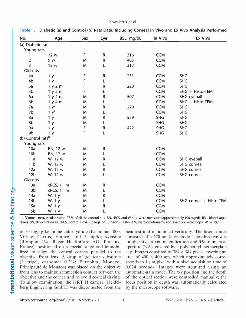
All animals were treated in accordance with theARVO Statement for the Use of Animals inOphthalmic and Vision Research. Twelve-week-old(n ¼ 3) and 1-year-old (n ¼ 6) GK rats thatspontaneously developed diabetes mellitus, from ourfacilities, were included and compared to age-matchednondiabetic rats from our facilities or purchased(Janvier; Le Genest-Saint-Isle, France) and roomedfor 1 week before inclusion in the study. Rat data andperformed analysis are given in Table 1.
Human Corneas
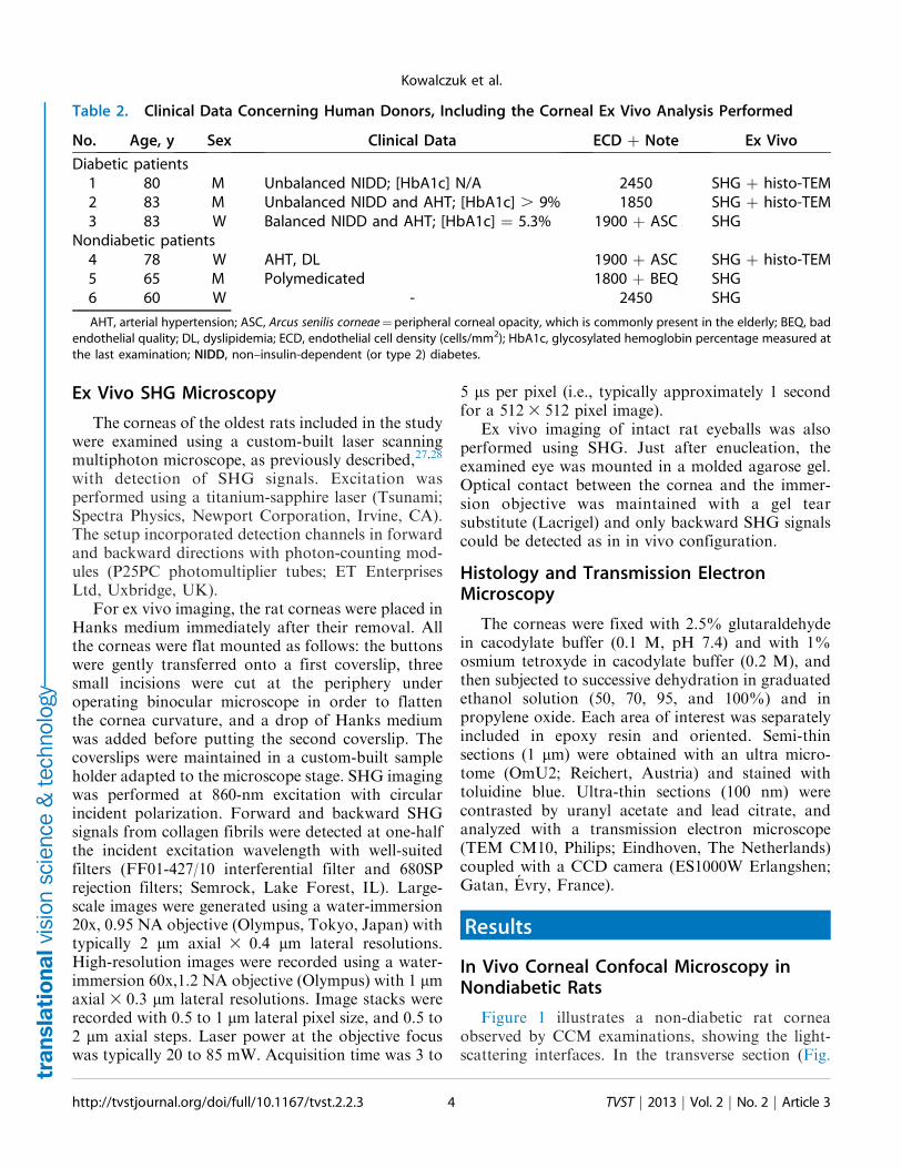
We obtained six human corneas assigned toscientific use,29 from an eye bank association (BanqueFrancaise des Yeux; Paris, France). Human corneaswere used under informed consent of the relatives,according to French bioethics laws. The study wasconducted according to the tenets of the Declarationof Helsinki for biomedical research involving humancorneas. Mean donor age was 75 years (3 women, 3men; age range, 60–83 years). As described in Table 2,three nondiabetic patients and three patients withtype 2 diabetes were included. Corneas were excisedwith 16- to 18-mm-diameter trephination, and imme-diately stored in 100 mL organ culture medium(CorneaMax; Eurobio, Courtabœuf, France) at 318Cin a dry incubator until the experiment, at least 2weeks later. Due to this storage, all corneas from bothdiabetic and nondiabetic donors were slightly edem-atous as demonstrated by their increased thickness(typically 700 to 900 lm) compared to physiologiccorneal thickness (approximately 500 lm).
In Vivo Corneal Confocal Microscopy
A confocal laser scanning system (HeidelbergRetina Tomograph II with Rostock Cornea Module[HRT II]; Heidelberg Engineering GmbH, Heidel-berg, Germany) was used to examine the rat corneasas previously described.30 Before in vivo CCMexamination, rats were anesthetized with a mixture
http://tvstjournal.org/doi/full/10.1167/tvst.2.2.3 TVST j 2013 j Vol. 2 j No. 2 j Article 32
Kowalczuk et al.
of 50 mg/kg ketamine chlorhydrate (Ketamine 1000,Virbac; Carros, France) and 5 mg/kg xylasine(Rompun 2%, Bayer HealthCare AG; Puteaux,France), positioned on a special stage and immobi-lized to align the central cornea parallel to theobjective front lens. A drop of gel tear substitute(Lacrigel, carbomer 0.2%; Europhta, Monaco,Principaute de Monaco) was placed on the objectivefront lens to maintain immersion contact between theobjective and the cornea and to avoid corneal drying.To allow examination, the HRT II camera (Heidel-berg Engineering GmbH) was disconnected from the
headrest and maintained vertically. The laser sourceconsisted of a 670 nm laser diode. The objective wasan objective at x60 magnification and 0.90 numericalaperture (NA), covered by a polymethyl methacrylatecap. Images consisted of 3843 384 pixels covering anarea of 400 3 400 lm, which approximately corre-sponds to 1 lm/pixel with a pixel acquisition time of0.024 seconds. Images were acquired using anautomatic-gain mode. The x-y position and the depthof the optical section were controlled manually; thefocus position in depth was automatically calculatedby the microscope software.
Table 1. Diabetic (a) and Control (b) Rats Data, Including Corneal In Vivo and Ex Vivo Analysis Performed
No. Age Sex Eye BSL, mg/dL In Vivo Ex Vivo
(a) Diabetic rats
Young rats
1 12 w F R 316 CCM
2 9 w M R 405 CCM
3 12 w M L 317 CCM
Old rats
4a 1 y F R 231 CCM SHG
4b 1 y F L CCM SHG
5a 1 y 2 m F R 220 CCM SHG
5b 1 y 2 m F L CCM SHG þ Histo-TEM
6a 1 y 4 m M R 507 CCM SHG eyeball
6b 1 y 4 m M L CCM SHG þ Histo-TEM
7a 1 ya M R 220 CCM SHG
7b 1 ya M L CCM SHG
8a 1 y M R 559 SHG SHG
8b 1 y M L SHG SHG
9a 1 y F R 422 SHG SHG
9b 1 y F L SHG SHG
(b) Control ratsb
Young rats
10a BN, 12 w M R CCM
10b BN, 12 w M L CCM
11a W, 12 w M R CCM SHG eyeball
11b W, 12 w M L CCM SHG cornea
12a W, 12 w M R CCM SHG cornea
12b W, 12 w M L CCM SHG cornea
Old rats
13a cRCS, 11 m M R CCM
13b cRCS, 11 m M L CCM
14a W, 1 y M R CCM
14b W, 1 y M L CCM SHG cornea þ Histo-TEM
15a W, 1 y M R CCM
15b W, 1 y M L CCMaCorneal neovascularization; bBSL of all the control rats–BN, cRCS, and W rats–were measured approximately 100 mg/dL. BSL, blood sugar
levels; BN, Brown-Norway; cRCS, control Royal College of Surgeons; Histo-TEM, histology-transmission electron microscopy; W, Wistar.
http://tvstjournal.org/doi/full/10.1167/tvst.2.2.3 TVST j 2013 j Vol. 2 j No. 2 j Article 33
Kowalczuk et al.
Ex Vivo SHG Microscopy
The corneas of the oldest rats included in the studywere examined using a custom-built laser scanningmultiphoton microscope, as previously described,27,28
with detection of SHG signals. Excitation wasperformed using a titanium-sapphire laser (Tsunami;Spectra Physics, Newport Corporation, Irvine, CA).The setup incorporated detection channels in forwardand backward directions with photon-counting mod-ules (P25PC photomultiplier tubes; ET EnterprisesLtd, Uxbridge, UK).
For ex vivo imaging, the rat corneas were placed inHanks medium immediately after their removal. Allthe corneas were flat mounted as follows: the buttonswere gently transferred onto a first coverslip, threesmall incisions were cut at the periphery underoperating binocular microscope in order to flattenthe cornea curvature, and a drop of Hanks mediumwas added before putting the second coverslip. Thecoverslips were maintained in a custom-built sampleholder adapted to the microscope stage. SHG imagingwas performed at 860-nm excitation with circularincident polarization. Forward and backward SHGsignals from collagen fibrils were detected at one-halfthe incident excitation wavelength with well-suitedfilters (FF01-427/10 interferential filter and 680SPrejection filters; Semrock, Lake Forest, IL). Large-scale images were generated using a water-immersion20x, 0.95 NA objective (Olympus, Tokyo, Japan) withtypically 2 lm axial 3 0.4 lm lateral resolutions.High-resolution images were recorded using a water-immersion 60x,1.2 NA objective (Olympus) with 1 lmaxial3 0.3 lm lateral resolutions. Image stacks wererecorded with 0.5 to 1 lm lateral pixel size, and 0.5 to2 lm axial steps. Laser power at the objective focuswas typically 20 to 85 mW. Acquisition time was 3 to
5 ls per pixel (i.e., typically approximately 1 secondfor a 5123 512 pixel image).
Ex vivo imaging of intact rat eyeballs was alsoperformed using SHG. Just after enucleation, theexamined eye was mounted in a molded agarose gel.Optical contact between the cornea and the immer-sion objective was maintained with a gel tearsubstitute (Lacrigel) and only backward SHG signalscould be detected as in in vivo configuration.
Histology and Transmission ElectronMicroscopy
The corneas were fixed with 2.5% glutaraldehydein cacodylate buffer (0.1 M, pH 7.4) and with 1%osmium tetroxyde in cacodylate buffer (0.2 M), andthen subjected to successive dehydration in graduatedethanol solution (50, 70, 95, and 100%) and inpropylene oxide. Each area of interest was separatelyincluded in epoxy resin and oriented. Semi-thinsections (1 lm) were obtained with an ultra micro-tome (OmU2; Reichert, Austria) and stained withtoluidine blue. Ultra-thin sections (100 nm) werecontrasted by uranyl acetate and lead citrate, andanalyzed with a transmission electron microscope(TEM CM10, Philips; Eindhoven, The Netherlands)coupled with a CCD camera (ES1000W Erlangshen;Gatan, Evry, France).
Results
In Vivo Corneal Confocal Microscopy inNondiabetic Rats
Figure 1 illustrates a non-diabetic rat corneaobserved by CCM examinations, showing the light-scattering interfaces. In the transverse section (Fig.
Table 2. Clinical Data Concerning Human Donors, Including the Corneal Ex Vivo Analysis Performed
No. Age, y Sex Clinical Data ECD þ Note Ex Vivo
Diabetic patients
1 80 M Unbalanced NIDD; [HbA1c] N/A 2450 SHG þ histo-TEM
2 83 M Unbalanced NIDD and AHT; [HbA1c] . 9% 1850 SHG þ histo-TEM
3 83 W Balanced NIDD and AHT; [HbA1c] ¼ 5.3% 1900 þ ASC SHG
Nondiabetic patients
4 78 W AHT, DL 1900 þ ASC SHG þ histo-TEM
5 65 M Polymedicated 1800 þ BEQ SHG
6 60 W - 2450 SHG
AHT, arterial hypertension; ASC, Arcus senilis corneae¼peripheral corneal opacity, which is commonly present in the elderly; BEQ, bad
endothelial quality; DL, dyslipidemia; ECD, endothelial cell density (cells/mm2); HbA1c, glycosylated hemoglobin percentage measured at
the last examination; NIDD, non–insulin-dependent (or type 2) diabetes.
http://tvstjournal.org/doi/full/10.1167/tvst.2.2.3 TVST j 2013 j Vol. 2 j No. 2 j Article 34
Kowalczuk et al.
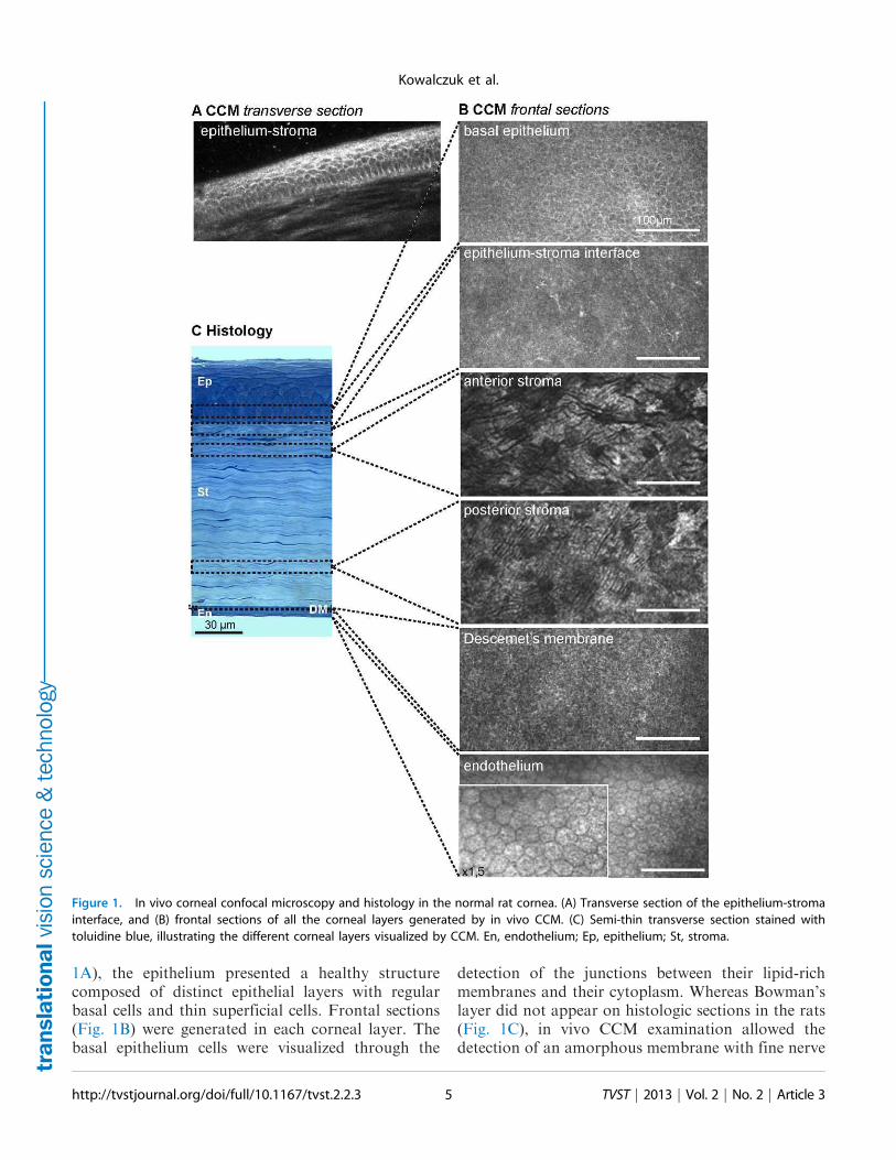
1A), the epithelium presented a healthy structure
composed of distinct epithelial layers with regularbasal cells and thin superficial cells. Frontal sections
(Fig. 1B) were generated in each corneal layer. Thebasal epithelium cells were visualized through the
detection of the junctions between their lipid-rich
membranes and their cytoplasm. Whereas Bowman’slayer did not appear on histologic sections in the rats
(Fig. 1C), in vivo CCM examination allowed thedetection of an amorphous membrane with fine nerve
Figure 1. In vivo corneal confocal microscopy and histology in the normal rat cornea. (A) Transverse section of the epithelium-stroma
interface, and (B) frontal sections of all the corneal layers generated by in vivo CCM. (C) Semi-thin transverse section stained with
toluidine blue, illustrating the different corneal layers visualized by CCM. En, endothelium; Ep, epithelium; St, stroma.
http://tvstjournal.org/doi/full/10.1167/tvst.2.2.3 TVST j 2013 j Vol. 2 j No. 2 j Article 35
Kowalczuk et al.
plexi (Fig. 1B). In the stroma, CCM allowed the
detection of the keratocytes. In the posterior cornea,
the DM appeared amorphous and the hexagonal
endothelial cells, with a 20 lm regular diameter,exhibited a honeycomb pattern.
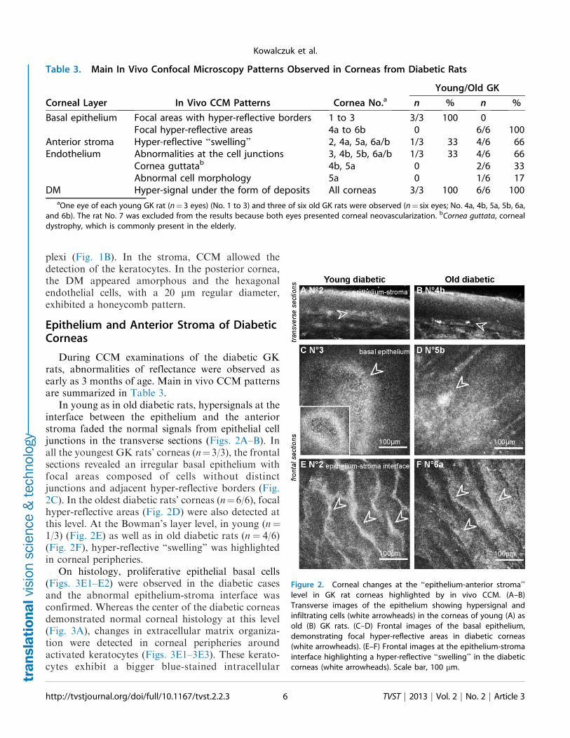
Epithelium and Anterior Stroma of DiabeticCorneas
During CCM examinations of the diabetic GK
rats, abnormalities of reflectance were observed as
early as 3 months of age. Main in vivo CCM patternsare summarized in Table 3.
In young as in old diabetic rats, hypersignals at the
interface between the epithelium and the anterior
stroma faded the normal signals from epithelial celljunctions in the transverse sections (Figs. 2A–B). In
all the youngest GK rats’ corneas (n¼3/3), the frontal
sections revealed an irregular basal epithelium with
focal areas composed of cells without distinct
junctions and adjacent hyper-reflective borders (Fig.
2C). In the oldest diabetic rats’ corneas (n¼6/6), focalhyper-reflective areas (Fig. 2D) were also detected at
this level. At the Bowman’s layer level, in young (n¼1/3) (Fig. 2E) as well as in old diabetic rats (n ¼ 4/6)
(Fig. 2F), hyper-reflective ‘‘swelling’’ was highlighted
in corneal peripheries.
On histology, proliferative epithelial basal cells
(Figs. 3E1–E2) were observed in the diabetic cases
and the abnormal epithelium-stroma interface was
confirmed. Whereas the center of the diabetic corneas
demonstrated normal corneal histology at this level(Fig. 3A), changes in extracellular matrix organiza-
tion were detected in corneal peripheries around
activated keratocytes (Figs. 3E1–3E3). These kerato-
cytes exhibit a bigger blue-stained intracellular
Table 3. Main In Vivo Confocal Microscopy Patterns Observed in Corneas from Diabetic Rats
Corneal Layer In Vivo CCM Patterns Cornea No.aYoung/Old GK
n % n %
Basal epithelium Focal areas with hyper-reflective borders 1 to 3 3/3 100 0
Focal hyper-reflective areas 4a to 6b 0 6/6 100
Anterior stroma Hyper-reflective ‘‘swelling’’ 2, 4a, 5a, 6a/b 1/3 33 4/6 66
Endothelium Abnormalities at the cell junctions 3, 4b, 5b, 6a/b 1/3 33 4/6 66
Cornea guttatab 4b, 5a 0 2/6 33
Abnormal cell morphology 5a 0 1/6 17
DM Hyper-signal under the form of deposits All corneas 3/3 100 6/6 100aOne eye of each young GK rat (n¼ 3 eyes) (No. 1 to 3) and three of six old GK rats were observed (n¼ six eyes; No. 4a, 4b, 5a, 5b, 6a,
and 6b). The rat No. 7 was excluded from the results because both eyes presented corneal neovascularization. bCornea guttata, corneal
dystrophy, which is commonly present in the elderly.
Figure 2. Corneal changes at the ‘‘epithelium-anterior stroma’’
level in GK rat corneas highlighted by in vivo CCM. (A–B)
Transverse images of the epithelium showing hypersignal and
infiltrating cells (white arrowheads) in the corneas of young (A) as
old (B) GK rats. (C–D) Frontal images of the basal epithelium,
demonstrating focal hyper-reflective areas in diabetic corneas
(white arrowheads). (E–F) Frontal images at the epithelium-stroma
interface highlighting a hyper-reflective ‘‘swelling’’ in the diabetic
corneas (white arrowheads). Scale bar, 100 lm.
http://tvstjournal.org/doi/full/10.1167/tvst.2.2.3 TVST j 2013 j Vol. 2 j No. 2 j Article 36
Kowalczuk et al.
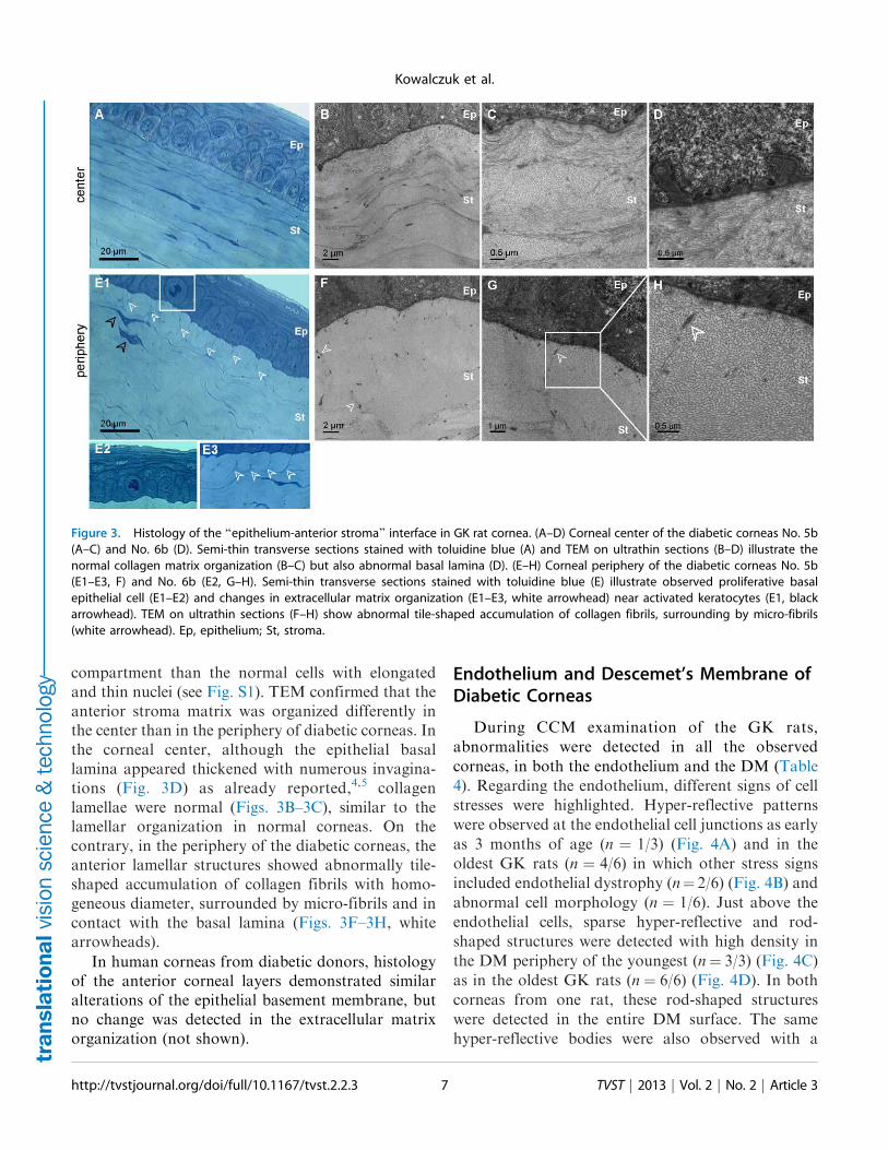
compartment than the normal cells with elongated
and thin nuclei (see Fig. S1). TEM confirmed that the
anterior stroma matrix was organized differently in
the center than in the periphery of diabetic corneas. In
the corneal center, although the epithelial basal
lamina appeared thickened with numerous invagina-
tions (Fig. 3D) as already reported,4,5 collagen
lamellae were normal (Figs. 3B–3C), similar to the
lamellar organization in normal corneas. On the
contrary, in the periphery of the diabetic corneas, the
anterior lamellar structures showed abnormally tile-
shaped accumulation of collagen fibrils with homo-
geneous diameter, surrounded by micro-fibrils and in
contact with the basal lamina (Figs. 3F–3H, white
arrowheads).
In human corneas from diabetic donors, histology
of the anterior corneal layers demonstrated similar
alterations of the epithelial basement membrane, but
no change was detected in the extracellular matrix
organization (not shown).
Endothelium and Descemet’s Membrane ofDiabetic Corneas
During CCM examination of the GK rats,
abnormalities were detected in all the observed
corneas, in both the endothelium and the DM (Table
4). Regarding the endothelium, different signs of cell
stresses were highlighted. Hyper-reflective patterns
were observed at the endothelial cell junctions as early
as 3 months of age (n ¼ 1/3) (Fig. 4A) and in the
oldest GK rats (n ¼ 4/6) in which other stress signs
included endothelial dystrophy (n¼ 2/6) (Fig. 4B) and
abnormal cell morphology (n ¼ 1/6). Just above the
endothelial cells, sparse hyper-reflective and rod-
shaped structures were detected with high density in
the DM periphery of the youngest (n¼ 3/3) (Fig. 4C)
as in the oldest GK rats (n ¼ 6/6) (Fig. 4D). In both
corneas from one rat, these rod-shaped structures
were detected in the entire DM surface. The same
hyper-reflective bodies were also observed with a
Figure 3. Histology of the ‘‘epithelium-anterior stroma’’ interface in GK rat cornea. (A–D) Corneal center of the diabetic corneas No. 5b
(A–C) and No. 6b (D). Semi-thin transverse sections stained with toluidine blue (A) and TEM on ultrathin sections (B–D) illustrate the
normal collagen matrix organization (B–C) but also abnormal basal lamina (D). (E–H) Corneal periphery of the diabetic corneas No. 5b
(E1–E3, F) and No. 6b (E2, G–H). Semi-thin transverse sections stained with toluidine blue (E) illustrate observed proliferative basal
epithelial cell (E1–E2) and changes in extracellular matrix organization (E1–E3, white arrowhead) near activated keratocytes (E1, black
arrowhead). TEM on ultrathin sections (F–H) show abnormal tile-shaped accumulation of collagen fibrils, surrounding by micro-fibrils
(white arrowhead). Ep, epithelium; St, stroma.
http://tvstjournal.org/doi/full/10.1167/tvst.2.2.3 TVST j 2013 j Vol. 2 j No. 2 j Article 37
Kowalczuk et al.
lesser extent in periphery of two corneas from oldcontrol rats (n¼ 2/6) (Fig. 4E).
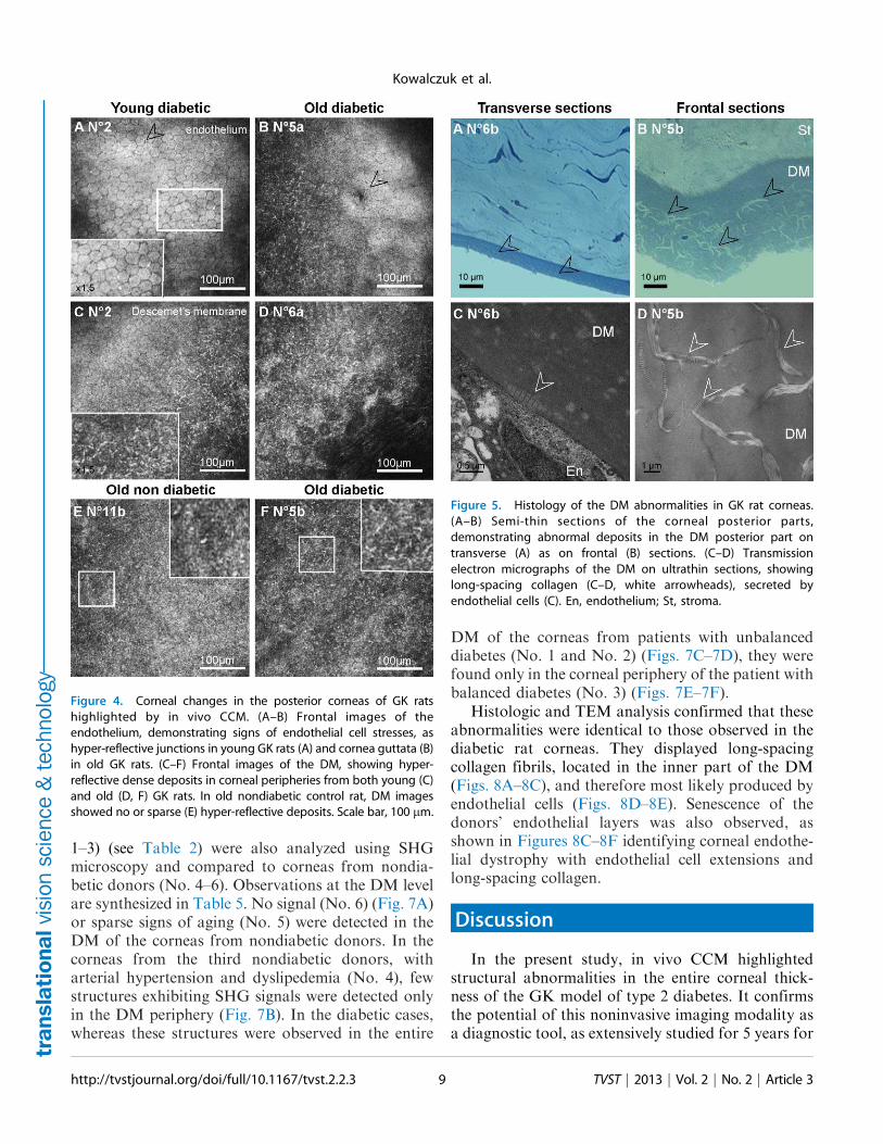
On histology, toluidine blue staining confirmed thepresence of abnormal deposits in the DM posteriorpart of the diabetic corneas (Figs. 5A–5B) but did notallow identifying their composition. This was per-formed during TEM analysis that revealed thepresence of long-spacing collagen fibrils at the samelevel (Figs. 5C–5D). In all the micrographs, thesepathologic collagen fibrils were distributed in theposterior part of the DM, and some of them wereobserved just near the endothelial cells as illustratedin Figure 5C, suggesting their secretion by these cells.
We then used ex vivo SHG microscopy to probespecifically these pathologic collagen fibrils with alarger field of view than in TEM and to characterizetheir 3D distribution in the DM. In the normalhuman cornea, ex vivo SHG microscopy reflects thedistribution of the type 1 collagen in the stroma, whilethe nonfibrillar collagen constituting the DM does
not exhibit SHG signals.27 In the DM of the GK ratcorneas, the hyper-reflective structures highlighted byin vivo CCM exhibited strong SHG signals, while, asexpected, other regions of the DM showed no signal.Their SHG-pattern was the same as previouslydescribed. The structures had a weak density in twoold control corneas (Fig. 6A) and were very dense inall diabetic cases (Fig. 6B). Moreover, acquisition ofimage stacks with 0.5 lm axial steps enabled 3Dreconstruction of DM abnormalities with excellentcontrast (Fig. 6C) (see Movie for 3D view). WhileSHG observations of flat-mounted corneas wereperformed using forward-detected SHG signals, theproof of feasibility of the abnormalities’ detectionfrom the epithelium was carried out in rat eyeballs.Figure 6D demonstrates that the structures in the DMof a diabetic rat cornea exhibited backward SHGsignals with the same high contrast, allowing epide-tection of these structures.
Three human corneas from diabetic patients (No.
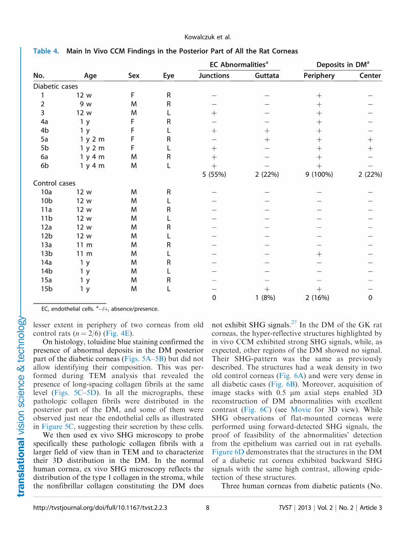
Table 4. Main In Vivo CCM Findings in the Posterior Part of All the Rat Corneas
No. Age Sex Eye
EC Abnormalitiesa Deposits in DMa
Junctions Guttata Periphery Center
Diabetic cases
1 12 w F R � � þ �2 9 w M R � � þ �3 12 w M L þ � þ �4a 1 y F R � � þ �4b 1 y F L þ þ þ �5a 1 y 2 m F R � þ þ þ5b 1 y 2 m F L þ � þ þ6a 1 y 4 m M R þ � þ �6b 1 y 4 m M L þ � þ �
5 (55%) 2 (22%) 9 (100%) 2 (22%)
Control cases
10a 12 w M R � � � �10b 12 w M L � � � �11a 12 w M R � � � �11b 12 w M L � � � �12a 12 w M R � � � �12b 12 w M L � � � �13a 11 m M R � � � �13b 11 m M L � � þ �14a 1 y M R � � � �14b 1 y M L � � � �15a 1 y M R � � � �15b 1 y M L � þ þ �
0 1 (8%) 2 (16%) 0
EC, endothelial cells. a�/þ, absence/presence.
http://tvstjournal.org/doi/full/10.1167/tvst.2.2.3 TVST j 2013 j Vol. 2 j No. 2 j Article 38
Kowalczuk et al.
1–3) (see Table 2) were also analyzed using SHG
microscopy and compared to corneas from nondia-
betic donors (No. 4–6). Observations at the DM level
are synthesized in Table 5. No signal (No. 6) (Fig. 7A)
or sparse signs of aging (No. 5) were detected in the
DM of the corneas from nondiabetic donors. In the
corneas from the third nondiabetic donors, with
arterial hypertension and dyslipedemia (No. 4), few
structures exhibiting SHG signals were detected only
in the DM periphery (Fig. 7B). In the diabetic cases,
whereas these structures were observed in the entire
DM of the corneas from patients with unbalanced
diabetes (No. 1 and No. 2) (Figs. 7C–7D), they were
found only in the corneal periphery of the patient with
balanced diabetes (No. 3) (Figs. 7E–7F).
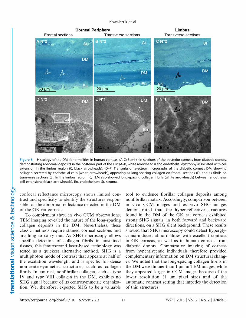
Histologic and TEM analysis confirmed that these
abnormalities were identical to those observed in the
diabetic rat corneas. They displayed long-spacing
collagen fibrils, located in the inner part of the DM
(Figs. 8A–8C), and therefore most likely produced by
endothelial cells (Figs. 8D–8E). Senescence of the
donors’ endothelial layers was also observed, as
shown in Figures 8C–8F identifying corneal endothe-
lial dystrophy with endothelial cell extensions and
long-spacing collagen.
Discussion
In the present study, in vivo CCM highlighted
structural abnormalities in the entire corneal thick-
ness of the GK model of type 2 diabetes. It confirms
the potential of this noninvasive imaging modality as
a diagnostic tool, as extensively studied for 5 years for
Figure 4. Corneal changes in the posterior corneas of GK rats
highlighted by in vivo CCM. (A–B) Frontal images of the
endothelium, demonstrating signs of endothelial cell stresses, as
hyper-reflective junctions in young GK rats (A) and cornea guttata (B)
in old GK rats. (C–F) Frontal images of the DM, showing hyper-
reflective dense deposits in corneal peripheries from both young (C)
and old (D, F) GK rats. In old nondiabetic control rat, DM images
showed no or sparse (E) hyper-reflective deposits. Scale bar, 100 lm.
Figure 5. Histology of the DM abnormalities in GK rat corneas.
(A–B) Semi-thin sections of the corneal posterior parts,
demonstrating abnormal deposits in the DM posterior part on
transverse (A) as on frontal (B) sections. (C–D) Transmission
electron micrographs of the DM on ultrathin sections, showing
long-spacing collagen (C–D, white arrowheads), secreted by
endothelial cells (C). En, endothelium; St, stroma.
http://tvstjournal.org/doi/full/10.1167/tvst.2.2.3 TVST j 2013 j Vol. 2 j No. 2 j Article 39
Kowalczuk et al.
diabetic neuropathy.31,32 For this purpose, in vivo
CCM is currently used in both preclinical33 and
clinical34 studies. The contrast mechanism of this
system relies on spatial variations of refractive indices,
like in light-scattering interfaces, as junctions between
cytoplasm or extracellular fluid and lipid-rich mem-
branes of cells, nucleus and mitochondria.35 CCM
allows easy recognition of the corneal nerves and, so,
the development of a new algorithm to perform image
rebuilding of the sub-basal nerve plexus.36 However,
Figure 6. Specific detection of the DM abnormalities using ex
vivo SHG microscopy in diabetic rat corneas. (A–B) False color
images generated by forward-detection of SHG signals in the flat-
mounted corneas from 1-year old control (A, 20x objective) and
diabetic (B, 60x objective) rats. (C) 3D reconstruction of the
structures forward-detected in the GK cornea No. 7. (D) False color
images generated by backward-detection of SHG signals in a
diabetic rat eyeball. DM, Descemet’s membrane; St, stroma.
Movie. 3D SHG view of the DM of the GK cornea No. 7 in false
colors (200 3 200 3 9 lm3). Strong SHG signals are emitted by
fibrillar collagen abnormalities, which allows their spatial
characterization. Note that the fibrils in the foreground appear
larger than in the background because of the 3D viewer.
Table 5. Main Ex Vivo SHG Findings in the DM of
Human Corneas
Patient Age, y Sex
Deposits in DM
Periphery Center
Diabetic patient
1. 80 M þþ þ2. 83 M þþþ þ3. 83 W þ �
Nondiabetic patient
4. 78 W þþþ N/A
5. 65 M þ �6. 60 W � �
�, absence; þ, few; þþ, great concentration; þþþ, covering the
whole field of view. N/A, no available data.
Figure 7. Ex vivo SHG images in the DM of human corneas. (A–B)
False color images of the corneas from nondiabetic donors,
showing sparse SHG signals in corneal periphery of one
hypertensive patient (B, in green; 20x objective). (C–F) False
color images of the corneas from two diabetic donors,
demonstrating the presence of SHG signals (in green) in the
entire DM in the case of unbalanced diabetes (C–D; 60x objective)
and only in the DM periphery in the case of balanced diabetes (E–
F; 20x objective). Scale bar, 20 lm.
http://tvstjournal.org/doi/full/10.1167/tvst.2.2.3 TVST j 2013 j Vol. 2 j No. 2 j Article 310
Kowalczuk et al.
confocal reflectance microscopy shows limited con-
trast and specificity to identify the structures respon-
sible for the abnormal reflectance detected in the DM
of the GK rat corneas.
To complement these in vivo CCM observations,
TEM imaging revealed the nature of the long-spacing
collagen deposits in the DM. Nevertheless, these
classic methods require stained corneal sections and
are long to carry out. As SHG microscopy allows
specific detection of collagen fibrils in unstained
tissues, this femtosecond laser-based technology was
tested as a quickest alternative method. SHG is a
multiphoton mode of contrast that appears at half of
the excitation wavelength and is specific for dense
non-centrosymmetric structures, such as collagen
fibrils. In contrast, nonfibrillar collagen, such as type
IV and type VIII collagen in the DM, exhibits no
SHG signal because of its centrosymmetric organiza-
tion. We, therefore, expected SHG to be a valuable
tool to evidence fibrillar collagen deposits among
nonfibrillar matrix. Accordingly, comparison between
in vivo CCM images and ex vivo SHG images
demonstrated that the hyper-reflective structures
found in the DM of the GK rat corneas exhibited
strong SHG signals, in both forward and backward
directions, on a SHG silent background. These results
showed that SHG microscopy could detect hypergly-
cemia-induced abnormalities with excellent contrast
in GK corneas, as well as in human corneas from
diabetic donors. Comparative imaging of corneas
from hyperglycemic individuals therefore provided
complementary information on DM structural chang-
es. We noted that the long-spacing collagen fibrils in
the DM were thinner than 1 lm in TEM images, while
they appeared larger in CCM images because of the
lower resolution (1 lm pixel size) and of the
automatic contrast setting that impedes the detection
of thin structures.
Figure 8. Histology of the DM abnormalities in human corneas. (A-C) Semi-thin sections of the posterior corneas from diabetic donors,
demonstrating abnormal deposits in the posterior part of the DM (A–B, white arrowheads) and endothelial dystrophy associated with cell
extension in the limbus region (C, black arrowheads). (D–F) Transmission electron micrographs of the diabetic corneas DM, showing
collagen secreted by endothelial cells (white arrowheads), appearing as long-spacing collagen on frontal sections (D) and as fibrils on
transverse sections (E). In the limbus region (F), TEM also showed long-spacing collagen fibrils (white arrowheads) between endothelial
cell extensions (black arrowheads). En, endothelium; St, stroma.
http://tvstjournal.org/doi/full/10.1167/tvst.2.2.3 TVST j 2013 j Vol. 2 j No. 2 j Article 311
Kowalczuk et al.
Backward-detected SHG imaging has been dem-onstrated in ex vivo ocular globes and could thusprovide a diagnosis for chronic hyperglycemia-induced effects in the corneal tissue. First develop-ments toward a clinical imaging system have beenreported recently28 and showed that in vivo SHGimaging can generate quantitative information aboutthe collagen architecture in the stroma. This femto-second laser-based imaging system could thus provideadditional diagnosis of structural abnormalities in thestroma, observed in other corneal pathologies.
From a pathophysiologic point of view, this studyhas highlighted focal abnormal reflectance in thebasal epithelium of diabetic corneas that is a sign ofdiabetic keratopathy. On histology, numerous epithe-lial basal cells were in a proliferative state, as reportedby Wakuta et al.l9 The increased proliferation ofepithelial cells in the cornea of GK rats wasdemonstrated to be associated with a delay in woundhealing and, thus, to affect the epithelial barrierfunction. These features were met again in thestreptozotocin-induced model of type 1 diabetes,37
and in diabetic patients using the fluorescein test6 orin vivo CCM.8 The well-known epithelial basementmembrane abnormality of diabetic corneas3–5 wasalso observed in our study, in both rat and humancorneas. The increased reflectivity measured at theinterface between the epithelial basal cell layer and theanterior stroma by Morishige et al39 and Takahashi etal40 could explain the hyper-reflectance detected onthe CCM transverse images of the GK rat epitheliumsgenerated in our experiments. From a biochemicalpoint of view, the abnormalities noticed at theepithelium-stroma interface in the cornea of GK ratsmight reflect the chronic hyperglycemia-inducedeffects, in particular the Maillard reactions. Thesereactions result in the formation of AGEs, which areresponsible for non-enzymatic glycation reactions onthe extracellular matrix proteins. Their effects in-crease with aging41 and are amplified by diabetes.42
Increased protein glycations12–14 and oxidativestress43 are involved in diabetic cornea alterations,such as decreased stability in the epithelial basementmembrane and changes in the collagen matrix of theanterior stroma. However, whether AGEs havecytotoxic effects mediated by the AGE receptors(RAGE) on corneal keratocytes remains to bestudied.
Regarding the posterior layers of the diabeticcorneas, various endothelial abnormalities, in partic-ular endothelial dystrophy and abnormal cell mor-phology, were noticed in the corneas of the oldest GK
rats. Corneal observations in diabetic patients usingCCM or specular microscopy have actually shownthat diabetes may disturb the stromal hydrationthrough decreased endothelial cell density,17 inparticular after cataract surgery,18 and polymegath-ism (changes in their size).19,20 The endothelium maythus be under greater metabolic stress in diabetic thanin healthy subjects. Endothelial cells are known to besensitive to AGEs44 and to express their receptors.45
Therefore, AGE formation may be responsible fornon-enzymatic glycation, leading to endothelial cellloss,45 but also for the RAGE pathway activation.This may induce changes in gene expression inendothelial cells and, so, the abnormal secretion oflong-spacing collagen in the DM of diabetic rat16,15
and human corneas.5 These pathologic collagen fibrilswere detected as hyper-reflective and rod-shapedstructures in the DM and in the endothelial celljunctions using in vivo CCM, and then, TEM whichallowed imaging their synthesis by the endothelialcells. Blood sugar levels revealed the hyperglycemicstate of the observed GK rats; however, they are notindicative of the metabolic effects of diabetes. Forsuch a purpose, glycosylated hemoglobin (HbA1c)should be measured. Even if this indicator was notfollowed at the time of our experiments, thepreliminary data currently measured in our facilitiesshow that HbA1c levels start to increase before 3months of age in GK rats in comparison with Wistarrats. Recent papers46,47 have also demonstrated that,whereas HbA1c slightly increased from 4 to 5% at 12weeks and then reached a plateau in Wistar rats,HbA1c levels in GK rats showed a delayed increaserelative to glucose at 4 weeks, and then graduallyincreased until the end of the studies, from 4 to 5%, to9% at 12 weeks46 or to over 11% at 20 weeks of age.47
These data allowed us to state that the depositsobserved in the DM at 12 weeks of age in GK ratcorneas were synthesized as soon as the diabetesinduced metabolic effects.
Moreover, these abnormalities were observed inhuman corneas from diabetic donors, with higherdensity in the cases of unbalanced diabetes, notably inthe cornea from a patient with a HbA1c level over9%, than in balanced diabetes, in the case of a donorwith a normal HbA1c level. These collagen depositshave also been observed in the corneas fromhypertensive old patients in our study, and reportedin other endothelial pathologies, such as iridocorneal-endothelial syndrome48,49 and Fuch’s dystrophy,48–51
in which AGEs accumulate as well as oxidativestress.52 Diabetes-induced tissue changes have been
http://tvstjournal.org/doi/full/10.1167/tvst.2.2.3 TVST j 2013 j Vol. 2 j No. 2 j Article 312
Kowalczuk et al.
characterized as accelerated aging in many organs,and also in collagen changes.53 Our observations let
us hypothesize that the abnormal collagen synthesis isan age-related phenomenon observed earlier andexacerbated in diabetic conditions and, probably, in
other metabolic diseases inducing an oxidative stresson corneal endothelial cells.
To conclude, we have used complementary imagingtechniques to demonstrate that diabetes alters thestructure of all corneal layers. In diabetic rat corneas,
we have mainly highlighted structural abnormalities atthe epithelium-stroma interface and fibrillar collagendeposits in the DM. This latter feature has also been
observed and characterized in human corneas usingSHGmicroscopy, and confirmed by TEM. Subclinicalabnormalities could predispose diabetic patients to
wound healing delay and also serve as early signs ofdiabetes-induced tissue structure modifications. Ourobservations in the DM demonstrated that diabetes
altered the collagen structure of this layer, with theformation of long-spacing collagen fibrils synthesized
by corneal endothelial cells. Whether these changescould be correlated to blood–sugar-level controlremains to be demonstrated. Moreover, we have
shown that SHG microscopy, an emerging multipho-ton imaging method that is highly specific for collagenfibrils, could provide a diagnosis for chronic hyper-
glycemia-induced effects in the corneal tissue. Furtherstudies are required to evaluate if diabetes-inducedcorneal collagen alterations can predict the develop-
ment of diabetic retinopathy.
Acknowledgments
The authors would like to thank Isabelle Souratiand Patrick Sabatier (Banque Francaise des Yeux,
Paris), Jean-Marc Legeais (Sorbonne Paris Cite, ParisDescartes University; AP-HP Hotel-Dieu Hospital,Department of Ophthalmology), and Patricia Crisanti
(INSERM UMRS 872, Team17: Physiopathology ofocular diseases, therapeutic innovations).
Laura Kowalczuk was supported by the ‘‘Agence
Nationale de la Recherche’’, ANR-08-TecSan-012‘‘NOUGAT.’’ Gael Latour was supported by
‘‘RTRA-Triangle de la Physique’’ and ‘‘Fondationde France - Fondation Berthe Fouassier.’’ The workat LOB was supported by ANR-10-INBS-04.
Disclosure: L. Kowalczuk, None; G. Latour, None;J.-L. Bourges, None; M. Savoldelli, None; J.-C.
Jeanny, None; K. Plamann, None; M.-C. Schanne-
Klein, None; F. Behar-Cohen, None
References
1. Brownlee M. Biochemistry and molecular cellbiology of diabetic complications. Nature.2001(6865);14:813–820.
2. Omri S, Behar-Cohen F, de Kozak Y, et al.Microglia/macrophages migrate through retinalepithelium barrier by a transcellular route indiabetic retinopathy: role of PKCf in the GotoKakizaki rat model. Am J Pathol. 2011(2);179:942–953.
3. Schultz RO, Van Horn DL, Peters MA, KlewinKM, Schutten WH. Diabetic keratopathy. TransAm Ophthalmol Soc. 1981;79:180–199.
4. Azar DT, Spurr-Michaud SJ, Tisdale AS, GipsonIK. Altered epithelial-basement membrane inter-actions in diabetic corneas. Arch Ophthalmol.1992(4);110:537–540.
5. Rehany U, Ishii Y, Lahav M, Rumelt S.Ultrastructural changes in corneas of diabeticpatients: an electron-microscopy study. Cornea.2000(4);19:534–538.
6. Gekka M, Miyata K, Nagai Y, et al. Cornealepithelial barrier function in diabetic, 110 pa-tients. Cornea. 2004(1);23:35–37.
7. Chang PY, Carrel H, Huang JS, et al. Decreaseddensity of corneal basal epithelium and subbasalcorneal nerve bundle changes in patients withdiabetic retinopathy. Am J Ophthalmol. 2006(3);142:488–490.
8. Chen WL, Lin CT, Ko PS, et al. In vivo confocalmicroscopic findings of corneal wound healingafter corneal epithelial debridement in diabeticvitrectomy. Ophthalmology. 2009(6);116:1038–1047.
9. Wakuta M, Morishige N, Chikama T, Seki K,Nagano T, Nishida T. Delayed wound closureand phenotypic changes in corneal epithelium ofthe spontaneously diabetic Goto-Kakizaki rat.Invest Ophthalmol Vis Sci. 2007(2);48:590–596.
10. Kaji Y, Usui T, Oshika T, et al. Advancedglycation end products in diabetic corneas. InvestOphthalmol Vis Sci. 2000(2);41:362–368.
11. McDermott AM, Xiao TL, Kern TS, Murphy CJ.Non-enzymatic glycation in corneas from normaland diabetic donors and its effects on epithelialcell attachment in vitro. Optometry. 2003(7);74:443–452.
12. Sady C, Khosrof S, Nagaraj R. AdvancedMaillard reaction and crosslinking of corneal
http://tvstjournal.org/doi/full/10.1167/tvst.2.2.3 TVST j 2013 j Vol. 2 j No. 2 j Article 313
Kowalczuk et al.
collagen in diabetes. Biochem Biophys Res Com-mun. 1995(3);214:793–797.
13. Sato E, Mori F, Igarashi S, et al. Cornealadvanced glycation end products increase inpatients with proliferative diabetic retinopathy.Diabetes Care. 2001(3);24:479–482.
14. Akimoto Y, Kawakami H, Yamamoto K, Mu-netomo E, Hida T, Hirano H. Elevated expres-sion of O-GlcNAc-modified proteins and O-GlcNAc transferase in corneas of diabetic Goto-Kakizaki rats. Invest Ophthalmol Vis Sci. 2003(9);44:3802–3809.
15. Rehany U, Ishii Y, Lahav M, Rumelt S. Collagenpleomorphism in Descemet’s membrane of strep-tozotocin-induced diabetic rats: an electron mi-croscopy study. Cornea. 2000(3);19:390–392.
16. Akimoto Y, Sawada H, Ohara-Imaizumi M,Nagamatsu S, Kawakami H. Change in long-spacing collagen in Descemet’s membrane ofdiabetic Goto-Kakizaki rats and its suppressionby antidiabetic agents. Exp Diabetes Res. 2008;2008:818341. doi: 10.1155/2008/818341.
17. Shenoy R, Khandekar R, Bialasiewicz A, AlMuniri A. Corneal endothelium in patients withdiabetes mellitus: a historical cohort study. Eur JOphthalmol. 2009(3);19:369–375.
18. Mathew PT, David S, Thomas N. Endothelial cellloss and central corneal thickness in patients withand without diabetes after manual small incisioncataract surgery. Cornea. 2011(4);30:424–428.
19. Lee JS, Oum BS, Choi HY, Lee JE, Cho BM.Differences in corneal thickness and cornealendothelium related to duration in diabetes. Eye(Lond). 2006(3);20:315–318.
20. Gonzalez-Meijome JM, Jorge J, Queiros A,Peixoto-de-Matos SC, Parafita MA. Two singledescriptors of endothelial polymegethism andpleomorphism. Graefes Arch Clin Exp Ophthal-mol. 2010(8);248:1159–1166.
21. Zipfel WR, Williams RM, Webb WW. Nonlinearmagic: multiphoton microscopy in the bioscienc-es. Nat Biotechnol. 2003(11);21:1369–1377.
22. Yeh AT, Nassif N, Zoumi A, Tromberg BJ.Selective corneal imaging using combined second-harmonic generation and two-photon excitedfluorescence. Opt Lett. 2002(23);27:2082–2084.
23. Han M, Giese G, Bille JF. Second harmonicgeneration imaging of collagen fibrils in corneaand sclera. Opt. Express. 2005(15);13:5791–5797.
24. Teng SW, Tan HY, Peng JL, et al. Multiphotonautofluorescence and second-harmonic genera-tion imaging of the ex vivo porcine eye. InvestOphthalmol Vis Sci. 2006(3);47:5251–5259.
25. Morishige N, Wahlert AJ, Kenney MC, et al.Second-harmonic imaging microscopy of normalhuman and keratoconus cornea. Invest Ophthal-mol Vis Sci. 2007(3);48:1087–1094.
26. Steven P, Hovakimyan M, Guthoff RF,Huttmann G, Stachs O. Imaging corneal cross-linking by autofluorescence 2-photon microsco-py, second harmonic generat ion, andfluorescence lifetime measurements. J CataractRefract Surg. 2010(10);36:2150–2159.
27. Aptel F, Olivier N, Deniset-Besseau A, et al.Multimodal nonlinear imaging of the humancornea. Invest Ophthalol Vis Sci. 2010(5);51:2459–2465.
28. Latour G, Gusachenko I, Kowalczuk L, LamarreI, Schanne-Klein M-C. In vivo structural imagingof the cornea by polarization-resolved secondharmonic microscopy. Biomed Opt Express.2012(1);3:1–15.
29. European Eye Bank Association (EEBA): EEBADirectory. 18th ed. EEBA, eds. Amsterdam,2010.
30. Labbe A, Liang H, Martin C, Brignole-BaudouinF, Warnet JM, Baudouin C. Comparative anat-omy of laboratory animal corneas with a new-generation high-resolution in vivo confocal mi-croscope. Curr Eye Res. 2006(6);31:501–509.
31. Schultz RO, Peters MA, Sobocinski K, Nassif K,Schultz KJ. Diabetic corneal neuropathy. TransAm Ophthalmol Soc. 1983;81:107–124.
32. Pritchard N, Edwards K, Shahidi AM, et al.Corneal markers of diabetic neuropathy. OculSurf. 2011(1);9:17–28.
33. Davidson EP, Coppey LJ, Holmes A, Yorek MA.Changes in corneal innervation and sensitivityand acetylcholine-mediated vascular relaxation ofthe posterior ciliary artery in a type 2 diabetic rat.Invest Ophthalmol Vis Sci. 2012, Epub ahead ofprint. doi: 10.1167/iovs.11-8806.
34. Tavakoli M, Kallinikos P, Iqbal A, et al. Cornealconfocal microscopy detects improvement incorneal nerve morphology with an improvementin risk factors for diabetic neuropathy. DiabetMed. 2011(10);28:1261–1267. doi: 10.1111/j.1464-5491.2011.03372.x.
35. Guthoff RF, Baudouin C, Stave J. Principles ofconfocal in vivo microscopy. In: Atlas of ConfocalLaser Scanning In-vivo Microscopy in Ophthal-mology. Heidelberg: Springer-Verlag Berlin Hei-delberg; 2006:3–22.
36. Allgeier S, Zhivov A, Eberle F, et al. Imagereconstruction of the subbasal nerve plexus within vivo confocal microscopy. Invest OphthalmolVis Sci. 2011(9); 52:5022–5028.
http://tvstjournal.org/doi/full/10.1167/tvst.2.2.3 TVST j 2013 j Vol. 2 j No. 2 j Article 314
Kowalczuk et al.
37. Yin J, Huang J, Chen C, Gao N, Wang F, Yu FS.Corneal complications in streptozocin-inducedtype I diabetic rats. Invest Ophthalmol Vis Sci.2011(9); 52:6589–6596. doi: 10.1167/iovs.11-7709.
38. Taylor HR, Kimsey RA. Corneal epithelialbasement membrane changes in diabetes. InvestOphthalmol Vis Sci. 1981(4);20:548–553.
39. Morishige N, Chikama TI, Sassa Y, Nishida T.Abnormal light scattering detected by confocalbiomicroscopy at the corneal epithelial basementmembrane of subjects with type II diabetes.Diabetologia. 2001(3);44:340–345.
40. Takahashi N, Wakuta M, Morishige N, ChikamaT, Nishida T, Sumii Y. Development of aninstrument for measurement of light scatteringat the corneal epithelial basement membrane indiabetic patients. Jpn J Ophthalmol. 2007(3);51:185–190.
41. Malik NS, Moss SJ, Ahmed N, Furth AJ, WallRS, Meek KM. Aging of the human cornealstroma: structural and biochemical changes.Biochim Biophys Acta. 1992(3);1138:222–228.
42. Stitt AW. The maillard reaction in eye diseases.Ann N Y Acad Sci. 2005;1043:582–597.
43. Kim J, Kim CS, Sohn E, Jeong IH, Kim H, KimJS. Involvement of advanced glycation endproducts, oxidative stress and nuclear factor-kappaB in the development of diabetic keratop-athy. Graefes Arch Clin Exp Ophthalmol. 2011(4);249:529–536.
44. Kaji Y, Amano S, Usui T, et al. Advancedglycation end products in Descemet’s membraneand their effect on corneal endothelial cell. CurrEye Res. 2001(6); 23:469–477.
45. Kaji Y, Amano S, Usui T, et al. Expression andfunction of receptors for advanced glycation endproducts in bovine corneal endothelial cells.Invest Ophthalmol Vis Sci. 2003(2);44:521–528.
46. Gao W, Bihorel S, DuBois DC, Almon RR,Jusko WJ. Mechanism-based disease progressionmodeling of type 2 diabetes in Goto-Kakizakirats. J Pharmacokinet Pharmacodyn. 2011(1);38:143–162.
47. Xue B, Sukumaran S, Nie J, Jusko WJ, DuboisDC, Almon RR. Adipose tissue deficiency andchronic inflammation in diabetic Goto-Kakizakirats. PLoS One. 2011(2);6:e17386.
48. Levy SG, McCartney AC, Sawada H, Dopping-Hepenstal PJ, Alexander RA, Moss J. Descemet’smembrane in the iridocorneal-endothelial syn-drome: morphology and composition. Exp EyeRes. 1995(3);61:323–333.
49. Levy SG, Moss J, Sawada H, Dopping-HepenstalPJ, McCartney AC. The composition of wide-spaced collagen in normal and diseased Desce-met’s membrane. Curr Eye Res. 1996(1);15:45–52.
50. Zhang C, Bell WR, Sundin OH, et al. Immuno-histochemistry and electron microscopy of early-onset fuchs corneal dystrophy in three cases withthe same L450W COL8A2 mutation. Trans AmOphthalmol Soc. 2006;104:85–97.
51. Gottsch JD, Zhang C, Sundin OH, Bell WR,Stark WJ, Green WR. Fuchs corneal dystrophy:aberrant collagen distribution in an L450Wmutant of the COL8A2 gene. Invest OphthalmolVis Sci. 2005(12);46:4504–4511.
52. Wang Z, Handa JT, Green WR, Stark WJ,Weinberg RS, Jun AS. Advanced glycation endproducts and receptors in Fuchs’ dystrophycorneas undergoing Descemet’s stripping withendothelial keratoplasty. Ophthalmology. 2007(8);1453:114–1460.
53. Hamlin CR, Kohn RR, Luschin JH. Apparentaccelerated aging of human collagen in diabetesmellitus. Diabetes. 1975(10);24:902–904.
http://tvstjournal.org/doi/full/10.1167/tvst.2.2.3 TVST j 2013 j Vol. 2 j No. 2 j Article 315
Kowalczuk et al.