Embed Size (px)
Citation preview
Multigeneration Reproductive ToxicityAssessment of 60-Hz Magnetic Fields Usinga Continuous Breeding Protocol in RatsBERNADETTE M. RYAN, 1* RICHARD R. SYMANSKI,1 LISA E. POMERANZ,1TIM R. JOHNSON,2 JAMES R. GAUGER,2 AND DAVID L. MCCORMICK1
1Life Sciences Department, IIT Research Institute, Chicago, Illinois 606162Electromagnetic and Electronic Systems Department, IIT Research Institute, Chicago, Illinois 60616
ABSTRACT Male and female reproductivefunctions have been proposed as possibly sensitivetargets for the biological effects of 60-Hz (powerfrequency) magnetic fields (MF). However, experimentaldata relevant to this hypothesized association are verylimited. In the present study, the ‘‘reproductive assess-ment by continuous breeding’’ design was used toidentify possible effects of MF exposure on reproduc-tive performance, fetal development, and early postna-tal growth in rats. Groups of age-matched Sprague-Dawley rats (40 breeding pairs/group) were exposedcontinuously (18.5 hr per day) to linearly polarized,transient-free 60-Hz MF at field strengths of 0 Gauss(G; sham control), 0.02 G, 2.0 G, or 10.0 G. Anadditional group of 40 breeding pairs received intermit-tent (1 hr on/1 hr off) exposure to 10.0 G fields. F0breeding pairs were exposed to MF or sham fields for 1week prior to mating, during a 14-week period ofcohabitation, and during a 3-week holding period aftercohabitation. The duration of the cohabitation periodwas selected to be sufficient for the delivery of five littersin the sham control group. Pups from the final F1 litterfrom each breeding pair were exposed to MF or shamfields until sexual maturity, were cohabitated in MF orsham fields for 7 days with nonsiblings from the sameexposure group, and were held in the MF or sham fieldsfor 22 days to permit delivery of F2 pups for evaluation.No evidence of exposure-related toxicity was identifiedin any rat in the F0, F1, or F2 generations. Fetal viabilityand body weights in all litters of groups exposed to MFwere comparable to those of sham controls. No signifi-cant differences between sham controls and MF-exposed groups were seen in any measure of reproduc-tive performance (litters/breeding pair, percent fertilepairs, latency to parturition, litter size, or sex ratio) ineither the F0 or F1 generation. Exposure of Sprague-Dawley rats to 60-Hz MF strengths of up to 10.0 Geither during their peak reproductive period (F0) orduring gestation and throughout their life span (F1) hasno biologically significant effects on reproductive perfor-mance. These results do not support the hypothesisthat exposure to pure, linearly polarized 60-Hz MF is asignificant reproductive or developmental toxicant. Tera-tology 59:156–162, 1999. r 1999 Wiley-Liss, Inc.
Magnetic fields (MF) generated by the production,transmission, and use of electricity are ubiquitous inindustrialized society. Although the public often associ-ates exposure to MF only with residential proximity toelectrical substations, transformers, and overheadpower transmission and distribution lines, significantlocal sources of MF include a wide range of electricaldevices and equipment in common use in both indus-trial and residential environments. In addition to elec-trical equipment used in industry, common sources ofextremely low-frequency magnetic fields include officeequipment such as video display terminals and laserprinters, and a wide range of common household appli-ances, including vacuum cleaners, toasters, electricshavers, hair dryers, and electric blankets (Gauger,’85). Because magnetic field intensity decreases withdistance from the source, MF measured in close proxim-ity to point sources of relatively low intensity (e.g.,household appliances, televisions, video display moni-tors) often exceed residential fields associated withoverhead power lines (Gauger, ’85; Bracken, ’93; Kavet,’95).
An important concern related to the possible healtheffects of human exposure to power frequency (50 and60 Hz) MF is their potential to induce adverse reproduc-tive outcomes (teratogenicity and/or impaired fertility).A significant experimental and epidemiological database is developing on which the possible risk of humandevelopmental toxicity associated with MF exposurecan be evaluated. The weight of the available evidencesuggests a lack of developmental effects associated within utero exposure limited to the period of organogen-esis. We previously reported on an extensive evaluationof the potential teratogenicity of 60-Hz magnetic fieldsin Sprague-Dawley rats (Ryan et al., ’96). The results ofour developmental toxicity study, when considered withthe results of other teratology bioassays (Rommereim
Grant sponsor: National Toxicology Program, National Institute ofEnvironmental Sciences, Department of Health and Human Services;Grant number: NO1-ES-25351.
*Correspondence to: Bernadette M. Ryan, Ph.D., Life Sciences Depart-ment, IIT Research Institute, 10 West 35th St., Chicago, IL 60616.
Received 4 May 1998; Accepted 3 November 1998
TERATOLOGY 59:156–162 (1999)
r 1999 WILEY-LISS, INC.

et al., ’96; reviews by Chernoff et al., ’92: Brent et al.,’93), suggest that exposure to MF is unlikely to pose asignificant risk to the developing fetus. By contrast,however, comparable data to support evaluations of thepossible relationship between MF exposure and alter-ations in reproductive performance and capacity arelargely unavailable. As a result, the potential reproduc-tive hazards that may be associated with exposures oflonger duration are still under debate. The lack ofconsensus concerning the possible reproductive toxicityof MF suggests a need for critical evaluation of theexisting data base, and emphasizes the requirementthat the biological effects of MF be evaluated systemati-cally, using well-studied and fully validated experimen-tal model systems, study designs of adequate statisticalpower, and an MF exposure environment that is exten-sively characterized and continuously monitored. It istoward these goals that the study described herein wasdesigned and conducted.
MATERIALS AND METHODS
Animals and animal husbandry
Male and female Sprague-Dawley rats (8 weeks oldat time of receipt) were purchased from Taconic Farms(Germantown, NY), and were held in quarantine for10–14 days prior to randomization into groups andinitiation of experimentation. Throughout the quaran-tine and exposure periods, rats were housed in polycar-bonate cages on certified hardwood bedding (BetaChipst, Northeastern Products, Warrensburg, NY). Dur-ing the quarantine period and for the first week of MFexposure (prior to cohabitation), rats were housed (bysex) in groups of 5/cage. During the cohabitation period,one male and one female were housed together; afterthe cohabitation period, females were single-housed topermit parturition, and males were again housed 5/cage.Rats had free access to certified NIH-07 open formuladiet (Zeigler Bros., Gardners, PA) and City of Chicagodrinking water at all times during the study.
Environmental conditions (temperature, humidity,and air flows) in all animal holding areas were moni-tored continuously. Temperature was maintained in therange of 72 6 3°F, and relative humidity was main-tained in the range of 35–65%. Fluorescent lighting wasprovided on a 12-hr light/dark cycle. Fluorescent lightballasts were located remotely in order that the mag-netic fields generated by the ballasts would not inter-fere with the MF environment in animal rooms.
Experimental design
The ‘‘reproductive assessment by continuous breed-ing’’ (RACB) protocol includes four related tasks thatare designed to evaluate fertility and reproductivecapacity; the requirement to conduct specific taskswithin the protocol is determined on the basis of thepresence or absence of preliminary range-finding data,and by breeding performance in other tasks. The pres-ent study included task 2 (continuous cohabitation
phase) and task 4 (assessment of F1 offspring). Task 1(range-finding) was not performed; MF levels evaluatedin these studies were selected on the basis of engineer-ing considerations and our previous findings that sub-chronic exposure to MF strengths of up to 10.0 Gauss(G) induces no significant toxicity in rats (Boorman etal., ’97). On the basis of the results of tasks 2 and 4, task3 (crossover mating) was determined to be unnecessary,and was not performed.
The range of MF flux densities used in this program(20 mG–10.0 G) was selected by a consensus panelconvened by the National Toxicology Program (Depart-ment of Health and Human Services). MF strengthswere selected on the basis of their relationship tohuman MF exposures, and on the basis of engineeringconsiderations. The highest MF intensity used, 10.0 G(10,000 mG), is a 5,000–20,000-fold multiple of the60-Hz MF strengths to which humans are routinelyexposed in residential environments (Kaune et al., ’87).Ten Gauss was also selected as a reasonable upper limitto MF intensities that could be generated experimen-tally without the induction of possible confounders suchas noise, vibration, and local temperature changes inthe exposure array. The lowest field strength used, 20mG, was selected to provide a 10–40-fold multiple ofresidential environmental exposures (Kaune et al., ’87);MF flux densities of 20 mG are also encountered insome industrial environments.
Groups of age-matched Sprague-Dawley rats (40breeding pairs/group) were randomized into experimen-tal groups. After randomization, all rats designated toreceive MF or sham exposure were transferred from thequarantine/breeding room to the appropriate exposureroom and were exposed continuously (18.5 hr per day)to linearly polarized, transient-free 60-Hz MF at fieldstrengths of 0 G (sham control), 0.02 G, 2.0 G, or 10.0 G.An additional group of 40 breeding pairs receivedintermittent (1 hr on/1 hr off) exposure to 10.0-G fields.Sham control breeding pairs were handled and housedin a manner identical to that of the MF-exposed pairs.Exposure modules (excluding modules in the shamcontrol room) were energized between the hours of11:00 AM–3:00 PM, C.S.T., and 4:30 PM–7:00 AM, C.S.T.;exposure system downtime allowed for routine hus-bandry operations and animal observation. Rats in theF0 (parental) generation were exposed to MF or shamfields for 18.5 hr/day for 1 week prior to mating, for 14weeks of cohabitation, and for a 3-week holding periodafter cohabitation. The 14-week period of cohabitationwas selected as sufficient to permit the production of 5litters per breeding pair in sham controls; the 3-weekholding period was designed to permit dams to undergonatural parturition.
In order to facilitate evaluation of offspring, exposureinitiation and cohabitation were staggered in all groups;10 rats/sex/group (10 F0 breeding pairs/group) wereentered into the study each day for 4 consecutive days.After 1 week of exposure in which rats were housedseparately by sex, cohabitation was accomplished by
REPRODUCTIVE TOXICITY ASSESSMENT OF MAGNETIC FIELDS 157
placing a single female rat from a given exposure groupinto the cage of a male from the same exposure group;breeding pairs were cohabitated for 14 weeks. Duringthe period of cohabitation, all offspring produced wereremoved from the cage, counted, sexed, weighed, andeuthanized by decapitation on the day that deliverywas complete (postnatal day 0).
Offspring delivered during the postcohabitation hold-ing period remained with their mothers until postnatalday 21; these pups were then weaned and used for task4 (F1 offspring assessment). After weaning, the finallitter from each breeding pair was randomly culled totwo males and two females. At sexual maturity (81 6 10days), 40 F1 female rats from each exposure group wererandomly assigned to cohabitation with 40 F1 nonsib-lings from the same exposure group. After a 7-daycohabitation period, F1 pairs were separated and heldfor 22 days, and females were observed for evidence ofparturition. At delivery, F2 pups were removed, counted,sexed, weighed, and euthanized via decapitation.
Magnetic field exposure and monitoringinstrumentation
The MF generation and monitoring equipment hasbeen described in great detail (Gauger et al., ’99; Ryanet al., ’96). Briefly, the MF generation and monitoringequipment was designed and constructed in collabora-tion with Electric Research and Management, Inc.(Pittsburgh, PA). The MF exposure system consisted offive identical field-generating coil sets, one located ineach of five identical animal exposure rooms. MF weremonitored continuously throughout the studies using aMultiwave (data collection and analysis system (Elec-tric Research and Management, Inc.).
Clinical observations and data collection
MF- and sham-exposed rats were observed for mortal-ity or evidence of moribundity on a daily basis duringquarantine, and twice daily during MF exposure peri-ods. In addition, pregnant dams were monitored aminimum of once daily to evaluate delivery status.Data for delivery status and offspring weights werecollected during the morning shutdown of the MFexposure system (7:00–11:00 AM, C.S.T.).
F0 dams were weighed during week 1 of exposure,postpartum (after delivery of each litter), at the end ofthe 14-week cohabitation period, and prior to terminalsacrifice. During the holding period after cohabitation,F1 offspring and maternal body weights were collectedon postnatal days 0, 4, 7, 14, and 21. F1 rats used in theoffspring assessment were weighed at the start ofmating and prior to terminal sacrifice.
Food consumption by the F0 (parental) generationwas monitored during week 1 (prior to cohabitation).During the postcohabitation holding period, maternalfood consumption was monitored during postnatal days0–4, 4–7, 7–14, and 14–21, and during the week prior tonecropsy.
Litter evaluation
On the day that delivery was completed for each litter(postnatal day 0), offspring were removed, counted,sexed, weighed, examined for gross external abnormali-ties, and euthanized by decapitation. The number oflitters produced per breeding pair, the number andpercent fertile pairs per group, the number and percentlive pups per litter, sex ratio, and mean body weight ofthe live offspring (by sex, and total) were determined.
Postmortem examination procedures
At the termination of each task protocol, rats fromthe F0 or F1 generation were euthanized by CO2 asphyxi-ation, and received a gross morphological examinationof the contents of the peritoneal and pleural cavities.The liver, kidneys, right testis, right epididymis, rightcauda epididymis, adrenals, prostate, seminal vesicles(containing their fluid and with coagulating glandsattached), and ovaries (without fallopian tubes) wereremoved, trimmed of adherent fat, weighed, and fixedin Bouin’s solution (testes and ovaries) or formalin. Thevagina, cervix, and uterus were collected and fixed informalin.
Statistical analysis
Statistical analysis of the data was performed usingthe breeding pair or litter as the independent samplingunit; for task 2, individual reproductive parameters foreach breeding pair were summed across litters. AKruskal-Wallis analysis of variance on ranks was usedto compare the number of litters per breeding pairamong treatment groups, and a one-way analysis ofvariance (ANOVA) or Kruskal-Wallis ANOVA was usedto compare number of days, interval between litters,and average interval in days until parturition wasobserved. A chi-square test was used to determineoverall significant differences in the number and per-cent fertile pairs among treatment groups.
The total number of newborn produced by a pair andthe overall mean body weight of live pups per pair wereanalyzed via one-way ANOVA using general linearmodels (GLM) procedures (Systat Version 5.0, SPSS,Inc., Chicago, IL). Overall mean body weights of livepups per pair were analyzed by covariate analysis,using litter size as the covariate. Prior to GLM analysis,a binomial arsine transformation was performed on thepercent live newborn per pair; transformed data werethen analyzed by a one-way ANOVA. Differences inparental mean body weight, food consumption, andorgan weights were analyzed by one-way ANOVA usingthe appropriate GLM. Intergroup pairwise compari-sons were performed using Tukey’s test. A significancelevel of P # 0.05 was used in all tests.
RESULTS
Magnetic field exposure conditions
Experimentally generated MF were within 2% of thetarget field strength at all times during the study.
158 B.M. RYAN ET AL.
During periods when exposure modules were ener-gized, mean MF strengths measured in the 0.02 G, 2.0G, and 10.0 G continuous, and 10.0 G intermittentexposure rooms were 0.020 G, 2.00 G, 9.93 G, and 9.89G, respectively. Mean MF strength in the sham controlexposure room was 0.0004 G (0.4 mG). MF strengths inthe quarantine room and in exposure rooms duringperiods when the exposure system was deenergizedranged from 0.0003 G (0.3 mG)–0.0009 G (0.9 mG).
Influence of magnetic field exposure on parentalsurvival and overall health
No evidence of toxicity associated with MF exposurewas observed in any study animal. Two F0 dams (one inthe 0.02 G group and one in the 10.0 G continuousgroup) died during the study; both deaths appeared tobe associated with complications during parturition. Inaddition, one sham control F0 male died during thepostcohabitation holding period when males had beenreturned to group housing. This death appeared to haveresulted from trauma inflicted by cagemates.
Nonspecific clinical signs were observed in low inci-dence in all experimental groups; none of the clinicalobservations were considered to be related to MFexposure. No differences in adult food consumption orin adult body weights were identified in comparisons ofMF- and sham-exposed groups in the F0 and F1 genera-
tions. At necropsy, a small but statistically significantincrease in absolute adrenal weight was seen in F1 malerats exposed intermittently to 10 G MF (0.052 6 0.001 gvs. 0.045 6 0.001 g in sham controls); relative adrenalweights were also significantly increased in male ratsin this group. However, because this increase wasquantitatively small, was not seen in male F1 rats inother MF exposure groups, was not seen in F1 femalesin any MF group, and was not seen in either sex in anyMF group in the F0 generation, this difference was notconsidered to be biologically significant. No other statis-tically significant differences in organ weights wereidentified. Consistent with the results of our previoussubchronic toxicity evaluation of MF (Boorman et al.,’97), gross pathology findings were unremarkable, andprovided no evidence of morphologic alterations associ-ated with MF exposure.
Influence of magnetic field exposure onreproductive function (F0 generation) and
offspring viability (F1 generation)
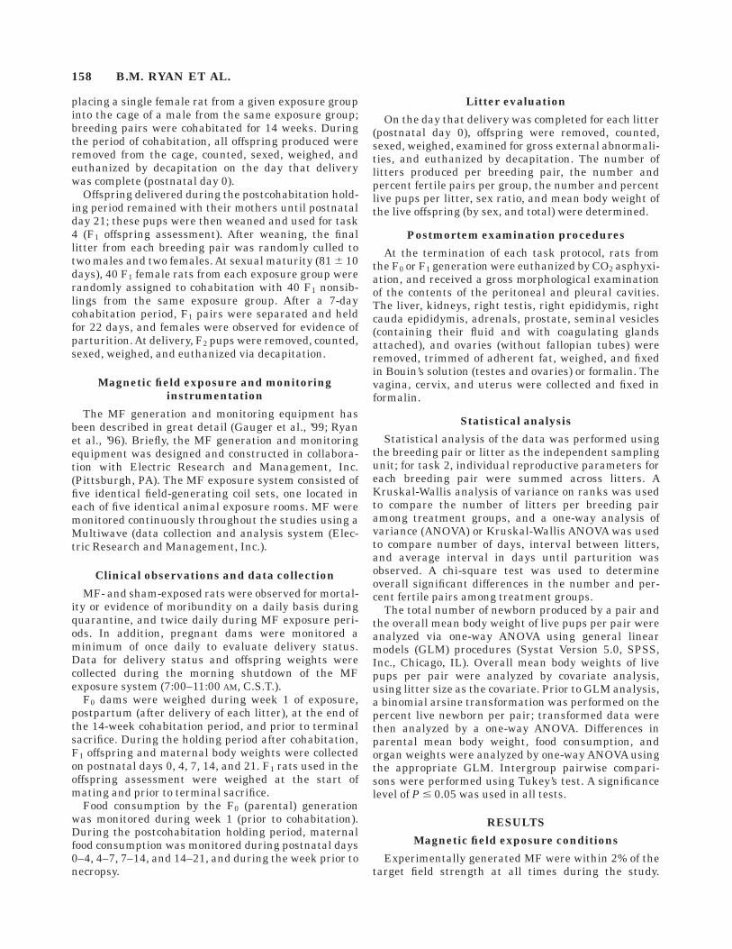
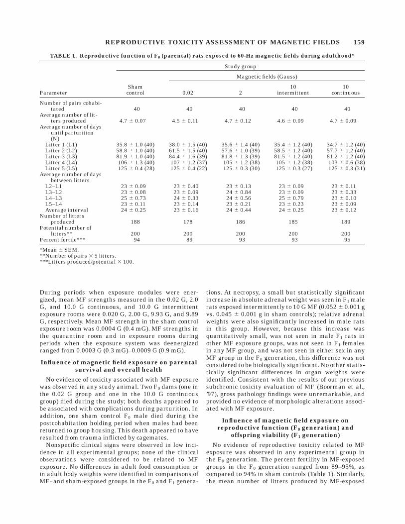
No evidence of reproductive toxicity related to MFexposure was observed in any experimental group inthe F0 generation. The percent fertility in MF-exposedgroups in the F0 generation ranged from 89–95%, ascompared to 94% in sham controls (Table 1). Similarly,the mean number of litters produced by MF-exposed
TABLE 1. Reproductive function of F0 (parental) rats exposed to 60-Hz magnetic fields during adulthood*
Parameter
Study group
Shamcontrol
Magnetic fields (Gauss)
0.02 210
intermittent10
continuous
Number of pairs cohabi-tated 40 40 40 40 40
Average number of lit-ters produced 4.7 6 0.07 4.5 6 0.11 4.7 6 0.12 4.6 6 0.09 4.7 6 0.09
Average number of daysuntil parturition(N)
Litter 1 (L1) 35.8 6 1.0 (40) 38.0 6 1.5 (40) 35.6 6 1.4 (40) 35.4 6 1.2 (40) 34.7 6 1.2 (40)Litter 2 (L2) 58.8 6 1.0 (40) 61.5 6 1.5 (40) 57.6 6 1.0 (39) 58.5 6 1.2 (40) 57.7 6 1.2 (40)Litter 3 (L3) 81.9 6 1.0 (40) 84.4 6 1.6 (39) 81.8 6 1.3 (39) 81.5 6 1.2 (40) 81.2 6 1.2 (40)Litter 4 (L4) 106 6 1.3 (40) 107 6 1.2 (37) 105 6 1.2 (38) 105 6 1.2 (38) 103 6 0.6 (38)Litter 5 (L5) 125 6 0.4 (28) 125 6 0.4 (22) 125 6 0.3 (30) 125 6 0.3 (27) 125 6 0.3 (31)
Average number of daysbetween litters
L2–L1 23 6 0.09 23 6 0.40 23 6 0.13 23 6 0.09 23 6 0.11L3–L2 23 6 0.08 23 6 0.09 24 6 0.84 23 6 0.09 23 6 0.33L4–L3 25 6 0.73 24 6 0.33 24 6 0.56 25 6 0.79 23 6 0.10L5–L4 23 6 0.11 23 6 0.14 23 6 0.21 23 6 0.23 23 6 0.09Average interval 24 6 0.25 23 6 0.16 24 6 0.44 24 6 0.25 23 6 0.12
Number of littersproduced 188 178 186 185 189
Potential number oflitters** 200 200 200 200 200
Percent fertile*** 94 89 93 93 95
*Mean 6 SEM.**Number of pairs 3 5 litters.***Litters produced/potential 3 100.
REPRODUCTIVE TOXICITY ASSESSMENT OF MAGNETIC FIELDS 159
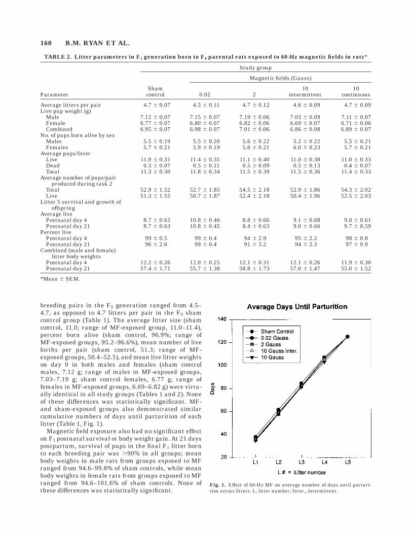
breeding pairs in the F0 generation ranged from 4.5–4.7, as opposed to 4.7 litters per pair in the F0 shamcontrol group (Table 1). The average litter size (shamcontrol, 11.0; range of MF-exposed group, 11.0–11.4),percent born alive (sham control, 96.9%; range ofMF-exposed groups, 95.2–96.6%), mean number of livebirths per pair (sham control, 51.3; range of MF-exposed groups, 50.4–52.5), and mean live litter weightson day 0 in both males and females (sham controlmales, 7.12 g; range of males in MF-exposed groups,7.03–7.19 g; sham control females, 6.77 g; range offemales in MF-exposed groups, 6.69–6.82 g) were virtu-ally identical in all study groups (Tables 1 and 2). Noneof these differences was statistically significant. MF-and sham-exposed groups also demonstrated similarcumulative numbers of days until parturition of eachlitter (Table 1, Fig. 1).
Magnetic field exposure also had no significant effecton F1 postnatal survival or body weight gain. At 21 dayspostpartum, survival of pups in the final F1 litter bornto each breeding pair was .90% in all groups; meanbody weights in male rats from groups exposed to MFranged from 94.6–99.8% of sham controls, while meanbody weights in female rats from groups exposed to MFranged from 94.6–101.6% of sham controls. None ofthese differences was statistically significant.
Fig. 1. Effect of 60-Hz MF on average number of days until parturi-tion across litters. L, litter number; Inter., intermittent.
TABLE 2. Litter parameters in F1 generation born to F0 parental rats exposed to 60-Hz magnetic fields in rats*
Parameter
Study group
Shamcontrol
Magnetic fields (Gauss)
0.02 210
intermittent10
continuous
Average litters per pair 4.7 6 0.07 4.5 6 0.11 4.7 6 0.12 4.6 6 0.09 4.7 6 0.09Live pup weight (g)
Male 7.12 6 0.07 7.15 6 0.07 7.19 6 0.06 7.03 6 0.09 7.11 6 0.07Female 6.77 6 0.07 6.80 6 0.07 6.82 6 0.06 6.69 6 0.07 6.71 6 0.06Combined 6.95 6 0.07 6.98 6 0.07 7.01 6 0.06 6.86 6 0.08 6.89 6 0.07
No. of pups born alive by sexMales 5.5 6 0.19 5.5 6 0.20 5.6 6 0.22 5.2 6 0.22 5.5 6 0.21Females 5.7 6 0.21 5.9 6 0.19 5.8 6 0.21 6.0 6 0.23 5.7 6 0.21
Average pups/litterLive 11.0 6 0.31 11.4 6 0.35 11.1 6 0.40 11.0 6 0.38 11.0 6 0.33Dead 0.3 6 0.07 0.5 6 0.11 0.5 6 0.09 0.5 6 0.13 0.4 6 0.07Total 11.3 6 0.30 11.8 6 0.34 11.5 6 0.39 11.5 6 0.36 11.4 6 0.33
Average number of pups/pairproduced during task 2
Total 52.9 6 1.52 52.7 6 1.85 54.5 6 2.18 52.9 6 1.86 54.3 6 2.02Live 51.3 6 1.55 50.7 6 1.87 52.4 6 2.18 50.4 6 1.96 52.5 6 2.03
Litter 5 survival and growth ofoffspring
Average livePostnatal day 4 8.7 6 0.62 10.8 6 0.46 8.8 6 0.66 9.1 6 0.68 9.8 6 0.61Postnatal day 21 8.7 6 0.63 10.8 6 0.45 8.4 6 0.63 9.0 6 0.66 9.7 6 0.59
Percent livePostnatal day 4 99 6 0.5 99 6 0.4 94 6 2.9 95 6 2.2 98 6 0.8Postnatal day 21 96 6 2.6 99 6 0.4 91 6 3.2 94 6 2.3 97 6 0.9
Combined (male and female)litter body weights
Postnatal day 4 12.2 6 0.26 12.0 6 0.25 12.1 6 0.31 12.1 6 0.26 11.9 6 0.30Postnatal day 21 57.4 6 1.71 55.7 6 1.38 58.8 6 1.73 57.0 6 1.47 55.0 6 1.52
*Mean 6 SEM.
160 B.M. RYAN ET AL.
Influence of magnetic field exposure onreproductive function (F1 generation) and
offspring viability (F2 generation)
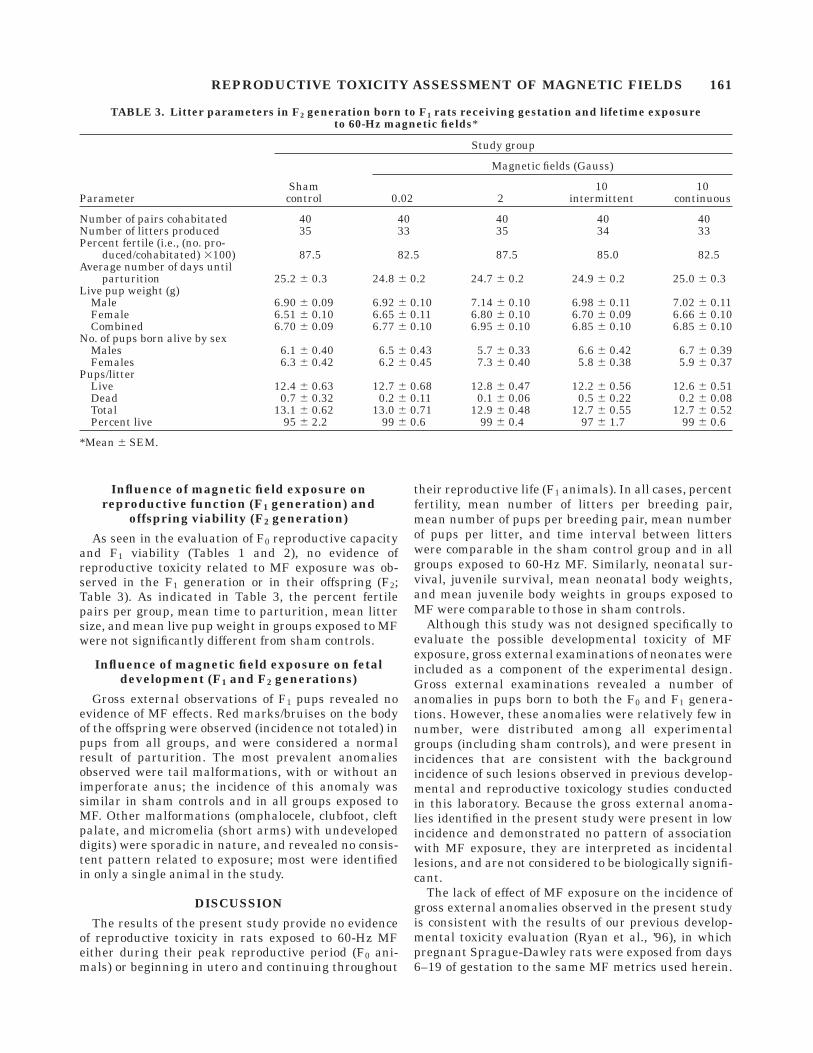
As seen in the evaluation of F0 reproductive capacityand F1 viability (Tables 1 and 2), no evidence ofreproductive toxicity related to MF exposure was ob-served in the F1 generation or in their offspring (F2;Table 3). As indicated in Table 3, the percent fertilepairs per group, mean time to parturition, mean littersize, and mean live pup weight in groups exposed to MFwere not significantly different from sham controls.
Influence of magnetic field exposure on fetaldevelopment (F1 and F2 generations)
Gross external observations of F1 pups revealed noevidence of MF effects. Red marks/bruises on the bodyof the offspring were observed (incidence not totaled) inpups from all groups, and were considered a normalresult of parturition. The most prevalent anomaliesobserved were tail malformations, with or without animperforate anus; the incidence of this anomaly wassimilar in sham controls and in all groups exposed toMF. Other malformations (omphalocele, clubfoot, cleftpalate, and micromelia (short arms) with undevelopeddigits) were sporadic in nature, and revealed no consis-tent pattern related to exposure; most were identifiedin only a single animal in the study.
DISCUSSION
The results of the present study provide no evidenceof reproductive toxicity in rats exposed to 60-Hz MFeither during their peak reproductive period (F0 ani-mals) or beginning in utero and continuing throughout
their reproductive life (F1 animals). In all cases, percentfertility, mean number of litters per breeding pair,mean number of pups per breeding pair, mean numberof pups per litter, and time interval between litterswere comparable in the sham control group and in allgroups exposed to 60-Hz MF. Similarly, neonatal sur-vival, juvenile survival, mean neonatal body weights,and mean juvenile body weights in groups exposed toMF were comparable to those in sham controls.
Although this study was not designed specifically toevaluate the possible developmental toxicity of MFexposure, gross external examinations of neonates wereincluded as a component of the experimental design.Gross external examinations revealed a number ofanomalies in pups born to both the F0 and F1 genera-tions. However, these anomalies were relatively few innumber, were distributed among all experimentalgroups (including sham controls), and were present inincidences that are consistent with the backgroundincidence of such lesions observed in previous develop-mental and reproductive toxicology studies conductedin this laboratory. Because the gross external anoma-lies identified in the present study were present in lowincidence and demonstrated no pattern of associationwith MF exposure, they are interpreted as incidentallesions, and are not considered to be biologically signifi-cant.
The lack of effect of MF exposure on the incidence ofgross external anomalies observed in the present studyis consistent with the results of our previous develop-mental toxicity evaluation (Ryan et al., ’96), in whichpregnant Sprague-Dawley rats were exposed from days6–19 of gestation to the same MF metrics used herein.
TABLE 3. Litter parameters in F2 generation born to F1 rats receiving gestation and lifetime exposureto 60-Hz magnetic fields*
Parameter
Study group
Shamcontrol
Magnetic fields (Gauss)
0.02 210
intermittent10
continuous
Number of pairs cohabitated 40 40 40 40 40Number of litters produced 35 33 35 34 33Percent fertile (i.e., (no. pro-
duced/cohabitated) 3100) 87.5 82.5 87.5 85.0 82.5Average number of days until
parturition 25.2 6 0.3 24.8 6 0.2 24.7 6 0.2 24.9 6 0.2 25.0 6 0.3Live pup weight (g)
Male 6.90 6 0.09 6.92 6 0.10 7.14 6 0.10 6.98 6 0.11 7.02 6 0.11Female 6.51 6 0.10 6.65 6 0.11 6.80 6 0.10 6.70 6 0.09 6.66 6 0.10Combined 6.70 6 0.09 6.77 6 0.10 6.95 6 0.10 6.85 6 0.10 6.85 6 0.10
No. of pups born alive by sexMales 6.1 6 0.40 6.5 6 0.43 5.7 6 0.33 6.6 6 0.42 6.7 6 0.39Females 6.3 6 0.42 6.2 6 0.45 7.3 6 0.40 5.8 6 0.38 5.9 6 0.37
Pups/litterLive 12.4 6 0.63 12.7 6 0.68 12.8 6 0.47 12.2 6 0.56 12.6 6 0.51Dead 0.7 6 0.32 0.2 6 0.11 0.1 6 0.06 0.5 6 0.22 0.2 6 0.08Total 13.1 6 0.62 13.0 6 0.71 12.9 6 0.48 12.7 6 0.55 12.7 6 0.52Percent live 95 6 2.2 99 6 0.6 99 6 0.4 97 6 1.7 99 6 0.6
*Mean 6 SEM.
REPRODUCTIVE TOXICITY ASSESSMENT OF MAGNETIC FIELDS 161
In that study, MF exposures were not associated withincreased incidences of gross external, visceral, skel-etal, or cephalic anomalies, or any biologically signifi-cant effect on fetal survival or body weight. Otherinvestigators have reported similar negative resultsfrom teratogenicity evaluations of MF (Rommereim etal., ’96).
The lack of parental toxicity observed in the presentstudy is also consistent with the results of our sub-chronic toxicity evaluations of MF in F344 rats andB6C3F1 mice (Boorman et al., ’97). In that study, youngadult rats and mice were exposed for 8 weeks to MFparameters that were identical to those used in thepresent study. MF exposure had no effect on survival,body weight gain, or a battery of hematology andclinical chemistry endpoints; furthermore, MF expo-sure induced no gross or microscopic pathology in any ofthe 50 tissues examined from each animal.
In the present study, only a single statistically signifi-cant difference was seen between sham controls andany group exposed to MF: absolute and relative adrenalweights in F1 male rats exposed intermittently to 10.0G fields were slightly but significantly increased fromadrenal weights seen in sham controls. This differencewas not seen in any other exposure group in the presentstudy; nor was it seen in male or female F0 rats or infemale F1 rats exposed intermittently to 10.0 G fields.Consistent with this lack of effect, no differences fromsham control adrenal weights were seen in any MF-exposed group in our previous subchronic toxicity evalu-ations (Boorman et al., ’97). On this basis, and becausethe observed change was quantitatively small, it isconsidered to be neither biologically significant norexposure-related.
The present study was conducted to address animportant data gap in our understanding of the pos-sible biological effects of MF. The experiment wasconducted using a well-defined animal model that hasbeen used widely to assess the reproductive and devel-opmental toxicity of chemical and physical agents(Gulati et al., ’91). Group sizes in the study wereincreased well beyond the standard for reproductivetoxicity evaluations in order to increase statisticalpower, and thereby increase the probability of detectingweak effects. It should be noted, however, that with itsfocus on pure, transient-free, linearly polarized MF, thedesign of the present study did not address the poten-tial adverse effects of MF exposure paradigms otherthan pure 60-Hz fields. As such, our results do notpreclude the possibility of adverse reproductive out-comes resulting from exposure to higher-order harmon-ics or magnetic field transients that are commonly
encountered in residential and occupational environ-ments.
The results of the present study do not support thehypothesis that exposure to 60-Hz MF has any signifi-cant adverse effects on adult reproductive capacity, thedeveloping fetus, or the neonate. On this basis, and inconsideration of the broad predictability of the experi-mental model used in these studies, it is concluded fromthese data that human exposure to pure 60-Hz MF isunlikely to pose significant risk to reproduction.
ACKNOWLEDGMENTS
The authors thank Tanya Bryan, Mary Harrington,and Ed Mallett for expert technical assistance, and Dr.Gary Boorman of the National Institute of Environmen-tal Health Sciences for his support and review of themanuscript.
LITERATURE CITEDBock RD. 1975. Multivariate statistical analysis in behavioral re-
search. New York: McGraw-Hill.Boorman GA, Gauger JR, Johnson TR, Tomlinson MJ, Findlay JC,
Travlos GS, McCormick DL. 1997. Eight-week toxicology study of 60Hz magnetic fields in F344 rats and B6C3F1 mice. Fundam ApplToxicol 35:55–63.
Bracken TD. 1993. Exposure assessment for power frequency electricand magnetic fields. Am Ind Hyg Assoc J 54:165–177.
Brent RL, Gordon WE, Bennett WR, Beckman DA. 1993. Reproductiveand teratologic effects of electromagnetic fields. Reprod Toxicol7:535–580.
Chernoff N, Rogers JM, Kavet R. 1992. A review of the literature onpotential reproductive and developmental toxicity of electric andmagnetic fields. Toxicology 74:91–126.
Gauger JR. 1985. Household appliance magnetic field survey. IEEETrans Power Apparatus Syst 104:2436–2445.
Gauger JR, Johnson TR, Stangel JE, Patterson RC, Williams DA,Harder JB, McCormick DL. 1999. Design, construction, and valida-tion of a large capacity rodent magnetic field exposure laboratory.Bioelectromagnetics (in press).
Gulati DK, Hope E, Teague J, Chapin RE. 1991. Reproductive toxicityassessment by continuous breeding in Sprague-Dawley rats: Acomparison of two study designs. Fundam Appl Toxicol 17:270–279.
Kaune WT, Stevens RG, Callahan NJ, Severson RK, Thomas DB.1987. Residential magnetic and electric fields. Bioelectromagnetics8:315–335.
Kavet R. 1995. Magnetic field exposure assessment. In: Blank M,editor. Electromagnetic fields: Biological interactions and mecha-nisms. Washington, DC: American Chemical Society. p 191–223.
Rommereim DN, Rommereim RL, Miller DL, Buschbom RL, AndersonLE. 1996. Developmental toxicology evaluation of 60-Hz horizontalmagnetic fields in rats. Appl Occup Environ Hyg 11:307–312.
Ryan BM, Mallett E Jr, Johnson TR, Gauger JR, McCormick DL. 1996.Developmental toxicity study of 60-Hz (power frequency) magneticfields in rats. Teratology 54:73–83.
Snedecor GW, Cochran WG. 1980. Statistical methods, 7th ed. Ames,IA: Iowa State University Press.
Winer BJ. 1962. Statistical principles in experimental design. NewYork: McGraw-Hill Book Co.
162 B.M. RYAN ET AL.